Next Issue
Volume 20, February-1
Previous Issue
Volume 20, January-1
 
 
ijms-logo

Journal Browser

Journal Browser

Int. J. Mol. Sci., Volume 20, Issue 2 (January-2 2019) – 224 articles

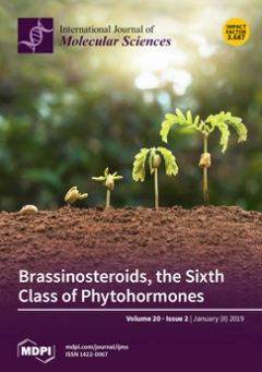
Cover Story (view full-size image): Phytohormones are natural chemical messengers that play critical roles in the regulation of plant growth and development as well as in responses to biotic and abiotic stress factors. The discovery, almost 40 years ago, of a new class of phytohormones, the brassinosteroids (BRs), opened a new era and introduced new perspectives in the regulation of agronomic traits through their use in agriculture. The present article provides an overview of BRs’ discovery and recent findings on their interactions with other phytohormones, in addition to clarifying how their network works to modulate plant growth, development, and responses to biotic and abiotic stresses. View this paper.
  • Issues are regarded as officially published after their release is announced to the table of contents alert mailing list.
  • You may sign up for e-mail alerts to receive table of contents of newly released issues.
  • PDF is the official format for papers published in both, html and pdf forms. To view the papers in pdf format, click on the "PDF Full-text" link, and use the free Adobe Reader to open them.
Order results
Result details
Section
Select all
Export citation of selected articles as:
11 pages, 1862 KiB  
Article
Hydrogen Indirectly Suppresses Increases in Hydrogen Peroxide in Cytoplasmic Hydroxyl Radical-Induced Cells and Suppresses Cellular Senescence
by Takahiro Sakai, Ryosuke Kurokawa, Shin-ichi Hirano and Jun Imai
Int. J. Mol. Sci. 2019, 20(2), 456; https://doi.org/10.3390/ijms20020456 - 21 Jan 2019
Cited by 16 | Viewed by 5688
Abstract
Bacteria inhabiting the human gut metabolize microbiota-accessible carbohydrates (MAC) contained in plant fibers and subsequently release metabolic products. Gut bacteria produce hydrogen (H2), which scavenges the hydroxyl radical (•OH). Because H2 diffuses within the cell, it is hypothesized that H [...] Read more.
Bacteria inhabiting the human gut metabolize microbiota-accessible carbohydrates (MAC) contained in plant fibers and subsequently release metabolic products. Gut bacteria produce hydrogen (H2), which scavenges the hydroxyl radical (•OH). Because H2 diffuses within the cell, it is hypothesized that H2 scavenges cytoplasmic •OH (cyto •OH) and suppresses cellular senescence. However, the mechanisms of cyto •OH-induced cellular senescence and the physiological role of gut bacteria-secreted H2 have not been elucidated. Based on the pyocyanin-stimulated cyto •OH-induced cellular senescence model, the mechanism by which cyto •OH causes cellular senescence was investigated by adding a supersaturated concentration of H2 into the cell culture medium. Cyto •OH-generated lipid peroxide caused glutathione (GSH) and heme shortage, increased hydrogen peroxide (H2O2), and induced cellular senescence via the phosphorylation of ataxia telangiectasia mutated kinase serine 1981 (p-ATMser1981)/p53 serine 15 (p-p53ser15)/p21 and phosphorylation of heme-regulated inhibitor (p-HRI)/phospho-eukaryotic translation initiation factor 2 subunit alpha serine 51 (p-eIF2α)/activating transcription factor 4 (ATF4)/p16 pathways. Further, H2 suppressed increased H2O2 by suppressing cyto •OH-mediated lipid peroxide formation and cellular senescence induction via two pathways. H2 produced by gut bacteria diffuses throughout the body to scavenge cyto •OH in cells. Therefore, it is highly likely that gut bacteria-produced H2 is involved in intracellular maintenance of the redox state, thereby suppressing cellular senescence and individual aging. Hence, H2 produced by intestinal bacteria may be involved in the suppression of aging. Full article
(This article belongs to the Special Issue Nutrition and Aging)
Show Figures

Figure 1

22 pages, 362 KiB  
Review
Do Neural Stem Cells Have a Choice? Heterogenic Outcome of Cell Fate Acquisition in Different Injury Models
by Felix Beyer, Iria Samper Agrelo and Patrick Küry
Int. J. Mol. Sci. 2019, 20(2), 455; https://doi.org/10.3390/ijms20020455 - 21 Jan 2019
Cited by 9 | Viewed by 5971
Abstract
The adult mammalian central nervous system (CNS) is generally considered as repair restricted organ with limited capacities to regenerate lost cells and to successfully integrate them into damaged nerve tracts. Despite the presence of endogenous immature cell types that can be activated upon [...] Read more.
The adult mammalian central nervous system (CNS) is generally considered as repair restricted organ with limited capacities to regenerate lost cells and to successfully integrate them into damaged nerve tracts. Despite the presence of endogenous immature cell types that can be activated upon injury or in disease cell replacement generally remains insufficient, undirected, or lost cell types are not properly generated. This limitation also accounts for the myelin repair capacity that still constitutes the default regenerative activity at least in inflammatory demyelinating conditions. Ever since the discovery of endogenous neural stem cells (NSCs) residing within specific niches of the adult brain, as well as the description of procedures to either isolate and propagate or artificially induce NSCs from various origins ex vivo, the field has been rejuvenated. Various sources of NSCs have been investigated and applied in current neuropathological paradigms aiming at the replacement of lost cells and the restoration of functionality based on successful integration. Whereas directing and supporting stem cells residing in brain niches constitutes one possible approach many investigations addressed their potential upon transplantation. Given the heterogeneity of these studies related to the nature of grafted cells, the local CNS environment, and applied implantation procedures we here set out to review and compare their applied protocols in order to evaluate rate-limiting parameters. Based on our compilation, we conclude that in healthy CNS tissue region specific cues dominate cell fate decisions. However, although increasing evidence points to the capacity of transplanted NSCs to reflect the regenerative need of an injury environment, a still heterogenic picture emerges when analyzing transplantation outcomes in injury or disease models. These are likely due to methodological differences despite preserved injury environments. Based on this meta-analysis, we suggest future NSC transplantation experiments to be conducted in a more comparable way to previous studies and that subsequent analyses must emphasize regional heterogeneity such as accounting for differences in gray versus white matter. Full article
(This article belongs to the Special Issue Roles of Stem Cells in Nerve Regeneration)
Show Figures

Graphical abstract

13 pages, 3795 KiB  
Article
HDAC1 and HDAC2 Double Knockout Triggers Cell Apoptosis in Advanced Thyroid Cancer
by Ching-Ling Lin, Ming-Lin Tsai, Chun-Yu Lin, Kai-Wen Hsu, Wen-Shyang Hsieh, Wei-Ming Chi, Li-Chi Huang and Chia-Hwa Lee
Int. J. Mol. Sci. 2019, 20(2), 454; https://doi.org/10.3390/ijms20020454 - 21 Jan 2019
Cited by 41 | Viewed by 6858
Abstract
Anaplastic thyroid carcinoma (ATC) and squamous thyroid carcinoma (STC) are both rare and advanced thyroid malignancies with a very poor prognosis and an average median survival time of 5 months and less than 20% of affected patients are alive 1 year after diagnosis. [...] Read more.
Anaplastic thyroid carcinoma (ATC) and squamous thyroid carcinoma (STC) are both rare and advanced thyroid malignancies with a very poor prognosis and an average median survival time of 5 months and less than 20% of affected patients are alive 1 year after diagnosis. The clinical management of both ATC and STC is very similar because they are not particularly responsive to radiotherapy and chemotherapy. This inspired us to explore a novel and effective clinically approved therapy for ATC treatment. Histone deacetylase inhibitor (HDACi) drugs are recently FDA-approved drug for malignancies, especially for blood cell cancers. Therefore, we investigated whether an HDACi drug acts as an effective anticancer drug for advanced thyroid cancers. Cell viability analysis of panobinostat treatment demonstrated a significant IC50 of 0.075 µM on SW579 STC cells. In addition, panobinostat exposure activated histone acetylation and triggered cell death mainly through cell cycle arrest and apoptosis-related protein activation. Using CRISPR/Cas9 to knock out HDAC1 and HDAC2 genes in SW579 cells, we observed that the histone acetylation level and cell cycle arrest were enhanced without any impact on cell growth. Furthermore, HDAC1 and HDAC2 double knockout (KO) cells showed dramatic cell apoptosis activation compared to HDAC1 and HDAC2 individual KO cells. This suggests expressional and biofunctional compensation between HDAC1 and HDAC2 on SW579 cells. This study provides strong evidence that panobinostat can potentially be used in the clinic of advanced thyroid cancer patients. Full article
(This article belongs to the Special Issue Roles of HDACs and HDAC Inhibitors in Human Cancers)
Show Figures

Figure 1

27 pages, 5930 KiB  
Review
Murine Models of Acute Myeloid Leukaemia
by Marwa Almosailleakh and Juerg Schwaller
Int. J. Mol. Sci. 2019, 20(2), 453; https://doi.org/10.3390/ijms20020453 - 21 Jan 2019
Cited by 47 | Viewed by 14670
Abstract
Acute myeloid leukaemia (AML) is a rare but severe form of human cancer that results from a limited number of functionally cooperating genetic abnormalities leading to uncontrolled proliferation and impaired differentiation of hematopoietic stem and progenitor cells. Before the identification of genetic driver [...] Read more.
Acute myeloid leukaemia (AML) is a rare but severe form of human cancer that results from a limited number of functionally cooperating genetic abnormalities leading to uncontrolled proliferation and impaired differentiation of hematopoietic stem and progenitor cells. Before the identification of genetic driver lesions, chemically, irradiation or viral infection-induced mouse leukaemia models provided platforms to test novel chemotherapeutics. Later, transgenic mouse models were established to test the in vivo transforming potential of newly cloned fusion genes and genetic aberrations detected in patients’ genomes. Hereby researchers constitutively or conditionally expressed the respective gene in the germline of the mouse or reconstituted the hematopoietic system of lethally irradiated mice with bone marrow virally expressing the mutation of interest. More recently, immune deficient mice have been explored to study patient-derived human AML cells in vivo. Unfortunately, although complementary to each other, none of the currently available strategies faithfully model the initiation and progression of the human disease. Nevertheless, fast advances in the fields of next generation sequencing, molecular technology and bioengineering are continuously contributing to the generation of better mouse models. Here we review the most important AML mouse models of each category, briefly describe their advantages and limitations and show how they have contributed to our understanding of the biology and to the development of novel therapies. Full article
(This article belongs to the Special Issue Genetics, Biology, and Treatment of Acute Myeloid Leukemia)
Show Figures

Figure 1

22 pages, 3132 KiB  
Article
Isodon rugosus (Wall. ex Benth.) Codd In Vitro Cultures: Establishment, Phytochemical Characterization and In Vitro Antioxidant and Anti-Aging Activities
by Bilal Haider Abbasi, Aisha Siddiquah, Duangjai Tungmunnithum, Shankhamala Bose, Muhammad Younas, Laurine Garros, Samantha Drouet, Nathalie Giglioli-Guivarc’h and Christophe Hano
Int. J. Mol. Sci. 2019, 20(2), 452; https://doi.org/10.3390/ijms20020452 - 21 Jan 2019
Cited by 39 | Viewed by 6947
Abstract
Isodon rugosus (Wall. ex Benth.) Codd accumulates large amounts of phenolics and pentacyclic triterpenes. The present study deals with the in vitro callus induction from stem and leaf explants of I. rugosus under various plant growth regulators (PGRs) for the production of antioxidant [...] Read more.
Isodon rugosus (Wall. ex Benth.) Codd accumulates large amounts of phenolics and pentacyclic triterpenes. The present study deals with the in vitro callus induction from stem and leaf explants of I. rugosus under various plant growth regulators (PGRs) for the production of antioxidant and anti-ageing compounds. Among all the tested PGRs, thidiazuron (TDZ) used alone or in conjunction with α-napthalene acetic acid (NAA) induced highest callogenesis in stem-derived explants, as compared to leaf-derived explants. Stem-derived callus culture displayed maximum total phenolic content and antioxidant activity under optimum hormonal combination (3.0 mg/L TDZ + 1.0 mg/L NAA). HPLC analysis revealed the presence of plectranthoic acid (373.92 µg/g DW), oleanolic acid (287.58 µg/g DW), betulinic acid (90.51 µg/g DW), caffeic acid (91.71 µg/g DW), and rosmarinic acid (1732.61 µg/g DW). Complete antioxidant and anti-aging potential of extracts with very contrasting phytochemical profiles were investigated. Correlation analyses revealed rosmarinic acid as the main contributor for antioxidant activity and anti-aging hyaluronidase, advance glycation end-products inhibitions and SIRT1 activation, whereas, pentacyclic triterpenoids were correlated with elastase, collagenase, and tyrosinase inhibitions. Altogether, these results clearly evidenced the great valorization potential of I. rugosus calli for the production of antioxidant and anti-aging bioactive extracts for cosmetic applications. Full article
(This article belongs to the Special Issue Health Promoting Effects of Phytochemicals)
Show Figures

Graphical abstract

15 pages, 1282 KiB  
Article
Comparative Phosphoproteomic Analysis of Barley Embryos with Different Dormancy during Imbibition
by Shinnosuke Ishikawa, José Barrero, Fuminori Takahashi, Scott Peck, Frank Gubler, Kazuo Shinozaki and Taishi Umezawa
Int. J. Mol. Sci. 2019, 20(2), 451; https://doi.org/10.3390/ijms20020451 - 21 Jan 2019
Cited by 9 | Viewed by 5274
Abstract
Dormancy is the mechanism that allows seeds to become temporally quiescent in order to select the right time and place to germinate. Like in other species, in barley, grain dormancy is gradually reduced during after-ripening. Phosphosignaling networks in barley grains were investigated by [...] Read more.
Dormancy is the mechanism that allows seeds to become temporally quiescent in order to select the right time and place to germinate. Like in other species, in barley, grain dormancy is gradually reduced during after-ripening. Phosphosignaling networks in barley grains were investigated by a large-scale analysis of phosphoproteins to examine potential changes in response pathways to after-ripening. We used freshly harvested (FH) and after-ripened (AR) barley grains which showed different dormancy levels. The LC-MS/MS analysis identified 2346 phosphopeptides in barley embryos, with 269 and 97 of them being up- or downregulated during imbibition, respectively. A number of phosphopeptides were differentially regulated between FH and AR samples, suggesting that phosphoproteomic profiles were quite different between FH and AR grains. Motif analysis suggested multiple protein kinases including SnRK2 and MAPK could be involved in such a difference between FH and AR samples. Taken together, our results revealed phosphosignaling pathways in barley grains during the water imbibition process. Full article
(This article belongs to the Special Issue Plant Proteomic Research 2.0)
Show Figures

Figure 1

17 pages, 2249 KiB  
Review
The Rice Alpha-Amylase, Conserved Regulator of Seed Maturation and Germination
by Rebecca Njeri Damaris, Zhongyuan Lin, Pingfang Yang and Dongli He
Int. J. Mol. Sci. 2019, 20(2), 450; https://doi.org/10.3390/ijms20020450 - 21 Jan 2019
Cited by 127 | Viewed by 17876
Abstract
Alpha-amylase, the major form of amylase with secondary carbohydrate binding sites, is a crucial enzyme throughout the growth period and life cycle of angiosperm. In rice, alpha-amylase isozymes are critical for the formation of the storage starch granule during seed maturation and motivate [...] Read more.
Alpha-amylase, the major form of amylase with secondary carbohydrate binding sites, is a crucial enzyme throughout the growth period and life cycle of angiosperm. In rice, alpha-amylase isozymes are critical for the formation of the storage starch granule during seed maturation and motivate the stored starch to nourish the developing seedling during seed germination which will directly affect the plant growth and field yield. Alpha-amylase has not yet been studied intensely to understand its classification, structure, expression trait, and expression regulation in rice and other crops. Among the 10-rice alpha-amylases, most were exclusively expressed in the developing seed embryo and induced in the seed germination process. During rice seed germination, the expression of alpha-amylase genes is known to be regulated negatively by sugar in embryos, however positively by gibberellin (GA) in endosperm through competitively binding to the specific promoter domain; besides, it is also controlled by a series of other abiotic or biotic factors, such as salinity. In this review, we overviewed the research progress of alpha-amylase with focus on seed germination and reflected on how in-depth work might elucidate its regulation and facilitate crop breeding as an efficient biomarker. Full article
(This article belongs to the Special Issue Seed Development, Dormancy and Germination)
Show Figures

Figure 1

47 pages, 26702 KiB  
Review
Bactericidal and Cytotoxic Properties of Silver Nanoparticles
by Chengzhu Liao, Yuchao Li and Sie Chin Tjong
Int. J. Mol. Sci. 2019, 20(2), 449; https://doi.org/10.3390/ijms20020449 - 21 Jan 2019
Cited by 700 | Viewed by 27754
Abstract
Silver nanoparticles (AgNPs) can be synthesized from a variety of techniques including physical, chemical and biological routes. They have been widely used as nanomaterials for manufacturing cosmetic and healthcare products, antimicrobial textiles, wound dressings, antitumor drug carriers, etc. due to their excellent antimicrobial [...] Read more.
Silver nanoparticles (AgNPs) can be synthesized from a variety of techniques including physical, chemical and biological routes. They have been widely used as nanomaterials for manufacturing cosmetic and healthcare products, antimicrobial textiles, wound dressings, antitumor drug carriers, etc. due to their excellent antimicrobial properties. Accordingly, AgNPs have gained access into our daily life, and the inevitable human exposure to these nanoparticles has raised concerns about their potential hazards to the environment, health, and safety in recent years. From in vitro cell cultivation tests, AgNPs have been reported to be toxic to several human cell lines including human bronchial epithelial cells, human umbilical vein endothelial cells, red blood cells, human peripheral blood mononuclear cells, immortal human keratinocytes, liver cells, etc. AgNPs induce a dose-, size- and time-dependent cytotoxicity, particularly for those with sizes ≤10 nm. Furthermore, AgNPs can cross the brain blood barrier of mice through the circulation system on the basis of in vivo animal tests. AgNPs tend to accumulate in mice organs such as liver, spleen, kidney and brain following intravenous, intraperitoneal, and intratracheal routes of administration. In this respect, AgNPs are considered a double-edged sword that can eliminate microorganisms but induce cytotoxicity in mammalian cells. This article provides a state-of-the-art review on the synthesis of AgNPs, and their applications in antimicrobial textile fabrics, food packaging films, and wound dressings. Particular attention is paid to the bactericidal activity and cytotoxic effect in mammalian cells. Full article
(This article belongs to the Special Issue Silver Nano/microparticles: Modification and Applications)
Show Figures

Graphical abstract

34 pages, 2776 KiB  
Review
Facets of Theiler’s Murine Encephalomyelitis Virus-Induced Diseases: An Update
by Ingo Gerhauser, Florian Hansmann, Malgorzata Ciurkiewicz, Wolfgang Löscher and Andreas Beineke
Int. J. Mol. Sci. 2019, 20(2), 448; https://doi.org/10.3390/ijms20020448 - 21 Jan 2019
Cited by 54 | Viewed by 7973
Abstract
Theiler’s murine encephalomyelitis virus (TMEV), a naturally occurring, enteric pathogen of mice is a Cardiovirus of the Picornaviridae family. Low neurovirulent TMEV strains such as BeAn cause a severe demyelinating disease in susceptible SJL mice following intracerebral infection. Furthermore, TMEV infections of C57BL/6 [...] Read more.
Theiler’s murine encephalomyelitis virus (TMEV), a naturally occurring, enteric pathogen of mice is a Cardiovirus of the Picornaviridae family. Low neurovirulent TMEV strains such as BeAn cause a severe demyelinating disease in susceptible SJL mice following intracerebral infection. Furthermore, TMEV infections of C57BL/6 mice cause acute polioencephalitis initiating a process of epileptogenesis that results in spontaneous recurrent epileptic seizures in approximately 50% of affected mice. Moreover, C3H mice develop cardiac lesions after an intraperitoneal high-dose application of TMEV. Consequently, TMEV-induced diseases are widely used as animal models for multiple sclerosis, epilepsy, and myocarditis. The present review summarizes morphological lesions and pathogenic mechanisms triggered by TMEV with a special focus on the development of hippocampal degeneration and seizures in C57BL/6 mice as well as demyelination in the spinal cord in SJL mice. Furthermore, a detailed description of innate and adaptive immune responses is given. TMEV studies provide novel insights into the complexity of organ- and mouse strain-specific immunopathology and help to identify factors critical for virus persistence. Full article
Show Figures

Figure 1

17 pages, 3644 KiB  
Article
The Expression of Thrombospondin-4 Correlates with Disease Severity in Osteoarthritic Knee Cartilage
by Kathrin Maly, Inna Schaible, Jana Riegger, Rolf E. Brenner, Andrea Meurer and Frank Zaucke
Int. J. Mol. Sci. 2019, 20(2), 447; https://doi.org/10.3390/ijms20020447 - 21 Jan 2019
Cited by 15 | Viewed by 5311
Abstract
Osteoarthritis (OA) is a progressive joint disease characterized by a continuous degradation of the cartilage extracellular matrix (ECM). The expression of the extracellular glycoprotein thrombospondin-4 (TSP-4) is known to be increased in injured tissues and involved in matrix remodeling, but its role in [...] Read more.
Osteoarthritis (OA) is a progressive joint disease characterized by a continuous degradation of the cartilage extracellular matrix (ECM). The expression of the extracellular glycoprotein thrombospondin-4 (TSP-4) is known to be increased in injured tissues and involved in matrix remodeling, but its role in articular cartilage and, in particular, in OA remains elusive. In the present study, we analyzed the expression and localization of TSP-4 in healthy and OA knee cartilage by reverse transcription polymerase chain reaction (RT-PCR), immunohistochemistry, and immunoblot. We found that TSP-4 protein expression is increased in OA and that expression levels correlate with OA severity. TSP-4 was not regulated at the transcriptional level but we detected changes in the anchorage of TSP-4 in the altered ECM using sequential protein extraction. We were also able to detect pentameric and fragmented TSP-4 in the serum of both healthy controls and OA patients. Here, the total protein amount was not significantly different but we identified specific degradation products that were more abundant in sera of OA patients. Future studies will reveal if these fragments have the potential to serve as OA-specific biomarkers. Full article
Show Figures

Graphical abstract

16 pages, 5152 KiB  
Article
Analysis of Mucopolysaccharidosis Type VI through Integrative Functional Metabolomics
by Abdellah Tebani, Lenaig Abily-Donval, Isabelle Schmitz-Afonso, Monique Piraud, Jérôme Ausseil, Farid Zerimech, Carine Pilon, Tony Pereira, Stéphane Marret, Carlos Afonso and Soumeya Bekri
Int. J. Mol. Sci. 2019, 20(2), 446; https://doi.org/10.3390/ijms20020446 - 21 Jan 2019
Cited by 21 | Viewed by 5437
Abstract
Metabolic phenotyping is poised as a powerful and promising tool for biomarker discovery in inherited metabolic diseases. However, few studies applied this approach to mcopolysaccharidoses (MPS). Thus, this innovative functional approach may unveil comprehensive impairments in MPS biology. This study explores mcopolysaccharidosis VI [...] Read more.
Metabolic phenotyping is poised as a powerful and promising tool for biomarker discovery in inherited metabolic diseases. However, few studies applied this approach to mcopolysaccharidoses (MPS). Thus, this innovative functional approach may unveil comprehensive impairments in MPS biology. This study explores mcopolysaccharidosis VI (MPS VI) or Maroteaux–Lamy syndrome (OMIM #253200) which is an autosomal recessive lysosomal storage disease caused by the deficiency of arylsulfatase B enzyme. Urine samples were collected from 16 MPS VI patients and 66 healthy control individuals. Untargeted metabolomics analysis was applied using ultra-high-performance liquid chromatography combined with ion mobility and high-resolution mass spectrometry. Furthermore, dermatan sulfate, amino acids, carnitine, and acylcarnitine profiles were quantified using liquid chromatography coupled to tandem mass spectrometry. Univariate analysis and multivariate data modeling were used for integrative analysis and discriminant metabolites selection. Pathway analysis was done to unveil impaired metabolism. The study revealed significant differential biochemical patterns using multivariate data modeling. Pathway analysis revealed that several major amino acid pathways were dysregulated in MPS VI. Integrative analysis of targeted and untargeted metabolomics data with in silico results yielded arginine-proline, histidine, and glutathione metabolism being the most affected. This study is one of the first metabolic phenotyping studies of MPS VI. The findings might shed light on molecular understanding of MPS pathophysiology to develop further MPS studies to enhance diagnosis and treatments of this rare condition. Full article
(This article belongs to the Section Molecular Pathology, Diagnostics, and Therapeutics)
Show Figures

Figure 1

13 pages, 2944 KiB  
Article
Obstructive Sleep Apnea Activates HIF-1 in a Hypoxia Dose-Dependent Manner in HCT116 Colorectal Carcinoma Cells
by Chloe-Anne Martinez, Bernadette Kerr, Charley Jin, Peter A. Cistulli and Kristina M. Cook
Int. J. Mol. Sci. 2019, 20(2), 445; https://doi.org/10.3390/ijms20020445 - 21 Jan 2019
Cited by 46 | Viewed by 9811
Abstract
Obstructive sleep apnea (OSA) affects a significant proportion of the population and is linked to increased rates of cancer development and a worse cancer outcome. OSA is characterized by nocturnal intermittent hypoxia and animal models of OSA-like intermittent hypoxia show increased tumor growth [...] Read more.
Obstructive sleep apnea (OSA) affects a significant proportion of the population and is linked to increased rates of cancer development and a worse cancer outcome. OSA is characterized by nocturnal intermittent hypoxia and animal models of OSA-like intermittent hypoxia show increased tumor growth and metastasis. Advanced tumors typically have regions of chronic hypoxia, activating the transcription factor, HIF-1, which controls the expression of genes involved in cancer progression. Rapid intermittent hypoxia from OSA has been proposed to increase HIF-1 activity and this may occur in tumors. The effect of exposing a developing tumor to OSA-like intermittent hypoxia is largely unknown. We have built a cell-based model of physiological OSA tissue oxygenation in order to study the effects of intermittent hypoxia in HCT116 colorectal cancer cells. We found that HIF-1α increases following intermittent hypoxia and that the expression of HIF-target genes increases, including those involved in glycolysis, the hypoxic pathway and extracellular matrix remodeling. Expression of these genes acts as a ‘hypoxic’ signature which is associated with a worse prognosis. The total dose of hypoxia determined the magnitude of change in the hypoxic signature rather than the frequency or duration of hypoxia-reoxygenation cycles per se. Finally, transcription of HIF1A mRNA differs in response to chronic and intermittent hypoxia suggesting that HIF-1α may be regulated at the transcriptional level in intermittent hypoxia and not just by the post-translational oxygen-dependent degradation pathway seen in chronic hypoxia. Full article
(This article belongs to the Special Issue Sleep Apnea and Intermittent Hypoxia)
Show Figures

Graphical abstract

25 pages, 306 KiB  
Review
Clinical Use and Molecular Action of Corticosteroids in the Pediatric Age
by Giovanna Ferrara, Maria Grazia Petrillo, Teresa Giani, Edoardo Marrani, Cesare Filippeschi, Teresa Oranges, Gabriele Simonini and Rolando Cimaz
Int. J. Mol. Sci. 2019, 20(2), 444; https://doi.org/10.3390/ijms20020444 - 21 Jan 2019
Cited by 55 | Viewed by 9043
Abstract
Corticosteroids are the mainstay of therapy for many pediatric disorders and sometimes are life-saving. Both endogenous and synthetic derivatives diffuse across the cell membrane and, by binding to their cognate glucocorticoid receptor, modulate a variety of physiological functions, such as glucose metabolism, immune [...] Read more.
Corticosteroids are the mainstay of therapy for many pediatric disorders and sometimes are life-saving. Both endogenous and synthetic derivatives diffuse across the cell membrane and, by binding to their cognate glucocorticoid receptor, modulate a variety of physiological functions, such as glucose metabolism, immune homeostasis, organ development, and the endocrine system. However, despite their proved and known efficacy, corticosteroids show a lot of side effects, among which growth retardation is of particular concern and specific for pediatric age. The aim of this review is to discuss the mechanism of action of corticosteroids, and how their genomic effects have both beneficial and adverse consequences. We will focus on the use of corticosteroids in different pediatric subspecialties and most common diseases, analyzing the most recent evidence. Full article
18 pages, 3478 KiB  
Article
Recurrent Stimulation of Natural Killer Cell Clones with K562 Expressing Membrane-Bound Interleukin-21 Affects Their Phenotype, Interferon-γ Production, and Lifespan
by Maria A. Streltsova, Sofya A. Erokhina, Leonid M. Kanevskiy, Maria V. Grechikhina, Polina A. Kobyzeva, Dean A. Lee, William G. Telford, Alexander M. Sapozhnikov and Elena I. Kovalenko
Int. J. Mol. Sci. 2019, 20(2), 443; https://doi.org/10.3390/ijms20020443 - 21 Jan 2019
Cited by 16 | Viewed by 6231
Abstract
A pattern of natural killer cell (NK cell) heterogeneity determines proliferative and functional responses to activating stimuli in individuals. Obtaining the progeny of a single cell by cloning the original population is one of the ways to study NK cell heterogeneity. In this [...] Read more.
A pattern of natural killer cell (NK cell) heterogeneity determines proliferative and functional responses to activating stimuli in individuals. Obtaining the progeny of a single cell by cloning the original population is one of the ways to study NK cell heterogeneity. In this work, we sorted single cells into a plate and stimulated them via interleukin (IL)-2 and gene-modified K562 feeder cells that expressed membrane-bound IL-21 (K562-mbIL21), which led to a generation of phenotypically confirmed and functionally active NK cell clones. Next, we applied two models of clone cultivation, which differently affected their phenotype, lifespan, and functional activity. The first model, which included weekly restimulation of clones with K562-mbIL21 and IL-2, resulted in the generation of relatively short-lived (5–7 weeks) clones of highly activated NK cells. Levels of human leukocyte antigen class II molecule—DR isotype (HLA-DR) expression in the expanded NK cells correlated strongly with interferon-γ (IFN-γ) production. The second model, in which NK cells were restimulated weekly with IL-2 alone and once on the sixth week with K562-mbIL21 and IL-2, produced long-lived clones (8–14 weeks) that expanded up to 107 cells with a lower ability to produce IFN-γ. Our method is applicable for studying variability in phenotype, proliferative, and functional activity of certain NK cell progeny in response to the stimulation, which may help in selecting NK cells best suited for clinical use. Full article
(This article belongs to the Special Issue The Interleukins in Health and Disease)
Show Figures

Graphical abstract

23 pages, 958 KiB  
Review
Arf GAPs as Regulators of the Actin Cytoskeleton—An Update
by Christine E. Tanna, Louisa B. Goss, Calvin G. Ludwig and Pei-Wen Chen
Int. J. Mol. Sci. 2019, 20(2), 442; https://doi.org/10.3390/ijms20020442 - 21 Jan 2019
Cited by 35 | Viewed by 8036
Abstract
Arf GTPase-activating proteins (Arf GAPs) control the activity of ADP-ribosylation factors (Arfs) by inducing GTP hydrolysis and participate in a diverse array of cellular functions both through mechanisms that are dependent on and independent of their Arf GAP activity. A number of these [...] Read more.
Arf GTPase-activating proteins (Arf GAPs) control the activity of ADP-ribosylation factors (Arfs) by inducing GTP hydrolysis and participate in a diverse array of cellular functions both through mechanisms that are dependent on and independent of their Arf GAP activity. A number of these functions hinge on the remodeling of actin filaments. Accordingly, some of the effects exerted by Arf GAPs involve proteins known to engage in regulation of the actin dynamics and architecture, such as Rho family proteins and nonmuscle myosin 2. Circular dorsal ruffles (CDRs), podosomes, invadopodia, lamellipodia, stress fibers and focal adhesions are among the actin-based structures regulated by Arf GAPs. Arf GAPs are thus important actors in broad functions like adhesion and motility, as well as the specialized functions of bone resorption, neurite outgrowth, and pathogen internalization by immune cells. Arf GAPs, with their multiple protein-protein interactions, membrane-binding domains and sites for post-translational modification, are good candidates for linking the changes in actin to the membrane. The findings discussed depict a family of proteins with a critical role in regulating actin dynamics to enable proper cell function. Full article
(This article belongs to the Special Issue Small GTPases)
Show Figures

Figure 1

24 pages, 4040 KiB  
Article
Exploration of the Effect of Blue Light on Functional Metabolite Accumulation in Longan Embryonic Calli via RNA Sequencing
by Hansheng Li, Yumeng Lyu, Xiaohui Chen, Congqiao Wang, Deheng Yao, Shanshan Ni, Yuling Lin, Yukun Chen, Zihao Zhang and Zhongxiong Lai
Int. J. Mol. Sci. 2019, 20(2), 441; https://doi.org/10.3390/ijms20020441 - 21 Jan 2019
Cited by 25 | Viewed by 5268
Abstract
Light is an important factor that affects the synthesis of functional metabolites in longan embryogenic calli (ECs). However, analysis of the effect of light on functional metabolites in longan ECs via RNA sequencing has rarely been reported and their light regulation network is [...] Read more.
Light is an important factor that affects the synthesis of functional metabolites in longan embryogenic calli (ECs). However, analysis of the effect of light on functional metabolites in longan ECs via RNA sequencing has rarely been reported and their light regulation network is unclear. The contents of various functional metabolites as well as the enzymatic activities of superoxide dismutase and peroxidase and the level of H2O2 in longan ECs were significantly higher under blue light treatment than under the other treatments (dark, white). In this study, we sequenced three mRNA libraries constructed from longan ECs subjected to different treatments. A total of 4463, 1639 and 1806 genes were differentially expressed in the dark versus blue (DB), dark versus white (DW) and white versus blue (WB) combinations, respectively. According to GO and KEGG analyses, most of the differentially expressed genes (DEGs) identified were involved in transmembrane transport, taurine and hypotaurine metabolism, calcium transport and so forth. Mapman analysis revealed that more DEGs were identified in each DB combination pathway than in DW combination pathways, indicating that blue light exerts a significantly stronger regulatory effect on longan EC metabolism than the other treatments. Based on previous research and transcriptome data mining, a blue light signaling network of genes that affect longan functional metabolites was constructed and HY5, PIF4 and MYC2 were shown to be the key regulatory genes in the network. The results of this study demonstrate that the expression levels of phase-specific genes vary with changes in longan EC functional metabolites. Full article
(This article belongs to the Section Molecular Plant Sciences)
Show Figures

Graphical abstract

12 pages, 3561 KiB  
Review
Structure and Mechanism of the Divalent Anion/Na+ Symporter
by Min Lu
Int. J. Mol. Sci. 2019, 20(2), 440; https://doi.org/10.3390/ijms20020440 - 21 Jan 2019
Cited by 6 | Viewed by 5473
Abstract
Integral membrane proteins of the divalent anion/Na+ symporter (DASS) family are conserved from bacteria to humans. DASS proteins typically mediate the coupled uptake of Na+ ions and dicarboxylate, tricarboxylate, or sulfate. Since the substrates for DASS include key intermediates and regulators [...] Read more.
Integral membrane proteins of the divalent anion/Na+ symporter (DASS) family are conserved from bacteria to humans. DASS proteins typically mediate the coupled uptake of Na+ ions and dicarboxylate, tricarboxylate, or sulfate. Since the substrates for DASS include key intermediates and regulators of energy metabolism, alterations of DASS function profoundly affect fat storage, energy expenditure and life span. Furthermore, loss-of-function mutations in a human DASS have been associated with neonatal epileptic encephalopathy. More recently, human DASS has also been implicated in the development of liver cancers. Therefore, human DASS proteins are potentially promising pharmacological targets for battling obesity, diabetes, kidney stone, fatty liver, as well as other metabolic and neurological disorders. Despite its clinical relevance, the mechanism by which DASS proteins recognize and transport anionic substrates remains unclear. Recently, the crystal structures of a bacterial DASS and its humanized variant have been published. This article reviews the mechanistic implications of these structures and suggests future work to better understand how the function of DASS can be modulated for potential therapeutic benefit. Full article
(This article belongs to the Section Molecular Biology)
Show Figures

Figure 1

16 pages, 5737 KiB  
Article
Antibacterial Composites of Cuprous Oxide Nanoparticles and Polyethylene
by Yanna Gurianov, Faina Nakonechny, Yael Albo and Marina Nisnevitch
Int. J. Mol. Sci. 2019, 20(2), 439; https://doi.org/10.3390/ijms20020439 - 21 Jan 2019
Cited by 28 | Viewed by 4830
Abstract
Cuprous oxide nanoparticles (Cu2ONPs) were used for preparing composites with linear low-density polyethylene (LLDPE) by co-extrusion, thermal adhesion, and attachment using ethyl cyanoacrylate, trimethoxyvinylsilane, and epoxy resin. The composites were examined by Scanning electron microscope and tested for their antibacterial activity [...] Read more.
Cuprous oxide nanoparticles (Cu2ONPs) were used for preparing composites with linear low-density polyethylene (LLDPE) by co-extrusion, thermal adhesion, and attachment using ethyl cyanoacrylate, trimethoxyvinylsilane, and epoxy resin. The composites were examined by Scanning electron microscope and tested for their antibacterial activity against Gram-positive Staphylococcus aureus and Gram-negative Escherichia coli. All of these composites—except for the one obtained by extrusion—eradicated cells of both bacteria within half an hour. The composite prepared by thermal adhesion of Cu2ONPs on LLDPE had the highest external exposure of nanoparticles and exhibited the highest activity against the bacteria. This composite and the one obtained using ethyl cyanoacrylate showed no leaching of copper ions into the aqueous phase. Copper ion leaching from composites prepared with trimethoxyvinylsilane and epoxy resin was very low. The antibacterial activity of the composites can be rated as follows: obtained by thermal adhesion > obtained using ethyl cyanoacrylate > obtained using trimethoxyvinylsilane > obtained using epoxy resin > obtained by extrusion. The composites with the highest activity are potential materials for tap water and wastewater disinfection. Full article
(This article belongs to the Special Issue Polymeric Systems as Antimicrobial or Antifouling Agents)
Show Figures

Figure 1

18 pages, 2103 KiB  
Article
Gut Microbiota and Predicted Metabolic Pathways in a Sample of Mexican Women Affected by Obesity and Obesity Plus Metabolic Syndrome
by Alejandra Chávez-Carbajal, Khemlal Nirmalkar, Ana Pérez-Lizaur, Fernando Hernández-Quiroz, Silvia Ramírez-del-Alto, Jaime García-Mena and César Hernández-Guerrero
Int. J. Mol. Sci. 2019, 20(2), 438; https://doi.org/10.3390/ijms20020438 - 21 Jan 2019
Cited by 140 | Viewed by 13289
Abstract
Obesity is an excessive fat accumulation that could lead to complications like metabolic syndrome. There are reports on gut microbiota and metabolic syndrome in relation to dietary, host genetics, and other environmental factors; however, it is necessary to explore the role of the [...] Read more.
Obesity is an excessive fat accumulation that could lead to complications like metabolic syndrome. There are reports on gut microbiota and metabolic syndrome in relation to dietary, host genetics, and other environmental factors; however, it is necessary to explore the role of the gut microbiota metabolic pathways in populations like Mexicans, where the prevalence of obesity and metabolic syndrome is high. This study identify alterations of the gut microbiota in a sample of healthy Mexican women (CO), women with obesity (OB), and women with obesity plus metabolic syndrome (OMS). We studied 67 women, characterizing their anthropometric and biochemical parameters along with their gut bacterial diversity by high-throughput DNA sequencing. Our results indicate that in OB or OMS women, Firmicutes was the most abundant bacterial phylum. We observed significant changes in abundances of bacteria belonging to the Ruminococcaceae, Lachnospiraceae, and Erysipelotrichaceae families and significant enrichment of gut bacteria from 16 different taxa that might explain the observed metabolic alterations between the groups. Finally, the predicted functional metagenome of the gut microbiota found in each category shows differences in metabolic pathways related to lipid metabolism. We demonstrate that Mexican women have a particular bacterial gut microbiota characteristic of each phenotype. There are bacteria that potentially explain the observed metabolic differences between the groups, and gut bacteria in OMS and OB conditions carry more genes of metabolic pathways implicated in lipid metabolism. Full article
(This article belongs to the Special Issue Bioactives and Nutraceuticals in Metabolic Syndrome)
Show Figures

Graphical abstract

21 pages, 16885 KiB  
Article
Lectin Sequence Distribution in QTLs from Rice (Oryza sativa) Suggest a Role in Morphological Traits and Stress Responses
by Mariya Tsaneva, Kristof De Schutter, Bruno Verstraeten and Els J.M. Van Damme
Int. J. Mol. Sci. 2019, 20(2), 437; https://doi.org/10.3390/ijms20020437 - 20 Jan 2019
Cited by 9 | Viewed by 4180
Abstract
Rice (Oryza sativa) is one of the main staple crops worldwide but suffers from important yield losses due to different abiotic and biotic stresses. Analysis of quantitative trait loci (QTL) is a classical genetic method which enables the creation of more [...] Read more.
Rice (Oryza sativa) is one of the main staple crops worldwide but suffers from important yield losses due to different abiotic and biotic stresses. Analysis of quantitative trait loci (QTL) is a classical genetic method which enables the creation of more resistant cultivars but does not yield information on the genes directly involved or responsible for the desired traits. Lectins are known as proteins with diverse functions in plants. Some of them are abundant proteins in seeds and are considered as storage/defense proteins while other lectins are known as stress-inducible proteins, implicated in stress perception and signal transduction as part of plant innate immunity. We investigated the distribution of lectin sequences in different QTL related to stress tolerance/resistance, morphology, and physiology through mapping of the lectin sequences and QTL regions on the chromosomes and subsequent statistical analysis. Furthermore, the domain structure and evolutionary relationships of the lectins in O. sativa spp. indica and japonica were investigated. Our results revealed that lectin sequences are statistically overrepresented in QTLs for (a)biotic resistance/tolerance as well as in QTLs related to economically important traits such as eating quality and sterility. These findings contribute to the characterization of the QTL sequences and can provide valuable information to the breeders. Full article
Show Figures

Graphical abstract

31 pages, 4446 KiB  
Article
Expression of Hypoxia-Inducible Factor 1α (HIF-1α) and Genes of Related Pathways in Altered Gravity
by Johannes Vogel, Cora Sandra Thiel, Svantje Tauber, Christian Stockmann, Max Gassmann and Oliver Ullrich
Int. J. Mol. Sci. 2019, 20(2), 436; https://doi.org/10.3390/ijms20020436 - 20 Jan 2019
Cited by 27 | Viewed by 6462
Abstract
Immune system deterioration in space represents a major risk, which has to be mitigated for exploration-class missions into the solar system. Altered gravitational forces have been shown to regulate adaptation processes in cells of the immune system, which are important for appropriate risk [...] Read more.
Immune system deterioration in space represents a major risk, which has to be mitigated for exploration-class missions into the solar system. Altered gravitational forces have been shown to regulate adaptation processes in cells of the immune system, which are important for appropriate risk management, monitoring and development of countermeasures. T lymphocytes and cells of the monocyte-macrophage system are highly migratory cell types that frequently encounter a wide range of oxygen tensions in human tissues and in hypoxic areas, even under homeostatic conditions. Hypoxia-inducible factor 1 and 2 (HIF’s) might have an important role in activation of T cells and cells of the monocyte-macrophages system. Thus, we investigated the regulation of HIF-dependent and, therefore, hypoxia-signaling systems in both cell types in altered gravity and performed transcript and protein analysis from parabolic flight and suborbital ballistic rocket experiments. We found that HIF-1α and HIF-1-dependent transcripts were differently regulated in altered gravity, whereas HIF-1α-dependent gene expression adapted after 5 min microgravity. Inter-platform comparisons identified PDK1 as highly responsive to gravitational changes in human U937 myelomonocytic cells and in Jurkat T cells. We suggest HIF-1 as a potential pharmacological target for counteracting immune system deterioration during space flight. Full article
(This article belongs to the Special Issue Adaptation of Living Organisms in Space: From Mammals to Plants)
Show Figures

Figure 1

17 pages, 2607 KiB  
Review
Current Trends in Fabrication of Biomaterials for Bone and Cartilage Regeneration: Materials Modifications and Biophysical Stimulations
by Agata Przekora
Int. J. Mol. Sci. 2019, 20(2), 435; https://doi.org/10.3390/ijms20020435 - 20 Jan 2019
Cited by 79 | Viewed by 7446
Abstract
The aim of engineering of biomaterials is to fabricate implantable biocompatible scaffold that would accelerate regeneration of the tissue and ideally protect the wound against biodevice-related infections, which may cause prolonged inflammation and biomaterial failure. To obtain antimicrobial and highly biocompatible scaffolds promoting [...] Read more.
The aim of engineering of biomaterials is to fabricate implantable biocompatible scaffold that would accelerate regeneration of the tissue and ideally protect the wound against biodevice-related infections, which may cause prolonged inflammation and biomaterial failure. To obtain antimicrobial and highly biocompatible scaffolds promoting cell adhesion and growth, materials scientists are still searching for novel modifications of biomaterials. This review presents current trends in the field of engineering of biomaterials concerning application of various modifications and biophysical stimulation of scaffolds to obtain implants allowing for fast regeneration process of bone and cartilage as well as providing long-lasting antimicrobial protection at the site of injury. The article describes metal ion and plasma modifications of biomaterials as well as post-surgery external stimulations of implants with ultrasound and magnetic field, providing accelerated regeneration process. Finally, the review summarizes recent findings concerning the use of piezoelectric biomaterials in regenerative medicine. Full article
(This article belongs to the Special Issue Biomaterials for Musculoskeletal System)
Show Figures

Graphical abstract

23 pages, 1913 KiB  
Review
Extracellular Vesicle-Mediated Cell–Cell Communication in the Nervous System: Focus on Neurological Diseases
by Celeste Caruso Bavisotto, Federica Scalia, Antonella Marino Gammazza, Daniela Carlisi, Fabio Bucchieri, Everly Conway de Macario, Alberto J. L. Macario, Francesco Cappello and Claudia Campanella
Int. J. Mol. Sci. 2019, 20(2), 434; https://doi.org/10.3390/ijms20020434 - 20 Jan 2019
Cited by 120 | Viewed by 12428
Abstract
Extracellular vesicles (EVs), including exosomes, are membranous particles released by cells into the extracellular space. They are involved in cell differentiation, tissue homeostasis, and organ remodelling in virtually all tissues, including the central nervous system (CNS). They are secreted by a range of [...] Read more.
Extracellular vesicles (EVs), including exosomes, are membranous particles released by cells into the extracellular space. They are involved in cell differentiation, tissue homeostasis, and organ remodelling in virtually all tissues, including the central nervous system (CNS). They are secreted by a range of cell types and via blood reaching other cells whose functioning they can modify because they transport and deliver active molecules, such as proteins of various types and functions, lipids, DNA, and miRNAs. Since they are relatively easy to isolate, exosomes can be characterized, and their composition elucidated and manipulated by bioengineering techniques. Consequently, exosomes appear as promising theranostics elements, applicable to accurately diagnosing pathological conditions, and assessing prognosis and response to treatment in a variety of disorders. Likewise, the characteristics and manageability of exosomes make them potential candidates for delivering selected molecules, e.g., therapeutic drugs, to specific target tissues. All these possible applications are pertinent to research in neurophysiology, as well as to the study of neurological disorders, including CNS tumors, and autoimmune and neurodegenerative diseases. In this brief review, we discuss what is known about the role and potential future applications of exosomes in the nervous system and its diseases, focusing on cell–cell communication in physiology and pathology. Full article
(This article belongs to the Special Issue Focus on Exosome-Based Cell-Cell Communication in Health and Disease)
Show Figures

Figure 1

12 pages, 1831 KiB  
Article
The Effect of Psyllium Husk on Intestinal Microbiota in Constipated Patients and Healthy Controls
by Jonna Jalanka, Giles Major, Kathryn Murray, Gulzar Singh, Adam Nowak, Caroline Kurtz, Inmaculada Silos-Santiago, Jeffrey M. Johnston, Willem M. de Vos and Robin Spiller
Int. J. Mol. Sci. 2019, 20(2), 433; https://doi.org/10.3390/ijms20020433 - 20 Jan 2019
Cited by 124 | Viewed by 29616
Abstract
Psyllium is a widely used treatment for constipation. It traps water in the intestine increasing stool water, easing defaecation and altering the colonic environment. We aimed to assess the impact of psyllium on faecal microbiota, whose key role in gut physiology is being [...] Read more.
Psyllium is a widely used treatment for constipation. It traps water in the intestine increasing stool water, easing defaecation and altering the colonic environment. We aimed to assess the impact of psyllium on faecal microbiota, whose key role in gut physiology is being increasingly recognised. We performed two randomised, placebo-controlled, double-blinded trials comparing 7 days of psyllium with a placebo (maltodextrin) in 8 healthy volunteers and 16 constipated patients respectively. We measured the patients’ gastrointestnal (GI) transit, faecal water content, short-chain fatty acid (SCFA) and the stool microbiota composition. While psyllium supplement had a small but significant effect on the microbial composition of healthy adults (increasing Veillonella and decreasing Subdoligranulum), in constipated subjects there were greater effects on the microbial composition (increased Lachnospira, Faecalibacterium, Phascolarctobacterium, Veillonella and Sutterella and decreased uncultured Coriobacteria and Christensenella) and alterations in the levels of acetate and propionate. We found several taxa to be associated with altered GI transit, SCFAs and faecal water content in these patients. Significant increases in three genera known to produce butyrate, Lachnospira, Roseburia and Faecalibacterium, correlated with increased faecal water. In summary, psyllium supplementation increased stool water and this was associated with significant changes in microbiota, most marked in constipated patients. Full article
(This article belongs to the Special Issue Microbiota, Food and Health)
Show Figures

Figure 1

18 pages, 616 KiB  
Review
Resistance to Cereal Cyst Nematodes in Wheat and Barley: An Emphasis on Classical and Modern Approaches
by Muhammad Amjad Ali, Mahpara Shahzadi, Adil Zahoor, Abdelfattah A. Dababat, Halil Toktay, Allah Bakhsh, Muhammad Azher Nawaz and Hongjie Li
Int. J. Mol. Sci. 2019, 20(2), 432; https://doi.org/10.3390/ijms20020432 - 19 Jan 2019
Cited by 55 | Viewed by 8205
Abstract
Cereal cyst nematodes (CCNs) are among the most important nematode pests that limit production of small grain cereals like wheat and barley. These nematodes alone are estimated to reduce production of crops by 10% globally. This necessitates a huge enhancement of nematode resistance [...] Read more.
Cereal cyst nematodes (CCNs) are among the most important nematode pests that limit production of small grain cereals like wheat and barley. These nematodes alone are estimated to reduce production of crops by 10% globally. This necessitates a huge enhancement of nematode resistance in cereal crops against CCNs. Nematode resistance in wheat and barley in combination with higher grain yields has been a preferential research area for cereal nematologists. This usually involved the targeted genetic exploitations through natural means of classical selection breeding of resistant genotypes and finding quantitative trait luci (QTLs) associated with resistance genes. These improvements were based on available genetic diversity among the crop plants. Recently, genome-wide association studies have widely been exploited to associate nematode resistance or susceptibility with particular regions of the genome. Use of biotechnological tools through the application of various transgenic strategies for enhancement of nematode resistance in various crop plants including wheat and barley had also been an important area of research. These modern approaches primarily include the use of gene silencing, exploitation of nematode effector genes, proteinase inhibitors, chemodisruptive peptides and a combination of one or more of these approaches. Furthermore, the perspective genome editing technologies including CRISPR-Cas9 could also be helpful for improving CCN resistance in wheat and barley. The information provided in this review will be helpful to enhance resistance against CCNs and will attract the attention of the scientific community towards this neglected area. Full article
(This article belongs to the Special Issue Molecular Advances in Wheat and Barley)
Show Figures

Figure 1

21 pages, 2041 KiB  
Article
Non-Targeted Metabolomics Reveals Sorghum Rhizosphere-Associated Exudates are Influenced by the Belowground Interaction of Substrate and Sorghum Genotype
by Sarah B. Miller, Adam L. Heuberger, Corey D. Broeckling and Courtney E. Jahn
Int. J. Mol. Sci. 2019, 20(2), 431; https://doi.org/10.3390/ijms20020431 - 19 Jan 2019
Cited by 44 | Viewed by 6744
Abstract
Root exudation is an important plant process by which roots release small molecules into the rhizosphere that serve in overall plant functioning. Yet, there is a major gap in our knowledge in translating plant root exudation in artificial systems (i.e., hydroponics, sterile media) [...] Read more.
Root exudation is an important plant process by which roots release small molecules into the rhizosphere that serve in overall plant functioning. Yet, there is a major gap in our knowledge in translating plant root exudation in artificial systems (i.e., hydroponics, sterile media) to crops, specifically for soils expected in field conditions. Sorghum (Sorghum bicolor L. Moench) root exudation was determined using both ultra-performance liquid chromatography and gas chromatography mass spectrometry-based non-targeted metabolomics to evaluate variation in exudate composition of two sorghum genotypes among three substrates (sand, clay, and soil). Above and belowground plant traits were measured to determine the interaction between sorghum genotype and belowground substrate. Plant growth and quantitative exudate composition were found to vary largely by substrate. Two types of changes to rhizosphere metabolites were observed: rhizosphere-enhanced metabolites (REMs) and rhizosphere-abated metabolites (RAMs). More REMs and RAMs were detected in sand and clay substrates compared to the soil substrate. This study demonstrates that belowground substrate influences the root exudate profile in sorghum, and that two sorghum genotypes exuded metabolites at different magnitudes. However, metabolite identification remains a major bottleneck in non-targeted metabolite profiling of the rhizosphere. Full article
(This article belongs to the Special Issue Plant Metabolism in Crops: A Systems Biology Perspective)
Show Figures

Graphical abstract

18 pages, 3830 KiB  
Article
Effect of Polymer Phase Transition Behavior on Temperature-Responsive Polymer-Modified Liposomes for siRNA Transfection
by Kenichi Nagase, Momoko Hasegawa, Eri Ayano, Yoshie Maitani and Hideko Kanazawa
Int. J. Mol. Sci. 2019, 20(2), 430; https://doi.org/10.3390/ijms20020430 - 19 Jan 2019
Cited by 50 | Viewed by 5895
Abstract
Small interfering RNAs (siRNAs) have been attracting significant attention owing to their gene silencing properties, which can be utilized to treat intractable diseases. In this study, two temperature-responsive liposomal siRNA carriers were prepared by modifying liposomes with different polymers—poly(N-isopropylacrylamide-co- [...] Read more.
Small interfering RNAs (siRNAs) have been attracting significant attention owing to their gene silencing properties, which can be utilized to treat intractable diseases. In this study, two temperature-responsive liposomal siRNA carriers were prepared by modifying liposomes with different polymers—poly(N-isopropylacrylamide-co-N,N-dimethylaminopropyl acrylamide) (P(NIPAAm-co-DMAPAAm)) and poly(N-isopropylacrylamide-co-N,N-dimethylacrylamide) P(NIPAAm-co-DMAAm). The phase transition of P(NIPAAm-co-DMAPAAm) was sharper than that of P(NIPAAm-co-DMAAm), which is attributed to the lower co-monomer content. The temperature dependent fixed aqueous layer thickness (FALT) of the prepared liposomes indicated that modifying liposomes with P(NIPAAm-co-DMAPAAm) led to a significant change in the thickness of the fixed aqueous monolayer between 37 °C and 42 °C; while P(NIPAAm-co-DMAAm) modification led to FALT changes over a broader temperature range. The temperature-responsive liposomes exhibited cellular uptake at 42 °C, but were not taken up by cells at 37 °C. This is likely because the thermoresponsive hydrophilic/hydrophobic changes at the liposome surface induced temperature-responsive cellular uptake. Additionally, siRNA transfection of cells for the prevention of luciferase and vascular endothelial growth factor (VEGF) expression was modulated by external temperature changes. P(NIPAAm-co-DMAPAAm) modified liposomes in particular exhibited effective siRNA transfection properties with low cytotoxicity compared with P(NIPAAm-co-DMAAm) modified analogues. These results indicated that the prepared temperature-responsive liposomes could be used as effective siRNA carriers whose transfection properties can be modulated by temperature. Full article
(This article belongs to the Special Issue Cell-Biomaterial Interaction 2019)
Show Figures

Graphical abstract

13 pages, 2294 KiB  
Article
BAP1 Status Determines the Sensitivity of Malignant Mesothelioma Cells to Gemcitabine Treatment
by Alice Guazzelli, Parisa Meysami, Emyr Bakker, Constantinos Demonacos, Antonio Giordano, Marija Krstic-Demonacos and Luciano Mutti
Int. J. Mol. Sci. 2019, 20(2), 429; https://doi.org/10.3390/ijms20020429 - 19 Jan 2019
Cited by 43 | Viewed by 6273
Abstract
Malignant mesothelioma (MMe) is a cancer with poor prognosis and resistance to standard treatments. Recent reports have highlighted the role of the BRCA1 associated protein 1 gene (BAP1) in the development of MMe. In this study, the chemosensitivity of human mesothelioma [...] Read more.
Malignant mesothelioma (MMe) is a cancer with poor prognosis and resistance to standard treatments. Recent reports have highlighted the role of the BRCA1 associated protein 1 gene (BAP1) in the development of MMe. In this study, the chemosensitivity of human mesothelioma cell lines carrying BAP1 wild-type (WT), mutant and silenced was analysed. The BAP1 mutant cells were significantly less sensitive than BAP1 WT cell lines to the clinically relevant drug gemcitabine. Silencing of BAP1 significantly increased resistance of MMe cells to gemcitabine. Cell cycle analysis suggested that gemcitabine induced Sub-G1 phase accumulation of the BAP1 WT cells and increased in the S-phase in both BAP1 WT and mutant cells. Analysis of the role of BAP1 in apoptosis suggested that gemcitabine induced early apoptosis in both BAP1 WT and BAP1 mutant cells but with a much higher degree in the WT cells. Effects on the population of cells in late apoptosis, which can mark necrosis and necroptosis, could not be seen in the mutant cells, highlighting the possibility that BAP1 plays a role in several types of cell death. Significantly decreased DNA damage in the form of double-strand breaks was observed in gemcitabine-treated BAP1 mutant cells, compared to BAP1 WT cells under the same conditions. After BAP1 silencing, a significant decrease in DNA damage in the form of double-strand breaks was observed compared to cells transfected with scramble siRNA. Taken together, the results presented in this manuscript shed light on the role of BAP1 in the response of MMe cells to gemcitabine treatment and in particular in the control of the DNA damage response, therefore providing a potential route for more efficient MMe therapy. Full article
(This article belongs to the Section Molecular Pathology, Diagnostics, and Therapeutics)
Show Figures

Figure 1

18 pages, 2254 KiB  
Article
Decidual Interleukin-22-Producing CD4+ T Cells (Th17/Th0/IL-22+ and Th17/Th2/IL-22+, Th2/IL-22+, Th0/IL-22+), Which Also Produce IL-4, Are Involved in the Success of Pregnancy
by Federica Logiodice, Letizia Lombardelli, Ornela Kullolli, Herman Haller, Enrico Maggi, Daniel Rukavina and Marie-Pierre Piccinni
Int. J. Mol. Sci. 2019, 20(2), 428; https://doi.org/10.3390/ijms20020428 - 19 Jan 2019
Cited by 40 | Viewed by 6053
Abstract
Trophoblast expressing paternal HLA-C resembles a semiallograft, and could be rejected by maternal T cells. IL-22 seems to be involved in allograft rejection and thus could be responsible for miscarriages. We examined the role of decidual IL-22-producing CD4+ T on human pregnancy. In [...] Read more.
Trophoblast expressing paternal HLA-C resembles a semiallograft, and could be rejected by maternal T cells. IL-22 seems to be involved in allograft rejection and thus could be responsible for miscarriages. We examined the role of decidual IL-22-producing CD4+ T on human pregnancy. In those experiencing successful pregnancy and those experiencing unexplained recurrent abortion (URA), the levels of IL-22 produced by decidual CD4+ T cells are higher than those of peripheral blood T cells. We found a correlation of IL-22 and IL-4 produced by decidual CD4+ T cells in those experiencing successful pregnancy, not in those experiencing URA. The correlation of IL-22 and IL-4 was also found in the serum of successful pregnancy. A prevalence of CD4+ T cells producing IL-22 and IL-4 (Th17/Th2/IL-22+, Th17/Th0/IL-22+, Th17/Th2/IL-22+, and Th0/IL-22+ cells) was observed in decidua of those experiencing successful pregnancy, whereas Th17/Th1/IL-22+ cells, which do not produce IL-4, are prevalent in those experiencing URA. Th17/Th2/IL-22+ and Th17/Th0/IL-22+ cells are exclusively present at the embryo implantation site where IL-4, GATA-3, IL-17A, ROR-C, IL-22, and AHR mRNA are expressed. T-bet and IFN-γ mRNA are found away from the implantation site. There is no pathogenic role of IL-22 when IL-4 is also produced by decidual CD4+ cells. Th17/Th2/IL-22+ and Th17/Th0/IL-22+ cells seem to be crucial for embryo implantation. Full article
(This article belongs to the Special Issue Reproductive Immunology: Cellular and Molecular Biology)
Show Figures

Figure 1

17 pages, 3905 KiB  
Article
Imaging of Metastatic Cancer Cells in Sentinel Lymph Nodes using Affibody Probes and Possibility of a Theranostic Approach
by Makoto Tsuchimochi, Haruka Yamaguchi, Kazuhide Hayama, Yasuo Okada, Tomoyuki Kawase, Takamasa Suzuki, Norio Tsubokawa, Noriaki Wada, Atsushi Ochiai, Satoshi Fujii and Hirofumi Fujii
Int. J. Mol. Sci. 2019, 20(2), 427; https://doi.org/10.3390/ijms20020427 - 19 Jan 2019
Cited by 9 | Viewed by 6016
Abstract
The accurate detection of lymph node metastases is essential for treatment success in early-stage malignant cancer. Sentinel lymph node (SLN) biopsy is the most effective procedure for detecting small or micrometastases that are undetectable by conventional imaging modalities. To demonstrate a new approach [...] Read more.
The accurate detection of lymph node metastases is essential for treatment success in early-stage malignant cancer. Sentinel lymph node (SLN) biopsy is the most effective procedure for detecting small or micrometastases that are undetectable by conventional imaging modalities. To demonstrate a new approach for developing a more efficient SLN biopsy procedure, we reported a two-stage imaging method combining lymphoscintigraphy and near-infrared (NIR) fluorescence imaging to depict metastatic cancer cells in SLNs in vivo. Furthermore, the theranostic potential of the combined procedure was examined by cell culture and xenograft mouse model. Anti-HER2 and anti-epidermal growth factor receptor (EGFR) affibody probes were used for NIR fluorescence imaging. Strong NIR fluorescence signal intensity of the anti-EGFR affibody probe was observed in SAS cells (EGFR positive). Radioactivity in the SLNs was clearly observed in the in vivo studies. High anti-EGFR affibody NIR fluorescence intensity was observed in the metastatic lymph nodes in mice. The addition of the IR700-conjugated anti-EGFR affibody to the culture medium decreased the proliferation of SAS cells. Decreased proliferation was shown in Ki-67 immunohistochemistry in xenograft tumors. Our data suggest that a two-stage combined imaging method using lymphoscintigraphy and affibody probes may offer the direct visualization of metastatic lymph nodes as an easily applied technique in SLN biopsy. Although further animal studies are required to assess the effect of treating lymphatic metastasis in this approach, our study results provide a foundation for the further development of this promising imaging and treatment strategy for earlier lymph node metastasis detection and treatment. Full article
(This article belongs to the Special Issue Bioactive Nanoparticles)
Show Figures

Figure 1

Previous Issue
Back to TopTop