Next Issue
Volume 21, June-1
Previous Issue
Volume 21, May-1
 
 
ijms-logo

Journal Browser

Journal Browser

Int. J. Mol. Sci., Volume 21, Issue 10 (May-2 2020) – 334 articles

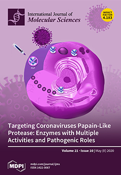
Cover Story (view full-size image): In the context of a cell infected by a coronavirus, a cartoon symbolizing a papain-like protease (PLP) is shown while interacting with other proteins to promote the infection and highlighted as a drug target. In our study, we review the cysteine protease, deubiquitinating and deISGylating activities of PLPs, involved in processing the viral polyprotein of the replication complex in its active elements and dysregulating the immune response of the host cell. Therefore, we also propose a list of PLP inhibitors, which may be a therapeutic option for coronavirus infections, including COVID-19. View this paper
  • Issues are regarded as officially published after their release is announced to the table of contents alert mailing list.
  • You may sign up for e-mail alerts to receive table of contents of newly released issues.
  • PDF is the official format for papers published in both, html and pdf forms. To view the papers in pdf format, click on the "PDF Full-text" link, and use the free Adobe Reader to open them.
Order results
Result details
Section
Select all
Export citation of selected articles as:
16 pages, 3421 KiB  
Article
Insights into the FMNAT Active Site of FAD Synthase: Aromaticity Is Essential for Flavin Binding and Catalysis
by Ana Serrano, Sonia Arilla-Luna and Milagros Medina
Int. J. Mol. Sci. 2020, 21(10), 3738; https://doi.org/10.3390/ijms21103738 - 25 May 2020
Cited by 6 | Viewed by 3282
Abstract
The last step in the biosynthesis of flavin adenine dinucleotide (FAD) is considered a target for the design of antimicrobial drugs because it is carried out by two non-homologous proteins in eukaryotic and prokaryotic organisms. Monofunctional FMN: adenylyltransferases (FMNAT) in Eukarya and FMNAT [...] Read more.
The last step in the biosynthesis of flavin adenine dinucleotide (FAD) is considered a target for the design of antimicrobial drugs because it is carried out by two non-homologous proteins in eukaryotic and prokaryotic organisms. Monofunctional FMN: adenylyltransferases (FMNAT) in Eukarya and FMNAT modules of bifunctional FAD synthases (FADS) in Prokarya belong to different structural families with dissimilar chemistry and binding modes for the substrates. In this study, we analyzed the relevance of the hydrophobic environment of the flavin isoalloxazine in the FMNAT active site of Corynebacterium ammoniagenes FADS (CaFADS) through the mutational analysis of its F62, Y106, and F128 residues. They form the isoalloxazine binding cavity and are highly conserved in the prokaryotic FADS family. The spectroscopic, steady-state kinetics and thermodynamic data presented indicate that distortion of aromaticity at the FMNAT isoalloxazine binding cavity prevents FMN and FAD from correct accommodation in their binding cavity and, as a consequence, decreases the efficiency of the FMNAT activity. Therefore, the side-chains of F62, Y106 and F128 are relevant in the formation of the catalytic competent complex during FMNAT catalysis in CaFADS. The introduced mutations also modulate the activity occurring at the riboflavin kinase (RFK) module of CaFADS, further evidencing the formation of quaternary assemblies during catalysis. Full article
(This article belongs to the Special Issue Flavin Adenine Dinucleotide (FAD): Biosynthesis and Function)
Show Figures

Graphical abstract

13 pages, 3081 KiB  
Article
(+)-Bornyl p-Coumarate Extracted from Stem of Piper betle Induced Apoptosis and Autophagy in Melanoma Cells
by Yu-Jen Wu, Tzu-Rong Su, Chi-I Chang, Chiy-Rong Chen, Kuo-Feng Hung and Cheng Liu
Int. J. Mol. Sci. 2020, 21(10), 3737; https://doi.org/10.3390/ijms21103737 - 25 May 2020
Cited by 13 | Viewed by 3585
Abstract
(+)-Bornyl p-coumarate is an active substance that is abundant in the Piper betle stem and has been shown to possess bioactivity against bacteria and a strong antioxidative effect. In the current study, we examined the actions of (+)-bornyl p-coumarate against A2058 [...] Read more.
(+)-Bornyl p-coumarate is an active substance that is abundant in the Piper betle stem and has been shown to possess bioactivity against bacteria and a strong antioxidative effect. In the current study, we examined the actions of (+)-bornyl p-coumarate against A2058 and A375 melanoma cells. The inhibition effects of (+)-bornyl p-coumarate on these cell lines were assessed by 3-(4,5-Dimethylthiazol-2-yl)-2,5- diphenyltetrazolium bromide (MTT) assay and the underlying mechanisms were identified by immunostaining, flow cytometry and western blotting of proteins associated with apoptosis and autophagy. Our results demonstrated that (+)-bornyl p-coumarate inhibited melanoma cell proliferation and caused loss of mitochondrial membrane potential, demonstrating treatment induced apoptosis. In addition, western blotting revealed that the process is mediated by caspase-dependent pathways, release of cytochrome C, activation of pro-apoptotic proteins (Bax, Bad and caspase-3/-9) and suppression of anti-apoptotic proteins (Bcl-2, Bcl-xl and Mcl-1). Also, the upregulated expressions of p-PERK, p-eIF2α, ATF4 and CCAAT/enhancer-binding protein (C/EBP)-homologous protein (CHOP) after treatment indicated that (+)-bornyl p-coumarate caused apoptosis via endoplasmic reticulum (ER) stress. Moreover, increased expressions of beclin-1, Atg3, Atg5, p62, LC3-I and LC3-II proteins and suppression by autophagic inhibitor 3-methyladenine (3-MA), indicated that (+)-bornyl p-coumarate triggered autophagy in the melanoma cells. In conclusion, our findings demonstrated that (+)-bornyl p-coumarate suppressed human melanoma cell growth and should be further investigated with regards to its potential use as a chemotherapy drug for the treatment of human melanoma. Full article
(This article belongs to the Section Molecular Toxicology)
Show Figures

Figure 1

16 pages, 2330 KiB  
Review
Connexin-Mediated Signaling at the Immunological Synapse
by Andrés Tittarelli, Mariela Navarrete, María Alejandra Gleisner, Peter Gebicke-Haerter and Flavio Salazar-Onfray
Int. J. Mol. Sci. 2020, 21(10), 3736; https://doi.org/10.3390/ijms21103736 - 25 May 2020
Cited by 21 | Viewed by 5680
Abstract
The immunological synapse (IS) is an intercellular communication platform, organized at the contact site of two adjacent cells, where at least one is an immune cell. Functional IS formation is fundamental for the modulation of the most relevant immune system activities, such as [...] Read more.
The immunological synapse (IS) is an intercellular communication platform, organized at the contact site of two adjacent cells, where at least one is an immune cell. Functional IS formation is fundamental for the modulation of the most relevant immune system activities, such as T cell activation by antigen presenting cells and T cell/natural killer (NK) cell-mediated target cell (infected or cancer) killing. Extensive evidence suggests that connexins, in particular connexin-43 (Cx43) hemichannels and/or gap junctions, regulate signaling events in different types of IS. Although the underlying mechanisms are not fully understood, the current evidence suggests that Cx43 channels could act as facilitators for calcium ions, cyclic adenosine monophosphate, and/or adenosine triphosphate uptake and/or release at the interface of interacting cells. These second messengers have relevant roles in the IS signaling during dendritic cell-mediated T and NK cell activation, regulatory T cell-mediated immune suppression, and cytotoxic T lymphocyte or NK cell-mediated target tumor cell killing. Additionally, as the cytoplasmic C-terminus domain of Cx43 interacts with a plethora of proteins, Cx43 may act as scaffolds for integration of various regulatory proteins at the IS, as suggested by the high number of Cx43-interacting proteins that translocate at these cell-cell interface domains. In this review, we provide an updated overview and analysis on the role and possible underlying mechanisms of Cx43 in IS signaling. Full article
(This article belongs to the Special Issue Signaling and Organelle Polarization at the Immunological Synapse)
Show Figures

Graphical abstract

16 pages, 2908 KiB  
Article
Oncogenic Potential of Bisphenol A and Common Environmental Contaminants in Human Mammary Epithelial Cells
by Vidhya A Nair, Satu Valo, Päivi Peltomäki, Khuloud Bajbouj and Wael M. Abdel-Rahman
Int. J. Mol. Sci. 2020, 21(10), 3735; https://doi.org/10.3390/ijms21103735 - 25 May 2020
Cited by 28 | Viewed by 4525
Abstract
There is an ample epidemiological evidence to support the role of environmental contaminants such as bisphenol A (BPA) in breast cancer development but the molecular mechanisms of their action are still not fully understood. Therefore, we sought to analyze the effects of three [...] Read more.
There is an ample epidemiological evidence to support the role of environmental contaminants such as bisphenol A (BPA) in breast cancer development but the molecular mechanisms of their action are still not fully understood. Therefore, we sought to analyze the effects of three common contaminants (BPA; 4-tert-octylphenol, OP; hexabromocyclododecane, HBCD) on mammary epithelial cell (HME1) and MCF7 breast cancer cell line. We also supplied some data on methoxychlor, MXC; 4-nonylphenol, NP; and 2-amino-1-methyl-6-phenylimidazo [4–b] pyridine, PhIP. We focused on testing the prolonged (two months) exposure to low nano-molar concentrations (0.0015–0.0048 nM) presumed to be oncogenic and found that they induced DNA damage (evidenced by upregulation of pH2A.X, pCHK1, pCHK2, p-P53) and disrupted the cell cycle. Some agents induced epigenetic (methylation) changes of tumor suppressor genes TIMP3, CHFR, ESR1, IGSF4, CDH13, and GSTP1. Obviously, the accumulation of these molecular alterations is an essential base for cancer development. Consistent with this, we observed that these agents increased cellular invasiveness through collagen. Cellular abilities to form colonies in soft agar were increased for MCF7. Toxic agents induced phosphorylation of protein kinase such as EGFR, CREB, STAT6, c-Jun, STAT3, HSP6, HSP27, AMPKα1, FAK, p53, GSK-3α/β, and P70S6 in HME1. Most of these proteins are involved in potential oncogenic pathways. Overall, these data clarify the molecular alterations that can be induced by some common environmental contaminants in mammary epithelial cells which could be a foundation to understand environmental carcinogenesis. Full article
Show Figures

Figure 1

34 pages, 4948 KiB  
Review
Adherent-Invasive E. coli: Update on the Lifestyle of a Troublemaker in Crohn’s Disease
by Mélissa Chervy, Nicolas Barnich and Jérémy Denizot
Int. J. Mol. Sci. 2020, 21(10), 3734; https://doi.org/10.3390/ijms21103734 - 25 May 2020
Cited by 65 | Viewed by 7856
Abstract
Besides genetic polymorphisms and environmental factors, the intestinal microbiota is an important factor in the etiology of Crohn’s disease (CD). Among microbiota alterations, a particular pathotype of Escherichia coli involved in the pathogenesis of CD abnormally colonizes the intestinal mucosa of patients: the [...] Read more.
Besides genetic polymorphisms and environmental factors, the intestinal microbiota is an important factor in the etiology of Crohn’s disease (CD). Among microbiota alterations, a particular pathotype of Escherichia coli involved in the pathogenesis of CD abnormally colonizes the intestinal mucosa of patients: the adherent-invasive Escherichia coli (AIEC) pathobiont bacteria, which have the abilities to adhere to and to invade intestinal epithelial cells (IECs), as well as to survive and replicate within macrophages. AIEC have been the subject of many studies in recent years to unveil some genes linked to AIEC virulence and to understand the impact of AIEC infection on the gut and consequently their involvement in CD. In this review, we describe the lifestyle of AIEC bacteria within the intestine, from the interaction with intestinal epithelial and immune cells with an emphasis on environmental and genetic factors favoring their implantation, to their lifestyle in the intestinal lumen. Finally, we discuss AIEC-targeting strategies such as the use of FimH antagonists, bacteriophages, or antibiotics, which could constitute therapeutic options to prevent and limit AIEC colonization in CD patients. Full article
(This article belongs to the Special Issue Molecular Mechanisms of Host-Pathogen Interaction)
Show Figures

Figure 1

20 pages, 3508 KiB  
Article
Oviductal Extracellular Vesicles Improve Post-Thaw Sperm Function in Red Wolves and Cheetahs
by Marcia de Almeida Monteiro Melo Ferraz, Jennifer Beth Nagashima, Michael James Noonan, Adrienne E. Crosier and Nucharin Songsasen
Int. J. Mol. Sci. 2020, 21(10), 3733; https://doi.org/10.3390/ijms21103733 - 25 May 2020
Cited by 24 | Viewed by 4927
Abstract
Artificial insemination (AI) is a valuable tool for ex situ wildlife conservation, allowing the re-infusion and dissemination of genetic material, even after death of the donor. However, the application of AI to species conservation is still limited, due mainly to the poor survival [...] Read more.
Artificial insemination (AI) is a valuable tool for ex situ wildlife conservation, allowing the re-infusion and dissemination of genetic material, even after death of the donor. However, the application of AI to species conservation is still limited, due mainly to the poor survival of cryopreserved sperm. Recent work demonstrated that oviductal extracellular vesicles (oEVs) improved cat sperm motility and reduced premature acrosomal exocytosis. Here, we build on these findings by describing the protein content of dog and cat oEVs and investigating whether the incubation of cryopreserved red wolf and cheetah sperm with oEVs during thawing improves sperm function. Both red wolf and cheetah sperm thawed with dog and cat oEVs, respectively, had more intact acrosomes than the non-EV controls. Moreover, red wolf sperm thawed in the presence of dog oEVs better maintained sperm motility over time (>15%) though such an improvement was not observed in cheetah sperm. Our work demonstrates that dog and cat oEVs carry proteins important for sperm function and improve post-thaw motility and/or acrosome integrity of red wolf and cheetah sperm in vitro. The findings show how oEVs can be a valuable tool for improving the success of AI with cryopreserved sperm in threatened species. Full article
(This article belongs to the Special Issue Extracellular Vesicles in Reproduction)
Show Figures

Figure 1

20 pages, 1742 KiB  
Review
Liquid Biopsy for the Diagnosis of Viral Hepatitis, Fatty Liver Steatosis, and Alcoholic Liver Diseases
by Ciniso Sylvester Shabangu, Jee-Fu Huang, Hui-Hua Hsiao, Ming-Lung Yu, Wan-Long Chuang and Shu-Chi Wang
Int. J. Mol. Sci. 2020, 21(10), 3732; https://doi.org/10.3390/ijms21103732 - 25 May 2020
Cited by 19 | Viewed by 4809
Abstract
During the progression from hepatitis to fibrosis, cirrhosis, and liver failure, the accumulation of stressed/damaged hepatocyte elements associated with liver inflammation is critical. The causes of hepatocyte injuries include viral hepatitis infections, alcoholic hepatitis, and non-alcoholic fatty liver disease. Hepatocyte-derived extracellular vesicles (Hep-EVs) [...] Read more.
During the progression from hepatitis to fibrosis, cirrhosis, and liver failure, the accumulation of stressed/damaged hepatocyte elements associated with liver inflammation is critical. The causes of hepatocyte injuries include viral hepatitis infections, alcoholic hepatitis, and non-alcoholic fatty liver disease. Hepatocyte-derived extracellular vesicles (Hep-EVs) released from stressed/damaged hepatocytes are partly responsible for liver disease progression and liver damage because they activate non-parenchymal cells and infiltrate inflammatory cells within the liver, which are in turn are an important source of EVs. This cell-to-cell signaling is prevalent during inflammation in many liver diseases. Accordingly, special emphasis should be placed on liquid biopsy methods for the long-term monitoring of chronic liver diseases. In the present review, we have highlighted various aspects of current liquid biopsy research into chronic liver diseases. We have also reviewed recent progress on liquid biopsies that focus on cell-free DNA (cfDNA), long non-coding RNA (lncRNA), and the proteins in EVs as potential diagnostic tools and novel therapeutic targets in patients with viral hepatitis, fatty liver steatosis, and alcoholic liver diseases. Full article
(This article belongs to the Section Molecular Pathology, Diagnostics, and Therapeutics)
Show Figures

Graphical abstract

14 pages, 2008 KiB  
Article
Mitochondrial pH Nanosensors for Metabolic Profiling of Breast Cancer Cell Lines
by Consuelo Ripoll, Mar Roldan, Rafael Contreras-Montoya, Juan J. Diaz-Mochon, Miguel Martin, Maria J. Ruedas-Rama and Angel Orte
Int. J. Mol. Sci. 2020, 21(10), 3731; https://doi.org/10.3390/ijms21103731 - 25 May 2020
Cited by 12 | Viewed by 3718
Abstract
The main role of mitochondria, as pivotal organelles for cellular metabolism, is the production of energy (ATP) through an oxidative phosphorylation system. During this process, the electron transport chain creates a proton gradient that drives the synthesis of ATP. One of the main [...] Read more.
The main role of mitochondria, as pivotal organelles for cellular metabolism, is the production of energy (ATP) through an oxidative phosphorylation system. During this process, the electron transport chain creates a proton gradient that drives the synthesis of ATP. One of the main features of tumoral cells is their altered metabolism, providing alternative routes to enhance proliferation and survival. Hence, it is of utmost importance to understand the relationship between mitochondrial pH, tumoral metabolism, and cancer. In this manuscript, we develop a highly specific nanosensor to accurately measure the intramitochondrial pH using fluorescence lifetime imaging microscopy (FLIM). Importantly, we have applied this nanosensor to establish differences that may be hallmarks of different metabolic pathways in breast cancer cell models, leading to the characterization of different metabophenotypes. Full article
Show Figures

Graphical abstract

37 pages, 1539 KiB  
Review
From Gene to Protein—How Bacterial Virulence Factors Manipulate Host Gene Expression During Infection
by Lea Denzer, Horst Schroten and Christian Schwerk
Int. J. Mol. Sci. 2020, 21(10), 3730; https://doi.org/10.3390/ijms21103730 - 25 May 2020
Cited by 33 | Viewed by 8528
Abstract
Bacteria evolved many strategies to survive and persist within host cells. Secretion of bacterial effectors enables bacteria not only to enter the host cell but also to manipulate host gene expression to circumvent clearance by the host immune response. Some effectors were also [...] Read more.
Bacteria evolved many strategies to survive and persist within host cells. Secretion of bacterial effectors enables bacteria not only to enter the host cell but also to manipulate host gene expression to circumvent clearance by the host immune response. Some effectors were also shown to evade the nucleus to manipulate epigenetic processes as well as transcription and mRNA procession and are therefore classified as nucleomodulins. Others were shown to interfere downstream with gene expression at the level of mRNA stability, favoring either mRNA stabilization or mRNA degradation, translation or protein stability, including mechanisms of protein activation and degradation. Finally, manipulation of innate immune signaling and nutrient supply creates a replicative niche that enables bacterial intracellular persistence and survival. In this review, we want to highlight the divergent strategies applied by intracellular bacteria to evade host immune responses through subversion of host gene expression via bacterial effectors. Since these virulence proteins mimic host cell enzymes or own novel enzymatic functions, characterizing their properties could help to understand the complex interactions between host and pathogen during infections. Additionally, these insights could propose potential targets for medical therapy. Full article
(This article belongs to the Special Issue Microbial Virulence Factors)
Show Figures

Figure 1

13 pages, 4212 KiB  
Article
A Novel Selective 11β-HSD1 Inhibitor, (E)-4-(2-(6-(2,6-Dichloro-4-(Trifluoromethyl)Phenyl)-4-Methyl-1,1-Dioxido-1,2,6-Thiadiazinan-2-yl)Acetamido)Adamantan-1-Carboxamide (KR-67607), Prevents BAC-Induced Dry Eye Syndrome
by Yoon-Ju Na, Kyoung Jin Choi, Won Hoon Jung, Sung Bum Park, Sein Kang, Jin Hee Ahn and Ki Young Kim
Int. J. Mol. Sci. 2020, 21(10), 3729; https://doi.org/10.3390/ijms21103729 - 25 May 2020
Cited by 8 | Viewed by 2849
Abstract
Dry eye syndrome is the most common eye disease and it is caused by various reasons. As the balance of the tear film that protects the eyes is broken due to various causes, it becomes impossible to properly protect the eyes. In this [...] Read more.
Dry eye syndrome is the most common eye disease and it is caused by various reasons. As the balance of the tear film that protects the eyes is broken due to various causes, it becomes impossible to properly protect the eyes. In this study, the protective effects and underlying mechanisms of topical (E)-4-(2-(6-(2,6-dichloro-4-(trifluoromethyl)phenyl)-4-methyl-1,1-dioxido-1,2,6-thiadiazinan-2-yl)acetamido)adamantan-1-carboxamide (KR-67607), a novel selective 11β-hydroxysteroid dehydrogenase 1 (11β-HSD1) inhibitor, were investigated in benzalkonium chloride (BAC)-induced dry eye syndrome. BAC-treated rat eyes induced significant increases in ocular surface damage, decreased corneal thickness, corneal basement membrane destruction in the conjunctival epithelium, and expression of pro-inflammatory cytokines tumor necrosis factor-α and 11β-HSD1. These effects of BAC were reversed by topical KR-67607 treatment. Furthermore, KR-67607 decreased 4-hydroxynonenal expression and increased antioxidant and mucus secretion in BAC-treated rat eyes. Taken together, a novel selective 11β-HSD1 inhibitor can prevent BAC-induced dry eye syndrome by inhibiting pro-inflammatory cytokine and reactive oxygen species expression via the inhibition of both 11β-HSD1 activity and expression. Full article
(This article belongs to the Section Biochemistry)
Show Figures

Graphical abstract

16 pages, 2805 KiB  
Article
Blood Oxidative Stress Modulates Alveolar Bone Loss in Chronically Stressed Rats
by Micaele Maria Lopes Castro, Priscila Cunha Nascimento, Deiweson Souza-Monteiro, Sávio Monteiro Santos, Mayra Barros Arouck, Vinicius Barreto Garcia, Raimundo Fernandes de Araújo, Jr., Aurigena Antunes de Araujo, Gabriela de Souza Balbinot, Fabrício Mezzomo Collares, Cassiano Kuchenbecker Rosing, Marta Chagas Monteiro, Cristiane Socorro Ferraz Maia and Rafael Rodrigues Lima
Int. J. Mol. Sci. 2020, 21(10), 3728; https://doi.org/10.3390/ijms21103728 - 25 May 2020
Cited by 10 | Viewed by 3519
Abstract
We aimed to investigate the effects of chronic stress (CS) on experimental periodontitis (EP) in rats. For this, 28 Wistar rats were divided into four groups: control, ligature-induced experimental periodontitis (EP), chronic stress (CS; by physical restraint model) and CS+EP (association of chronic [...] Read more.
We aimed to investigate the effects of chronic stress (CS) on experimental periodontitis (EP) in rats. For this, 28 Wistar rats were divided into four groups: control, ligature-induced experimental periodontitis (EP), chronic stress (CS; by physical restraint model) and CS+EP (association of chronic stress and ligature-induced periodontitis). The experimental period lasted 30 days, including exposure to CS every day and ligature was performed on the 15th experimental day. After 30 days, the animals were submitted to the behavioral test of the elevated plus maze (EPM). Next, rats were euthanized for blood and mandible collection in order to evaluate the oxidative biochemistry (by nitric oxide (NO), reduced-glutathione activity (GSH), and thiobarbituric acid reactive substance levels (TBARS)) and alveolar bone characterization (by morphometric, micro-CT, and immunohistochemistry), respectively. The behavioral parameters evaluated in EPM indicated higher anxiogenic activity in the CS and CS+EP, groups, which is a behavioral reflex of CS. The results showed that CS was able to change the blood oxidative biochemistry in CS and CS+EP groups, decrease GSH activity in the blood, and increase the NO and TBARS concentrations. Thus, CS induces oxidative blood imbalance, which can potentialize or generate morphological, structural, and metabolic damages to the alveolar bone. Full article
(This article belongs to the Special Issue Molecular Mechanisms of Periodontal Disease)
Show Figures

Figure 1

11 pages, 4336 KiB  
Communication
CRISPR-Cas9-Based Mutagenesis of the Mucormycosis-Causing Fungus Lichtheimia corymbifera
by Sandugash Ibragimova, Csilla Szebenyi, Rita Sinka, Elham I. Alzyoud, Mónika Homa, Csaba Vágvölgyi, Gábor Nagy and Tamás Papp
Int. J. Mol. Sci. 2020, 21(10), 3727; https://doi.org/10.3390/ijms21103727 - 25 May 2020
Cited by 10 | Viewed by 3753
Abstract
Lichtheimia corymbifera is considered as one of the most frequent agents of mucormycosis. The lack of efficient genetic manipulation tools hampers the characterization of the pathomechanisms and virulence factors of this opportunistic pathogenic fungus. Although such techniques have been described for certain species, [...] Read more.
Lichtheimia corymbifera is considered as one of the most frequent agents of mucormycosis. The lack of efficient genetic manipulation tools hampers the characterization of the pathomechanisms and virulence factors of this opportunistic pathogenic fungus. Although such techniques have been described for certain species, the performance of targeted mutagenesis and the construction of stable transformants have remained a great challenge in Mucorales fungi. In the present study, a plasmid-free CRISPR-Cas9 system was applied to carry out a targeted gene disruption in L. corymbifera. The described method is based on the non-homologous end-joining repair of the double-strand break caused by the Cas9 enzyme. Using this method, short, one-to-five nucleotide long-targeted deletions could be induced in the orotidine 5′-phosphate decarboxylase gene (pyrG) and, as a result, uracil auxotrophic strains were constructed. These strains are applicable as recipient strains in future gene manipulation studies. As we know, this is the first genetic modification of this clinically relevant fungus. Full article
(This article belongs to the Section Molecular Microbiology)
Show Figures

Figure 1

27 pages, 1301 KiB  
Review
Mechanisms of Apoptosis Resistance to NK Cell-Mediated Cytotoxicity in Cancer
by Christian Sordo-Bahamonde, Seila Lorenzo-Herrero, Ángel R. Payer, Segundo Gonzalez and Alejandro López-Soto
Int. J. Mol. Sci. 2020, 21(10), 3726; https://doi.org/10.3390/ijms21103726 - 25 May 2020
Cited by 65 | Viewed by 10909
Abstract
Natural killer (NK) cells are major contributors to immunosurveillance and control of tumor development by inducing apoptosis of malignant cells. Among the main mechanisms involved in NK cell-mediated cytotoxicity, the death receptor pathway and the release of granules containing perforin/granzymes stand out due [...] Read more.
Natural killer (NK) cells are major contributors to immunosurveillance and control of tumor development by inducing apoptosis of malignant cells. Among the main mechanisms involved in NK cell-mediated cytotoxicity, the death receptor pathway and the release of granules containing perforin/granzymes stand out due to their efficacy in eliminating tumor cells. However, accumulated evidence suggest a profound immune suppression in the context of tumor progression affecting effector cells, such as NK cells, leading to decreased cytotoxicity. This diminished capability, together with the development of resistance to apoptosis by cancer cells, favor the loss of immunogenicity and promote immunosuppression, thus partially inducing NK cell-mediated killing resistance. Altered expression patterns of pro- and anti-apoptotic proteins along with genetic background comprise the main mechanisms of resistance to NK cell-related apoptosis. Herein, we summarize the main effector cytotoxic mechanisms against tumor cells, as well as the major resistance strategies acquired by tumor cells that hamper the extrinsic and intrinsic apoptotic pathways related to NK cell-mediated killing. Full article
(This article belongs to the Special Issue Survival Pathways Involved in Resistance to Apoptosis in Cancer)
Show Figures

Figure 1

20 pages, 1760 KiB  
Review
Unravelling the Biology of Adult Cardiac Stem Cell-Derived Exosomes to Foster Endogenous Cardiac Regeneration and Repair
by Teresa Mancuso, Antonella Barone, Alessandro Salatino, Claudia Molinaro, Fabiola Marino, Mariangela Scalise, Michele Torella, Antonella De Angelis, Konrad Urbanek, Daniele Torella and Eleonora Cianflone
Int. J. Mol. Sci. 2020, 21(10), 3725; https://doi.org/10.3390/ijms21103725 - 25 May 2020
Cited by 26 | Viewed by 4914
Abstract
Cardiac remuscularization has been the stated goal of the field of regenerative cardiology since its inception. Along with the refreshment of lost and dysfunctional cardiac muscle cells, the field of cell therapy has expanded in scope encompassing also the potential of the injected [...] Read more.
Cardiac remuscularization has been the stated goal of the field of regenerative cardiology since its inception. Along with the refreshment of lost and dysfunctional cardiac muscle cells, the field of cell therapy has expanded in scope encompassing also the potential of the injected cells as cardioprotective and cardio-reparative agents for cardiovascular diseases. The latter has been the result of the findings that cell therapies so far tested in clinical trials exert their beneficial effects through paracrine mechanisms acting on the endogenous myocardial reparative/regenerative potential. The endogenous regenerative potential of the adult heart is still highly debated. While it has been widely accepted that adult cardiomyocytes (CMs) are renewed throughout life either in response to wear and tear and after injury, the rate and origin of this phenomenon are yet to be clarified. The adult heart harbors resident cardiac/stem progenitor cells (CSCs/CPCs), whose discovery and characterization were initially sufficient to explain CM renewal in response to physiological and pathological stresses, when also considering that adult CMs are terminally differentiated cells. The role of CSCs in CM formation in the adult heart has been however questioned by some recent genetic fate map studies, which have been proved to have serious limitations. Nevertheless, uncontested evidence shows that clonal CSCs are effective transplantable regenerative agents either for their direct myogenic differentiation and for their paracrine effects in the allogeneic setting. In particular, the paracrine potential of CSCs has been the focus of the recent investigation, whereby CSC-derived exosomes appear to harbor relevant regenerative and reparative signals underlying the beneficial effects of CSC transplantation. This review focuses on recent advances in our knowledge about the biological role of exosomes in heart tissue homeostasis and repair with the idea to use them as tools for new therapeutic biotechnologies for “cell-less” effective cardiac regeneration approaches. Full article
(This article belongs to the Section Molecular Biology)
Show Figures

Figure 1

18 pages, 4703 KiB  
Article
Studies on the Mechanisms of Anti-Inflammatory Activity of Heparin- and Hyaluronan-Containing Multilayer Coatings—Targeting NF-κB Signalling Pathway
by Hala Alkhoury, Adrian Hautmann, Bodo Fuhrmann, Frank Syrowatka, Frank Erdmann, Guoying Zhou, Sanja Stojanović, Stevo Najman and Thomas Groth
Int. J. Mol. Sci. 2020, 21(10), 3724; https://doi.org/10.3390/ijms21103724 - 25 May 2020
Cited by 18 | Viewed by 8004
Abstract
The use of implants can be hampered by chronic inflammatory reactions, which may result in failure of the implanted device. To prevent such an outcome, the present study examines the anti-inflammatory properties of surface coatings made of either hyaluronic acid (HA) or heparin [...] Read more.
The use of implants can be hampered by chronic inflammatory reactions, which may result in failure of the implanted device. To prevent such an outcome, the present study examines the anti-inflammatory properties of surface coatings made of either hyaluronic acid (HA) or heparin (Hep) in combination with chitosan (Chi) prepared as multilayers through the layer-by-layer (LbL) technique. The properties of glycosaminoglycan (GAG)-modified surfaces were characterized in terms of surface topography, thickness and wettability. Results showed a higher thickness and hydrophilicity after multilayer formation compared to poly (ethylene imine) control samples. Moreover, multilayers containing either HA or Hep dampened the inflammatory response visible by reduced adhesion, formation of multinucleated giant cells (MNGCs) and IL-1β release, which was studied using THP-1 derived macrophages. Furthermore, investigations regarding the mechanism of anti-inflammatory activity of GAG were focused on nuclear transcription factor-кB (NF-κB)-related signal transduction. Immunofluorescence staining of the p65 subunit of NF-κB and immunoblotting were performed that showed a significant decrease in NF-κB level in macrophages on GAG-based multilayers. Additionally, the association of FITC-labelled GAG was evaluated by confocal laser scanning microscopy and flow cytometry showing that macrophages were able to associate with and take up HA and Hep. Overall, the Hep-based multilayers demonstrated the most suppressive effect making this system most promising to control macrophage activation after implantation of medical devices. The results provide an insight on the anti-inflammatory effects of GAG not only based on their physicochemical properties, but also related to their mechanism of action toward NF-κB signal transduction. Full article
(This article belongs to the Collection Feature Papers in Materials Science)
Show Figures

Graphical abstract

30 pages, 3474 KiB  
Review
Fractalkine/CX3CL1 in Neoplastic Processes
by Jan Korbecki, Donata Simińska, Klaudyna Kojder, Szymon Grochans, Izabela Gutowska, Dariusz Chlubek and Irena Baranowska-Bosiacka
Int. J. Mol. Sci. 2020, 21(10), 3723; https://doi.org/10.3390/ijms21103723 - 25 May 2020
Cited by 72 | Viewed by 10620
Abstract
Fractalkine/CX3C chemokine ligand 1 (CX3CL1) is a chemokine involved in the anticancer function of lymphocytes—mainly NK cells, T cells and dendritic cells. Its increased levels in tumors improve the prognosis for cancer patients, although it is also associated with a poorer prognosis in [...] Read more.
Fractalkine/CX3C chemokine ligand 1 (CX3CL1) is a chemokine involved in the anticancer function of lymphocytes—mainly NK cells, T cells and dendritic cells. Its increased levels in tumors improve the prognosis for cancer patients, although it is also associated with a poorer prognosis in some types of cancers, such as pancreatic ductal adenocarcinoma. This work focuses on the ‘hallmarks of cancer’ involving CX3CL1 and its receptor CX3CR1. First, we describe signal transduction from CX3CR1 and the role of epidermal growth factor receptor (EGFR) in this process. Next, we present the role of CX3CL1 in the context of cancer, with the focus on angiogenesis, apoptosis resistance and migration and invasion of cancer cells. In particular, we discuss perineural invasion, spinal metastasis and bone metastasis of cancers such as breast cancer, pancreatic cancer and prostate cancer. We extensively discuss the importance of CX3CL1 in the interaction with different cells in the tumor niche: tumor-associated macrophages (TAM), myeloid-derived suppressor cells (MDSC) and microglia. We present the role of CX3CL1 in the development of active human cytomegalovirus (HCMV) infection in glioblastoma multiforme (GBM) brain tumors. Finally, we discuss the possible use of CX3CL1 in immunotherapy. Full article
(This article belongs to the Section Biochemistry)
Show Figures

Graphical abstract

12 pages, 3656 KiB  
Article
Early Detection of Pancreatic Intraepithelial Neoplasias (PanINs) in Transgenic Mouse Model by Hyperpolarized 13C Metabolic Magnetic Resonance Spectroscopy
by Prasanta Dutta, Susana Castro Pando, Marilina Mascaro, Erick Riquelme, Michelle Zoltan, Niki M. Zacharias, Seth T. Gammon, David Piwnica-Worms, Mark D. Pagel, Subrata Sen, Anirban Maitra, Shayan Shams, Florencia McAllister and Pratip K. Bhattacharya
Int. J. Mol. Sci. 2020, 21(10), 3722; https://doi.org/10.3390/ijms21103722 - 25 May 2020
Cited by 11 | Viewed by 3998
Abstract
While pancreatic cancer (PC) survival rates have recently shown modest improvement, the disease remains largely incurable. Early detection of pancreatic cancer may result in improved outcomes and therefore, methods for early detection of cancer, even premalignant lesions, may provide more favorable outcomes. Pancreatic [...] Read more.
While pancreatic cancer (PC) survival rates have recently shown modest improvement, the disease remains largely incurable. Early detection of pancreatic cancer may result in improved outcomes and therefore, methods for early detection of cancer, even premalignant lesions, may provide more favorable outcomes. Pancreatic intraepithelial neoplasias (PanINs) have been identified as premalignant precursor lesions to pancreatic cancer. However, conventional imaging methods used for screening high-risk populations do not have the sensitivity to detect PanINs. Here, we have employed hyperpolarized metabolic imaging in vivo and nuclear magnetic resonance (1H-NMR) metabolomics ex vivo to identify and understand metabolic changes, towards enabling detection of early PanINs and progression to advanced PanINs lesions that precede pancreatic cancer formation. Progression of disease from tissue containing predominantly low-grade PanINs to tissue with high-grade PanINs showed a decreasing alanine/lactate ratio from high-resolution NMR metabolomics ex vivo. Hyperpolarized magnetic resonance spectroscopy (HP-MRS) allows over 10,000-fold sensitivity enhancement relative to conventional magnetic resonance. Real-time HP-MRS was employed to measure non-invasively changes of alanine and lactate metabolites with disease progression and in control mice in vivo, following injection of hyperpolarized [1-13C] pyruvate. The alanine-to-lactate signal intensity ratio was found to decrease as the disease progressed from low-grade PanINs to high-grade PanINs. The biochemical changes of alanine transaminase (ALT) and lactate dehydrogenase (LDH) enzyme activity were assessed. These results demonstrate that there are significant alterations of ALT and LDH activities during the transformation from early to advanced PanINs lesions. Furthermore, we demonstrate that real-time conversion kinetic rate constants (kPA and kPL) can be used as metabolic imaging biomarkers of pancreatic premalignant lesions. Findings from this emerging HP-MRS technique can be translated to the clinic for detection of pancreatic premalignant lesion in high-risk populations. Full article
Show Figures

Figure 1

21 pages, 657 KiB  
Review
S-Adenosine Methionine (SAMe) and Valproic Acid (VPA) as Epigenetic Modulators: Special Emphasis on their Interactions Affecting Nervous Tissue during Pregnancy
by Asher Ornoy, Maria Becker, Liza Weinstein-Fudim and Zivanit Ergaz
Int. J. Mol. Sci. 2020, 21(10), 3721; https://doi.org/10.3390/ijms21103721 - 25 May 2020
Cited by 20 | Viewed by 5661
Abstract
S-adenosylmethionine (SAMe) is involved in many transmethylation reactions in most living organisms and is also required in the synthesis of several substances such as monoamine neurotransmitters and the N-methyl-D-aspartate (NMDA) receptor. Due to its important role as an epigenetic modulator, we discuss [...] Read more.
S-adenosylmethionine (SAMe) is involved in many transmethylation reactions in most living organisms and is also required in the synthesis of several substances such as monoamine neurotransmitters and the N-methyl-D-aspartate (NMDA) receptor. Due to its important role as an epigenetic modulator, we discuss in some length the process of DNA methylation and demethylation and the critical periods of epigenetic modifications in the embryo, fetus, and thereafter. We also discuss the effects of SAMe deficiency and the attempts to use SAMe for therapeutic purposes such as the treatment of major depressive disorder, Alzheimer disease, and other neuropsychiatric disorders. SAMe is an approved food additive and as such is also used during pregnancy. Yet, there seems to scanty data on the possible effects of SAMe on the developing embryo and fetus. Valproic acid (VPA) is a well-tolerated and effective antiepileptic drug that is also used as a mood stabilizer. Due to its high teratogenicity, it is contraindicated in pregnancy. A major mechanism of its action is histone deacetylase inhibition, and therefore, it acts as an epigenetic modulator, mainly on the brain. This prompted clinical trials using VPA for additional indications i.e., treating degenerative brain disease such as Alzheimer disease, dementia, HIV, and even cancer. Therefore, we discuss the possible effects of VPA and SAMe on the conceptus and early postnatally, during periods of susceptibility to epigenetic modifications. VPA is also used as an inducer of autistic-like behavior in rodents and was found by us to modify gene expression when administered during the first postnatal week but not when administered to the pregnant dams on day 12 of gestation. In contrast, SAMe modified gene expression when administered on day 12 of pregnancy but not postnatally. If administered together, VPA prevented the changes in gene expression induced by prenatal SAMe administration, and SAMe prevented the gene expression changes and autistic-like behavior induced by early postnatal VPA. It is concluded that both VPA and SAMe are powerful epigenetic modifiers with antagonistic actions on the brain that will probably be used in the future more extensively for the treatment of a variety of epigenetic diseases of the nervous system. Full article
(This article belongs to the Special Issue Psychoactive Substances in Neuronal Development)
Show Figures

Figure 1

18 pages, 2201 KiB  
Article
DNA Damage Regulates Senescence-Associated Extracellular Vesicle Release via the Ceramide Pathway to Prevent Excessive Inflammatory Responses
by Kazuhiro Hitomi, Ryo Okada, Tze Mun Loo, Kenichi Miyata, Asako J. Nakamura and Akiko Takahashi
Int. J. Mol. Sci. 2020, 21(10), 3720; https://doi.org/10.3390/ijms21103720 - 25 May 2020
Cited by 49 | Viewed by 6807
Abstract
DNA damage, caused by various oncogenic stresses, can induce cell death or cellular senescence as an important tumor suppressor mechanism. Senescent cells display the features of a senescence-associated secretory phenotype (SASP), secreting inflammatory proteins into surrounding tissues, and contributing to various age-related pathologies. [...] Read more.
DNA damage, caused by various oncogenic stresses, can induce cell death or cellular senescence as an important tumor suppressor mechanism. Senescent cells display the features of a senescence-associated secretory phenotype (SASP), secreting inflammatory proteins into surrounding tissues, and contributing to various age-related pathologies. In addition to this inflammatory protein secretion, the release of extracellular vesicles (EVs) is also upregulated in senescent cells. However, the molecular mechanism underlying this phenomenon remains unclear. Here, we show that DNA damage activates the ceramide synthetic pathway, via the downregulation of sphingomyelin synthase 2 (SMS2) and the upregulation of neutral sphingomyelinase 2 (nSMase2), leading to an increase in senescence-associated EV (SA-EV) biogenesis. The EV biogenesis pathway, together with the autophagy-mediated degradation pathway, functions to block apoptosis by removing cytoplasmic DNA fragments derived from chromosomal DNA or bacterial infections. Our data suggest that this SA-EV pathway may play a prominent role in cellular homeostasis, particularly in senescent cells. In summary, DNA damage provokes SA-EV release by activating the ceramide pathway to protect cells from excessive inflammatory responses. Full article
(This article belongs to the Special Issue Recognition of DNA Lesions)
Show Figures

Graphical abstract

35 pages, 1012 KiB  
Review
Mitochondrial Dysfunctions: A Red Thread across Neurodegenerative Diseases
by Serena Stanga, Anna Caretto, Marina Boido and Alessandro Vercelli
Int. J. Mol. Sci. 2020, 21(10), 3719; https://doi.org/10.3390/ijms21103719 - 25 May 2020
Cited by 65 | Viewed by 9185
Abstract
Mitochondria play a central role in a plethora of processes related to the maintenance of cellular homeostasis and genomic integrity. They contribute to preserving the optimal functioning of cells and protecting them from potential DNA damage which could result in mutations and disease. [...] Read more.
Mitochondria play a central role in a plethora of processes related to the maintenance of cellular homeostasis and genomic integrity. They contribute to preserving the optimal functioning of cells and protecting them from potential DNA damage which could result in mutations and disease. However, perturbations of the system due to senescence or environmental factors induce alterations of the physiological balance and lead to the impairment of mitochondrial functions. After the description of the crucial roles of mitochondria for cell survival and activity, the core of this review focuses on the “mitochondrial switch” which occurs at the onset of neuronal degeneration. We dissect the pathways related to mitochondrial dysfunctions which are shared among the most frequent or disabling neurodegenerative diseases such as Alzheimer’s, Parkinson’s, and Huntington’s, Amyotrophic Lateral Sclerosis, and Spinal Muscular Atrophy. Can mitochondrial dysfunctions (affecting their morphology and activities) represent the early event eliciting the shift towards pathological neurobiological processes? Can mitochondria represent a common target against neurodegeneration? We also review here the drugs that target mitochondria in neurodegenerative diseases. Full article
(This article belongs to the Special Issue Targeting Mitochondria in Aging and Disease)
Show Figures

Graphical abstract

15 pages, 1898 KiB  
Review
NTRK Fusions, from the Diagnostic Algorithm to Innovative Treatment in the Era of Precision Medicine
by Federica Zito Marino, Francesca Pagliuca, Andrea Ronchi, Immacolata Cozzolino, Marco Montella, Massimiliano Berretta, Maria Elena Errico, Vittoria Donofrio, Roberto Bianco and Renato Franco
Int. J. Mol. Sci. 2020, 21(10), 3718; https://doi.org/10.3390/ijms21103718 - 25 May 2020
Cited by 53 | Viewed by 8837
Abstract
In the era of precision medicine, the identification of several predictive biomarkers and the development of innovative therapies have dramatically increased the request of tests to identify specific targets on cytological or histological samples, revolutionizing the management of the tumoral biomaterials. The Food [...] Read more.
In the era of precision medicine, the identification of several predictive biomarkers and the development of innovative therapies have dramatically increased the request of tests to identify specific targets on cytological or histological samples, revolutionizing the management of the tumoral biomaterials. The Food and Drug Administration (FDA) has recently approved a selective neurotrophic tyrosine receptor kinase (NTRK) inhibitor, larotrectinib. Contemporarily, the development of multi-kinase inhibitors with activity in tumors carrying TRK fusions is ongoing. Chromosomal translocations involving the NTRK1, NTRK2, and NTRK3 genes result in constitutive activation and aberrant expression of TRK kinases in numerous cancer types. In this context, the identification of tumors harboring TRK fusions is crucial. Several methods of detection are currently available. We revise the advantages and disadvantages of different techniques used for identifying TRK alterations, including immunohistochemistry, fluorescence in situ hybridization, reverse transcriptase polymerase chain reaction, and next generation sequencing-based approaches. Finally, we propose a diagnostic algorithm based on histology and the relative frequency of TRK fusions in each specific tumor, considering also the economic feasibility in the clinical practice. Full article
(This article belongs to the Special Issue Tumor Biobanks at the Service of Precision Medicine)
Show Figures

Figure 1

6 pages, 1397 KiB  
Editorial
RNAs in Brain and Heart Diseases
by Dimitris Beis, Inga Zerr, Fabio Martelli, Wolfram Doehner and Yvan Devaux
Int. J. Mol. Sci. 2020, 21(10), 3717; https://doi.org/10.3390/ijms21103717 - 25 May 2020
Cited by 5 | Viewed by 3630
Abstract
In the era of single-cell analysis, one always has to keep in mind the systemic nature of various diseases and how these diseases could be optimally studied. Comorbidities of the heart in neurological diseases as well as of the brain in cardiovascular diseases [...] Read more.
In the era of single-cell analysis, one always has to keep in mind the systemic nature of various diseases and how these diseases could be optimally studied. Comorbidities of the heart in neurological diseases as well as of the brain in cardiovascular diseases are prevalent, but how interactions in the brain–heart axis affect disease development and progression has been poorly addressed. Several brain and heart diseases share common risk factors. A better understanding of the brain–heart interactions will provide better insights for future treatment and personalization of healthcare, for heart failure patients’ benefit notably. We review here emerging evidence that studying noncoding RNAs in the brain–heart axis could be pivotal in understanding these interactions. We also introduce the Special Issue of the International Journal of Molecular Sciences RNAs in Brain and Heart Diseases—EU-CardioRNA COST Action. Full article
(This article belongs to the Special Issue RNAs in Brain and Heart Diseases - EU-CardioRNA COST Action)
Show Figures

Figure 1

15 pages, 18709 KiB  
Article
Epigallocatechin 3-Gallate Has a Neuroprotective Effect in Retinas of Rabbits with Ischemia/Reperfusion through the Activation of Nrf2/HO-1
by Josué Rivera-Pérez, Martín Martínez-Rosas, César A. Conde-Castañón, Julia D. Toscano-Garibay, Nancy J. Ruiz-Pérez, Pedro L. Flores, Elvia Mera Jiménez and Javier Flores-Estrada
Int. J. Mol. Sci. 2020, 21(10), 3716; https://doi.org/10.3390/ijms21103716 - 25 May 2020
Cited by 21 | Viewed by 3823
Abstract
Retinal ischemia-reperfusion (rI/R) generates an oxidative condition causing the death of neuronal cells. Epigallocatechin 3-gallate (EGCG) has antioxidant and anti-inflammatory properties. Nonetheless, its correlation with the pathway of nuclear factor erythroid 2-related factor 2/heme oxygenase-1 (Nrf2/HO-1) for the protection of the retina is [...] Read more.
Retinal ischemia-reperfusion (rI/R) generates an oxidative condition causing the death of neuronal cells. Epigallocatechin 3-gallate (EGCG) has antioxidant and anti-inflammatory properties. Nonetheless, its correlation with the pathway of nuclear factor erythroid 2-related factor 2/heme oxygenase-1 (Nrf2/HO-1) for the protection of the retina is unknown. We aimed to evaluate the neuroprotective efficacy of single-doses of EGCG in rI/R and its association with Nrf2/Ho-1 expression. In albino rabbits, rI/R was induced and single-doses of EGCG in saline (0–30 mg/kg) were intravenously administered to select an optimal EGCG concentration that protects from retina damage. To reach this goal, retinal structural changes, gliosis by glial fibrillary acidic protein (GFAP) immunostaining, and lipid peroxidation level by TBARS (thiobarbituric acid reactive substance) assay were determined. EGCG in a dose of 15 mg/kg (E15) presented the lowest levels of histological damage, gliosis, and oxidative stress in the studied groups. To determine the neuroprotective efficacy of E15 in a timeline (6, 24, and 48 h after rI/R), and its association with the Nrf2/HO-1 pathway, the following assays were done by immunofluorescence: apoptosis (TUNEL assay), necrosis (high-mobility group box-1; HMGB1), Nrf2, and HO-1. In addition, the Ho-1 mRNA (qPCR) and lipid peroxidation levels were evaluated. E15 showed a protective effect during the first 6 h, compared to 24 and 48 h after rI/R, as revealed by a decrease in the levels of all damage markers. Nuclear translocation Nrf2 and HO-1 staining were increased, including Ho-1 mRNA levels. In conclusion, a single dose of E15 decreases the death of neuronal cells induced by oxidative stress during the first 6 h after rI/R. This protective effect is associated with the nuclear translocation of Nrf2 and with an elevation of Ho-1 expression. Full article
Show Figures

Figure 1

18 pages, 818 KiB  
Review
Animal Models to Translate Phage Therapy to Human Medicine
by Alessia Brix, Marco Cafora, Massimo Aureli and Anna Pistocchi
Int. J. Mol. Sci. 2020, 21(10), 3715; https://doi.org/10.3390/ijms21103715 - 25 May 2020
Cited by 33 | Viewed by 7366
Abstract
Phagotherapy, the use of bacteriophages to fight bacterial infections as an alternative to antibiotic treatments, has become of increasing interest in the last years. This is mainly due to the diffusion of multi-drug resistant (MDR) bacterial infections that constitute a serious issue for [...] Read more.
Phagotherapy, the use of bacteriophages to fight bacterial infections as an alternative to antibiotic treatments, has become of increasing interest in the last years. This is mainly due to the diffusion of multi-drug resistant (MDR) bacterial infections that constitute a serious issue for public health. Phage therapy is gaining favor due to its success in agriculture and veterinary treatments and its extensive utilization for human therapeutic protocols in the Eastern world. In the last decades, some clinical trials and compassionate treatments have also been performed in the Western world, indicating that phage therapy is getting closer to its introduction in standard therapy protocols. However, several questions concerning the use of phages in human therapeutic treatments are still present and need to be addressed. In this review, we illustrate the state of art of phage therapy and examine the role of animal models to translate these treatments to humans. Full article
(This article belongs to the Section Molecular Microbiology)
Show Figures

Figure 1

20 pages, 3116 KiB  
Article
An Evaluation of the In Vitro Roles and Mechanisms of Silibinin in Reducing Pyrazinamide- and Isoniazid-Induced Hepatocellular Damage
by Zhang-He Goh, Jie Kai Tee and Han Kiat Ho
Int. J. Mol. Sci. 2020, 21(10), 3714; https://doi.org/10.3390/ijms21103714 - 25 May 2020
Cited by 11 | Viewed by 4669
Abstract
Tuberculosis remains a significant infectious lung disease that affects millions of patients worldwide. Despite numerous existing drug regimens for tuberculosis, drug-induced liver injury is a major challenge that limits the effectiveness of these therapeutics. Two drugs that form the backbone of the commonly [...] Read more.
Tuberculosis remains a significant infectious lung disease that affects millions of patients worldwide. Despite numerous existing drug regimens for tuberculosis, drug-induced liver injury is a major challenge that limits the effectiveness of these therapeutics. Two drugs that form the backbone of the commonly administered quadruple antitubercular regimen, that is, pyrazinamide (PZA) and isoniazid (INH), are associated with such hepatotoxicity. Yet, we lack safe and effective alternatives to the antitubercular regimen. Consequently, current research largely focuses on exploiting the hepatoprotective effect of nutraceutical compounds as complementary therapy. Silibinin, a herbal product widely believed to protect against various liver diseases, potentially provides a useful solution given its hepatoprotective mechanisms. In our study, we identified silibinin’s role in mitigating PZA- and INH-induced hepatotoxicity and elucidated a deeper mechanistic understanding of silibinin’s hepatoprotective ability. Silibinin preserved the viability of human foetal hepatocyte line LO2 when co-administered with 80 mM INH and decreased apoptosis induced by a combination of 40 mM INH and 10 mM PZA by reducing oxidative damage to mitochondria, proteins, and lipids. Taken together, this proof-of-concept forms the rational basis for the further investigation of silibinin’s hepatoprotective effect in subsequent preclinical studies and clinical trials. Full article
(This article belongs to the Special Issue Molecular Mechanisms and Biomarkers in Drug-Induced Organ Injury)
Show Figures

Graphical abstract

16 pages, 2506 KiB  
Article
Role of the Ang2–Tie2 Axis in Vascular Damage Driven by High Glucose or Nucleoside Diphosphate Kinase B Deficiency
by Anupriya Chatterjee, Rachana Eshwaran, Hongpeng Huang, Di Zhao, Martina Schmidt, Thomas Wieland and Yuxi Feng
Int. J. Mol. Sci. 2020, 21(10), 3713; https://doi.org/10.3390/ijms21103713 - 25 May 2020
Cited by 6 | Viewed by 3658
Abstract
Ablation of nucleoside diphosphate kinase B (NDPK-B) in mice causes a breakdown of the neurovascular unit in the retina, mimicking diabetic retinopathy. The NDPK-B deficiency-induced vascular damage is mediated by excessive angiopoietin 2 (Ang2). Herein, the potential involvement of its receptor, Tie2, was [...] Read more.
Ablation of nucleoside diphosphate kinase B (NDPK-B) in mice causes a breakdown of the neurovascular unit in the retina, mimicking diabetic retinopathy. The NDPK-B deficiency-induced vascular damage is mediated by excessive angiopoietin 2 (Ang2). Herein, the potential involvement of its receptor, Tie2, was investigated. NDPK-B-deficient mouse retinas showed an upregulation of Tie2, specifically in the deep capillary layer. A similar upregulation of Tie2 was observed in cultured endothelial cells (ECs) from different origins upon NDPK-B depletion, whereas high glucose (HG) treatment did not alter Tie2 expression. Immunofluorescence staining and subcellular fractionation showed that the majority of Tie2 upregulation occurred at the plasma membrane. Similar to HG, however, NDPK-B depletion reduced Tie2 tyrosine phosphorylation. Compared to HG, a stronger increase of Ang2 was observed in NDPK-B depleted ECs. Treatment of ECs with soluble Tie2 or siRNA-mediated Tie2 knockdown attenuated NDPK-B depletion- but not HG-induced Ang2 upregulation. Like NDPK-B depletion, overexpression of recombinant Ang2 in ECs enhanced Ang2 secretion and concomitantly promoted the upregulation of Tie2. Thus, we identified a new mechanism showing that after reaching a threshold level of secretion, Ang2 sustains its own expression and secretion by a Tie2-dependent positive feedback loop. Full article
Show Figures

Figure 1

25 pages, 10473 KiB  
Review
Unravelling the Potential of Graphene Quantum Dots in Biomedicine and Neuroscience
by Giordano Perini, Valentina Palmieri, Gabriele Ciasca, Marco De Spirito and Massimiliano Papi
Int. J. Mol. Sci. 2020, 21(10), 3712; https://doi.org/10.3390/ijms21103712 - 25 May 2020
Cited by 85 | Viewed by 8539
Abstract
Quantum dots (QDs) are semiconducting nanoparticles that have been gaining ground in various applications, including the biomedical field, thanks to their unique optical properties. Recently, graphene quantum dots (GQDs) have earned attention in biomedicine and nanomedicine, thanks to their higher biocompatibility and low [...] Read more.
Quantum dots (QDs) are semiconducting nanoparticles that have been gaining ground in various applications, including the biomedical field, thanks to their unique optical properties. Recently, graphene quantum dots (GQDs) have earned attention in biomedicine and nanomedicine, thanks to their higher biocompatibility and low cytotoxicity compared to other QDs. GQDs share the optical properties of QD and have proven ability to cross the blood-brain barrier (BBB). For this reason, GQDs are now being employed to deepen our knowledge in neuroscience diagnostics and therapeutics. Their size and surface chemistry that ease the loading of chemotherapeutic drugs, makes them ideal drug delivery systems through the bloodstream, across the BBB, up to the brain. GQDs-based neuroimaging techniques and theranostic applications, such as photothermal and photodynamic therapy alone or in combination with chemotherapy, have been designed. In this review, optical properties and biocompatibility of GQDs will be described. Then, the ability of GQDs to overtake the BBB and reach the brain will be discussed. At last, applications of GQDs in bioimaging, photophysical therapies and drug delivery to the central nervous system will be considered, unraveling their potential in the neuroscientific field. Full article
(This article belongs to the Special Issue Graphene-Based Materials: Biological and Biomedical Applications)
Show Figures

Figure 1

13 pages, 1908 KiB  
Review
Emerging Roles of Estrogen-Regulated Enhancer and Long Non-Coding RNAs
by Melina J. Sedano, Alana L. Harrison, Mina Zilaie, Chandrima Das, Ramesh Choudhari, Enrique Ramos and Shrikanth S. Gadad
Int. J. Mol. Sci. 2020, 21(10), 3711; https://doi.org/10.3390/ijms21103711 - 25 May 2020
Cited by 16 | Viewed by 4925
Abstract
Genome-wide RNA sequencing has shown that only a small fraction of the human genome is transcribed into protein-coding mRNAs. While once thought to be “junk” DNA, recent findings indicate that the rest of the genome encodes many types of non-coding RNA molecules with [...] Read more.
Genome-wide RNA sequencing has shown that only a small fraction of the human genome is transcribed into protein-coding mRNAs. While once thought to be “junk” DNA, recent findings indicate that the rest of the genome encodes many types of non-coding RNA molecules with a myriad of functions still being determined. Among the non-coding RNAs, long non-coding RNAs (lncRNA) and enhancer RNAs (eRNA) are found to be most copious. While their exact biological functions and mechanisms of action are currently unknown, technologies such as next-generation RNA sequencing (RNA-seq) and global nuclear run-on sequencing (GRO-seq) have begun deciphering their expression patterns and biological significance. In addition to their identification, it has been shown that the expression of long non-coding RNAs and enhancer RNAs can vary due to spatial, temporal, developmental, or hormonal variations. In this review, we explore newly reported information on estrogen-regulated eRNAs and lncRNAs and their associated biological functions to help outline their markedly prominent roles in estrogen-dependent signaling. Full article
(This article belongs to the Collection Feature Papers in Molecular Genetics and Genomics)
Show Figures

Figure 1

16 pages, 1534 KiB  
Review
The Mammalian High Mobility Group Protein AT-Hook 2 (HMGA2): Biochemical and Biophysical Properties, and Its Association with Adipogenesis
by Linjia Su, Zifang Deng and Fenfei Leng
Int. J. Mol. Sci. 2020, 21(10), 3710; https://doi.org/10.3390/ijms21103710 - 25 May 2020
Cited by 21 | Viewed by 4648
Abstract
The mammalian high-mobility-group protein AT-hook 2 (HMGA2) is a small DNA-binding protein and consists of three “AT-hook” DNA-binding motifs and a negatively charged C-terminal motif. It is a multifunctional nuclear protein directly linked to obesity, human height, stem cell youth, human intelligence, and [...] Read more.
The mammalian high-mobility-group protein AT-hook 2 (HMGA2) is a small DNA-binding protein and consists of three “AT-hook” DNA-binding motifs and a negatively charged C-terminal motif. It is a multifunctional nuclear protein directly linked to obesity, human height, stem cell youth, human intelligence, and tumorigenesis. Biochemical and biophysical studies showed that HMGA2 is an intrinsically disordered protein (IDP) and could form homodimers in aqueous buffer solution. The “AT-hook” DNA-binding motifs specifically bind to the minor groove of AT-rich DNA sequences and induce DNA-bending. HMGA2 plays an important role in adipogenesis most likely through stimulating the proliferative expansion of preadipocytes and also through regulating the expression of transcriptional factor Peroxisome proliferator-activated receptor γ (PPARγ) at the clonal expansion step from preadipocytes to adipocytes. Current evidence suggests that a main function of HMGA2 is to maintain stemness and renewal capacity of stem cells by which HMGA2 binds to chromosome and lock chromosome into a specific state, to allow the human embryonic stem cells to maintain their stem cell potency. Due to the importance of HMGA2 in adipogenesis and tumorigenesis, HMGA2 is considered a potential therapeutic target for anticancer and anti-obesity drugs. Efforts are taken to identify inhibitors targeting HMGA2. Full article
(This article belongs to the Special Issue HMG Proteins in Development and Disease)
Show Figures

Graphical abstract

35 pages, 12204 KiB  
Article
Intrinsic Disorder in Tetratricopeptide Repeat Proteins
by Nathan W. Van Bibber, Cornelia Haerle, Roy Khalife, Bin Xue and Vladimir N. Uversky
Int. J. Mol. Sci. 2020, 21(10), 3709; https://doi.org/10.3390/ijms21103709 - 25 May 2020
Cited by 11 | Viewed by 4243
Abstract
Among the realm of repeat containing proteins that commonly serve as “scaffolds” promoting protein-protein interactions, there is a family of proteins containing between 2 and 20 tetratricopeptide repeats (TPRs), which are functional motifs consisting of 34 amino acids. The most distinguishing feature of [...] Read more.
Among the realm of repeat containing proteins that commonly serve as “scaffolds” promoting protein-protein interactions, there is a family of proteins containing between 2 and 20 tetratricopeptide repeats (TPRs), which are functional motifs consisting of 34 amino acids. The most distinguishing feature of TPR domains is their ability to stack continuously one upon the other, with these stacked repeats being able to affect interaction with binding partners either sequentially or in combination. It is known that many repeat-containing proteins are characterized by high levels of intrinsic disorder, and that many protein tandem repeats can be intrinsically disordered. Furthermore, it seems that TPR-containing proteins share many characteristics with hybrid proteins containing ordered domains and intrinsically disordered protein regions. However, there has not been a systematic analysis of the intrinsic disorder status of TPR proteins. To fill this gap, we analyzed 166 human TPR proteins to determine the degree to which proteins containing TPR motifs are affected by intrinsic disorder. Our analysis revealed that these proteins are characterized by different levels of intrinsic disorder and contain functional disordered regions that are utilized for protein-protein interactions and often serve as targets of various posttranslational modifications. Full article
(This article belongs to the Collection Feature Papers in Molecular Biophysics)
Show Figures

Figure 1

Previous Issue
Next Issue
Back to TopTop