Article

New Haloterpenes from the Marine Red Alga Laurencia papillosa: Structure Elucidation and Biological Activity

by
Mohamed Shaaban
1,2,*,
Ghada S. E. Abou-El-Wafa
3,
Christopher Golz
2 and
Hartmut Laatsch
2,*
1
National Research Centre, Chemistry of Natural Compounds Department, Division of Pharmaceutical Industries, El-Behoos St. 33, Dokki, Cairo 12622, Egypt
2
Institute of Organic and Biomolecular Chemistry, University of Göttingen, Tammannstrasse 2, D-37077 Göttingen, Germany
3
Department of Botany, Faculty of Science, Mansoura University, Algomhuria st. 60, El-Mansoura 35516, Egypt
*
Authors to whom correspondence should be addressed.
Mar. Drugs 2021, 19(1), 35; https://doi.org/10.3390/md19010035
Submission received: 12 December 2020 / Revised: 7 January 2021 / Accepted: 9 January 2021 / Published: 14 January 2021

Abstract

:
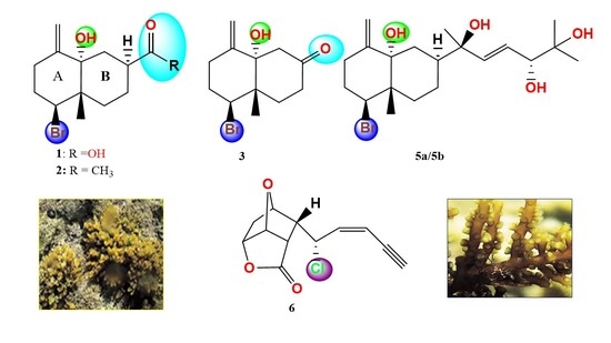
Analysis of the air-dried marine red alga Laurencia papillosa, collected near Ras-Bakr at the Suez gulf (Red Sea) in Egypt delivered five new halogenated terpene derivatives: aplysiolic acid (1), 7-acetyl-aplysiol (2), aplysiol-7-one (3), 11,14-dihydroaplysia-5,11,14,15-tetrol (5a), and a new maneonene derivative 6, named 5-epi-maneolactone. The chemical structures of these metabolites were characterized employing spectroscopic methods, and the relative and absolute configurations were determined by comparison of experimental and ab initio-calculated NMR, NOE, ECD, and ORD data, and by X-ray diffraction of 2 and 6. The antimicrobial activities of the crude extract and compounds 13, 5a and 6 were studied.

Graphical Abstract

1. Introduction

Among the red algae, the genus Laurencia is known to produce the largest number and diversity of secondary metabolites, making it the world’s chemically most complex seaweed genus [1]. Furthermore, several Laurencia metabolites have exhibited noteworthy antibacterial [2,3,4,5] insecticidal [3,6], antifungal [7], and antiviral activity [8], in addition to their anti-inflammatory, antiproliferative, antifouling, antifeedant, cytotoxic, ichthyotoxic, and insecticidal properties [9]. Certain Laurencia species have shown to be an unprecedented source of halogenated secondary metabolites, predominantly sesquiterpenes [10], diterpenes [11], and C15 non-terpenoids, including halogenated cyclic enyne ethers and related allenes [12,13,14]. An example is the halogenated diterpene aplysiadiol (4), which was obtained from the marine mollusk Aplysia kurodai [15] and a Malaysian Laurencia species, and exhibited potent antibacterial activities [16].
In the course of our research to isolate and investigate bioactive metabolites from Laurencia algae collected from the shallow waters of the Red Sea on Egyptian coasts, a series of four halogenated bioactive decalin derivatives was isolated, namely aplysiolic acid (1), 7-acetyl-aplysiol (2), aplysiol-7-one (3), 11,14-dihydroaplysia-5,11,14,15-tetrol (5a), and a further chloro compound 6 (Figure 1). We named the latter 5-epi-maneolactone (6), because of the similarity with the maneonenes, bioactive C15 acetogenins from L. obtusa [17,18]. Additionally, thyrsiferol [19], 10-hydroxykahukuene B [20], cholesterol, hexadecanoic acid, thymine, and uracil were gained from the dichloromethane-methanol extract of L. papillosa. The chemical structures of these compounds were determined by extensive 1D and 2D NMR and ESI HR mass measurements. Their relative and absolute configurations were derived by 1D and 2D NMR measurements, from ab initio calculations of ECD, ORD, 13C NMR data, by geometry-derived NOE predictions, and by X-ray diffraction in case of 2 and 6. The antimicrobial activity of the crude extract and of the new compounds was studied using a set of diverse microorganisms.

2. Results and Discussion

2.1. Working up and Structure Identification

The dichloromethane-methanol extract of air-dried L. papillosa algae was fractionated by a series of chromatographic purification steps implying silica gel and size exclusion columns, to afford the haloterpenes 13, 5ac, and 6 (Figure 1), and additionally thyrsiferol [19], 10-hydroxykahukuene B [20], cholesterol, hexadecanoic acid, thymine, and uracil. The physico-chemical properties of the new compounds are listed in Table 1.

2.1.1. Aplysiolic Acid

Compound 1 was isolated as an optically active colorless solid, exhibiting neither UV-absorbance nor fluorescence on silica gel. On TLC, it was detected as a pink spot after spraying with anisaldehyde/sulfuric acid, tentatively pointing to a terpenoidal moiety. The (−)-ESI mass spectrum showed [M − H] ion signals of nearly equal intensities at m/z 301 and 303 Dalton, and a triple ion-peak at m/z 605 [2M − H], indicating the presence of one bromine atom. The (−)-ESI HR mass spectrum suggested the molecular formula C13H19BrO3, indicating four double bond equivalents (DBE) (Table 1).
The 13C NMR spectrum (Table 2) showed a total of 13 resonances, amongst them ten in the aliphatic, two in the olefinic, and one in the carbonyl region. The 1H NMR pattern exhibited two singlets at δ 4.90 and 4.81, which were assigned by H,H COSY and HMQC spectra as exo-methylene protons (δC 110.2). The respective quaternary carbon signal appeared at δ 148.5 and was assigned by HMBC correlations (Figure 2). The carbonyl group, together with one methyl, six methylene, and two methine groups and three fully substituted carbons, sums up to C13H17BrO. Respectively, 1 should contain two rings, and the remaining two hydrogen and oxygen atoms are forming hydroxyl groups.
Based on the H,H COSY correlations, two fragments were identified in 1, a 1,1,3-trisubstituted propane (-CH2-CH2-CH-), and a 1,2,4-trisubstituted butane (-CH2-CH-CH2-CH2-) unit, respectively. The terminal methylene protons in the first fragment (δ 2.70, 2.15) showed 2J and 3J HMBC correlations with the olefinic carbons C-4 and C-13, confirming their direct neighborhood. One of these methylene protons (δ 2.70) showed a 4J COSY signal with the exo-methylene protons. The methine proton H-1 (δ 4.72) exhibited 2J and 3J correlations towards C-10 (δ 43.0) and the methyl singlet of C-12 (δ 14.8), respectively, suggesting a connection of C-10 with C-1 and Me-12. If C-5 is hydroxylated (δ 76.2), the downfield shift of C-1 (δ 62.9) fits best on its substitution with bromine [21]. Among others, the methylene group CH2-3 showed a 3J HMBC correlation towards C-5, which itself gave cross signals with the methylene protons H2-13 and Me-12, so that an exomethylene-cyclohexane (ring A) was formed.
Further HMBC and NOESY correlations (Figure 2 and Figure 3) indicated that the butanediyl fragment formed a second cyclohexane (ring B) via C-10 (δ 43.0) and C-5 (δ 76.2). CH2-9 correlated with C-10 and Me-12 and showed all other expected HMBC and NOESY correlations. In a similar way, the ring closure between CH2-6 and C-5 was confirmed. According to 2J and 3J HMBC correlations from Hax,β-6 (δ 2.04), Hα-7 (δ 2.94), and Hax,β-8 (δ 1.66, weak) with C-11 (δ 181.0), the missing atoms are forming a carboxyl group at C-7.
Due to the low flexibility of 1, its NOESY signals were surprisingly clear and easy to interpret. Me-12 showed correlations with Hax,β-2, Hax,β -6, Hax,β-8, and Heq,β-9 but not with H-1, Hα-2, Hα-6, Hα-7, and Hα-8, so that all correlating protons should be in a syn-facial position with the methyl group. The NOE between H-7 and Heq,α-6, Heq,α-8, Hax,α-9, and of H-1 with Heq,α-2, Hax,α-3, Hax,α-9, but not with Me-12, indicated their anti-position with respect to Me-12, resulting in the rel-(1S,7S,10S)-configuration. DFT-calculations of the 1-geometry showed that the strong NOE effect between (Z)-H-13 (δ 4.81) and H2-6 (δ 2.04, 1.83), and between Hβ-6 (2.04) and Me-12 (0.96) is not explainable by a cis-decalin, but requires definitely a trans-decalin with a rel-(5S,10S) configuration (see Figures S2 and S3). As the sign of the ORD data calculated for all-(S)-1 agreed with the experimental data, the decalin was elucidated as 5-bromo-8a-hydroxy-4a-methyl-8-methylene-decahydronaphthalene-2-carboxylic acid (1), having the absolute all-(S)-configuration. We named it aplysiolic acid, with respect to the similarity with aplysiadiol (4) [15]. For aplysiadiol (4) and anhydroaplysiadiol [21], the same relative configuration as for 1 was published, and we confirmed their absolute configuration by agreement of calculated and published negative sign of their optical rotation as all-(S) as well (Table S4).

2.1.2. 7-Acetyl-aplysiol

An additional optically active colorless solid had similar chromatographic properties as 1, but with noticeably less polarity. The ESI HR mass spectrum indicated the molecular formula C14H21BrO2 with four double bond equivalents (DBE) as well. However, an OH group in 1 was formally exchanged against a methyl group. Respectively, a methyl singlet (δH 2.13) and its corresponding carbon (28.3) were visible. The carboxyl signal in 1 (181.0) was exchanged in 2 against a keto carbonyl at δC 211.8, but the remaining 1H and 13C NMR signals were very similar in both compounds. On the basis of long-range 2D NMR couplings (Figure 2) and NOESY experiments (Figure 4), the structure was assigned as 2, which we named 7-acetyl-aplysiol. From this ketone, the acid 1 may be formed in a haloform reaction (Einhorn reaction). With respect to the negative optical rotation, the decalin system of 2 should have the same absolute all-(S)-configuration as 1, which also agrees with biosynthetic considerations. However, as some doubt remained after comparison of the calculated and experimental ECD spectra, a crystal of 2 was analyzed additionally by X-ray diffraction (Figure S6), which reassured the all-(S)-configuration.

2.1.3. Aplysiol-7-one

A third closely related decalin gave the molecular formula C12H17BrO2 by ESI HR, containing 4 DBE as well. The 1H and 13C NMR spectra of 3 resembled those of 1 and 2; however, the signals of CH-7 (in 1 and 2) and CH3-14 (in 2) were absent, and the ABX signal of CH2-6 in 1 and 2 had changed into an AB signal. As indicated by the HMBC correlations and the downfield shifts of CH2-6 and CH2-8, the carbonyl signal at δC 209.0 was due to a ß-decalone. Furthermore, long-range 2D NMR couplings (Figure 2) confirmed structure 3, which we named as aplysiol-7-one. In addition, the all-(S)-configuration was plausible for NOESY experiments (Figure 4) and biosynthetic reasons; it was, however, not further analyzed due to an inseparable contamination by 10-hydroxykahukuene B [20].

2.1.4. 11,14-Dihydroaplysia-5,11,14,15-tetrols

A further brominated terpenoide with moderate polarity was isolated as an optically active resin with color reactions similar as of 1 (Table 1). The NMR spectrum (Table 3) of this compound showed 20 13C signals. At high magnification, however, each of them appeared as a group of up to four signals in distances of <0.2 ppm (Figures S34 and S35), so that a mixture of four stereoisomers or otherwise closely related compounds was expected; this was confirmed by the 1H NMR signal of the Δ12 double bond (Figure S11) and also by analytical HPLC analyses, where three components in the ratio of ~1:1:1 were separated. By HPLC/HRMS, two stereoisomers C20H33BrO4 (5a,b) and a slightly more polar component C20H33BrO5 (5c) were detected (Figures S12 and S13). The latter compound seems to be a 11,14-dihydroaplysiapentol; we need, however, to isolate further material to fully elucidate the structure (see Supporting Information).
Both molecular formulas indicated four DBE (Table 1), and the COSY, HSQC, HMBC (Figure 2), and NOESY (Figure S15) data confirmed the same brominated rel-(1S,5S,7S,10S)-exomethylene-decalin skeleton as in 13. However, instead of the carboxy or the acetyl group, respectively, a 6-methyl-hept-3-ene-2,5,6-triol-2-yl side chain was found: the singlet of Me-18 (δH 1.27) displayed HMBC correlations with C-7, C-11 (δC 75.4), and the olefinic carbon C-12 (δC 139.8). Further COSY and HMBC correlations completed the chain from CH-7 to Me-16/Me-17. Accordingly, structures 5a/b were elucidated as further new aplysiadiol derivatives [15] and named, respectively, as 11,14-dihydroaplysia-5,11,14,15-tetrols. Also here, the same absolute configuration as in 13 was assumed for the decalin system for biosynthetic reasons, so that only the stereochemistry of the side chain remained open. On this basis, the absolute configuration of the main component 5a was determined as (1S,5S,7S,10S,11R,14R) by correlation of NOE signals with ab-initio calculated atom distances (Table S6 and Figure S7); the other diastereomers could not be assigned, due to overlapping signals.
Aplysiadiol derivatives are biosynthetically regarded as extended sesquiterpenes; they represent examples of the rare prenylated eudesmane type, which commonly occur in terrestrial plants [22]. A few compounds of this type have been isolated from marine mollusks [15], brown algae [23], and soft corals [24,25,26].

2.1.5. 5-epi-Maneolactone

Compound 6 displayed neither UV absorbance nor fluorescence on TLC, but turned brownish gray with anisaldehyde/sulfuric acid on heating. The molecular weight was determined by ESI MS: two quasi-molecular ion peaks in the ratio of 1:0.33 at m/z 238 and 240 indicated the presence of one chlorine atom. The corresponding molecular formula C12H11ClO3 was determined by ESI HRMS and indicated seven DBE (Table 1). The IR spectrum of 6 displayed a characteristic vibration signal (ν = 2361 cm−1) of an acetylenic bond. Two absorption bands at ν =1781 and 1592 cm−1 indicated the presence of a lactone carbonyl and an olefinic double bond, respectively, so that three rings were expected.
The 1H NMR methine signal at δH 3.36 did not show an HSQC correlation; however, a large HMBC coupling with the CH carbon at δC 86.5 and a smaller one with a Cq at δC 78.2 was noted. Together with strong long-range correlations with a cis-double bond (δC 113.8, 139.2, J = 10.5 Hz), this indicated a terminal acetylene in conjugation with a double bond. According to further HMBC and COSY correlations, the (Z)-enyne unit was connected with the methines C-5 and C-6, and the latter additionally with CH-7 and CH-11 (Figure 5). A detailed analysis resulted in a ((Z)-pent-2-en-4-ynyl)-cyclohexane, where the shift assignments of positions CH-7, -9, and -10 remained open due to their overlapping 1H NMR signals (Table 4).
According to AntiBase [27], only two groups of metabolites with these structural features have been described before, the lembynes [2,28], and the maneonenes [17,18]. All were isolated from Laurencia spp. If chlorine was present, it was found in position C-5; also the shifts of 6 were explained best by chlorine at C-5. HMBC correlations of the upfield methine protons CH-6 and CH-11 with the signal at δC 173.5 connected the ester carbonyl C-12 with C-11, and the correlation with the oxymethine CH-9 (or CH-7) via oxygen closed a lactone ring with C-12. The remaining oxygen atom bridges the oxymethine groups CH-10 with CH-7 or CH-9, so that all DBE were used. Further correlation analyses by means of COCON [29] delivered four structure options, with structure 6 being the only plausible one (Figure 5 and Figure S16). A suitable crystal allowed us finally to confirm this structure including the absolute configuration unambiguously by X-ray diffraction (Figure 6). DFT calculations afforded the same absolute configuration, so that the validity of the computational methods applied here were additionally confirmed (see Supporting Information). We named compound 6 as 5-epi-maneolactone, as it looks like an oxidative cleavage product of the cis-maneonenes. It should be mentioned, however, that C-5 is (R)-configured in these compounds, oppositely to the (5S)-configured 6.

2.2. Biological Activities

Antimicrobial activity testing of the crude extract of the red alga L. papillosa was carried out against ten microorganisms using the agar diffusion technique with paper platelets: Bacillus subtilis ATCC6051, Staphylococcus aureus, Streptomyces viridochromogenes Tü 57, Streptococcus pyogenes, Escherichia coli, Shigella sp., Proteus sp., Candida albicans, Mucor miehei Tü 284, and Chlorella vulgaris. The crude extract showed at 400 μg/disk a strong antibacterial activity against the Gram-positive Streptomyces viridochromogenes Tü 57 (30 mm). Among the tested compounds 13, 5, and 6, aplysiolic acid (1) and aplysiol-7-one (3) were only moderately active against S. aureus (10.5 mm) at a concentration of 40 μg/disk.

3. Materials and Methods

3.1. General Procedures

IR spectra: FT/IR-4100 Infrared Spectrometer (Jasco, Inc., Easton, MD, USA). NMR spectra were measured with Varian Unity 300 and Varian Inova 600 spectrometers (Varian, Palo Alto, CA, USA). Optical rotations: Polarimeter (Perkin-Elmer, model 343, Perkin-Elmer Life and Analytical Sciences, Shelton, CT, USA). Electron spray ionization mass spectrometry (ESI MS): Finnigan LCQ ion trap mass spectrometer coupled with a Flux Instruments (Basel, Switzerland) quaternary pump Rheos 4000 and an HP 1100 HPLC (Nucleosil column EC 125/2, 100-5, C 18) with autosampler (Jasco 851-AS, Jasco, Inc., Easton, MD, USA) and a diode array detector (Finnigan Surveyor LC System, San Jose, CA, USA). High-resolution mass spectra (HRMS) were recorded by ESI MS on a MicrOTOF mass spectrometer (Bruker Daltonics, Billerica, MA, USA) or a LTQ Orbitrap XL (Thermo Fisher Scientific wissenschaftliche Geräte GmbH, 20354-Hamburg, Germany), equipped with an HPLC instrument (Thermo Scientific Accela HPLC System Markham, Ontario, Canada); for further details, see mass spectra in Supporting Information, e.g., Figures S12 and S17. Rf values are listed in Table 1 and were determined on Polygram SIL G/UV254 (Macherey & Nagel, Düren, Germany). Size exclusion chromatography was performed on Sephadex LH-20 (Lipophilic Sephadex; Amersham Biosciences, Ltd., purchased from Sigma-Aldrich Chemie, Steinheim, Germany).

3.2. Collection and Taxonomy of the Marine Alga

The red alga L. papillosa (Forsk., Grev) was collected in summer 2009 at Ras Abu-Bakr, 65 km north of Ras Gharib on the Suez-Gulf, Red Sea, Egypt. The identification was carried out by G. S. Abou-El Wafa according to Nasr’s method [30,31]. A reference specimen of the alga is kept at the Department of Botany, Faculty of Science, Mansoura University, Egypt. Samples of L. papillosa were separated from epiphytes and the algal material rinsed in tap water and distilled water. The sample was then spread on string nets, allowed to dry in air, ground, and stored in closed bottles at room temperature.

3.3. Extraction and Isolation of the Bioactive Constituents

The air-dried algal sample (~850 g) was extracted at room temperature with dichloromethane/methanol (1:1, 48 h for each batch). The extract was concentrated under reduced pressure to gain 8.83 g of residue, which was applied to column chromatography on silica gel (SC) using a gradient elution starting first with cyclohexane, then cyclohexane-CH2Cl2 and finally with CH2Cl2-MeOH. By TLC monitoring (UV light 254 and 366 nm, Merck, 64579 Gernsheim, Germany) and by using anisaldehyde/sulfuric acid spray reagent, five fractions were obtained: I (1.73 g), II (0.32 g), III (0.73 g), IV (1.86 g), and V (1.92 g). Further SC (silica gel, cyclohexane-CH2Cl2) of fraction I afforded cholesterol (46 mg) and hexadecanoic acid (66.7 mg) as colorless solids. Purification of fraction II (SC on silica gel eluted with a cyclohexane/DCM/MeOH gradient and subsequently Sephadex LH20 (CH2Cl2/40% MeOH) afforded compound 5 as a pale-yellow oil (165 mg) and 6 as a colorless solid (6 mg), respectively. SC of fraction IV on silica gel with a DCM/MeOH gradient and subsequently Sephadex LH20 (CH2Cl2/40% MeOH)) resulted in 7-acetyl-aplysiol (2, 19.4 mg), aplysiol-7-one (3, 4.8 mg) and 10-hydroxykahukuene B (5 mg) as colorless solids. SC of fraction V on silica gel (CH2Cl2/MeOH), followed by PTLC (DCM/10% MeOH) and Sephadex LH20 (MeOH) delivered aplysiolic acid (1, 1.4 mg), thyrsiferol (1.5 mg), uracil (1 mg), and thymine (1 mg) as colorless solids. Detailed 2D NMR correlations (H,H COSY and HMBC) of compounds 13, 5a and 6 have been given in the Supplementary Data file, Figures S5–S55.

3.4. Antimicrobial Assay

Antimicrobial assays using the agar diffusion test [32] were performed as described previously [33]. M. miehei Tü 284 and S. viridochromogenes Tü 57 were obtained from the collection of H. Zähner (University of Tübingen, Germany), and Chlorella vulgaris was provided by the Algal Collection Göttingen. B. subtilis ATCC 6051 was obtained from the American Type Culture Collection, while S. aureus, E. coli and C. albicans are clinical isolates from Göttingen hospitals. Strains are kept in the strain collection of H. Laatsch, Institute of Organic and Biomolecular Chemistry, Georg-August University, Göttingen, Germany.

3.5. Ab Initio Calculations

DFT calculations were performed as described previously [34].

3.6. Crystal Structure Determination of 7-acetylaplysiol (2) and 5-epi-maneolactone (6)

The structures of 2 and 6 were determined by single-crystal X-ray diffraction on two dual source equipped Bruker D8 Venture four-circle-diffractometer (Bruker AXS GmbH, Karlsruhe, Germany). 7-Acetylaplysiol (2), C14H21BrO2, was crystallized from methanol as yellow plates in the orthorhombic crystal system in the non-centrosymmetric cpase group P212121; 5-epi-maneolactone (6), C12H11ClO3, was crystallized from CHCl3/20% MeOH as colorless blocks in the monoclinic crystal system in the non-centrosymmetric space group P21. Absolute configuration could be determined reliably for both compounds with Flack’s parameter of 0.001(5) and −0.014(7) for 7-acetylaplysiol (2) and 5-epi-maneolactone (6), respectively. Full crystallographic information can be retrieved from the CIF file and the Supporting Information. CCDC 2041564 (2) and 2008525 (6) contain the supplementary crystallographic data for this paper. These data can be obtained free of charge from The Cambridge Crystallographic Data Centre via www.ccdc.cam.ac.uk/data request/cif. For further data, refer to the Supporting Information.

4. Conclusions

Six haloterpenes, namely aplysiolic acid (1), 7-acetyl-aplysiol (2), aplysiol-7-one (3), 11,14-dihydroaplysia-5,11,14,15-tetrol (5a), the epimer 5b and 5-epi-maneolactone (6), along with thyrsiferol, 10-hydroxykahukuene B, cholesterol, hexadecanoic acid, thymine, and uracil were isolated from the marine red alga L. papillosa. The chemical structures of the new metabolites were characterized by employing spectroscopic methods (1D, 2D NMR, and ESI HR mass measurements). The relative and absolute configurations of the new compounds were determined by ab initio calculations of ECD, ORD, and NMR data, and for 2 and 6, additionally by X-ray diffraction. In a set of microorganisms, the crude extract was strongly active against Streptomyces viridochromogenes Tü 57 (30 mm), whereas the activity of the pure metabolites was low.

Supplementary Materials

NMR spectra and other supplementary data associated with this article can be found in the online version at https://www.mdpi.com/1660-3397/19/1/35/s1.

Author Contributions

Conceptualization, M.S., G.S.E.A.-E.-W. and H.L.; methodology, G.S.E.A.-E.-W. and M.S.; validation, M.S. and H.L.; formal analysis, M.S., H.L. and C.G.; investigation, M.S. and H.L.; resources, G.S.E.A.-E.-W.; data curation, M.S. and H.L.; writing—Original draft preparation, M.S. and H.L.; writing—Review and editing, M.S. and H.L.; visualization, M.S., C.G. and H.L.; supervision, M.S.; project administration, M.S.; funding acquisition, M.S.; All authors have read and agreed to the published version of the manuscript.

Funding

M. Shaaban thanks the German Academic Exchange Service (DAAD) for a short-term fellowship (2015–2016).

Institutional Review Board Statement

Not applicable.

Informed Consent Statement

Not applicable.

Data Availability Statement

Further data are available on request from the corresponding authors.

Acknowledgments

The authors are thankful to Dr. H. Frauendorf (http://zentrale-analytik.chemie.uni-goettingen.de/index.htm) and Dr. M. John (https://www.uni-goettingen.de/de/nmr-abteilung/18904.html) for the mass and NMR spectra, and to Prof. Dr. Manuel Alcarazo (IOBC, Göttingen, https://www.uni-goettingen.de/de/prof.+dr.+manuel+alcarazo/522472.html) for providing his X-ray facilities; the diffractometer was kindly made available by the German Research Foundation (DFG, registration number INST 186/1237-1). We thank F. Lissy for biological activity tests.

Conflicts of Interest

The authors declare no conflict of interest.

Author Statement

In memoriam of Dietrich Laatsch, †20 May, 2020.

Abbreviations

COSY: Correlation Spectroscopy; DFT: Density Functional Theory (quantum-mechanical modeling method); ECD: Electronic Circular Dichroism; ESI HRMS ElectroSpray Ionization High Resolution Mass Spectrometry; HMBC spectrum: heteronuclear multiple bond correlation spectrum; HMQC spectrum: Heteronuclear Multiple Quantum Coherence spectrum; HPLC: High Pressure Liquid Chromatography; NMR: Nuclear Magnetic Resonance; NOE: Nuclear Overhauser Effect; NOESY: Nuclear Overhauser Enhancement Spectroscopy; ORD: Optical Rotation Dispersion; TLC: Thin Layer Chromatography.

References

  1. Pereira, R.C.; Dagama, B.A.P.; Teixeira, V.L.; Yoneshigue-Valentin, Y. Ecological roles of natural products of the Brazilian red seaweed Laurencia obtusa. Braz. J. Biol. 2003, 63, 665–672. [Google Scholar] [CrossRef] [PubMed]
  2. Vairappan, C.S.; Daitoh, M.; Suzuki, M.; Abe, T.; Masuda, M. Antibacterial halogenated metabolites from the Malaysian Laurencia species. Phytochemistry 2001, 58, 291–297. [Google Scholar] [CrossRef]
  3. Vairappan, C.S.; Kawamoto, T.; Miwa, H.; Suzuki, M. Potent antibacterial activity of halogenated compounds against antibiotic-resistant bacteria. Planta Med. 2004, 70, 1087–1090. [Google Scholar] [CrossRef] [PubMed] [Green Version]
  4. Manilal, A.; Sujith, S.; Selvin, J.; Kiran, G.S.; Shakir, C.; Gandhimathi, R.; Panikkar, M.V.N. Biopotentials of seaweeds collected from southwest coast of India. J. Mar. Sci. Technol. 2009, 17, 67–73. [Google Scholar]
  5. Shanmughapriya, S.; Manilal, A.; Sujith, S.; Selvin, J.; Kiran, G.S.; Natarajaseenivasan, K. Antimicrobial activity of seaweeds extracts against multi-resistant pathogens. Ann. Microbiol. 2008, 58, 535–541. [Google Scholar] [CrossRef]
  6. El Sayed, K.A.; Dunbar, D.C.; Perry, T.L.; Wilkins, S.P.; Hamann, M.T.; Greenplate, J.T.; Wideman, M.A. Marine natural products as prototype insecticidal agents. J. Agric. Food Chem. 1997, 45, 2735–2739. [Google Scholar] [CrossRef]
  7. Koenig, G.M.; Wright, A.D. Sesquiterpene content of the antibacterial dichloromethane extract of the marine red alga Laurencia obtusa. Planta Med. 1997, 63, 186–187. [Google Scholar] [CrossRef]
  8. Sakemi, S.; Higa, T.; Jefford, C.W.; Bernardinelli, G.; Venustatriol, G. A new anti-viral triterpene tetracyclic ether from Laurencia venusta. Tetrahedron Lett. 1986, 27, 4287–4290. [Google Scholar] [CrossRef]
  9. De Oliveira, A.L.L.; de Felício, R.; Debonsi, H.M. Marine natural products: Chemical and biological potential of seaweeds and their endophytic fungi. Rev. Bras. Farmacogn. 2012, 22, 906–920. [Google Scholar] [CrossRef] [Green Version]
  10. Davyt, D.; Fernandez, R.; Suescun, L.; Mombrú, A.W.; Saldanña, J.; Dominguez, L.; Fujii, M.T.; Manta, E. Bisabolanes from the red alga Laurencia scoparia. J. Nat. Prod. 2006, 69, 1113–1116. [Google Scholar] [CrossRef]
  11. Ji, N.Y.; Li, X.M.; Cui, X.M.; Wang, B.G. Two new brominated diterpenes from Laurencia decumbens. Chin. Chem. Lett. 2007, 18, 957–959. [Google Scholar] [CrossRef]
  12. Iliopoulou, D.; Mihopoulos, N.; Vagias, C.; Papazafiri, P.; Roussis, V. Novel cytotoxic brominated diterpenes from the red alga Laurencia obtusa. J. Org. Chem. 2003, 68, 7667–7674. [Google Scholar] [CrossRef] [PubMed]
  13. Lyakhova, E.G.; Kalinovsky, A.I.; Dmitrenok, A.S.; Kolesnikova, S.A.; Fedorov, S.N.; Vaskovsky, V.E.; Stonik, V.A. Structures and absolute stereochemistry of nipponallene and neonipponallene, new brominated allenes from the red alga Laurencia nipponica. Tetrahedron Lett. 2006, 47, 6549–6552. [Google Scholar] [CrossRef]
  14. Kladi, M.; Vagias, C.; Stavri, M.; Rahman, M.M.; Gibbons, S.; Roussis, V. C15 acetogenins with antistaphylococcal activity from the red alga Laurencia glandulifera. Phytochem. Lett. 2008, 1, 31–36. [Google Scholar] [CrossRef]
  15. Ojika, M.; Yoshida, Y.; Okumura, M.; Ieda, S.; Yamada, K. Aplysiadiol, a new brominated diterpene from the marine mollusk Aplysia kurodai. J. Nat. Prod. 1990, 53, 1619–1622. [Google Scholar] [CrossRef]
  16. Vairappan, C.S.; Ishii, T.; Lee, T.K.; Suzuki, M.; Zhaoqi, Z. Antibacterial Activities of a New Brominated Diterpene from Borneon Laurencia spp. Mar. Drugs 2010, 8, 1743–1749. [Google Scholar] [CrossRef]
  17. Waraszkiewicz, S.M.; Sun, H.H.; Erickson, K.L.; Finer, J.; Clardy, J. C15 halogenated compounds from the Hawaiian marine alga Laurencia nidifica. Maneonenes and isomaneonenes. J. Org. Chem. 1978, 43, 3194–3204. [Google Scholar] [CrossRef]
  18. Ayyad, S.-E.N.; Al-Footy, K.O.; Alarif, W.M.; Sobahi, T.R.; Bassaif, S.A.; Makki, M.S.; Asiri, A.M.; Halwani, A.A.Y.; Badria, A.F.; Badria, F.A.-R. Bioactive C15 acetogenins from the red alga Laurencia obtusa. Chem. Pharm. Bull. 2011, 59, 1294–1298. [Google Scholar] [CrossRef] [PubMed] [Green Version]
  19. Ji, N.-Y.; Li, X.-M.; Xie, H.; Ding, J.; Lia, K.; Ding, L.-P.; Wang, B.-G. Highly Oxygenated Triterpenoids from the Marine Red Alga Laurencia mariannensis (Rhodomelaceae). Helv. Chim. Acta 2008, 91, 1940–1946. [Google Scholar] [CrossRef]
  20. Ji, N.-Y.; Li, X.-M.; Li, K.; Ding, L.-P.; Gloer, J.B.; Wang, B.-G. Diterpenes, Sesquiterpenes, and a C15-Acetogenin from the Marine Red Alga Laurencia Mariannensis. J. Nat. Prod. 2007, 70, 1901–1905. [Google Scholar] [CrossRef]
  21. Takahashi, Y.; Suzuki, M.; Abe, T.; Masuda, M. Anhydroaplysiadiol from Laurencia japonensis. Phytochemistry 1998, 48, 987–990. [Google Scholar] [CrossRef]
  22. Zhao, Y.; Yue, J.-M.; He, Y.-N.; Lin, Z.-W.; Sun, H.-D. Eleven New Eudesmane Derivatives from Laggera pterodonta. J. Nat. Prod. 1997, 60, 545–549. [Google Scholar] [CrossRef]
  23. Sun, H.H.; Waraszkiewicz, S.M.; Erickson, K.L.; Finer, K.L.; Clardy, J. Dictyoxepin and Dictyolene, Two New Diterpenes from the Marine Alga Dictyota acutiloba (Phaeophyta). J. Am. Chem. Soc. 1977, 99, 3516–3517. [Google Scholar] [CrossRef] [PubMed]
  24. Bowden, B.F.; Coll, J.C.; Liyanage, N.; Mitcell, S.J.; Stokie, G.J.; Blount, J.F. Studies of Australian soft corals. VIII. A chemical and crystallographic study of a novel bicyclic diterpene alcohol with a rearranged skeleton from an unknown species of soft coral. Aust. J. Chem. 1978, 31, 2039–2047. [Google Scholar] [CrossRef]
  25. Poet, S.E.; Ravi, B.N. Three new diterpenes from a soft coral Nephthea species. Aust. J. Chem. 1982, 35, 77–83. [Google Scholar] [CrossRef]
  26. Goll, J.C.; Bowden, B.F.; Konig, G.M.; Braslau, R.; Price, I.R. Studies of Australian Soft Corals. The Natural Products Chemistry of Alcyonacean Soft Corals with Special Reference to the Genus Lobophytum. Bull. Soc. Chim. Belg. 1986, 95, 815. [Google Scholar]
  27. Laatsch, H. AntiBase: The Natural Compound Identifier; Wiley-VCH Verlag: Weinheim, Germany, 2017. [Google Scholar]
  28. Vairappan, C.S.; Suzuki, M.; Abe, T.; Masuda, M. Halogenated metabolites with antibacterial activity from the Okinawan Laurencia species. Phytochemistry 2001, 58, 517–523. [Google Scholar] [CrossRef]
  29. Lindel, T.; Junker, J.; Koeck, M. 2D-NMR-guided constitutional analysis of organic compounds employing the computer program COCON. Eur. J. Org. Chem. 1999, 1999, 573–577. [Google Scholar] [CrossRef]
  30. Nasr, A.H. The Marine Algae of Alexandria. 1—A Report on Some Marine Algae Collected from the Vicinity of Alexandria; Notes and Memoirs No. 36; Government Press: Bulaq, Cairo, 1940; p. 33. [Google Scholar]
  31. Abou-El Wafa, G.S.E.; El-Naggar, M.E.E. Studies on the Biological Activities of Some Species of Egyptian Marine Algae, Private Communication; Mansoura University: El-Mansoura, Egypt, 2005. [Google Scholar]
  32. Burkholder, P.R.; Burkholder, L.M.; Almodovar, L.R. Antibiotic activity of some marine algae of Puerto Rico. Bot. Mar. 1960, 2, 149–156. [Google Scholar] [CrossRef]
  33. Sajid, I.; Fondja, Y.C.B.; Shaaban, K.A.; Hasnain, S.; Laatsch, H. Antifungal and antibacterial activities of indigenous Streptomyces isolates from saline farmlands: Prescreening, ribotyping and metabolic diversity. World J. Microbiol. Biotechnol. 2009, 25, 601–610. [Google Scholar] [CrossRef]
  34. Shaaban, K.A.; Shaaban, M.; Rahman, H.; Grün-Wollny, I.; Laatsch, H. Karamomycins, A.-C: Novel 2-Naphthalen-2-yl-thiazoles from Nonomuraea endophytica sp. J. Nat. Prod. 2019, 82, 870–877. [Google Scholar] [CrossRef] [PubMed]
Figure 1. Structures of new haloterpenes 13, 5a, 6 from L. papillosa.
Marinedrugs 19 00035 g001
Figure 2. H,H COSY (3J Marinedrugs 19 00035 i001, 4J Marinedrugs 19 00035 i002) and HMBC ( Marinedrugs 19 00035 i003, Marinedrugs 19 00035 i004) correlations of 13, 5a.
Marinedrugs 19 00035 g002
Figure 3. NOESY correlations ( Marinedrugs 19 00035 i005 = front side, β-orientation, Marinedrugs 19 00035 i006 = weak); Marinedrugs 19 00035 i007 = backside, α-orientation;) of all-(S)-aplysiolic acid (1); proton shifts = values at the atoms. For atom distances, see Table S1.
Marinedrugs 19 00035 g003
Figure 4. Main NOESY correlations ( Marinedrugs 19 00035 i008 = front side, β-orientation Marinedrugs 19 00035 i009 = weak; Marinedrugs 19 00035 i010 = backside, α-orientation; Marinedrugs 19 00035 i011 geminal correlations) of all-(S)-7-acetyl-aplysiol (2) and all-(S)-aplysiol-7-one (3).
Marinedrugs 19 00035 g004
Figure 5. (a) H,H COSY ( Marinedrugs 19 00035 i012 Marinedrugs 19 00035 i013) and selected HMBC ( Marinedrugs 19 00035 i014) correlations of 5-epi-maneolactone (6, left), (b) NOESY connectivities ( Marinedrugs 19 00035 i015) of 5-epi-maneolactone (6, right).
Marinedrugs 19 00035 g005
Figure 6. Crystal structure and absolute configuration of 5-epi-maneolactone (6) by X-ray diffraction.
Marinedrugs 19 00035 g006
Table 1. Physico-chemical properties of compounds 13ac, 5a and 6.
Aplysiolic Acid (1)7-Acetyl-aplysiol (2)Aplysiol-7-one (3)Dihydroaplysiatetrol (5a)5-epi-Maneolactone (6)
Appearancecolorless solidcolorless solidcolorless solidcolorless oilcolorless solid
Rf0.26 a 0.28 b0.39 b0.30 a 0.50 b
Anisaldehyde/
sulfuric acid
pink, turning later to violetpink, turning later to violetpink, turning later to violetpink, turning later to violet brownish gray
Molecular formulaC13H19BrO3C14H21BrO2C12H17BrO2C20H33BrO4C12H11ClO3
(+)-ESIMS:
m/z (%)
323/325 [M + Na]+ (100:95.4), 623/625/627 [2M + Na]+, (10:31:10)296/298 [M + Na]+ (88:100), 569 [2M + Na]+439/441 [M + Na]+ (100:97), 855/857/859 [2M + Na]+, (20:57:19)261/263 [M + Na]+ (100:31), 499 [2M + Na]+ (4)
(−)-ESIMS:
m/z (%)
301/303 [M − H] (100:90.5), 603/605/607 [2M − H](37:57:21) 335/337/339 [M + Cl] (69:100:25) 461/463 [M + 2Na − 3H] (100:97), 831/833/855 [2M − H], (25:52:27)
(+)-ESIHRMS: (m/z) 323.0615 [M + Na]+ (calc. 323.0617 for C14H21BrNaO2), 625.1326 [2M + Na]+ (calc. 625.1327 for C28H42Br2NaO4) 439.1454 [M + Na]+
(calc. 439.1454 for C20H33BrO4Na)
261.0299 [M + Na]+ (calc. 261.0289 for C12H11ClO3Na)
(−)-ESIHRMS (m/z)301.0441 [M − H]- (calc. 301.0444 for C13H18BrO3)335.0418 [M + H] (calc. 335.0418 for C14H21BrClO2)
IR (KBr) ν cm−1 3259, 2361, 2182, 1782, 1592, 1358, 1160, 1018, 895, 841, 794, 665
[α]20D (MeOH)−34.8 (c, 0.13)−55.4 (c, 0.24) − 31.9 (c, 0.12)−155.8 (c, 0.26)
a CH2Cl2/10% MeOH; b CH2Cl2/3% MeOH.
Table 2. 13C (125 MHz) and 1H (600 MHz) NMR data of aplysiolic acid (1) and related analogues 2, 3 in CDCl3.
Nr.123
δCδH (J in Hz) δCδH (J in Hz) δCδH (J in Hz)
162.94.72 (dd, 12.4, 4.7)63.24.69 (dd, 12.3, 4.7)61.54.67 (dd, 12.7, 4.5)
233.92.20 (m), 2.13 (m)33.92.15 (m), 2.08 (m)34.02.24 (m), 2.09 (m)
332.52.70 (dddd, 13.5, 9.6, 5.7, 2.1)2.15 (m)32.52.11 (m), 2.67 (tdt, 13.0, 5.6, 2.0)32.12.64 (ddd, 13.6, 5.0, 2.8), 2.17 (ddd, 13.6, 5.0, 2.0)
4148.5 148.6 147.7
576.2 76.3 79.6
634.12.04 (dd, 13.8, 12.8), 1.83 (m)33.11.65 (ddd, 14.0, 3.9, 1.3), 1.91 (m)48.52.86 (d, 14.6), 2.38 (m)
738.22.94 (tt, 12.9, 4.1)46.32.90 (tt, 12.7, 4.0)209.9
823.51.90 (m), 1.66 (m)23.41.80 (m), 1.45 (m)37.52.41 (m)
932.01.77 (m)32.21.74 (m)33.32.08 (m)
1043.0 43.0 43.4
11181.0 211.8 110.94.94 (d, 2.0), 4.74 (d, 1.5)
1214.80.96 (s)14.70.89 (s)14.81.18 (s)
13110.24.90 (dd, 2.0, 1.0), 4.81 (d, 1.5)110.04.86 (m), 4.79 (t, 1.3)
14- 28.32.13 (s)
“-” means that there is no carbon and therefore also no shift, as seen in the structure. An alternative is just to omit the hyphen.
Table 3. 13C (125 MHz) and 1H NMR (300 MHz) data of dihydroaplysiatetrols (5a and 5b) in CD3OD. For the 13C shifts, we used two digits behind the decimal point to differentiate between 5a and its epimer 5b); in Figure S15, the average of shifts for 5a and 5b was used with only one digit.
Nr.5aEpimer 5b
δCδH (J in Hz)δCδH (J in Hz)
165.234.72 (m)65.234.72 (m)
235.612.14 (m), 2.10 (m)35.612.13 (m), 2.08 (m)
333.692.77 (m), 2.12 (m)33.692.77 (m), 2.09 (m)
4151.64 151.61
577.28 77.27
632.751.66 (m), 1.75 (m)32.751.66 (m), 1.73
743.481.95 (m)43.651.95 (m)
822.221.33, 1.67 (m)22.931.33, 1.60 (m)
934.031.68 (m)33.961.67 (m)
1044.17 44.17
1175.39 75.38
12139.835.79 (d, 16.3)140.465.77 (d, 15.6)
13128.085.74 (dd, 15–16, 6–7) 127.955.71 (dd, 15–16, 6–7)
1480.333.85 (m)80.393.86 (m)
1573.69 73.66
1625.97 1.15 (s)26.071.15 (s)
1725.111.15 (s)25.161.16 (s)
1826.301.27 (s)26.221.27 (s)
1915.210.89 (s)15.210.891 (s)
20109.154.84 (s), 4.74 (s)109.174.83 (s), 4.75 (s)
Table 4. 13C (125 MHz) and 1H NMR (300 MHz) data of 5-epi-maneolactone (6) in CDCl3.
Nr.δCδH (J in Hz)
186.53.36 (dd, 2.4, 1.0)
278.2
3113.85.71 (ddd, 10.5, 2.5, 0.5)
4139.26.01 (ddd, 10.5, 9.5, 1.1)
555.34.81 (m)
652.92.83 (dddd, 12.1, 10.4, 4.1, 1.7)
780.04.83 (m)
833.92.11 (d,18.0), 2.02 (m)
978.94.85 (m)
1084.15.36 (td, 5.0, 0.9)
1142.92.71 (ddd, 10.2, 5.1, 0.8)
12173.5
Publisher’s Note: MDPI stays neutral with regard to jurisdictional claims in published maps and institutional affiliations.