Article

Biological Secondary Metabolites from the Lumnitzera littorea-Derived Fungus Penicillium oxalicum HLLG-13

1
Key Laboratory of Tropical Medicinal Resource Chemistry of Ministry of Education, College of Chemistry and Chemical Engineering, Hainan Normal University, Haikou 571158, China
2
Key Laboratory of Tropical Medicinal Plant Chemistry of Hainan Province, Haikou 571158, China
3
Guangxi Key Laboratory of Marine Drugs, Institute of Marine Drugs, Guangxi University of Chinese Medicine, Nanning 530200, China
*
Author to whom correspondence should be addressed.
These authors contributed equally to this work.
Mar. Drugs 2023, 21(1), 22; https://doi.org/10.3390/md21010022
Submission received: 12 December 2022 / Revised: 21 December 2022 / Accepted: 23 December 2022 / Published: 27 December 2022

Abstract

:
Five new compounds, including two cyclopiane diterpenes conidiogenones J and K (12), a steroid andrastin H (5), an alkaloid (Z)-4-(5-acetoxy-N-hydroxy-3-methylpent-2-enamido) butanoate (6), and an aliphatic acid (Z)-5-acetoxy-3-methylpent-2-enoic acid (7), together with ten known compounds (34 and 815) were isolated from the EtOAc extract of the fermentation broth of the Lumnitzera littorea-derived fungus Penicillium oxalicum HLLG-13. Their structures were elucidated by 1D, 2D NMR, and HR-ESI-MS spectral analyses. The absolute configurations of 1, 2, 5, and 8 were determined by quantum chemical electronic circular dichroism (ECD) calculations, and the absolute configuration of 8 was determined for the first time. Compound 15 was a new natural product, and its NMR data were reported for the first time. Compounds 5 and 914 exhibited antibacterial activities against Staphylococcus epidermidis and Candida albicans, with MIC values ranging from 6.25 to 25 μg/ mL. Compounds 16 and 914 showed significant growth inhibition activities against newly hatched Helicoverpa armigera Hubner larvae, with IC50 values ranging from 50 to 200 μg/mL.

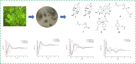
Graphical Abstract

1. Introduction

Lumnitzera littorea (Jack) Voigt is a mangrove tree that has been included on the list of national key protected wild plants (the first batch) (Level II) approved by the State Council of China on 4 August 1999. According to the literature reports, different types of active compounds from Lumnitzera have been isolated, such as hepatoprotective flavonoids and phenolic glycosides [1], antileishmanial macrolides [2], cytotoxic polyketones [3], and anti-angiogenic and anti-inflammatory neolignans [4]. Due to the shortage of L. littorea, the study of bioactive secondary metabolites from the L. littorea-derived endophytic fungus is necessary. Only four articles about the secondary metabolites from the endophytic fungi of Lumnitzera have been reported [5,6,7,8], including an article about antibacterial terpenoids [5], one about cytotoxic polyketones [6], another about cytotoxic oxygenated meroterpenoids [7], and one about steroids with α-glucosidase inhibitory activity [8].
During our exploration of the structurally diverse and bioactive compounds from mangrove-derived fungi, some new bioactive compounds have been found [9,10,11,12]. In the previous study, cytotoxic oxygenated meroterpenoids and steroids with α-glucosidase inhibitory activity had been isolated from the secondary metabolites of two endophytic fungi: Penicillium sp. HLLG-122 and Penicillium sclerotiorum HLL113, which were both isolated from the roots of the L. littorea. [7,8]. In our continuing research, the endophytic fungus Penicillium oxalicum HLLG-13, obtained from the roots of L. littorea and collected from the Tielugang Mangrove Reserve in Sanya, was selected for further research because its EtOAc extract showed antibacterial activity and growth inhibition activity against newly hatched H. armigera Hubner larvae. Five new compounds (12 and 57) and ten known compounds (34 and 815) (Figure 1) were isolated from the EtOAc extract of the fermentation broth from P. oxalicum HLLG-13. In this study, we report the isolation, structure elucidation, antibacterial activity, and growth inhibition activity of these compounds against newly hatched H. armigera Hubner larvae.

2. Results and Discussion

Compound 1 was isolated as a brownish yellow oil. Its molecular formula was established by HR-ESI-MS (m/z 325.2135 [M + Na]+, calcd. for 325.2143) to be C20H30O2 with six degrees of unsaturation (Supplementary Materials). 1H NMR data (Table 1) showed two olefinic proton signals at δH 5.94 (1H, dd, J = 10.0, 1.2 Hz, H-2) and 7.08 (1H, dd, J = 10.0, 5.6 Hz, H-3); five methyl signals at δH 1.26 (3H, d, J = 7.3 Hz, H-16), 1.20 (3H, s, H-17), 1.12 (3H, s, H-18), 1.12 (3H, s, H-19), and 0.98 (3H, s, H-20); four methine signals at δH 2.79 (1H, m, H-4), 2.41 (1H, m, H-6), 4.08 (1H, dd, J = 10.4, 7.2 Hz, H-13), and 1.64 (1H, m, H-15); and six methylene signals at δH 1.63 (1H, m, H-7a), 1.23 (1H, m, H-7b), 2.06 (2H, m, H-8), 1.74 (2H, m, H-10), 2.18 (1H, d, J = 14.8 Hz, H-12a), and 1.49 (1H, d, J = 14.8 Hz, H-12b). The 13C NMR data (Table 2) combined with the DEPT spectrum exhibited twenty carbon resonances, including one carbonyl carbon at δC 208.0 (C-1); two olefinic carbons at δC 128.0 (C-2) and 157.1 (C-3); five methyl carbons at δC 18.8 (C-16), 21.5 (C-17), 35.1 (C-18), 23.1 (C-19), and 29.5 (C-20); four methylene carbons at δC 35.6 (C-7), 40.0 (C-8), 50.1 (C-10), and 43.5 (C-12); four methine carbons at δC 40.2 (C-4), 55.8 (C-6), 78.5 (C-13), and 74.0 (C-15); and four quaternary carbons at δC 61.9 (C-5), 58.7 (C-9), 36.6 (C-11), and 57.4 (C-14). These signals were closely related to those of 3 [13], except for the presence of one hydroxyl-methylene group at δH 4.08 (1H, dd, J = 10.4, 7.2 Hz, H-13) and δC 78.5 (C-13), and one methyl signal at δH 1.12 (3H, s, H-20) and δC 29.5 (C-20) in 1. The absence of one hydroxymethyl group at δH 3.32 (3H, d, J = 10.9 Hz) and δC 72.0 in 3 indicated that the methylene at C-13 in 3 was replaced by a hydroxyl-methylene group in 1, and the hydroxymethyl group at C-20 in 3 was replaced by a methyl group in 1. These results were further confirmed by the 1H-1H COSY and HMBC spectra. The 1H-1H COSY correlation between H-12 to H-13, combined with the HMBC correlations from H-12 to C-13, H-13 to C-19, H-15 to C-19, H-19 to C-15/C-20, and H-20 to C-13/C-15/C-19, confirmed the structure (Figure 2). Hereto, the planar structure of 1 was elucidated.
The relative configuration of 1 was elucidated by the NOESY correlations (Figure 3). The NOE relationships of H-4 to H-13/Me-17 and Me-20 to H-6/H-13 indicated that H-4, H-6, H-13, Me-17, and Me-20 were in α-orientation. The relationships of H-15 to Me-16/Me-19 and H-10 to H-16/Me-18 indicated that H-10, H-15, Me-16, Me-18, and Me-19 were in β-configuration. Thus, the relative configuration of 1 was determined to be 4R*, 5R*, 6R*, 9R*, 11R*, 13R*, and 15R*.
In order to determine the absolute configuration of 1, the theoretical electronic circular dichroism (ECD) spectra of two possible stereoisomers of (4R, 5R, 6R, 9R, 11R, 13R, 15R)-1 (1a) and its enantiomer (1b) were calculated using time-dependent density-functional theory (TDDFT) calculation, and the calculated ECD curve of isomer 1a revealed good agreement with the experimental one (Figure 4). Therefore, the absolute configuration of 1 were assigned as 4R, 5R, 6R, 9R, 11R, 13R, and 15R and named as conidiogenone J.
Compound 2 was isolated as a brownish yellow oil. HR-ESI-MS, the 1H NMR, and 13C NMR data (Table 1 and Table 2) showed that 2 was almost the same as 1; thus, compound 2 has the same planar structure as 1. The relative configuration of 2 was determined by the NOESY correlations (Figure 3). The NOE relationships of H-4 to H-6/H-13 and H-13 to Me-20 indicated that H-4, H-6, H-13, and Me-20 were in α-orientation. The relationships of H-15 to Me-16/Me-19, H-10 to Me-16/Me-17, and Me-18 to Me-19 indicated that H-10, H-15, Me-16, Me-17, Me-18, and Me-19 were in β-configuration. Thus, the relative configuration of 2 was determined to be 4R*, 5R*, 6R*, 9S*, 11R*, 13R*, and 15R*. The calculated ECD spectrum of (4R, 5R, 6R, 9S, 11R, 13R, 15R)-2 (2a) showed good agreement with the experimental spectrum of 2 (Figure 4). Therefore, the absolute configuration of 2 were assigned as 2a and named as conidiogenone K.
Compound 5 was obtained as a brownish yellow oil, and it was determined to have the molecular formula C26H36O6 on the basis of positive HR-ESI-MS (m/z 467.2404 [M + Na]+, calcd. for 467.2410) with nine degrees of unsaturation. 1H NMR spectrum (Table 1) of 5 revealed the presence of one singlet olefinic proton signal at δH 5.21 (1H, s, H-11); one aldehyde proton signal at δH 10.07 (1H, s, H-21); one methoxy signal at δH 3.49 (3H, s, H-25); six methyl signals at δH 0.89 (3H, s, H-18), 0.67 (3H, s, H-19), 1.11 (3H, s, H-20), 1.65 (3H, s, H-22), 1.02 (3H, s, H-23), and 1.50 (3H, s, H-26); three methine signals at δH 3.17 (1H, m, H-3), 1.76 (1H, m, H-5), and 2.00 (1H, q, J = 2.7 Hz, H-9); and eight methylene signals at δH 2.07 (1H, t, J = 13.3 Hz, H-1a), 1.36 (1H, t, J = 13.3 Hz, H-1b), 1.36 (1H, m, H-2a), 1.02 (1H, m, H-2b), 2.00 (1H, m, H-6a), 1.53 (1H, d, J = 13.2 Hz, H-6b), 2.93 (1H, t, J = 13.2 Hz, H-7a), and 2.06 (1H, t, J = 13.2 Hz, H-7b). The 13C NMR data (Table 2), combined with the DEPT spectrum of 5, revealed the presence of twenty-six carbon, including one aldehyde carbon at δC 206.0 (C-21); one ester carbon at δC 170.8 (C-24); one carbonyl carbon at δC 195.5 (C-17); one oxygenated olefinic carbon at δC 186.2 (C-15); three olefinic carbons at δC 122.0 (C-11), 135.5 (C-12), and 111.5 (C-16); one oxomethyl carbon at δC 51.6 (C-25); six methyl carbons at δC 27.4 (C-18), 21.3 (C-19), 19.1 (C-20), 19.7 (C-22), 15.5 (C-23), and 6.9 (C-26); four methylene carbons at δC 27.0 (C-1), 25.8 (C-2), 16.5 (C-6), and 32.2 (C-7); three methine carbons at δC 73.5 (C-3), 46.4 (C-5), and 53.1 (C-9); and five quaternary carbons at δC 37.4 (C-4), 41.2 (C-8), 51.7 (C-10), 55.7 (C-13), and 66.9 (C-14). The above data showed that the planar structure of 5 was similar to that of andrastin E [14]. The obvious differences were the appearance of one aldehyde signal at δH 10.07 (1H, s) for H-21, instead of one methyl signal at δH 0.90 (3H, s) in the 1H NMR spectrum. Furthermore, the 13C NMR data from one aldehyde carbon at δC 206.0 (CH) for C-21 were observed, instead of one methyl carbon at δC 16.7 (CH3) in andrastin E, indicating the methyl of andrastin E was replaced by an aldehyde group in 5. The HMBC correlations (Figure 2) from H-2 to C-21, H-5 to C-21, H-9 to C-21, and H-21 to C-1/C-10 verified the statement above. Hereto, the planar structure of 5 was elucidated.
The relative configuration of compound 5 was determined by the NOESY correlations (Figure 3). The NOE relationships of H-21 to H-19/H-9/H-20, H-9 to H-5/H-23, and Me-25 to Me-23 indicated that H-5, H-9, Me-19, Me-20, H-21, Me-23, and Me-25 were in α-orientation. The relationships of H-3 to H-18 indicated that H-3 and Me-18 were in β-configuration. Thus, the relative configurations of 5 were determined to be 3R*, 5S*, 8S*, 9S*, 10S*, 13R*, and 14R*.
The absolute configuration was assigned by the experimental and calculated ECD spectra. The ECD spectrum of (3R, 5S, 8S, 9S, 10S, 13R, 14R)-5 (5a) and its enantiomer (5b) were calculated using TDDFT in MeOH. As shown in Figure 4, the calculated spectrum of 5a agreed well with the experimental spectrum. Therefore, the absolute configuration of 5 was assigned as 5a and named as andrastin H.
Compound 6 was obtained as a brownish yellow oil, and its molecular formula was determined to be C13H21NO6 on the basis of negative HR-ESI-MS (m/z 286.1290 [M − H], calcd. for 286.1291), indicating four degrees of unsaturation. 1H NMR data (Table 1) displayed one olefinic hydrogen signal at δH 6.30 (1H, br s, H-6); five methylene signals at δH 4.11 (2H, t, J = 7.2 Hz, H-3), 3.53 (2H, t, J = 6.8 Hz, H-8), 2.79 (2H, m, H-4), 2.30 (2H, t, J = 7.6 Hz, H-10), and 1.77 (2H, m, H-9); and three methyl signals at δH 3.58 (3H, s, H-12), 1.98 (3H, s, H-1), and 1.87 (3H, br s, H-13). A total of thirteen carbon signals (including three carbonyl carbons at δC 170.2 (C-2), 166.2 (C-7), and 173.0 (C-11); two olefinic carbons at δC 149.5 (C-5) and 117.6 (C-6); five methylene carbons at δC 62.4 (C-3), 31.9 (C-4), 46.2 (C-8), 21.8 (C-9), and 30.4 (C-10); one methoxy group at δC 51.2 (C-12); and two methyl groups at δC 20.7 (C-1) and 25.2 (C-13)) were exhibited in the 13C NMR data (Table 2), combined with the DEPT spectrum. The 1H-1H COSY correlations from H-3 to H-4, H-8 to H-9, and H-9 to H-10, combined with the HMBC correlations (Figure 2) from H-1 to C-2, H-3 to C-2/C-5, H-4 to C-6/C-13, H-8 to C-7/C-10, H-9 to C-11, H-10 to C-8, H-12 to C-11, and H-13 to C-4/C-6. Therefore, the planar structure of 6 was elucidated as showed in Figure 1. The Z-configuration of the double bond was determined from the correlation of H-6 to Me-13 in the NOESY spectrum (Figure 3). Thus, the structure of 6 was established and named as methyl (Z)-4-(5-acetoxy-N-hydroxy-3-methylpent-2-enamido) butanoate.
Compound 7 was isolated as a yellow oil with the molecular formula C8H12O4 (three degrees of unsaturation) by the HR-ESI-MS spectrum (m/z 173.0807 [M + H]+, calcd. for 173.0814). An analysis of the 1H NMR data (Table 1) indicated that 7 has an olefinic hydrogen signal at δH 5.77 (1H, br s, H-2), two methylene signals at δH 4.21 (2H, t, J = 6.8 Hz, H-5) and 2.95 (2H, t, J = 6.8 Hz, H-4), and two methyl signals at δH 2.01 (3H, s, H-7) and 1.95 (3H, br s, H-8). The 13C NMR data (Table 2), combined with the DEPT spectrum, exhibited eight carbon resonances, including two carbonyl carbons at δC 169.4 (C-1) and 172.7 (C-6), two olefinic carbons at δC 119.5 (C-2) and 157.1 (C-3), two methylene carbons at δC 33.3 (C-4) and 63.8 (C-5), and two methyl groups at δC 20.8 (C-7) and 25.7 (C-8). The 1H-1H COSY correlation from H-4 to H-5, combined with the HMBC correlations (Figure 2) from H-2 to C-4/C-8, H-4 to C-2/C-8, H-5 to C-3/C-6, H-7 to C-6, and H-8 to C-2. On the basis of these results, the planar structure of 7 was elucidated. The Z-configuration of the double bond was determined from the correlation of H-2 to Me-8 in the NOESY spectrum (Figure 3). Thus, the structure of 7 was established and named as (Z)-5-acetoxy-3-methylpent-2-enoic acid.
Compound 8 was isolated as a yellow amorphous powder. Its molecular formula was established by HR-ESI-MS (m/z 379.1529 [M + Na]+, calcd. for 379.1521) to be C21H24O5 with ten degrees of unsaturation. Compared with that of the literature [15], the 1D NMR data (Table 1 and Table 2) of 8 closely resembled those of stocksiloate, which was isolated from Vincetoxicum stocksii, and the absolute configuration remained to be determined due to certain limitations. In order to determine the absolute configuration of 8, the theoretical ECD spectra of two possible stereoisomers of 2R and 2S were calculated using TDDFT calculation, and the calculated ECD curve of isomer 2R revealed a good agreement with the experimental one (Figure 4). Therefore, the absolute configuration of 8 was assigned as 2R-form and named as methyl 2R-stocksiloate.
Compound 15 was isolated as a white amorphous powder, and its molecular formula was established as C10H12O4 by HR-ESI-MS (m/z 195.0660 [M − H], calcd. for 195.0657) with five degrees of unsaturation. The 1H NMR (Table 1) showed three aromatic hydrogen signals at δH 6.75 (1H, d, J = 3.2 Hz, H-2), 6.80 (1H, d, J = 8.8 Hz, H-5), and 6.70 (1H, dd, J = 3.2, 8.8 Hz, H-6); one methylene signal at δH 5.06 (2H, s, H-8); and two methyl signals at δH 3.77 (3H, s, H-7) and 2.07 (3H, s, H-10). The 13C NMR data (Table 2) combined with the DEPT spectrum exhibited ten carbon resonances, including a carbonyl carbon at δC 172.8 (C-9); a benzene moiety at δC 152.2 (C-1), 117.3 (C-2), 126.5 (C-3), 152.1 (C-4), 113.1 (C-5), and 116.3 (C-6); a methoxy carbon at δC 56.6 (C-7); a methylene carbon at δC 62.7 (C-8); and a methyl carbon at δC 20.8 (C-10). The 1H-1H COSY correlation from H-5 to H-6 was combined with the HMBC correlations from H-2 to C-4/C-6/C-8, H-5 to C-1/C-3, H-6 to C-2/C-4, H-8 to C-2/C-3/C-4/C-9, H-7 to C-1, and H-10 to C-9 (Figure 2). Thus, compound 15 was identified as (2-hydroxy-5-methoxyphenyl) methyl acetate. According to the available literature, compound 15 was obtained as the intermediate in the synthesis reaction [16,17,18,19]; thus, it was a new natural product reported here for the first time, and its NMR data were reported for the first time.
By comparing physical and spectroscopic data with literatures, the eight known compounds were identified as conidiogenone D (3) [13], conidiogenone C (4) [13], demethylincisterol A3 (9) [20], ergosterol (10) [21], Δ7-sitosterol (11) [22], (−)-β-sitosterol (12) [23], 7-deacetoxyyanuthone A (13) [24], and (1S,5R,6S)-5-Hydroxy-4-methyl-1-[(2E,6E)-3,7,11-trimethyl-2,6,10-dodecatrien-1-yl]-7-oxabicyclo[4.1.0]hept-3-en-2-one (14) [25]. Compounds 14 were cyclopiane diterpenes with a unique 6/5/5/5 tetracyclic skeleton, and this type of compound is very rarely found from a natural source. Only thirteen compounds have been found in the existing literature, and most of them were mainly isolated as secondary metabolites from the genus of Penicillum [13,26,27,28,29,30].
The antibacterial activities of all compounds were determined against eight pathogenic bacteria (S. aureus (ATCC 25923), E. coli (ATCC 25922), C. albicans (ATCC 14053), S. epidermidis (ATCC 49134), P. aeruginosa (ATCC 17749), V. harveyi (ATCC 25919), V. alginolyticus (ATCC 33787), and V. parahaemolyticus (ATCC 27969)) by the microplate assay method [31]. Compounds 5 and 914 exhibited obvious antibacterial activities against S. epidermidis and C. albicans, with the MIC values ranging from 6.25 to 25 μg/ mL (Table 3).
The growth inhibition activities against newly hatched H. armigera Hubner larvae were tested using the assay described by Guo [32]. Compound 5 exhibited obvious insecticidal activity against newly hatched H. armigera Hubner larvae, with an IC50 value of 50 μg/mL, which was equivalent to that of the positive control (azadirachtin); and compounds 14, 6, and 914 also showed growth inhibition activities against newly hatched H. armigera Hubner larvae, with IC50 values ranging from 100 to 200 μg/ mL (Table 4).

3. Materials and Methods

3.1. General Experimental Procedures

Optical rotations were measured on an Anton paar MCP 5100 modular circular polarimeter (JASCO, Tokyo, Japan). ECD spectra were recorded on a Boilogic Mos-500 spectrometer (JASCO, Tokyo, Japan). IR spectra were recorded on a Nicolet 6700 spectrophotometer (Thermo Scientific, Madison, WI, USA). UV spectra were recorded on a Beckman DU 640 spectrophotometer (JASCO, Tokyo, Japan). The 1D and 2D NMR spectra were obtained with a Bruker AV spectrometer (400 MHZ for 1H and 100 MHZ for 13C, (Bruker Corporation, Basel, Switzerland) or a JNM-ECZS spectrometer (600 HMZ for 1H and 125 MHZ for 13C, (JEOL, Tokyo, Japan), using Methanol-d4 or DMSO-d6 as a solvent. TMS was used as an internal standard. HR-ESI-MS spectra were measured on a Bruker APEX II spectrometer (Billerica, MA, USA). Silica gel (Qing Dao Hai Yang Chemical Group Co., Qingdao, China; 200–300 mesh) and octadecylsilyl silica gel (YMC; 12 μm–50 μm) were used for column chromatography (CC). Precoated silica gel plates (Yan Tai Zi Fu Chemical Group Co., Yan Tai, China; G60, F-254) were used for thin layer chromatography (TLC). Semi-preparative HPLC was performed on an Agilent 1260 LC series with a DAD detector using an Agilent Eclipse XDB-C18 column (250 × 9.4 mm, 5 μm, Agilent Corporation, Santa Clara, CA, USA).

3.2. Fungal Materials

The fungus HLLG-13 was isolated from the roots of the mangrove L. littorea (Jack) Voigt. The L. littorea was collected in Tielugang, Sanya city, Hainan province, in November 2018 and identified by Yukai Chen, associate professor of the College of Life Sciences, Hainan Normal University. This strain was deposited in the Key Laboratory of Tropical Medicinal Resource Chemistry of Ministry of Education, College of Chemistry and Chemical Engineering, Hainan Normal University, Haikou, Hainan, China. The fungus was identified according to its morphological traits and a molecular protocol by amplification and sequencing of the DNA of the ITS region of the rRNA gene. Its base pair ITS sequence had 99% sequence identity to that of P. oxalicum. Therefore, the fungal strain was identified as P. oxalicum. The sequence data have been submitted to GenBank, with an accession number of OK560165.

3.3. Fermentation, Extraction, and Isolation

The seed culture was prepared in a potato liquid medium (30 g sea salt in 1 L of potato infusion in 1 L × 4 Erlenmeyer flasks, each containing 300 mL seed medium), and incubated on a rotary shaker (160 rpm) for 3 days at 28 °C. In total, 50 mL seed culture was then transferred into 1 L Erlenmeyer flasks with a solid rice medium, for a total of 200 bottles of fermentation (each flask contained 80 g rice, 3.0 g sea salt, and 100 mL water) at 28 °C for 28 days. The medium was extracted repeatedly with EtOAc to obtain the corresponding extracts.
The EtOAc extracts were concentrated in vacuo to yield an oily residue (61.27 g). The total crude extract was subjected to silica gel column chromatography (CC) eluted with petroleum ether/EtOAc (v/v, gradient 100:0–0:100) and EtOAc/CH3OH (v/v, gradient 100:0–0:100) to generate nine fractions (Fr.1–Fr.9). Fr.2 (12.7 g) was fractionated by silica gel CC eluted with petroleum ether/EtOAc (v/v, gradient 100:0–0:100) to obtain six fractions (Fr.2-1–Fr.2-6) by the TLC analysis. We obtained 11 (9.43 mg) by crystallization from Fr2-1. Fr.3 (4.6 g) was fractionated by silica gel CC eluted with petroleum ether/EtOAc (v/v, gradient 100:0–0:100) to obtain five fractions (Fr.3-1–Fr.3-5) by the TLC analysis. We obtained 10 (12.32 mg) by crystallization from Fr.3-1, and 12 (8.25 mg) was obtained by crystallization from Fr.3-3. Fr.4 (3.2 g) was fractionated by reverse phase silica gel using a gradient elution of CH3OH/H2O system (1:9-10:0) to obtain thirteen fractions (Fr.4-1–Fr.4-13) by the TLC analysis. Fr.4-1 (722.8 mg) was further purified by semi-preparative HPLC using Agilent Eclipse XDB-C18 (250 × 9.4 mm, 5 μm) with CH3CN/H2O (32:68, v/v) to obtain four fractions (Fr.4-1-1–Fr.4-1-4). Fr.4-1-1 (206.3 mg) was further purified by semi-preparative HPLC using Agilent Eclipse XDB-C18 (250 × 9.4 mm, 5 μm) with CH3OH/H2O (38:62, v/v) to obtain 7 (37.62 mg). Fr.4-1-3 (43.2 mg) was further purified by semi-preparative HPLC using Agilent Eclipse XDB-C18 (250 × 9.4 mm, 5 μm) with CH3CN/H2O (22:78, v/v) to obtain 15 (3.99 mg). Fr.4-5 (348.2 mg) was further purified by semi-preparative HPLC using Agilent Eclipse XDB-C18 (250 × 9.4 mm, 5 μm) with CH3OH/H2O (62:38, v/v) to obtain 1 (12.49 mg), 2 (10.6 mg), and 3 (41.04 mg). Fr.4-7 (106.3 mg) was further purified by semi-preparative HPLC using Agilent Eclipse XDB-C18 (250 × 9.4 mm, 5 μm) with CH3CN/H2O (48:52, v/v) to obtain 4 (11.52 mg). Fr.4-11 (405.7 mg) was further purified by semi-preparative HPLC using Agilent Eclipse XDB-C18 (250 × 9.4 mm, 5 μm) with CH3CN/H2O (72:28, v/v) to obtain 9 (9.57 mg), 13 (140.49 mg), and 14 (23.18 mg). Fr.5 (4.1 g) was fractionated by reverse phase silica gel using a gradient elution of CH3OH/H2O system (1:9–10:0) to obtain nine fractions (Fr.5-1–Fr.5-9) by the TLC analysis. Fr.5-6 (124.8 mg) was further purified by semi-preparative HPLC using Agilent Eclipse XDB-C18 (250 × 9.4 mm, 5 μm) with CH3CN/H2O (46:54, v/v) to obtain 8 (20.09 mg). Fr.6 (3.9 g) was fractionated by reverse phase silica gel using a gradient elution of CH3OH/H2O system (1:9–10:0) to obtain fifteen fractions (Fr.6-1–Fr.6-15) by the TLC analysis. Fr.6-4 (77.6 mg) was further purified by semi-preparative HPLC using Agilent Eclipse XDB-C18 (250 × 9.4 mm, 5 μm) with CH3CN/H2O (19:81, v/v) to obtain 6 (7.23 mg). Fr.6-9 (802.5 mg) was further purified by semi-preparative HPLC using Agilent Eclipse XDB-C18 (250 × 9.4 mm, 5 μm) with CH3CN/H2O (2%CH3COOH) (32:64, v/v) to obtain 5 (198.0 mg).
Conidiogenone J (1): brownish yellow oil; [α]25D +13.8 (c 0.10, MeOH); UV (MeOH) λmax (log ε) 261 (0.56); IR (KBr) νmax 3426, 2952, 2865, 1661, 1453, 1386, 1272, 1073, 1024 cm−1; CD (c 0.0013, MeOH) λmax (Δε) 197 (−11.56), 232 (+6.38), 295 (−1.35), 344 (+1.76) nm; 1H and 13C NMR data, see Table 1 and Table 2; HR-ESI-MS m/z: 325.2135 [M + Na]+ (C20H30O2Na, calcd. for 325.2143).
Conidiogenone K (2): brownish yellow oil; [α]25D +8.6 (c 0.10, MeOH); UV (MeOH) λmax (log ε) 261 (0.52); IR (KBr) νmax 3423, 2956, 2863, 1657, 1446, 1382, 1275, 1068, 1020 cm−1; CD (c 0.002, MeOH) λmax (Δε) 200 (−23.01), 235 (+8.12), 299 (−1.52), 346 (+2.30) nm; 1H and 13C NMR data, see Table 1 and Table 2; HR-ESI-MS m/z: 325.2134 [M + Na]+ (C20H30O2Na, calcd. for 325.2143).
Andrastin H (5): brownish yellow oil; [α]25D +67.6 (c 0.10, MeOH); UV (MeOH) λmax (log ε) 205 (2.46), 242 (1.82); IR (KBr) νmax 3442, 2955, 2886, 1744, 1692, 1611, 1463, 1382, 1328, 1215, 1147, 1020, 1007 cm−1; CD (c 0.002, MeOH) λmax (Δε) 201 (−58.01), 238 (+19.65), 3.9 (−10.99) nm; 1H and 13C NMR data, see Table 1 and Table 2; HR-ESI-MS m/z: 467.2404 [M + Na]+ (C26H36O6Na, calcd. for 467.2410).
(Z)-4-(5-acetoxy-N-hydroxy-3-methylpent-2-enamido) butanoate (6): brownish yellow oil; [α]25D +23.8 (c 0.10, MeOH); UV (MeOH) λmax (log ε) 219 (2.38); IR (KBr) νmax 3415, 2929, 1735, 1618 cm−1; 1H and 13C NMR data, see Table 1 and Table 2; HR-ESI-MS m/z: 286.1290 [M − H] (C13H20NO6, calcd. for 286.1291).
(Z)-5-acetoxy-3-methylpent-2-enoic acid (7): yellow oil; [α]25D +14.2 (c 0.10, MeOH); UV (MeOH) λmax (log ε) 215 (1.37); IR (KBr) νmax 3473, 3414, 1639, 1243 cm−1; 1H and 13C NMR data, see Table 1 and Table 2; HR-ESI-MS m/z: 173.0807 [M + H]+, (C8H13O4, calcd. for 173.0814).
2R-stocksiloate (8): yellow amorphous powder; m.p. 162–163 °C; [α]25D +68.4 (c 0.10, MeOH); UV (MeOH) λmax (log ε) 206 (1.48), 306 (0.95); IR (KBr) νmax 3419, 1740, 1613, 1514, 1265 cm−1; CD (c 0.0015, MeOH) λmax (Δε) 206 (+37.52), 232 (−4.10), 256 (+4.52), 286 (−5.62), 323 (+4.66) nm; 1H and 13C NMR data, see Table 1 and Table 2; HR-ESI-MS m/z: 379.1529 [M + Na]+ (C21H24O5Na, calcd. for 379.1521).
(2-hydroxy-5-methoxyphenyl) methyl acetate (15): white amorphous powder; m.p. 126–127 °C; [α]25D +12.6 (c 0.10, MeOH); UV (MeOH) λmax (log ε) 208 (1.85), 226 (1.95), 295 (1.07); IR (KBr) νmax 3534, 3442, 1697, 1264 cm−1; 1H and 13C NMR data, see Table 1 and Table 2; HR-ESI-MS m/z: 195.0660 [M − H] (C10H11O4, calcd. for 195.0657).

3.4. Biological Assays

3.4.1. Antibacterial Activity

The antibacterial activities of all compounds against eight pathogenic bacteria (S. aureus (ATCC 25923), E. coli (ATCC 25922), C. albicans (ATCC 14053), S. epidermidis (ATCC 49134), P. aeruginosa (ATCC 17749), V. harveyi (ATCC 25919), V. alginolyticus (ATCC 33787), and V. parahaemolyticus (ATCC 27969)) were determined by the microplate assay method. The activated pathogenic bacteria were inoculated into the nutrient broth medium. The concentration of the test group and positive control was 1 mg/mL. The antibacterial effect was evaluated by full wavelength multifunctional microplate reader measurement at 630 nm; the broth medium containing pathogenic bacteria was used as the blank group and DMSO as the negative control; and ciprofloxacin was used as the positive control.

3.4.2. Growth Inhibition Activities against Newly Hatched H. armigera Hubner Larvae

In the test, there were three groups, each containing three neonate larvae of H. armigera Hubner, and the tested compounds were dissolved in DMSO at a concentration of 1 mg/mL. The insecticidal activity was investigated by adding the serial dilution of the isolated compounds and the positive control azadirachtin at 200, 100, and 50 μL/well, with 3 replicates per treatment to the artificial diet for the newly hatched larvae, and the bioassay diet was placed into six-well plates. Newly hatched larvae were incubated at 25 °C and a relative humidity of 80%. DMSO was used as the negative control, azadirachtin was used as the positive control, and an artificial diet was used as the blank control. The number of dead larvae was recorded on the second, fourth, sixth, and eighth day after treatment.

4. Conclusions

Five new compounds, including two cyclopiane diterpenes conidiogenones J and K (12), a steroid andrastin H (5), an alkaloid (Z)-4-(5-acetoxy-N-hydroxy-3-methylpent-2-enamido) butanoate (6), and an aliphatic acid (Z)-5-acetoxy-3-methylpent-2-enoic acid (7), together with ten known compounds (34 and 815) were isolated from the EtOAc extracts of the fermentation broth of the L. littorea-derived fungus P. oxalicum HLLG-13. Compounds 5 and 914 exhibited strong antibacterial activities against S. epidermidis and C. albicans, with MIC values ranging from 6.25 to 25 μg/ mL. Compounds 16 and 914 exhibited significant growth inhibition activities against newly hatched H. armigera Hubner larvae, with IC50 values ranging from 50 to 100 μg/ mL.

Supplementary Materials

The following are available online: https://www.mdpi.com/article/10.3390/md21010022/s1, Figures S1–S54: HR-ESI-MS, 1D and 2D NMR spectra of compounds 1, 2, 5, 6, 7, 8 and 15. S55: 1H and 13C NMR data of the known compounds 34, 914.

Author Contributions

Y.W. and Z.X. performed the experiments for the isolation and structure elucidation and prepared the manuscript; Q.B. contributed to the antibacterial activity and growth inhibition activity against newly hatched H. armigera Hubner larvae; X.Z. and M.B. contributed to part of the structure determination; W.C., C.Z. and G.C. supervised the research work and revised the manuscript. All authors have read and agreed to the published version of the manuscript.

Funding

This project was supported by the Hainan Provincial Natural Science Foundation of China (No. 221RC1054); the National Natural Science Foundation of China (Nos. 22177023 and 41866005); the Key Science and Technology Program of Hainan Province (No. ZDKJ202008); the specific research fund of the Innovation Platform for Academicians of Hainan Province (No. YSPTZX202030).

Conflicts of Interest

The authors declare no competing financial interest.

References

  1. Darwish, A.G.G.; Samy, M.N.; Sugimoto, S.; Otsuka, H.; Abdel-Salam, H.; Matsunami, K. Effects of Hepatoprotective Compounds from the Leaves of Lumnitzera racemosa on Acetaminophen-Induced Liver Damage in Vitro. Chem. Pharm. Bull. 2016, 64, 360–365. [Google Scholar] [CrossRef] [PubMed] [Green Version]
  2. Ahmed, G.G.D.; Mamdouh, N.S.; Sachiko, S.; Hideaki, O.; Hosni, A.S.; Katsuyoshi, M. A New macrolactone, racemolide along with seven known compounds with biological activities from mangrove plant, Lumnitzera racemose. Nat. Prod. Commun. 2019, 14. [Google Scholar] [CrossRef] [Green Version]
  3. Nguyen, P.T.; Bui, T.T.L.; Chau, N.D.; Bui, H.T.; Eun, J.K.; Hee, K.K.; Sang, H.L.; Hae, D.J.; Nguyen, T.C.; Nguyen, V.T.; et al. In vitro evaluation of the antioxidant and cytotoxic activities of constituents of the mangrove Lumnitzera racemosa Willd. Arch. Pharm. Res. 2015, 38, 446–455. [Google Scholar] [PubMed]
  4. Yu, S.Y.; Wang, S.W.; Hwang, T.L.; Wei, B.L.; Su, C.J.; Chang, F.R.; Cheng, Y.B. Components from the Leaves and Twigs of Mangrove Lumnitzera racemosa with Anti-Angiogenic and Anti-Inflammatory Effects. Mar. Drugs 2018, 16, 404. [Google Scholar] [CrossRef] [PubMed] [Green Version]
  5. Meng, L.H.; Li, X.M.; Liu, Y.; Wang, B.G. Penicibilaenes A and B, sesquiterpenes with a tricyclo[6.3.1.01,5]dodecane skeleton from the marine isolate of Penicillium bilaiae MA-267. Org. Lett. 2014, 16, 6052–6055. [Google Scholar] [CrossRef] [PubMed]
  6. Meng, L.H.; Li, X.M.; Lv, C.T.; Li, C.S.; Xu, G.M.; Huang, C.G.; Wang, B.G. Sulfur-containing cytotoxic curvularin macrolides from Penicillium sumatrense MA-92, a fungus obtained from the rhizosphere of the mangrove Lumnitzera racemose. J. Nat. Prod. 2013, 76, 2145–2149. [Google Scholar] [CrossRef]
  7. Qin, Y.Y.; Zou, L.H.; Lei, X.G.; Su, J.W.; Yang, R.X.; Xie, W.J.; Li, W.S.; Chen, G.Y. OSMAC strategy integrated with molecular networking discovery peniciacetals A−I, nine new meroterpenoids from the mangrove-derived fungus Penicillium sp. HLLG-122. Bioorg. Chem. 2023, 130, 106271. [Google Scholar] [CrossRef]
  8. Yang, J.Y.; Tang, M.M.; Chen, L.; Lai, X.Y.; Zhuo, X.; Zhou, X.M.; Chen, G.Y. Study on the secondary metabolites of endophytic Penicillium sclerotiorum HLL113. Chin. J. Org. Chem. 2022, 42, 896–900. [Google Scholar] [CrossRef]
  9. Zheng, C.J.; Bai, M.; Zhou, X.M.; Huang, G.L.; Shao, T.M.; Luo, Y.P.; Niu, Z.G.; Niu, Y.Y.; Chen, G.Y.; Han, C.R. Penicilindoles A-C, Cytotoxic Indole Diterpenes from the Mangrove-Derived Fungus Eupenicillium sp. HJ002. J. Nat. Prod. 2018, 81, 1045–1049. [Google Scholar] [CrossRef]
  10. Cai, J.; Zhu, X.C.; Zeng, W.N.; Wang, B.; Luo, Y.P.; Liu, J.; Chen, M.J.; Li, G.Y.; Huang, G.L.; Chen, G.Y.; et al. Talaromarins A–F: Six New Isocoumarins from Mangrove-Derived Fungus Talaromyces flavus TGGP35. Mar. Drugs 2022, 20, 361. [Google Scholar] [CrossRef]
  11. Bai, M.; Zheng, C.J.; Tang, D.Q.; Zhang, F.; Wang, H.Y.; Chen, G.Y. Two new secondary metabolites from a mangrove-derived fungus Cladosporium sp. JS1-2. J. Antibiot. 2019, 72, 779–782. [Google Scholar] [CrossRef]
  12. Bai, M.; Huang, G.L.; Mei, R.Q.; Wang, B.; Luo, Y.P.; Nong, X.H.; Chen, G.Y.; Zheng, C.J. Bioactive Lactones from the Mangrove-Derived Fungus Penicillium sp. TGM112. Mar. Drugs 2019, 17, 433. [Google Scholar] [CrossRef] [Green Version]
  13. Du, L.; Li, D.; Zhu, T.; Cai, S.; Wang, F.; Xiao, X.; Gu, Q. New alkaloids and diterpenes from a deep ocean sediment derived fungus Penicillium sp. Tetrahedron 2009, 65, 1033–1039. [Google Scholar] [CrossRef]
  14. Yudai, M.; Takayoshi, A.; Ikuro, A. Reconstituted biosynthesis of fungal meroterpenoid andrastin A. Tetrahedron 2013, 69, 8199–8204. [Google Scholar]
  15. Saima, K.; Muhammad, I.T.; Naheed, R.; Mamona, N.; Mahreen, M.; Liaquat, A.; Rasool, B.T.; Muhammad, S.J. Rarely occurring natural products isolated from Vincetoxicum stocksii. Chem. Soc. Pak. 2019, 41, 695–700. [Google Scholar]
  16. Tian, T.; Li, L.Q.; Xue, J.J.; Zhang, J.; Li, Y.J. Enantioselective Syntheses of Spiroketals via a Tandem Reaction of Cu(I)-Catalyzed Cycloetherification and Hydrogen-Bond-Induced [4 + 2] Cyclization. Org. Chem. 2015, 80, 4189–4200. [Google Scholar] [CrossRef]
  17. Li, X.; Xue, J.J.; Huang, C.S.; Li, Y. Copper(I)-Catalyzed hydroalkoxylation/hydrogen-bonding-induced asymmetric hetero-diels–alder cycloaddition cascade: An approach to aromatic spiroketals. Chem. Asian J. 2012, 7, 903–906. [Google Scholar] [CrossRef]
  18. Selvanathan, A.; Vladimir, V.P. Dual Reactivity of Hydroxy- and Methoxy- Substituted o-Quinone Methides in Aqueous Solutions: Hydration versus Tautomerization. J. Org. Chem. 2010, 75, 7338–7346. [Google Scholar]
  19. Bray, C.D. An Approach to Benzannelated [5,6]-Spiroketals. Synlett 2008, 16, 2500–2502. [Google Scholar] [CrossRef]
  20. Wang, C.F.; Huang, X.F.; Xiao, H.X.; Hao, Y.J.; Xu, L.; Yan, Q.X.; Zou, Z.B.; Xie, C.L.; Xu, Y.Q.; Yang, X.W. Chemical Constituents of the Marine Fungus Penicillium sp. MCCC 3A00228. Chem. Biodivers. 2021, 18, 2100697. [Google Scholar] [CrossRef]
  21. Abdelhameed, R.F.A.; Habib, E.S.; Goda, M.S.; Fahim, J.R.; Hassanean, H.A.; Eltamany, E.E.; Ibrahim, A.K.; AboulMagd, A.M.; Fayez, S.; Abd, E.; et al. Thalassosterol, a New Cytotoxic Aromatase Inhibitor Ergosterol Derivative from the Red Sea Seagrass Thalassodendron ciliatum. Mar. Drugs 2020, 18, 354. [Google Scholar] [CrossRef] [PubMed]
  22. Tettamanzi, M.C.; Jares, E.A.; Iannone, L.M.; Pomilio, A.B. Constituents of Senecio crassiflorus. Fitoterapia 1994, 65, 468. [Google Scholar]
  23. Lorensi, G.H.; Oliveira, R.S.; Leal, A.P.; Zanatta, A.P.; de Almea, C.G.M.; Barreto, Y.C.; Rosa, M.E.; Vieira, P.B.; Ramos, C.J.B.; Victoria, F.C.; et al. Entomotoxic Activity of Prasiola crispa (Antarctic Algae) in Nauphoeta cinerea Cockroaches: Identification of Main Steroidal Compounds. Mar. Drugs 2019, 17, 573. [Google Scholar] [CrossRef] [PubMed] [Green Version]
  24. Liu, S.; Su, M.; Song, S.J.; Hong, J.; Chung, H.Y.; Jung, J.H. An anti-inflammatory PPAR-γ agonist from the jellyfish-derived fungus Penicillium chrysogenum J08NF-4. J. Nat. Prod. 2018, 81, 356–363. [Google Scholar] [CrossRef]
  25. Kusakabe, K.; Honmura, Y.; Uesugi, S.; Tonouchi, A.; Maeda, H.; Kimura, K.; Koshino, H.; Hashimoto, M. Neomacrophorin X, a [4.4.3]Propellane-Type Meroterpenoid from Trichoderma sp. 1212-03. J. Nat. Prod. 2017, 80, 1484–1492. [Google Scholar] [CrossRef]
  26. Zhang, S.T.; He, Y.; Li, F.L.; Lin, S.; Yang, B.Y.; Mo, S.Y.; Li, H.Q.; Wang, J.P.; Qi, C.X.; Hu, Z.X. Bioassay-directed isolation of antibacterial metabolites from an arthropod-derived Penicillium chrysogenum. J. Nat. Prod. 2020, 83, 3397–3403. [Google Scholar] [CrossRef]
  27. Li, F.L.; Sun, W.G.; Zhang, S.T.; Gao, W.X.; Lin, S.; Yang, B.Y.; Chai, C.W.; Li, H.Q.; Wang, J.P.; Hu, Z.X. New cyclopiane diterpenes with anti-inflammatory activity from the sea sediment-derived fungus Penicillium sp. TJ403-2. Chin. Chem. Lett. 2020, 31, 197–201. [Google Scholar] [CrossRef]
  28. Mazlan, N.W.; Clements, C.; Edrada, E.R.A. Targeted Isolation of Anti-Trypanosomal Naphthofuran-Quinone Compounds from the Mangrove Plant Avicennia lanata. Mar. Drugs 2020, 18, 661. [Google Scholar] [CrossRef]
  29. Cheng, Z.B.; Li, Y.L.; Xu, W.; Liu, W.; Liu, L.J.; Zhu, D.G.; Kang, Y.; Luo, Z.H.; Li, Q. Three new cyclopiane-type diterpenes from a deep-sea derived fungus Penicillium sp. YPGA11 and their effects against human esophageal carcinoma cells. Bioorg. Chem. 2019, 91, 103129. [Google Scholar] [CrossRef]
  30. Xie, C.L.; Zhang, D.; Xia, J.M.; Hu, C.C.; Lin, T.; Lin, Y.K.; Wang, G.H.; Tian, W.J.; Li, Z.P.; Zhang, X.K.; et al. Steroids from the Deep-Sea-Derived Fungus Penicillium granulatum MCCC 3A00475 Induced Apoptosis via Retinoid X Receptor (RXR)-α Pathway. Mar. Drugs 2019, 17, 178. [Google Scholar] [CrossRef] [Green Version]
  31. Pierce, C.G.; Uppuluri, P.; Tristan, A.R.; Wormley, F.L.; Mowat, E.; Ramage, G.; Lopez-Ribot, J.L. A simple and reproducible 96-well plate-based method for the formation of fungal biofilms and its application to antifungal susceptibility testing. Nat. Protoc. 2008, 3, 1494–1500. [Google Scholar] [CrossRef]
  32. Guo, Z.K.; Gai, C.J.; Cai, C.H.; Chen, L.L.; Liu, S.B.; Zeng, Y.B.; Yuan, J.Z.; Mei, W.L.; Dai, H.F. Metabolites with Insecticidal Activity from Aspergillus fumigatus JRJ111048 Isolated from Mangrove Plant Acrostichum specioum Endemic to Hainan Island. Mar. Drugs 2017, 15, 381. [Google Scholar] [CrossRef]
Figure 1. The structures of compounds 115.
Marinedrugs 21 00022 g001
Figure 2. 1H-1H COSY and key HMBC correlations for 12, 58, and 15.
Marinedrugs 21 00022 g002
Figure 3. Key NOESY correlations for 12 and 57.
Marinedrugs 21 00022 g003
Figure 4. Experimental CD spectra and the calculated ECD spectra of compounds 12, 5, and 8.
Marinedrugs 21 00022 g004
Table 1. 1H NMR spectroscopic data (400/600 MHz) (δ in ppm, J in Hz) for 12, 58, and 15.
Position12567815
1 2.07, t (13.3)1.98, s
1.36, t (13.3)
25.94, dd (10.0, 1.2)5.96, dd (10.0, 0.8)1.36, m 5.77, s 6.75, d (3.2)
1.02, m
37.08, dd (10.0, 5.6)7.13, dd (10.0, 6.0)3.17, m4.11, t (7.2) 3.42, d (8.5)
42.79, m2.74, m 2.79, m2.95, t (6.8)
5 1.76, m 4.21, t (6.8) 6.80, d (8.8)
62.41, m2.30, dd (9.4, 5.2)2.00, m6.30, br s 6.48, d (8.2)6.70, dd (3.2, 8.8)
1.53, d (13.2)
71.63, m1.57, m2.93, t (13.2) 2.01, s6.55, dd (2.0, 8.2)3.77, s
1.23, m1.21, m2.06, t (13.2)
82.06, m2.04, m 3.53, t (6.8)1.95, br s 5.06, s
1.73, m1.72, m
9 2.00, q (2.7)1.77, m 6.41, d (2.0)
101.74, m2.06, m 2.30, t (7.6) 3.06, d (7.2)2.07, s
1.68, d (14.4)
11 5.27, s 5.06, m
122.18, d (14.8)2.02, m 3.58, s
1.49, d (14.8)1.66, m
134.08, dd (10.4, 7.2)3.94, dd (9.7, 8.6) 1.87, br s 1.57, s
14 1.66, s
151.64, d (6.1)1.52, d (5.2)
161.26, d (7.3)1.24, d (7.2) 7.61, d (8.7)
171.20, s1.16, s 6.88, d (8.7)
181.12, s1.35, s0.89, s
191.12, s0.98, s0.67, s 6.88, d (8.7)
200.98, s1.00, s1.11, s 7.61, d (8.7)
21 10.07, s 3.78, s
22 1.65, s
23 1.02, s
24
25 3.49, s
26 1.50, s
Table 2. 13C NMR spectroscopic data (100/150MHz) for 12, 58, and 15.
Position12567815
1208.0, C208.2, C27.0, CH220.7, CH3169.4, C171.7, C152.2, C
2128.0, CH128.0, CH25.8, CH2170.2, C119.5, CH86.9, C117.3, CH
3157.1, CH157.2, CH73.5, CH62.4, CH2157.1, C39.6, CH2126.5, C
440.2, CH45.8, CH37.4, C31.9, CH233.3, CH2125.0, C152.1, C
561.9, C59.8, C46.4, CH149.5, C63.8, CH2155.0, C113.1, CH
655.8, CH49.9, CH16.5, CH2117.6, CH172.7, C115.0, CH116.3, CH
735.6, CH235.5, CH232.2, CH2166.2, C20.8, CH3129.8, CH56.6, CH3
840.0, CH240.0, CH241.2, C46.2, CH225.7, CH3128.4, C62.7, CH2
958.7, C58.7, C53.1, CH21.8, CH2 132.4, CH172.8, C
1050.1, CH247.8, CH251.7, C30.4, CH2 28.7, CH220.8, CH3
1136.6, C38.9, C122.0, CH173.0, C 123.5, CH
1243.5, CH247.7, CH2135.5, C51.2, CH3 133.0, C
1378.5, CH80.8, CH55.7, C25.2, CH3 17.8, CH3
1457.4, C57.6, C66.9, C 25.9, CH3
1574.0, CH73.9, CH186.2, C 128.4, C
1618.8, CH319.2, CH3111.5, C 130.3, CH
1721.5, CH321.5, CH3195.5, C 116.6, CH
1835.1, CH333.8, CH327.4, CH3 159.2, C
1923.1, CH324.1, CH321.3, CH3 116.6, CH
2029.5, CH324.8, CH319.1, CH3 130.3, CH
21 206.0, CH 53.8, CH3
22 19.7, CH3
23 15.5, CH3
24 170.8, C
25 51.6, CH3
26 6.9, CH3
Table 3. Antibacterial activity of compounds 5 and 914.
CompoundsMIC (μg/ mL)
S. epidermidisC. albicans
512.56.25
96.256.25
102525
1112.525
12>5025
1312.56.25
146.256.25
Ciprofloxacin a0.3130.313
a Ciprofloxacin was used as a positive control.
Table 4. Growth inhibition activities of 16 and 914 against newly hatched H. armigera Hubner larvae.
CompoundsIC50 (μg/ mL)
1200
2200
3100
4100
550
6200
9200
10100
11200
12100
13200
14200
Azadirachtin b50
b Azadirachtin was used as a positive control.
Disclaimer/Publisher’s Note: The statements, opinions and data contained in all publications are solely those of the individual author(s) and contributor(s) and not of MDPI and/or the editor(s). MDPI and/or the editor(s) disclaim responsibility for any injury to people or property resulting from any ideas, methods, instructions or products referred to in the content.