Article

Optical Properties Investigation of Upconverting K2Gd(PO4)(WO4):20%Yb3+,Tm3+ Phosphors

by
Julija Grigorjevaite
* and
Arturas Katelnikovas
*
Institute of Chemistry, Faculty of Chemistry and Geosciences, Vilnius University, Naugarduko 24, LT-03225 Vilnius, Lithuania
*
Authors to whom correspondence should be addressed.
Materials 2023, 16(3), 1305; https://doi.org/10.3390/ma16031305
Submission received: 23 December 2022 / Revised: 22 January 2023 / Accepted: 1 February 2023 / Published: 3 February 2023

Abstract

:
Nowadays, scientists are interested in inorganic luminescence materials that can be excited with UV or NIR radiation and emit in the visible range. Such inorganic materials can be successfully used as luminescent or anti-counterfeiting pigments. In this work, we report the synthesis and optical properties investigation of solely Tm3+ doped and Yb3+/Tm3+ co-doped K2Gd(PO4)(WO4) phosphors. The single-phase samples were prepared using a solid-state reaction method. The Tm3+ concentration was changed from 0.5% to 5%. Downshifting and upconversion emission studies were performed under 360 nm and 980 nm excitation, respectively. Yb3+ ions were used as sensitizers in the K2Gd(PO4)(WO4) phosphors to transfer the captured energy to Tm3+ ions. It turned out that under UV excitation, phosphors emitted in the blue spectral area regardless of the presence or absence of Yb3+. However, a very strong deep-red (~800 nm) emission was observed when Yb3+ and Tm3+-containing samples were excited with a 980 nm wavelength laser. It is interesting that the highest upconversion emission in the UV/Visible range was achieved for 20% Yb3+, 0.5% Tm3+ doped sample, whereas the sample co-doped with 20% Yb3+, 2% Tm3+ showed the most intensive UC emission band in the NIR range. The materials were characterized using powder X-ray diffraction and scanning electron microscopy. Optical properties were studied using steady-state and kinetic downshifting and upconversion photoluminescence spectroscopy.

Graphical Abstract

1. Introduction

The lanthanides doped inorganic upconversion (UC) materials with excellent optical properties have significant applications in wide fields, for example, temperature sensing [1], solar light conversion [2], optical sensors [3], or security applications [4], bioimaging [5], optical heating [6], optogenetics [7], nanoscopy [8], nanoscale optical writing [9], etc. Inorganic phosphors, compared with organic dyes or quantum dots, have several advantages, including a longer excited state lifetime, sharp emission bandwidths, and cheaper and more environmentally friendly synthesis [4]. UC is a process where at least two low-energy photons produce high-energy photons. Usually, the inorganic upconverting luminescent materials contain two incorporated RE3+ ions: one as a sensitizer, typically Yb3+, and another as an emitter, for example, Ho3+ [10,11], Tm3+ [12,13], etc. Yb3+ possesses a simple energy level structure, including one ground (2F7/2) and one excited (2F5/2) state level. Additionally, the excited state of Yb3+ is higher than the metastable energy levels of emitters in UC materials, for example, Er3+, Ho3+, or Tm3+. In this case, the energy of 2F7/22F5/2 transition is capable of exciting other rare-earth (RE3+) elements, and UC emitters can release emissions.
The UC process in the phosphors could be achieved through co-doped RE3+ ions into a suitable host lattice. The K2Gd(PO4)(WO4), as a novel phosphate-tungstate compound, has been suitable for application in important optical material fields [14]. Tungstate and phosphate could be used as hosts due to their excellent structure and high thermal stability. Thus, in tungstate, the average distance between the luminescent centers is larger, which may cause a reduced concentration quenching of RE3+ [15], which indicates that this host lattice has a high tolerance for heavily doped RE3+ [16]. Thus, the RE3+ concentration could be an important factor for the luminescence properties, and the proper ratio between a sensitizer and an emitter should be chosen.
The purpose of achieving blue light under 980 nm excitation inspired us to synthesize materials co-doped with Yb3+-Tm3+ because this RE3+ pair is the best combination for blue emission because, among the rare-earth ions, Tm3+ is one of the most studied RE3+ for blue emission based upon upconversion mechanism. In this work, co-doped crystalline K2Gd(PO4)(WO4):20%Yb3+,Tm3+ phosphors are synthesized by a simple solid-state reaction and analyzed for the first time to the best of our knowledge. Further study of K2Gd(PO4)(WO4) samples exhibits both downshifting and upconversion photoluminescence under 360 nm and 980 nm excitation, respectively. The obtained results of the synthesized materials show great potential for NIR-excited security pigments application.

2. Materials and Methods

Two series of K2Gd(PO4)(WO4) (further abbreviated as KGPW) samples were solely doped with Tm3+ and co-doped with 20% Yb3+ and Tm3+. The Tm3+ concentration was 0%, 0.5%, 1%, 2%, and 5% with respect to Gd3+. All samples were prepared by the solid-state reaction method. The stoichiometric amounts of reagents, namely, Gd2O3 (99.99% Tailorlux, Münster, Germany), K2CO3 (99+% Acros Organics, Geel, Belgium), NH4H2PO4 (99% Reachem Slovakia, Petržalka, Slovakia), WO3 (99+% Acros Organics), Yb2O3 (99.99% Alfa Aesar, Haverhill, MA, USA), and Tm2O3 (99.99% Alfa Aesar) were weighed, poured to an agate mortar, and moistened with some acetone. The moist mixture of the materials was homogenized until all the acetone evaporated. The dry homogenous powders were placed in the porcelain crucibles and sintered for 10 h at 873 K. To obtain single-phase compounds, the sintering procedure was repeated two more times.
The phase purity of the prepared compounds was checked using a Rigaku MiniFlexII diffractometer (Tokyo, Japan). The XRD patterns were collected in the 2θ range of 5° to 80°. IR spectra were obtained within the range of 4000 to 400 cm−1 using a Bruker Alpha ATR spectrometer (Ettlingen, Germany) with a resolution of 4 cm−1. The samples were also examined using the FE-SEM SU-70 microscope from Hitachi (Tokyo, Japan). Room temperature and temperature-dependent optical properties of the synthesized compounds were investigated using the FLS980 spectrometer from Edinburgh Instruments (Livingston, UK). The used spectrometer settings are given in Tables S1–S4.
The lattice parameters of the synthesized compounds were calculated from the XRD patterns using the Rietveld refinement method. FullProf Suite software (version 2 December 2022) was used for calculations. The background was set as a 24-term Chebychev-type function. A pseudo-Voigt peak shape was used for the peak profiles. The scale factor, instrument zero, unit cell parameters, preferred orientation, atomic coordinates, and the peak shape (u, v, w, γ0, and γ1) parameters were also refined. K2Ho(PO4)(WO4) (PDF-4+ (ICDD) 04-015-9304) compound, reported by Terebilenko et al. [17], was used for Rietveld fits.

3. Results and Discussion

Overall, two different series of samples were prepared. One contained KGPW doped solely with Tm3+, whereas the second one contained samples co-doped with Yb3+ and Tm3+. The Tm3+ concentration in both series varied from 0.5% to 5%. The concentration of Yb3+ in the samples was 20%. Tm3+-containing samples could be directly excited by UV radiation (~360 nm) (3H61D2 transition (blue upward arrow in Figure 1). The Yb3+ co-doped samples, on the other hand, could also be indirectly excited with a 980 nm wavelength laser. Here, the Yb3+ ions absorb the laser radiation (7F7/22F5/2 transition) and transfer the captured energy to Tm3+, which, after receiving several quants of energy, emits in the UV and visible (Vis) spectral areas. The schematic diagram showing the main Yb3+ and Tm3+ energy levels involved in the downshifting (DS) and upconversion (UC) processes is depicted in Figure 1. Due to the rather large amount of energy levels, Tm3+ can emit in a wide range, i.e., from UV to deep-red and even infrared (IR) [18,19].
The phase purity of all synthesized compounds was checked by recording their powder XRD patterns. The lattice parameters of the synthesized compounds were calculated from the XRD patterns using the Rietveld refinement method. The Rietveld refinement of undoped KGPW, KGPW:5%Tm3+, KGPW:20%Yb3+, and KGPW:20%Yb3+,5%Tm3+ specimens are given in Figure 2. The calculated unit cell parameters of undoped KGPW, KGPW:5%Tm3+, KGPW:20%Yb3+, and KGPW:20%Yb3+,5%Tm3+ specimens are summarized in Table S5. The unit cell parameters decrease with increasing Yb3+ and Tm3+ concentration since both ions are smaller than Gd3+. The recorded XRD patterns matched well with the reference pattern, and no additional peaks were observed; therefore, we can conclude that single-phase materials were obtained. K2Gd(PO4)(WO4) is isostructural with the reported K2Ho(PO4)(WO4) compound. The crystal structure of the prepared materials is orthorhombic, and the space group is Ibca (#73) [17]. The crystal structure of these compounds is built from isolated PO4 and WO4 tetrahedral units and K+ and Gd3+ eight-fold coordinated polyhedra. Considering the same charge and very close ionic radii, we assumed that VIIIYb3+ (0.985 Å) and VIIITm3+ (0.994 Å) replaced VIIIGd3+ (1.053 Å) ions [20].
The SEM images of KGPW:5%Tm3+, KGPW:20%Yb3+, and KGPW:20%Yb3+,5%Tm3+ samples are shown in Figure S1. The SEM images show that samples consist of irregularly shaped and agglomerated particles. No obvious changes in particle shape and size with changing the dopant concentration were observed.
The IR spectra of KGPW and KGPW:20%Yb3+ are shown in Figure S2. Both spectra possess several sets of absorption lines in the range of 1100–400 cm−1. The three sharp absorption lines at 650–450 cm−1 are assigned to the bending vibrations of PO4. The strong absorption band ranging from 900 to 700 cm−1 is attributed to the stretching vibrations of Mo−O within MoO4 tetrahedral units. The strong absorption band at ca. 1075 is ascribed to asymmetric vibrations of PO4 tetrahedral units, whereas the band at ca. 935 cm−1 is ascribed to symmetric ones [21].
The reflection spectra of undoped KGPW, KGPW:20%Yb3+, and KGPW:5%Tm3+ are presented in Figure 3. The reflection spectrum of KGPW:20%Yb3+,5%Tm3+ is identical to the one of KGPW:5%Tm3+; therefore, it was not shown. All samples possessed a white body color showing that the samples do not absorb in the VIS range. It also should be mentioned that the reflectance at longer wavelengths is almost 100% showing low defect concentration in the synthesized materials.
The reflection spectra were measured in a 250–750 nm range. The reflection spectra of Tm3+ doped samples contain three typical Tm3+ absorption lines, i.e., 3H61D2 (ca. 355–370 nm), 3H61G4 (ca. 460–486 nm), and 3H63F2,3 (ca. 655–715 nm) [22]. The broad absorption band in the UV range (around 250–300 nm) could be assigned to the charge transfer from O2− to W6+ transition in the host lattice [23].
The excitation (λem = 450 nm) spectra of KGPW:Tm3+ and KGPW:20%Yb3+,Tm3+ (where the Tm3+ concentration is changed from 0.5% to 5%) samples were recorded from 250 to 430 nm and are shown in Figure 4a,c, respectively. The excitation spectra contain one band at 360 nm originating from the typical Tm3+ ground state 3H6 absorption to the excited state 1D2. In both cases, the highest intensity was achieved with a 5% Tm3+ doped sample. Relatively lower excitation intensity in co-doped samples could be explained due to Tm3+ → Yb3+ energy transfer [24]. The same tendency was observed in emission spectra (λex = 360 nm). The highest emission intensity was observed for KGPW:5%Tm3+ and KGPW:20%Yb3+,5%Tm3+ samples (please refer to Figure 4b,d, respectively). The insets in Figure 4b,d show the normalized integrated emission intensity of the prepared samples and reveal that the emission intensity increases with increasing Tm3+ concentration and reaches maximum intensity when Tm3+ concentration is the highest. There are few sets of emission lines in the down-conversion emission spectra: the intense blue emission at 440–463 nm corresponds to the 1D23F4 transition, whereas much weaker emission lines at 463–485 nm, 650–670 nm, 740–760 nm, and 780–800 nm correspond to the 1G43H6, 1G43F4, 1D23F3, and 3H43H6 transitions, respectively [25]. The most intense emission line observed at 450 nm could be explained due to the directly excited 1D2 energy level with 360 nm excitation. Upon 360 nm excitation, phosphors doped with Tm3+ emit intense blue emission through 1D23F4 transition and that corresponds well with CIE 1931 chromaticity coordinates, depicted in Figure S3. The color coordinates of all the synthesized samples under 360 nm are located in the blue spectral region and are near the perimeter of the CIE 1931 color space diagram. This indicates high color purity. The precise values of color coordinates are tabulated in Table S6.
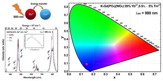
The influence of Tm3+ concentration on the UC emission intensity was also investigated. The UC emission spectra of KGPW:20%Yb3+,Tm3+ (where Tm3+ concentration is 0.5%, 2%, and 5%) samples under 980 nm laser excitation are given in Figure 5. The observed emission bands in the UV, visible, and near-IR range can be attributed to 1D23H6 (at 355–365 nm), 1D23F4 (at 445–458 nm), 1G43H6 (458–496 nm), 1G43F4 (at 625–670 nm, respectively), and 3H43H6 (at 755–844 nm) transition. The substantial change in UC emission spectra was observed as a function of Tm3+ concentration. Interestingly, the intensity of the bands in the UV and visible range do not follow the same trend as the emission band in the NIR range. The 0.5% Tm3+ doped sample showed the strongest emission in the UV/Visible range. On the other hand, the 2% Tm3+ doped sample yielded the most intensive UC emission band in the NIR range. This sample also showed the strongest overall UC emission, as shown in the inset graph of Figure 5. When Tm3+ concentration increases, the Yb3+ → Tm3+ energy becomes more efficient because the average distance between these ions decreases. However, a further increase in Tm3+ concentration leads to a decrease in 3H43H6 emission intensity. Typically, decreasing emission intensity with increasing Tm3+ concentration is attributed to the concentration quenching [26]. Furthermore, with increasing Tm3+ concentration the probability of energy back transfer from Tm3+ to Yb3+ increases (1G4 (Tm3+) + 2F7/2 (Yb3+) → 3H5 (Tm3+) + 2F5/2 (Yb3+)), thus, depopulating the 1G4 (Tm3+) level and reducing blue emission [27]. Such energy back transfer also increases the population of 3F4 (Tm3+) level due to non-radiative relaxation from the 3H5 (Tm3+). The populated 3F4 level can again receive the energy from Yb3+ and be excited to 3F2 level (Tm3+) (2F5/2 (Yb3+) + 3F4 (Tm3+) → 2F7/2 (Yb3+) + 3F2 (Tm3+)). Then 3F2 energy level can populate the 3H4 level through non-radiative relaxation, thus, increasing the 3H43H6 emission in the NIR region [28].
For a further understanding of the DC process, the PL decay curves for the most intense DC transition 1D23F4ex = 360 nm, λem = 450 nm) as a function of Tm3+ concentration were recorded. The mono exponential PL decay curves for KGPW:Tm3+ and KGPW:20%Yb3+,Tm3+ samples were obtained, as depicted in Figure 6a,b, respectively. With increasing Tm3+ concentration, the PL decay curves get steeper, showing that the effective PL lifetime (τeff) values decrease. This, indeed, was confirmed after calculating the τeff values [29]:
τ e f f = 0 I ( t ) t d t 0 I ( t ) d t
Here, I(t) is emission intensity at a given time t. The increasing Tm3+ concentration leads to decreasing τeff values which, in turn, also indicate the decreasing internal quantum efficiency of Tm3+.
For a better understanding of the UC process, the PL decay curves of two main Tm3+ emission peaks, namely, 1G43H6ex = 980 nm, λem = 478 nm) (see Figure 7) and 3H43H6ex = 980 nm, λem = 800 nm) (see Figure 8), were measured. The bi-exponential PL decay curves were observed in both cases.
With increasing Tm3+ concentration, the UC PL decay curves of the main emission peaks become steeper, suggesting that τeff values decrease. The calculated τeff values decreased from 201 μs to 107 μs for 1G43H6 transition and from 267 μs to 101 μs for 3H43H6 transition when Tm3+ concentration was increased from 0.5% to 5%. The calculated rise time values for 1G43H6 transition decreased from 73 μs to 38 μs when Tm3+ concentration was increased from 0.5 % to 5%. Energy transfer from Yb3+ to Tm3+ increases with increasing Tm3+ concentration, which is reflected by a shorter rise time in the KGPW:20% Yb3+ with a higher concentration of Tm3+. The calculated PL rise and τeff values are tabulated in Table S7.
To evaluate the Tm3+ concentration-dependent PL lifetime values of Yb3+, the samples were excited with a 980 nm laser, and the PL decay curves for Yb3+ 2F5/22F7/2 transition were recorded. The Yb3+ emission was monitored at 1050 nm. The recorded PL decay curves as a function of Tm3+ concentration are depicted in Figure 9. As it was expected, the PL lifetime values of the mentioned Yb3+ transition drastically decreased from 1312 ± 24 μs to 416 ± 4 μs when Tm3+ concentration was increased from 0% to 5%. Such an expected decrease in Yb3+ 2F5/22F7/2 transition PL lifetime with increasing Tm3+ concentration is caused by Yb3+ → Tm3+ energy transfer. The Yb3+ → Tm3+ energy transfer efficiency (ηtr) was determined from the Yb3+ PL lifetime values using this equation [30]:
η t r = ( 1 τ Y b T m τ Y b ) × 100 %
Here, τYb−Tm and τYb are Yb3+ PL lifetime values of 2F5/22F7/2 transition in the presence and absence of Tm3+, respectively. The ηtr coherently increases from 44% to 68% when changing Tm3+ concentration from 0.5% to 5%, respectively. The exact PL lifetime values, together with calculated ηtr values, are given in Figure 9 insets and Table S8.
Finally, in order to represent an emission color of the synthesized material under different excitation wavelengths, the response of the standard human eye should be considered. For this reason, the color coordinates (in CIE 1931 color space) of the samples, excited with 360 and 980 nm wavelength radiation, were calculated and are given in Figure 10a,b, respectively. Additionally, the exact color coordinate values are given in Table S6. The color coordinates are invariant upon the Tm3+ concentrations but have a slight dependence on the excitation wavelength. The color coordinates of KGPW:20%Yb3+,Tm3+ samples excited with 980 nm wavelength laser shift upwards if compared to color coordinates obtained for 360 nm excitation. The shift is caused by the fact that the strongest emission lines at 360 nm excitation are at 450 nm (1D23F4 transition), whereas for 980 nm wavelength laser excitation, the 1G43H6 transition at ~475 nm is the strongest one. Color coordinates for samples excited with 360 nm radiation are closer to the perimeter of the CIE 1931 color space diagram showing higher color purity. However, color coordinates slightly shift to the center of the color space diagram if samples are excited with a 980 nm wavelength laser. The shift is caused by relatively strong emission from 1G43F4 transition at ~650 nm if compared to samples excited with 360 nm (please refer to Figure 4d and Figure 5).

4. Conclusions

In summary, we have successfully synthesized single-phase K2Gd(PO4)(WO4):Tm3+ and K2Gd(PO4)(WO4):20%Yb3+,Tm3+ powders, where Tm3+ concentration varied from 0.5% to 5%. Under 360 nm excitation, there was no concentration quenching (at least up to 5% Tm3+) in solely Tm3+ doped samples. The highest upconversion emission in the UV/Visible range was achieved with 20%Yb3+,0.5%Tm3+ concentration. However, the sample co-doped with 20%Yb3+,2%Tm3+ shows the most intense UC emission band in the NIR range, which could be explained by a more efficient Yb3+ → Tm3+ energy transfer. Thus, the color coordinates are invariant upon the Tm3+ doping concentrations and take place in the blue region.

Supplementary Materials

The following supporting information can be downloaded at: https://www.mdpi.com/article/10.3390/ma16031305/s1, Table S1. Spectrometer settings for measuring reflection spectra of KGPW:20%Yb3+,Tm3+ phosphors. Table S2. Spectrometer settings for measuring excitation spectra of KGPW:20%Yb3+,Tm3+ phosphors. Table S3. Spectrometer settings for measuring DC emission spectra of KGPW:20%Yb3+,Tm3+ phosphors. Table S4. Spectrometer settings for measuring UC emission spectra of KGPW:20%Yb3+,Tm3+ phosphors. Table S5. Lattice parameters of KGPW, KGPW:5%Tm3+, KGPW:20%Yb3+, and KGPW:20%Yb3+,5%Ho3+ samples derived from Rietveld refinement analysis. Table S6. Color coordinates (CIE 1931 color space) of KGPW:Tm3+ and KGPW:20%Yb3+,Tm3+ as a function of Tm3+ concentration and excitation wavelength. Table S7. UC PL rise time and lifetime values of KGPW:Tm3+ and KGPW:20%Yb3+,Tm3+ phosphors as a function of Tm3+ concentration, emission wavelength, and excitation wavelength. Table S8. The calculated PL lifetime values (λex = 980 nm, λem = 1050 nm) of Yb3+ emission in KGPW:20%Yb3+,Tm3+, and Yb3+ → Tm3+ energy transfer efficiency (ηtr) as a function of Tm3+ concentration. Figure S1. SEM images of KGPW:5%Yb3+ (a), KGPW:20%Yb3+ (b), and KGPW:20%Yb3+,5%Tm3+ (c). Figure S2. IR spectra of undoped KGPW (a) and KGPW:20%Yb3+ (b). Figure S3. CIE 1931 color space diagram and color coordinates of KGPW:Tm3+ as a function of Tm3+ concentration under 360 nm excitation.

Author Contributions

Conceptualization, A.K.; investigation, J.G.; writing—original draft preparation, J.G.; writing—review and editing, A.K.; visualization J.G. and A.K.; funding acquisition, A.K. All authors have read and agreed to the published version of the manuscript.

Funding

This research was funded by a grant (No. D-2018-0703 “Controlling the up-conversion emission by tuning band gap of the host matrix”) from the Research Council of Lithuania.

Institutional Review Board Statement

Not applicable.

Informed Consent Statement

Not Applicable.

Data Availability Statement

The data presented in this study are available on request from the corresponding author.

Acknowledgments

The authors gratefully thank Andrius Pakalniskis (Vilnius University) for taking SEM images.

Conflicts of Interest

The authors declare no conflict of interest.

References

  1. Zhang, Y.H.; Wang, B.; Liu, Y.H.; Bai, G.Y.; Fu, Z.L.; Liu, H. Upconversion luminescence and temperature sensing characteristics of Yb3+/Tm3+:KLa(MoO4)2 phosphors. Dalton Trans. 2021, 50, 1239–1245. [Google Scholar] [CrossRef] [PubMed]
  2. Sinha, S.; Kumar, K. Studies on up/down-conversion emission of Yb3+ sensitized Er3+ doped MLa2(MoO4)4 (M = Ba, Sr and Ca) phosphors for thermometry and optical heating. Opt. Mater. 2018, 75, 770–780. [Google Scholar] [CrossRef]
  3. Wen, X.; Tang, G.W.; Yang, Q.; Chen, X.D.; Qian, Q.; Zhang, Q.Y.; Yang, Z.M. Highly Tm3+ doped germanate glass and its single mode fiber for 2.0 mu m laser. Sci. Rep. 2016, 6, 20344. [Google Scholar] [CrossRef] [PubMed]
  4. Kumari, A.; Mondal, M.; Rai, V.K.; Singh, S.N. Photoluminescence study in Ho3+/Tm3+/Yb3+/Li+:Gd2(MoO4)3 nanophosphors for near white light emitting diode and security ink applications. Methods Appl. Fluoresc. 2018, 6, 015003. [Google Scholar] [CrossRef]
  5. Maurizio, S.L.; Tessitore, G.; Kramer, K.W.; Capobianco, J.A. BaYF5:Yb3+, Tm3+ Upconverting Nanoparticles with Improved Population of the Visible and Near-Infrared Emitting States: Implications for Bioimaging. ACS Appl. Nano Mater. 2021, 4, 5301–5308. [Google Scholar] [CrossRef]
  6. Li, A.M.; Li, X.B.; Wang, J.L.; Guo, Y.S.; Li, C.G.; Chen, W.G.; Wang, Z.W.; Tang, Y.A. Multifunctional alpha-NaYbF4:Tm3+ nanocrystals with intense ultraviolet self-sensitized upconversion luminescence and highly efficient optical heating. Ceram. Int. 2022, 48, 22961–22966. [Google Scholar] [CrossRef]
  7. Chen, S.; Weitemier, A.Z.; Zeng, X.; He, L.M.; Wang, X.Y.; Tao, Y.Q.; Huang, A.J.Y.; Hashimotodani, Y.; Kano, M.; Iwasaki, H.; et al. Near-infrared deep brain stimulation via upconversion nanoparticle-mediated optogenetics. Science 2018, 359, 679–683. [Google Scholar] [CrossRef]
  8. Liu, Y.J.; Lu, Y.Q.; Yang, X.S.; Zheng, X.L.; Wen, S.H.; Wang, F.; Vidal, X.; Zhao, J.B.; Liu, D.M.; Zhou, Z.G.; et al. Amplified stimulated emission in upconversion nanoparticles for super-resolution nanoscopy. Nature 2017, 543, 229–233. [Google Scholar] [CrossRef]
  9. Lamon, S.; Wu, Y.; Zhang, Q.; Liu, X.; Gu, M. Nanoscale optical writing through upconversion resonance energy transfer. Sci. Adv. 2021, 7, eabe2209. [Google Scholar] [CrossRef]
  10. Li, K.; Van Deun, R. Mutual energy transfer luminescent properties in novel CsGd(MoO4)2:Yb3+,Er3+/Ho3+ phosphors for solid-state lighting and solar cells. Phys. Chem. Chem. Phys. 2019, 21, 4746–4754. [Google Scholar] [CrossRef]
  11. Mahata, M.K.; Koppe, T.; Kumar, K.; Hofsass, H.; Vetter, U. Upconversion photoluminescence of Ho3+-Yb3+ doped barium titanate nanocrystallites: Optical tools for structural phase detection and temperature probing. Sci. Rep. 2020, 10, 8775. [Google Scholar] [CrossRef]
  12. Li, M.; Liu, X.Y.; Liu, L.; Ma, B.; Li, B.X.; Zhao, X.D.; Tong, W.M.; Wang, X.F. beta-NaYF4: Yb, Tm: Upconversion properties by controlling the transition probabilities at the same energy level. Inorg. Chem. Front. 2016, 3, 1082–1090. [Google Scholar] [CrossRef]
  13. Yu, H.Q.; Jiang, P.P.; Chen, B.J.; Sun, J.S.; Cheng, L.H.; Li, X.P.; Zhang, J.S.; Xu, S. Electrospinning preparation and upconversion luminescence of Y2Ti2O7:Tm/Yb nanofibers. Appl. Phys. A 2020, 126, 690. [Google Scholar] [CrossRef]
  14. Fan, G.; Tian, Z.C.; Wang, X.J.; Tang, S.L.; Chen, Y.N. High quantum efficiency red-emitting K2Gd(PO4)(WO4): Sm3+ phosphor: Preparation, characterization and photoluminescence properties. J. Mater. Sci. Mater. Electron. 2018, 29, 17681–17688. [Google Scholar] [CrossRef]
  15. Han, L.L.; Zhao, L.; Zhang, J.; Wang, Y.Z.; Guo, L.N.; Wang, Y.H. Structure and luminescence properties of the novel multifunctional K2Y(WO4)(PO4):Ln3+ (Ln = Tb, Eu, Yb, Er, Tm and Ho) phosphors. RSC Adv. 2013, 3, 21824–21831. [Google Scholar] [CrossRef]
  16. Zhang, X.G.; He, P.; Zhou, L.Y.; Shi, J.X.; Gong, M.L. Energy transfer and luminescent properties of a green-to-red color tunable Tb3+, Eu3+ co-doped K2Y(WO4)(PO4) phosphor. Mater. Res. Bull. 2014, 60, 300–307. [Google Scholar] [CrossRef]
  17. Terebilenko, K.V.; Zatovsky, I.V.; Baumer, V.N.; Slobodyanik, N.S.; Shishkin, O.V. K2Ho(PO4)(WO4). Acta Crystallogr. Sect. E-Crystallogr. Commun. 2008, 64, I75. [Google Scholar] [CrossRef] [PubMed]
  18. Cheng, T.; Marin, R.; Skripka, A.; Vetrone, F. Small and Bright Lithium-Based Upconverting Nanoparticles. J. Am. Chem. Soc. 2018, 140, 12890–12899. [Google Scholar] [CrossRef] [PubMed]
  19. Li, W.C.; He, Q.; Xu, J.X.; Shao, C.Y.; Sun, S.Y.; Fan, S.H.; Hu, L.L. Efficient NIR to NIR up-conversion in LiYE4:Yb3+Tm3+ micro-octahedrons by modified hydrothermal method. J. Lumin. 2020, 227, 117396. [Google Scholar] [CrossRef]
  20. Shannon, R.D. Revised Effective Ionic Radii and Systematic Studies of Interatomic Distances in Halides and Chalcogenides. Acta Crystallogr. 1976, A32, 751–767. [Google Scholar] [CrossRef]
  21. Zatovsky, I.V.; Terebilenko, K.V.; Slobodyanik, N.S.; Baumer, V.N.; Shishkin, O.V. Synthesis, characterization and crystal structure of K2Bi(PO4)(MoO4). J. Solid State Chem. 2006, 179, 3550–3555. [Google Scholar] [CrossRef]
  22. Carnall, W.T.; Crosswhite, H.; Crosswhite, H.M. Energy Level Structure and Transition Probabilities in the Spectra of the Trivalent Lanthanides in LaF3. In Argonne National Laboratory Report; U.S. Department of Energy Office of Scientific and Technical Information: Washington, DC, USA, 1977. [Google Scholar]
  23. Han, B.; Liu, B.K.; Zhang, J.; Shi, H.Z. Luminescence properties of novel Ba2MgWO6:Eu3+ and g-C3N4/Ba2MgWO6:Eu3+ phosphors. Optik 2017, 131, 764–768. [Google Scholar] [CrossRef]
  24. Zheng, Y.; Deng, L.Z.; Li, J.P.; Jia, T.Q.; Qiu, J.R.; Sun, Z.R.; Zhang, S.A. Controlling multiphoton excited energy transfer from Tm3+ to Yb3+ ions by a phase-shaped femtosecond laser field. Photonics Res. 2019, 7, 486–492. [Google Scholar] [CrossRef]
  25. El-Maaref, A.A.; Wahab, E.A.A.; Shaaban, K.S.; Abdelawwad, M.; Koubisy, M.S.I.; Borcsok, J.; Yousef, E.S. Visible and mid-infrared spectral emissions and radiative rates calculations of Tm3+ doped BBLC glass. Spectrochim. Acta A Mol. Biomol. Spectrosc. 2020, 242, 118774. [Google Scholar] [CrossRef]
  26. Halubek-Gluchowska, K.; Szymanski, D.; Tran, T.N.L.; Ferrari, M.; Lukowiak, A. Upconversion Luminescence of Silica-Calcia Nanoparticles Co-doped with Tm3+ and Yb3+ Ions. Materials 2021, 14, 937. [Google Scholar] [CrossRef] [PubMed]
  27. Huang, S.H.; Xu, J.; Zhang, Z.G.; Zhang, X.; Wang, L.Z.; Gai, S.L.; He, F.; Niu, N.; Zhang, M.L.; Yang, P.P. Rapid, morphologically controllable, large-scale synthesis of uniform Y(OH)3 and tunable luminescent properties of Y2O3:Yb3+/Ln3+ (Ln = Er, Tm and Ho). J. Mater. Chem. 2012, 22, 16136–16144. [Google Scholar] [CrossRef]
  28. Yi, Z.G.; Li, X.L.; Xue, Z.L.; Liang, X.; Lu, W.; Peng, H.; Liu, H.R.; Zeng, S.J.; Hao, J.H. Remarkable NIR Enhancement of Multifunctional Nanoprobes for In Vivo Trimodal Bioimaging and Upconversion Optical/T2-Weighted MRI-Guided Small Tumor Diagnosis. Adv. Funct. Mater. 2015, 25, 7119–7129. [Google Scholar] [CrossRef]
  29. Lahoz, F.; Martin, I.R.; Mendez-Ramos, J.; Nunez, P. Dopant distribution in a Tm3+-Yb3+ codoped silica based glass ceramic: An infrared-laser induced upconversion study. J. Chem. Phys. 2004, 120, 6180–6190. [Google Scholar] [CrossRef] [PubMed]
  30. Lupei, A.; Lupei, V.; Ikesue, A.; Gheorghe, C.; Hau, S. Nd → Yb energy transfer in (Nd, Yb):Y2O3 transparent ceramics. Opt. Mater. 2010, 32, 1333–1336. [Google Scholar] [CrossRef]
Figure 1. The schematic energy level structure of Yb3+ and Tm3+.
Materials 16 01305 g001
Figure 2. Rietveld refinement of undoped KGPW (a), KGPW: 5%Tm3+ (b), KGPW: 20%Yb3+ (c), and KGPW: 20%Yb3+,5%Tm3+ (d) XRD patterns.
Materials 16 01305 g002
Figure 3. Reflection spectra of undoped KGPW, KGPW: 20%Yb3+, and KGPW: 5%Tm3+ samples.
Materials 16 01305 g003
Figure 4. Excitation (λem = 450 nm) and emission (λex = 360 nm) spectra of KGPW: Tm3+ (a,b), and KGPW: 20%Yb3+,Tm3+ (c,d), respectively. Both insets show normalized integrated emission of the prepared samples.
Materials 16 01305 g004
Figure 5. Upconversion emission spectra of KGPW: 20%Yb3+,Tm3+ as a function of Tm3+ concentration (λex = 980 nm). The inset shows the Tm3+ concentration-dependent normalized integrated emission.
Materials 16 01305 g005
Figure 6. DC PL decay curves (λex = 360 nm, λem = 450 nm) of KGPW: Tm3+ (a) and KGPW: 20%Yb3+,Tm3+ (b). Both insets show Tm3+ concentration-dependent τeff values.
Materials 16 01305 g006
Figure 7. Tm3+ concentration-dependent: UC PL decay curves (λex = 980 nm, λem = 478 nm) of: KGPW: 20%Yb3+,Tm3+ (a), PL rise time (b), and τeff (c).
Materials 16 01305 g007
Figure 8. Tm3+ concentration-dependent UC PL decay curves (λex = 980 nm, λem = 800 nm) of KGPW: 20%Yb3+,Tm3+. Tm3+ concentration-dependent τeff values are given in the inset.
Materials 16 01305 g008
Figure 9. Tm3+ concentration-dependent PL decay curves of Yb3+ in KGPW: 20%Yb3+,Tm3+ compounds. The inset graphs show Tm3+ concentration-dependent τeff and ηtr values.
Materials 16 01305 g009
Figure 10. Tm3+ concentration-dependent color coordinates (in CIE 1931 color space) of KGPW: 20%Yb3+,Tm3+ samples excited with 360 nm radiation (a) and 980 nm wavelength laser (b).
Materials 16 01305 g010
Disclaimer/Publisher’s Note: The statements, opinions and data contained in all publications are solely those of the individual author(s) and contributor(s) and not of MDPI and/or the editor(s). MDPI and/or the editor(s) disclaim responsibility for any injury to people or property resulting from any ideas, methods, instructions or products referred to in the content.