Article

Bioactivities and Chemical Compositions of Cinnamomum burmannii Bark Extracts (Lauraceae)

by
Prasetyorini Djarot
1,*,
Yulianita
2,
Novi Fajar Utami
2,
Aditya Mahesa Putra
2,
Yurena Irma Mulya Putri
2,
Sri Melia Muhardianty
2,
Tiara Akasi Suciyani
2 and
Asep Syaepulrohman
3
1
Department of Biology, Faculty of Mathematic and Natural Sciences, Universitas Pakuan, Kota Bogor 16129, Indonesia
2
Department of Pharmacy, Faculty of Mathematic and Natural Sciences, Universitas Pakuan, Kota Bogor 16129, Indonesia
3
Department of Computer Sciences, Faculty of Mathematic and Natural Sciences, Universitas Pakuan, Kota Bogor 16129, Indonesia
*
Author to whom correspondence should be addressed.
Sustainability 2023, 15(2), 1696; https://doi.org/10.3390/su15021696
Submission received: 14 December 2022 / Revised: 10 January 2023 / Accepted: 11 January 2023 / Published: 16 January 2023

Abstract

:
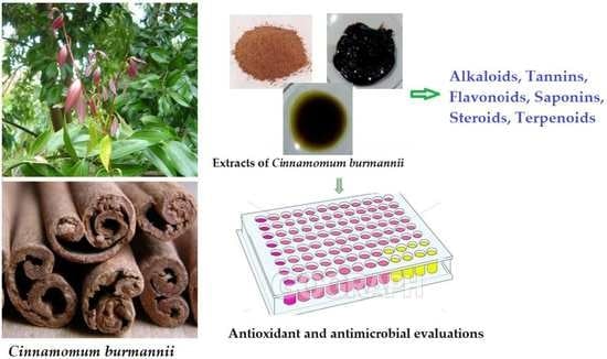
Cinnamomum burmanni has antifungal and antibacterial properties, including alkaloids, tannins, flavonoids, saponins, terpenoids, and essential oil content of cinnamaldehyde, eugenol, and safrole. This study aims to determine the antibacterial properties against Streptococcus mutans and Staphylococcus aureus, antifungal properties against Candida albicans and Candida tropicalis, antioxidant, and flavonoid content of microwave-assisted extraction (MAE) extracts from C. burmanni bark. This study began with the MAE extraction of C. burmanni, followed by qualitative phytochemical tests on the alkaloids, tannins, flavonoids, saponins, and terpenoid groups. Furthermore, using the UV-Vis spectrophotometry method, a quantitative phytochemical test was performed to determine the levels of flavonoids. The 1,1-diphenyl-2-pikrilhidrazil (DPPH) method was used for the antioxidant test, the agar dilution method for the minimum inhibitory concentration (MIC), and the paper disc diffusion method for the width of the inhibitory area (LDH). The positive antifungal control was nystatin, while the antibacterial control was amoxicillin, and both negative controls were 1% dimethyl sulfoxide (DMSO). The antifungal and antibacterial components were found to be 3% n-hexane extract from the bark of C. burmanni, with an inhibitory area width (IAW) of 13.83 mm. The best antioxidant results were the ethanol extract with a very active category IC50 of 8.533 ppm, 5.90%, and the highest ethanol extract containing flavonoid test results.

Graphical Abstract

1. Introduction

Cinnamomum burmanni is one of the herbal plants in Indonesia that is cheap and easy to obtain. Its bark and leaves contain secondary metabolites in the form of cinnamaldehyde in the essential oil, saponins, and flavonoids, which have been widely used as a reliever of flatulence, canker sores, and as a cooking spice [1,2]. These compounds also possess the attributes of antioxidants. The bark of C. burmanii mixed with 50% ethanol extract had antibacterial activity against ten types of bacteria. Furthermore, the bark extract of C. burmanii contained cinnamaldehyde compounds that provided antibacterial effects [3,4].
This herbal plant and the essential oil produced from the bark of C. burmanii have good antifungal activity against C. albicans and can kill microorganisms (antiseptic) [5]. Previous research showed that the minimum inhibitory concentration (MIC) of n-hexane and ethyl acetate of C. albicans was 2.5%, while that of the ethanol extract was 20%. Gupta et al. stated that the secondary metabolites used as antimicrobial and antifungal against B. cereus, S. aureus, E. coli, P. aeruginosa, C. albicans, S. mutans, and Klebsiella sp. are cinnamaldehyde and cinnamic acid [6,7].
Staphylococcus aureus is one of the pathogenic bacteria that cause bacteremia, endocarditis, osteomyelitis, and several skin diseases. S. aureus infection of the skin causes ulcers, impetigo, cellulitis, and staphylococcal scalded skin syndrome [8]. S. aureus is one of the bacteria that are resistant to drugs [9]. Likewise, the S. mutant can cause dental caries, and a survey in America showed that almost 100% of children have this disease. According to Dias et al. [2], mouthwash preparations containing C. burmanii oil can inhibit bacteria with an area of inhibition of up to 13.80 mm. The previous study of ethanol extract of C. burmanni stem bark at concentrations of 6.25, 12.5, and 25% showed antibacterial activity in vitro and in mouthwash [10,11]. Similar results also reported that C. burmanni extract could inhibit S. mutans and had better antibacterial activity than ginger extract against S. mutans. Shan et al. [12] stated that C. burmanii bark oil showed better antibacterial activity against gram-positive bacteria than gram-negative bacteria because it contains phenolic compounds [10,13].
According to previous research, acute infections that attacked various body tissues, such as the mouth, vagina, and nails, were caused by superficial or systemic C. albicans infection in humans, with 75% and 35% caused by C. albicans and C. tropicalis, respectively. Bacterial growth was inhibited using n-hexane extract with a concentration of 10% and a width of 14.50 mm. In addition to the width of the inhibition area, the colonies of C. albicans did not grow [14,15,16].
The method currently being developed is microwave-assisted extraction (MAE), which is relatively new, with microwaves used to speed the extraction process by rapidly heating the solvent [17,18]. Hu et al. stated that the antibacterial activity of MAE is better than the maceration method [19,20]. In addition, the extraction process produces higher flavonoids and phenols than a maceration. Zuofa et al. stated that the optimum results of the extraction process with MAE were obtained at a 50% power ratio of 1:40 and an extraction time of 15 min, and the yield was 32.2% [21].
Flavonoids are aromatic compounds which have been obtained from 70% of ethyl acetate and ethanol extract of the stem bark of C. burmanii. This method is used to determine the concentration of AlCl3 with the quercetin standard [22,23]. Antioxidant activity can be influenced by the content of active compounds contained in the extract, including flavonoids. Furthermore, a polyphenol compound with antioxidant properties can donate one electron to free radicals without damaging body cells.
Free radicals are chemical compounds with one or more unpaired electrons; hence, they are highly reactive. The ethanol extract of C. burmanii stem bark extracted by the maceration method has antioxidant activity with an IC50 of 10.398 ppm. The factors that affect the antioxidant activity and flavonoid content are the extraction method and solvent used. The factors that should be considered in the extraction process include the property of the compound extracted from the type of solvent and the method utilized. The MAE method has numerous advantages, namely faster extraction time, use of less solvent, and relatively higher yield than conventional methods [24,25,26,27,28].
Microbial infection is a severe global problem, and its treatment poses a serious risk to human life due to multi-drug resistance. Petrov warned that antibiotic resistance could kill 10 million people by 2050, so the discovery of new antibiotics is now needed to prevent death due to multi-drug-resistant pathogens [29,30,31].
The ionization process that occurs in the microwave is more evenly distributed. Therefore, it can break down plant cell walls. Previous research showed that the extraction of pine bark with MAE is influenced by the type of solvent and the time during the extraction process. This study used a temperature of ±47 °C for 15 min with a maximum power of 300 watts [32,33]. Different solvents can also affect flavonoid levels in a sample. The red galangal rhizome contained flavonoid content from an n-hexane and ethyl extract of 2.6% and 18.5%. Clove leaf ethanol extract has a flavonoid content of 7.3% [34]. Based on the data on the flavonoid content which is affected by the solvent, it is necessary to study the levels of flavonoids and the antimicrobial activity of the extracts against S. mutant and S. aureus, as well as C. albicans and C. tropicalis. Therefore, this study aims to obtain alternative herbal medicines, such as antifungal, antibacterial, and antioxidant, to overcome the resistance of several bacteria to antibiotics [35,36].

2. Materials and Methods

2.1. Plant Material, Chemicals, and Reagents

The material used was dry C. burmanii bark obtained from the Research Institute for Spices and Medicinal Plants and determined at the Indonesian Institute of Sciences, Botanical Gardens Plant Conservation Center, Bogor. Others include isolates of S. mutans, S. aureus, C. albicans, and C. tropicalis fungi from the Microbiology Laboratory at Bogor Agricultural University.

2.2. Chemicals and Reagents

The chemicals and reagents that were used are amoxicillin and nystatin as a positive control for antibacterial and antifungal testing from the Kimia Farma brand. Furthermore, dimethyl sulfoxide 1% was used as a negative control, in addition to hydrochloric acid, 96% ethanol, iron (III) chloride solution, magnesium powder, nutrient agar medium, potato dextrose agar medium, Bouchardat reagent, Dragendorf, Mayer, from German Sigma. The solvents include n-hexane, ethyl acetate, ethanol, methanol, DPPH powder, quercetin, aluminum chloride, sodium acetate, and magnesium powder. The study was conducted in several stages, the first by making Simplicia powder. The second by extracting with the MAE method with three solvents, followed by the third stage, which qualitatively screens the extracts to determine the content of alkaloids, flavonoids, tannins, saponins, steroids, and terpenoids. The fourth stage is determining the extracted flavonoid content, while the fifth step is testing the antioxidant activity using the DPPH method. The sixth stage is the minimum inhibitory concentration (MIC) test, and the seventh is the inhibitory area width (IAW) test.

2.3. The Tools

The tools that were used include microwave (Samsung® Company, Suwon, South Korea), digital balance (Lab Pro® Company, Knoxville, TN, USA), analytical balance (A&D Company, Tokyo, Japan), UV-Vis spectrophotometer (Jasco V-730® Company, Easton, MD, United States), the crucible, thermometer, furnace (Daihan Scientific Furnace® Company, Kangwon-do, South Korea), oven (Memmert® Company, Pune, India), Mesh 40, filter paper, aluminum foil, and glassware (Pyrex® Company, Corning, NY, USA), autoclave (All American® Company, Hillsville, VA, USA), Bunsen burners, steam plate, Petri dish (Pyrex®), grinder (Airlux® Company, Taoyuan, Taiwan), incubator (Memmert®), paper disc (Whatman number 40, Maidstone, UK) diameter 6 mm, micropipette (Proline plus® Company, Dallas, TX, USA), oven (Memmert®), 40 mesh sieve, and furnace (Vulcan A-550® Company, Washington, NY, USA).

2.4. Extracts Preparations

Firstly, cinnamon bark is cleaned and dried in an oven at 50 °C, ground, and sieved using a 40-mesh sieve to obtain powdered Simplicia. The compound was characterized by measuring yield, moisture content, ash content, and organoleptic characteristics, including color, aroma, and taste. The yield was calculated using Equation (1). The gravimetric method conducted the water and ash contents, which were calculated using Equations (2) and (3), respectively.
% Yield   of   Simplicia   powder = T h e   w e i g h t   o f   p o w d e r   S i m p l i c i a   o b t a i n e d   T h e   w e i g h t   o f   c i n n a m o n   b a r k   S i m p l i c i a   ×   100 %
The calculation of the percentage of the water content of Cinnamon bark powder Simplicia was performed using the following equation:
% Water   content =   T h e   w e i g h t   b e f o r e   h e a t i n g     T h e   w e i g h t   a f t e r   h e a t i n g     T h e   w e i g h t   b e f o r e   h e a t i n g   ×   100 %
The calculation of the percentage of ash content of Cinnamon bark powder was performed using the following equation:
% Ash   content = ( T h e   w e i g h t   b e f o r e   g l o w i n g ) ( E m p t y   e x c h a n g e   r a t e ) P o w d e r   S i m p l i c i a   w e i g h t   ×   100 %

2.5. Extracts Preparations Using MAE Method

The extraction process was conducted using the MAE method with three different polar solvents, namely n-hexane (non-polar), ethyl acetate (semi-polar), and 96% ethanol (polar). The extraction process was conducted in stages, and each extract was 180 g of Simplicia, which was placed in a 3500 mL Erlenmeyer filled with 60 g of powder in 200 mL of n-hexane solvent. The solution was extracted in a microwave for 15 min at a power of 300 watts and a temperature of ±47 °C.
Radiation was alternately conducted in the microwave from 2 to 3 min to maintain the temperature until a total of 15 min was obtained for one stage. Next, the filtrate was separated from the residue and collected in one container. The residue was added again with 200 mL of n-hexane solvent and re-extracted using the same temperature, power, and time to obtain a solvent at a 1:10 ratio. Furthermore, the remaining powder was extracted with n-hexane solvent using the same strategy. After extraction with n-hexane solvent, the residue was dried in an oven at 50 °C and extracted using the same procedure for ethyl acetate and 96% ethanol as solvent. The extracted filtrate from each solvent was separately accommodated and then evaporated with vacuum drying at a temperature of 50 °C until it became a thick extract. The overall extraction results produced three types of thick extracts, namely n-hexane, ethyl acetate, and ethanol, with each calculated to determine the yield using Equation (4).
% Extract   yield = e x t r a c t   w e i g h   o b t a i n e d   w e i g h t   o f   S i m p l i c i a   c i n n a m o n   b a r k   p o w d e r     ×   100 %

2.6. Extract Phytochemical Screening

The extract was tested for water and ash contents using the gravimetric method. Phytochemical screening of extracts included the alkaloid, flavonoid, tannin, saponin, steroid, and terpenoid tests qualitatively using the methods developed by Banu and Cathrine and Pandhi et al. [37,38].
The alkaloids test was conducted by weighing 20 mg of the extract, adding 1 mL of 2 N hydrochloric acid and 9 mL of distilled water, heating over a water bath for 2 min, cooling, and filtering. Three drops of the filtrate were taken and put on a watch glass, with two drops of Bouchardat (potassium iodide) added to the reaction. The formation of a brown-to-black precipitate indicated the presence of alkaloid compounds. Three drops of the filtrate were put in a watch glass, adding two drops of Mayer’s reagent comprising potassium mercury iodide. The formation of a white or yellow precipitate, which dissolves in ethanol, indicates the presence of alkaloid compounds.
Additionally, three drops of the filtrate were taken with two drops of Dragendroff’s reagent comprising potassium bismuth nitrate added to form a brown precipitate, indicating the presence of alkaloid compounds. The flavonoid test was conducted by evaporating the sample to dryness, adding 2–3 drops of ethanol, Mg powder, and a few drops of 5 M hydrochloric acid. The formation of a red-to-purple color indicates the presence of flavonoids.
The tannin test was conducted with 500 mg of each sample dissolved in distilled water with 1% gelatin in 10% sodium chloride. The formation of white color on the tube indicated a positive result. As much as 0.5 g of each sample was dissolved in distilled water and then added to the FeCl3 solution. The formation of a blue-black or green-brown color indicated a positive result.
The saponin test was conducted by weighing a sample of 500 mg in a test tube, adding 10 mL of hot water, cooling it, and then shaking it for 10 s. The presence of saponins was indicated by the formation of foam for not less than 10 min and as high as 1–10 cm after one drop of 2 N HCL was added.
Steroid and terpenoid tests were conducted by weighing 50 mg of the sample dissolved in n-hexane. The formation of a red ring when 1 mL of CH3COOH and 0.5 mL of concentrated H2SO4 were added, and the appearance of a green-to-blue color indicated the presence of steroids.

2.7. Determination of Flavonoid Level

The flavonoid level was determined using the colorimetric method with aluminum chloride (AlCl3) as a reagent. The compound used as the standard was quercetin because it is a flavonoid of the flavonol group, which has keto and hydroxyl groups on the C-4 and neighboring C-3 or C-5 atoms, respectively. The test and series solutions of the C. burmanii stem bark extract and quercetin were measured to determine the absorption, optimum wavelength, and time obtained on a spectrophotometer. The resulting absorbance was entered into the regression equation of the quercetin standard curve. Equation (5) was used to calculate the levels of flavonoids as follows:
% Flavonoid   levels = C ( ppm )   ×   v o l u m e   ( mL ) ×   f p   ×   10 6 w e i g h t   o f   s i m p l i c i a ( w e i g h t   o f   S i m p l i c i a   ×   w a t e r   c o n t e n t   o f   e x t r a c t ) × 100 %
Determination of the flavonoid content was conducted by weighing 50 mg of ethanol dissolved in a 50 mL volumetric flask and shaking until a homogenous solution was obtained. Approximately 10 mL of the extracted solution was added to 15 mL of ethanol using a pipette. Then, 1 mL of 10% AlCl3, 1 mL of sodium acetate, and distilled water were added and homogenized. The solution was cooled to room temperature to determine the optimum time and measure the maximum wavelength absorption using a UV-Vis spectrophotometer [39]. The resulting absorbance was then entered into the linear regression equation from the standard quercetin curve, and then the total flavonoid content was calculated.

2.8. Antioxidant Test

The antioxidant test was conducted to measure the free radical scavenging by the compound being tested using the DPPH method. Antioxidant activity occurs by DPPH reacting with antioxidant hydrogen atoms; therefore, the compound changes color from purple to yellow [40]. The percentage value of the cinnamon bark extract test solution series and the vitamin C and blank comparison solution were measured to determine the optimum wavelength and time obtained on a spectrophotometer. The percentage inhibition value was calculated using the inhibition concentration (IC) value of 50%, which was the sample concentration capable of reducing its radicals by 50%. The percentage of inhibition was plotted in a graph, and from the graph obtained, the equation of the linear regression line Y = bx + a, where y = 50 and x shows IC50 [41]. The IC50 value is calculated by Equation (6) as follows:
%   inhibition = ( blank   absorbance   ( DPPH ) )     ( absorbance   of   extract   sample   or   vitamin   c ) ( b l a n k   a b s o r b a n c e   ( D P P H )   ×   100

2.9. Antimicrobial Test

2.9.1. Antibacterial and Antifungal Test Preparation

The tools used were glasses, paper discs, ose, volume pipettes, and tweezers which were sterilized first by autoclaving at 121 °C for 15 min. Glass utensils were put in the oven until they were ready to use. The bacteria used in this study were S. mutants, S. aureus, and the fungi C. albicans and C. tropicalis. The bacteria and fungi were rejuvenated using nutrient agar (NA) media and potato dextrose agar (PDA). The nutrient agar (NA) as much as 23 g was weighed to make the media, then dissolved in 1 L of distilled water, and heated while stirring with a magnetic stirrer until it boiled and became homogeneous. Furthermore, 9.75 g of potato dextrose agar (PDA) was weighed and dissolved in 250 mL of distilled water, then heated and stirred with a magnetic stirrer until it boiled and became homogenous. Both compounds were then sterilized using an autoclave at 121 °C for 15 min at 1.5 atm pressure. The media were heated, poured into a 20 mL Petri dish, closed tightly, and stored. Bacterial and fungal cultures were rejuvenated by scratching a needle containing the culture on the agar surface, then they were incubated at 37 °C for 24 h. Bacterial and fungal suspensions were prepared using cultures 24 h old, which were then suspended in 5 mL of sterile physiological NaCl solution with the turbidity measured using a standard 0.5 Mc Farland [42,43,44].

2.9.2. Test Solution and Disc Paper Preparation

The preparation of test solutions from each extract for bacteria and fungi was performed by making a stock solution of C. burmanii extract in 1% DMSO. Concentration series of 0.5, 1, 2.5, and 5% were used to test the minimum inhibitory concentration (MIC) of the stock solution. A series of concentrations of 5, 10, and 20% was used to test the width of the inhibitory zone (IAW) according to the results of the minimum inhibitory concentration (MIC) in 1% DMSO. The positive control used for the antibacterial test was amoxicillin with a concentration of 50 ppm, and the negative was 1% DMSO. The positive control for the antifungal test was Nystatin with a concentration of 350 ppm, and the negative was DMSO 1% [45]. Disc paper was made from Whatman filter paper with a 6 mm diameter and sterilized. Furthermore, sterile disc paper was soaked in the active ingredients for 30 min to determine the positive and negative control. Then it is dried in an oven for 24 h at a temperature of 40–50 °C.

2.9.3. Determination of Minimum Inhibitory Concentration

Sterile BHI (brain heart infusion) media were inserted into a 20 mL Petri dish at 45 °C to test the minimum inhibitory concentration (MIC) for bacteria using the agar dilution method. In addition, 1 mL of n-hexane extract with concentrations of 0.25, 0.5, 1, and 2%, and 1, 2, 4, and 8% was separately added to the Petri dishes, followed by ethyl acetate extract and 20, 30, 40, and 50% ethanol extract. After that, 0.2 mL of bacterial culture was added, homogenized, and solidified. The solution was incubated for 24 h at a temperature of 20–25 °C to determine the smallest concentration not overgrown with bacteria, known as the minimum inhibitory concentration (MIC). The test was conducted on 3 extracts, namely n-hexane, ethyl acetate, and 96% ethanol. The same method was used to test fungi’s minimum inhibitory concentration (MIC), using PDA (potato dextrose agar) media and inoculated with fungal cultures.

2.9.4. Resistance Width Test

The inhibitory area width (IAW) was tested using the disc paper diffusion method. IAW testing for S. mutans and S. aureus used nutrient agar (NA) media and C. albicans and C. tropicalis used sterile PDA (potato dextrose agar). Furthermore, a suspension of bacteria and fungi was added to 0.2 mL of each Petri dish according to the test microbe. The solution was homogenized circularly with the paper disc containing the extract placed on the media surface using sterile tweezers at a distance of 20 mm from the edge of the Petri dish. It was further incubated at 37 °C for 24 h. The IAW test was conducted for 3 different types of extracts, namely n-hexane, ethyl acetate, and 96% ethanol extract, using the same method.
After 24 h, the clear zone formed on the Petri dish was measured using a caliper with an accuracy of 0.05 mm to determine the clear zone of the disc, along with the positive and negative controls. The IAW was determined by measuring the diameter of the clear zone formed and subtracting it from the diameter of the paper disc (6 mm) before dividing it by 2. The IAW for fungi was tested using PDA (potato dextrose agar) solid media with fungal microbial cultures [46].

3. Results and Discussion

3.1. Powder and Extract Manufacturing

The powder simplification and extraction process using the MAE method with three different solvents obtained three different filtrates, with each dried in a vacuum to produce a thick extract. The three extracts’ organoleptic tests were dark brown, with a strong cinnamon aroma with a slightly spicy, sweet taste, and the n-hexane extract looked a bit oily and watery compared with the other two extracts, as shown in Figure 1. The yield of cinnamon bark extract from each solvent is presented in Table 1.
The yield results indicate that the solvent’s polarity affects the yield obtained. Ethanol extract obtained the largest yield compared with extracts from ethyl acetate and n-hexane solvents. This is because ethanol has a stronger polar group than non-polar groups and from its chemical structure, which comprises hydroxyl (polar) and carbon (non-polar). Polar solvents have a greater ability to extract organic compounds, such as essential oils; therefore, the extract looks oily. The description of Simplicia powder and the extract is shown in Figure 1.
Figure 1. Simplicia and the MAE of cinnamon bark; Simplicia (A), ethanol extract (B), ethyl acetate extract (C), n-hexane extract (D).
Sustainability 15 01696 g001

3.2. The Results of Phytochemical and Flavonoid Determination

Phytochemical identification was conducted by qualitative analysis to determine the content of secondary metabolites contained in samples of cinnamon bark, Simplicia powder, n-hexane extract, ethyl acetate, and 96% ethanol. The results of the phytochemical test of cinnamon bark are shown in Table 2.
The results of the flavonoid determination levels obtained a maximum wavelength of quercetin 432.5 nm with an incubation time of 25 min. This means that at 25 min, the optimum incubation time for the compound to react with the maximum and the most stable absorbance value was determined. Flavonoid levels were tested on n-hexane, ethyl acetate, and ethanol extracts with an optimum incubation time of 25 min in a dark room. The measurements were conducted using UV-Vis spectrophotometry at a wavelength of 432.5 nm. The results obtained were calculated by dilution factor and percent of moisture content, as shown in Table 3.
Flavonoids are polyphenolic compounds with antioxidant properties. Therefore, the higher their content, the greater the IC50 value in the sample. According to the test results, the highest and lowest levels, 5.90% and 3.033%, were found in the ethanol and n-hexane extracts, respectively. This is because polar flavonoid compounds are more easily attracted to polar solvents, such as ethanol. According to Abreu, excessive solvents can cause thermal stress during the extraction process. The exposed microwaves will be concentrated in the solvent with a decrease in the effectiveness of microwaves in breaking down cells, thereby leading to lower levels of n-hexane flavonoid extracts than ethyl acetate and ethanol extracts [47].

3.3. Antioxidant Test Result

The antioxidant test results showed that the highest absorbance value is at a wavelength of 514.5 nm with an optimum incubation time of 30 min. The antioxidant activity was measured with vitamin C used as a standard. It is one of the secondary antioxidants capable of capturing free radicals and preventing chain reactions. In addition, vitamin C has the working principle of reducing two free radicals from DPPH by releasing two hydrogen atoms to form non-radical DPPH compounds.
Several concentrations were used to determine the standard curve of vitamin C and its antioxidant activity. According to the linear regression results, Y = 4.8951x + 11.561, standard vitamin C has an IC50 value of 7.85 ppm, meaning it has very active antioxidant activity. The n-hexane extract antioxidant activity, ethyl acetate, and cinnamon bark ethanol extract were tested using the DPPH method. Cinnamon bark extract was made in a concentration series to obtain the absorbance value using UV-Vis spectrophotometry at a wavelength of 514.5 nm to determine the percentage value of inhibition. Therefore, it obtained the IC50 value of each test sample, as shown in Table 4.
According to the test results, the ethanol extract has an antioxidant activity of 7.85 ppm, which is similar to standard vitamin C. This means that the ethanol extract of cinnamon bark has very active antioxidant activity. Molyneux [41] stated that a compound has very active antioxidant activity assuming the IC50 value is less than 50 ppm. The antioxidant activity test results are also similar to the study conducted by Dewi on P. ostreatus extract using the microwave-assisted extraction method with an IC50 value of 14.66 ppm [48].
The IC50 value analysis showed that the ethanolic extract of cinnamon bark had more active antioxidant activity than the ethyl acetate and n-hexane extracts. This is due to differences in the levels of flavonoid compounds from each extracted sample using antioxidants; therefore, it can be used to inhibit DPPH radicals and has a good effect on health. The macerated extract of cinnamon bark has a different IC50 value, where the n-hexane, ethyl acetate, and ethanol extracts are 313.628, 163.010, and 76.490 ppm. This is also influenced by the flavonoid compounds in the bark of C. burmanii, which were not completely attracted during the extraction process, thereby leading to fewer hydroxyl groups in the flavonoid structure compared to Cinnamon bark extracted using microwave-assisted [3].

3.4. Antibacterial Test Result

3.4.1. Minimum Inhibitory Concentration (MIC) Test Results

Generally, the MIC test for bacteria and fungi n-hexane extract was better than ethyl acetate and ethanol extracts. The MIC of S. mutans and S. aureus showed that the n-hexane and ethyl acetate extracts had low values at concentrations of 0.5, 1.5, and 1% for S. mutans, S. aureus, C. Albicans, and C. tropicalis. In the ethanol extract, up to a concentration of more than 50%, bacteria and fungi were not found in MIC. This indicated that the colony could still grow at a concentration of more than 50%. The difference in the MIC concentration of each extract tended to occur because the solvent’s varying polarity determined the yield. The MIC test results of Cinnamon bark extract against bacteria and fungi are shown in Table 5.
The difference in solvent polarity can affect the concentration of the extracted active compound and the MIC. The minimum inhibitory concentration can be used as a reference in the IAW test. The smaller the concentration of extract obtained in the MIC test, the better the antibacterial activity. The compounds with antifungal activity are essential oils from the bark of C. burmanii against C. Albicans. Meanwhile, essential oils that can be extracted by non-polar and semi-polar solvents, in this case, are n-hexane and ethyl acetate. Similarly, the MIC of C. burmanii stem bark extracted using the multilevel maceration method for the n-hexane and ethyl acetate extracts produced 2.5%, while for the ethanol extract, 25% was obtained.

3.4.2. Inhibitory Area Width (IAW) Test Results

The IAW test was conducted to determine the effect of the concentration of the extract on the antibacterial power. This was conducted using the paper disc diffusion method because it is more sensitive to antimicrobial compounds whose activity is unknown. According to Banjara et al., growth inhibition in this method was indicated by forming a clear zone around the paper disc [49]. The antibacterial activity category was determined. The diameter of the inhibition zone of 5, 5–10, 11–20, and 21 mm are categorized as weak, moderate, strong, and very strong (Table 6).
The test results showed that the three extracts have antibacterial activity against S. mutans and were categorized from weak to medium. The concentration of the extract had a significant effect on IAW. The higher the n-hexane extract concentration, the wider the IAW. Although IAW produced the widest concentration at 4%, this result was still in the weak category and under positive control. Similarly, even though the ethyl acetate extract produced the best extract concentration at 4% extract with 3.66 mm, it was in the weak category. The lowest IAW test results were ethanol extract with a concentration of 15% against S. mutans.
One of the non-polar n-hexane solvents that attracted compounds from the bark of C. burmanii was cinnamaldehyde, which could inhibit energy metabolism in bacteria and has been widely reported to have antibacterial properties. This was evidenced by the synthetic inhibition of the cell wall of S. mutans bacteria and biosynthetic enzymes used for energy formation [50].
In addition to cinnamaldehyde compounds, the bark of C. burmanii contains other secondary metabolites, such as alkaloids, flavonoids, tannins, saponins, and terpenoids. Flavonoids are polar compounds and belong to the phenol group that contains a hydroxyl on a carbon ring and functions as an antimicrobial and antiviral [51]. Flavonoid compounds can change the physical and chemical properties of cytoplasm-containing proteins and denature bacterial cell walls by binding to proteins through hydrogen bonds. It also interferes with membrane permeability function, active transport function, and control of protein composition.
Terpenoids have the same polarity as the phenol group and bind to fats and carbohydrates, causing the permeability of bacterial cell membranes to be disturbed. Tannins are complex organic compounds acting as antimicrobials, reacting with cell membranes, inactivating enzymes, destroying bacteria, and functioning as bacterial genetic material. The alkaloids have a mechanism of action by influencing the osmotic pressure between bacteria and their environment [52]. Polyphenols are phenol group compounds that play a role in damaging the cytoplasmic membrane of bacteria. Therefore, it causes instability in the controlling protein composition function of bacterial cells.
The IAW test on S. aureus and S. mutants showed that the extract concentration had a significant effect. All the extracts tested showed antibacterial properties from weak to moderate, and the higher its concentration, the greater the IAW S. aureus. Duncan’s test showed that the extract concentration produced the highest IAW at a concentration of 6% with a strong category and quantitatively exceeded the control. The mouthwash preparations containing C. burmanii oil can inhibit bacteria with an IAW of up to 13.80 mm. In contrast to the n-hexane extract, the ethyl acetate extract significantly affected IAW until the category was under positive control, while the ethanol extract was in the weak category. The results of IAW testing in cinnamon bark extract against S. aureus bacteria are shown in Table 7.
Antibacterial activity is influenced by the extracted polarity compound of the solvent, and each compound can have a different effect in inhibiting bacterial growth [53]. The effective concentration ion intense antibacterial activity is in the inhibition zone. Therefore, the 6% concentration of n-hexane extract of C. burmanii stem bark effectively inhibits the growth of S. aureus. The antibacterial activity of the n-hexane extract was more significant than the ethyl acetate and 96% ethanol extracts because it contained many essential oils, such as cinnamaldehyde, which are effective antibacterial and antifungal.
Pracheeta et al. stated that flavonoids inhibit bacterial growth by damaging cell walls and cytoplasmic membranes. The ability of saponins to act as an antibacterial compound is because they cause leakage of proteins and enzymes from within the cell [54]. Similarly, Wiyanto [55] stated that steroids could inhibit microbes by damaging the plasma membrane and causing the release of the cytoplasm due to leakage, leading to cell death [55]. According to Rahmawati et al. [56], antibacterial compounds containing terpenoids damage the cell wall structure and interfere with the work of active transport as well as the strength of protons in the bacterial cytoplasmic membrane.
In a study of active antifungal compounds conducted to determine the category of antifungal activity by Davis and Stout, IAW is in the weak, medium, and strong categories when it is <5, 5–10, and 10–20 mm, respectively. The results of the variance analysis showed that the concentration of n-hexane extract had a very significant effect on the IAW of C. albicans. The higher the extract concentration, the greater the IAW. Hence the n-hexane extract with concentrations of 2% and 3% has an average inhibition area of 11.33 mm and 13.83 mm in the strong category and higher than the positive control. The ethyl acetate extract also showed that the concentration had a very significant effect on IAW C. albicans. The 32% ethyl acetate extract indicates significantly inhibited bacterial growth with an IAW of 13.67 mm above the positive control.
Meanwhile, the ethanol extracts up to a concentration of 70% did not form an inhibition zone; therefore, increasing the test concentration will not affect its utilization. This extraction process did not have antifungal activity against C. albicans. The extraction using a multilevel maceration method obtained n-hexane extract concentrations of 2.5% and 5%, which are included in the weak category (<5 mm) and 10% in the strong category (0–20 mm). Ethyl acetate extract at a concentration of 10% (5–10 mm) and 96% ethanol extract (<5 mm) were included in the medium and weak categories.
The variance analysis results showed that the concentration of n-hexane extract had a very significant effect on IAW C. tropicalis, with 3% having an average inhibition area of 10 mm and in the strong category with a positive control value of 4.83 mm. In addition, the extract ethyl acetate results in variance analysis showed that the concentration also had a very significant effect on IAW C. tropicalis at a concentration of 32% in an IAW of 7 mm with a positive control value of 4.83 mm. IAW test results for ethanol extract up to a concentration of 70% were not produced. The average value of the IAW test results for C. burmanii bark extract against C. albicans and C. tropicalis is shown in Table 8.
The positive control treatment using nystatin 350 ppm had an average inhibition area of 4.83 mm and was included in the weak category. Nystatin is an antifungal recommended for patients infected with the fungi Candida sp. Meanwhile, the negative control treatment was 1% DMSO due to the use of DMSO as the extracting solvent without antifungal activity. It was also used to dissolve the extract in polar or non-polar compounds and has no antifungal activity.
Essential oil compounds can inhibit fungal growth; therefore, the antifungal effect is the presence of phenolic groups in essential oils to form complexes with proteins in cell membranes, thereby leading to clumping. This process undergoes denaturation, which can reduce cell membrane permeability and interfere with the transport of nutrients into cells while inhibiting fungi growth. In addition to essential oils, tannin compounds can also inhibit fungi growth because they prevent the synthesis of chitin, which is used to form cell walls in fungi and damage cell membranes [57]. The mechanism of tannin action compounds in inhibiting fungi growth prevents the biosynthesis of ergosterol, the main sterol constituent of fungi cell membranes. Sterols are structural and regulatory components found in eukaryotic cell membranes and the final product of sterol biosynthesis in fungi cells. It is similar to cholesterol in mammals, which plays a role in the permeability of fungal cell membranes [58].

4. Conclusions

In conclusion, the use of the microwave-assisted extraction (MAE) extract of the stem bark of C. burmanii with n-hexane as solvent is very effective in inhibiting the growth of S. mutans, S. aureus, and C. albicans, as well as C. tropicalis fungi. On average, the positive control potential for inhibitory area width (IAW) was developed into antibacterial and antifungal herbal medicines. The n-hexane extract as antifungal C. albicans with a concentration of 3% produced the best inhibitory area width (IAW) result of 13.67 mm.

Author Contributions

Conceptualization P.D., Y., N.F.U. and A.M.P.; methodology P.D. and Y.I.M.P.; validation P.D. and S.M.M.; formal analysis T.A.S. and A.S.; investigation P.D.; resources P.D.; data curation P.D. and Y., writing—original draft preparation P.D. and A.M.P.; writing—review and editing P.D., A.S., and N.F.U.; Software A.S.; supervision P.D. All authors have read and agreed to the published version of the manuscript.

Funding

This research received no external funding.

Institutional Review Board Statement

Not applicable.

Data Availability Statement

Data can be found in this article.

Acknowledgments

The authors are grateful to the Chairman of the Pakuan Siliwangi Foundation, the builder of Pakuan University, Bibin Rubini M.Pd as Chancellor, and Ani Iryani, MS, as Chair of the Institute for Research and Community Service (LPPM) of Pakuan University, which has provided internal research funding facilities so that this research could be conducted.

Conflicts of Interest

The authors declare no conflict of interest.

References

  1. Al-Dhubiab, B.E. Pharmaceutical applications and phytochemical profile of Cinnamomum burmannii. Pharmacogn. Rev. 2012, 6, 125–131. [Google Scholar] [CrossRef] [PubMed] [Green Version]
  2. Ardini, D.; Mulatasih, E.R. Streptococcus mutans antibacterial study: Mouthwash preparations formulation using Cinnamon and betel leaf essential oils (Cinnamomum burmannii) (Piper betle L). Int. J. Innov. Creat. Chang. 2020, 13, 85–95. [Google Scholar]
  3. Huang, S.; Pan, Y.; Gan, D.; Ouyang, X.; Tang, S.; Ekunwe, S.I.N.; Wang, H. Antioxidant activities and UV-protective properties of melanin from the berry of Cinnamomum burmannii and Osmanthus fragrans. Med. Chem. Res. 2011, 20, 475–481. [Google Scholar] [CrossRef]
  4. Foudah, A.I.; Shakeel, F.; Alqarni, M.H.; Ross, S.A.; Salkini, M.A.; Alam, P. Simultaneous estimation of cinnamaldehyde and eugenol in essential oils and traditional and ultrasound-assisted extracts of different species of Cinnamon using a sustainable/green HPTLC technique. Molecules 2021, 26, 2054. [Google Scholar] [CrossRef]
  5. Prabhakar, U.; Rajendran, L.S. GC/MS Spectroscopic analysis of some bioactive antimicrobial compounds from Cinnamomum zeylanicum. J. Med. Plants 2008, 5, 1–21. [Google Scholar]
  6. Gunia-Krzyżak, A.; Słoczyńska, K.; Popiół, J.; Koczurkiewicz, P.; Marona, H.; Pękala, E. Cinnamic acid derivatives in cosmetics: Current use and future prospects. Int. J. Cosmet. Sci. 2018, 40, 356–366. [Google Scholar] [CrossRef] [Green Version]
  7. Mishra, P.; Gupta, P.; Pruthi, V. Cinnamaldehyde incorporated gellan/PVA electrospun nanofibers for eradicating Candida biofilm. Mater. Sci. Eng. C 2021, 119, 111450. [Google Scholar] [CrossRef]
  8. Gemeinder, J.L.P.; de Barros, N.R.; Pegorin, G.S.; Singulani, J.d.L.; Borges, F.A.; del Arco, M.C.G.; Giannini, M.J.S.M.; Almeida, A.M.F.; Salvador, S.L.d.S.; Herculano, R.D. Gentamicin encapsulated within a biopolymer for the treatment of Staphylococcus aureus and Escherichia coli infected skin ulcers. J. Biomater. Sci. Polym. Ed. 2021, 32, 93–111. [Google Scholar] [CrossRef]
  9. Rajendrasozhan, S.; Moll, H.E.; Snoussi, M.; Romeilah, R.M.; Shalaby, E.A.; Younes, K.M.; El-Beltagi, H.S. Phytochemical screening and antimicrobial activity of various extracts of aerial parts of Rhanterium epapposum. Processes 2021, 9, 1351. [Google Scholar] [CrossRef]
  10. Guo, Y.; Song, G.; Sun, M.; Wang, J.; Wang, Y. Prevalence and therapies of antibiotic-resistance in Staphylococcus aureus. Front. Cell. Infect. Microbiol. 2020, 10, 107. [Google Scholar] [CrossRef] [Green Version]
  11. Mitchell, T.J. The pathogenesis of streptococcal infections: From tooth decay to meningitis. Nat. Rev. Microbiol. 2003, 1, 219–230. [Google Scholar] [CrossRef] [PubMed]
  12. Shan, B.; Cai, Y.Z.; Brooks, J.D.; Corke, H. Antibacterial properties and major bioactive components of Cinnamon stick (Cinnamomum burmannii): Activity against foodborne pathogenic bacteria. J. Agric. Food Chem. 2007, 55, 5484–5490. [Google Scholar] [CrossRef] [PubMed]
  13. Waty, S.; Suryanto, D. Yurnaliza Antibacterial activity of Cinnamon ethanol extract (Cinnamomum burmannii) and its application as a mouthwash to inhibit Streptococcus growth. IOP Conf. Ser. Earth Environ. Sci. 2018, 130, 012049. [Google Scholar] [CrossRef]
  14. Lamoth, F.; Lockhart, S.R.; Berkow, E.L.; Calandra, T. Changes in the epidemiological landscape of invasive candidiasis. J. Antimicrob. Chemother. 2018, 73, i4–i13. [Google Scholar] [CrossRef] [Green Version]
  15. Bertholf, M.E.; Stafford, M.J. Colonization of Candida albicans in vagina, rectum, and mouth. J. Fam. Pract. 1983, 16, 919–924. [Google Scholar]
  16. Fanello, S.; Bouchara, J.P.; Sauteron, M.; Delbos, V.; Parot, E.; Marot-Leblond, A.; Moalic, E.; Le Flohicc, A.M.; Brangerd, B. Predictive value of oral colonization by Candida yeasts for the onset of a nosocomial infection in elderly hospitalized patients. J. Med. Microbiol. 2006, 55, 223–228. [Google Scholar] [CrossRef] [Green Version]
  17. Desai, M.; Parikh, J.; Parikh, P.A. Extraction of natural products using microwaves as a heat source. Sep. Purif. Rev. 2010, 39, 1–32. [Google Scholar] [CrossRef]
  18. Delazar, A.; Nahar, L.; Hamedeyazdan, S.; Sarker, S.D. Microwave-assisted extraction in natural products isolation. Methods Mol. Biol. 2012, 864, 89–115. [Google Scholar] [CrossRef]
  19. Hu, X.; Luo, Y.; Zhou, Q. Simultaneous analysis of selected typical antibiotics in manure by microwave-assisted extraction and LC-MS n. Chromatographia 2010, 71, 217–223. [Google Scholar] [CrossRef]
  20. Teng, H.; Lee, W.Y. Antibacterial and antioxidant activities and chemical compositions of volatile oils extracted from Schisandra chinensis Baill. seeds using simultaneous distillation extraction method, and comparison with Soxhlet and microwave-assisted extraction. Biosci. Biotechnol. Biochem. 2014, 78, 79–85. [Google Scholar] [CrossRef] [Green Version]
  21. Zhang, Z.; Lv, G.; Pan, H.; Shi, L.; Fan, L. Optimization of the microwave-assisted extraction process for polysaccharides in himematsutake (Agaricus blazei Murrill) and evaluation of their antioxidant activities. Food Sci. Technol. Res. 2011, 17, 461–470. [Google Scholar] [CrossRef] [Green Version]
  22. Irfan, A.; Imran, M.; Khalid, M.; Sami Ullah, M.; Khalid, N.; Assiri, M.A.; Thomas, R.; Muthu, S.; Raza Basra, M.A.; Hussein, M.; et al. Phenolic and flavonoid contents in malva sylvestris and exploration of active drugs as antioxidant and anti-COVID19 by quantum chemical and molecular docking studies. J. Saudi Chem. Soc. 2021, 25, 101277. [Google Scholar] [CrossRef]
  23. Phong, H.X.; Viet, N.T.; Quyen, N.T.N.; Van Thinh, P.; Trung, N.M.; Ngan, T.T.K. Phytochemical screening, total phenolic, flavonoid contents, and antioxidant activities of four spices commonly used in Vietnamese traditional medicine. Mater. Today Proc. 2022, 56, A1–A5. [Google Scholar] [CrossRef]
  24. Aboulghazi, A.; Bakour, M.; Fadil, M.; Lyoussi, B. Simultaneous optimization of extraction yield, phenolic compounds and antioxidant activity of Moroccan propolis extracts: Improvement of ultrasound-assisted technique using response surface methodology. Process. Artic. 2022, 10, 297. [Google Scholar] [CrossRef]
  25. Kumar, D.; Ladaniya, M.S.; Gurjar, M.; Kumar, S.; Mendke, S. Quantification of flavonoids, phenols and antioxidant potential from dropped citrus reticulata blanco fruits influenced by drying techniques. Molecules 2021, 26, 4159. [Google Scholar] [CrossRef] [PubMed]
  26. Muzykiewicz-Szyma, A.; Nowak, A.; Wira, D.; .Klimowicz, A. The effect of brewing process parameters on antioxidant activity and caffeine content in infusions of roasted and different countries. Mol. Artic. 2021, 26, 3681. [Google Scholar] [CrossRef]
  27. Parcheta, M.; Swisłocka, R. Recent developments in effective antioxidants: The structure and antioxidant properties. Materials 2021, 14, 1984. [Google Scholar] [CrossRef]
  28. Verma, S.; Gupta, A.; Singh, S. Evaluation radical scavenging activity of punica grantum and citrus plant waste peel hydro alcoholic extracts from improved. Processing 2021, 11, 99–104. [Google Scholar] [CrossRef]
  29. Petrov, M.M. Antimicrobial resistance and antibiotic consumption. Int. J. Infect. Dis. Epidemiol. Antimicrob. 2022, 3, 3–4. [Google Scholar] [CrossRef]
  30. Fukusaki, E.; Kobayashi, A.; Marfori, E.C.; Kajiyama, S.I.; Fukusaki, E.-I. Lansioside D, a new triterpenoid glycoside antibiotic from the fruit peel of Lansium domesticum Correa. J. Pharmacogn. Phytochem. 2015, 3, 140–143. [Google Scholar]
  31. Arenas-Chávez, C.A.; de Hollanda, L.M.; Arce-Esquivel, A.A.; Alvarez-Risco, A.; Del-Aguila-Arcentales, S.; Yáñez, J.A.; Vera-Gonzales, C. Antibacterial and antifungal activity of functionalized cotton fabric with nanocomposite based on silver nanoparticles and carboxymethyl chitosan. Processes 2022, 10, 1088. [Google Scholar] [CrossRef]
  32. Hadi, M.S.; Sulistyowati, E. The comparison antibacterial effect combination of n-hexane extract Zingiber officinale and Alpinia purpura compared with nalidixic acid. J. Bio. Comm. Med. 2022, 9, 1. [Google Scholar]
  33. Sillero, L.; Prado, R.; Welton, T.; Labidi, J. Energy and environmental analysis of flavonoids extraction from bark using alternative solvents. J. Clean. Prod. 2021, 308, 127286. [Google Scholar] [CrossRef]
  34. Elisha, E.; Hf, A.; Ae, I.; Kk, A.; Lc, U.; Ao, S.; Pa, U.; An, Y.; Jo, A. Antimicrobial activity of clove plant flower bud extract (Syzygium aromaticum) on Salmonella typhi. Sch. J. Appl. Med. Sci. 2022, 6691, 698–708. [Google Scholar] [CrossRef]
  35. Berman, J. Candida albicans. Curr. Biol. 2012, 22, R620–R622. [Google Scholar] [CrossRef] [PubMed]
  36. Berman, J.; Hadany, L. Does stress induce (para)sex? implications for Candida albicans evolution. Trends Genet. 2012, 28, 197–203. [Google Scholar] [CrossRef] [PubMed] [Green Version]
  37. Banu, K.S.; Cathrine, L. General techniques involved in phytochemical analysis. Int. J. Adv. Res. Chem. Sci. 2015, 2, 25–32. [Google Scholar]
  38. Pandhi, S.; May, T.; Bandiola, B.; Ramos, I.; Poonia, A. Phytochemical screening of jamun seeds using different extraction methods phytochemical screening of Syzygium cumini (Myrtaceae) leaf ext ract s using different solvent. Pharma Innov. J. 2019, 8, 226–231. [Google Scholar]
  39. Cong, Y.; Zhang, J.; Chen, F.; Anpo, M. Synthesis and characterization of nanophotocatalyst with high visible light activity. J. Phys. Chem. C 2007, 111, 6976–6982. [Google Scholar] [CrossRef]
  40. Xiao, F.; Xu, T.; Lu, B.; Liu, R. Guidelines for antioxidant assays for food components. Food Front. 2020, 1, 60–69. [Google Scholar] [CrossRef] [Green Version]
  41. Molyneux, P. Molineux 07-DPPH. Songklanakarin J. Sci. Technol. 2004, 26, 211–219. [Google Scholar]
  42. Maharani, R.; Napitupulu, O.I.; Dirgantara, J.M.; Hidayat, A.T.; Sumiarsa, D.; Harneti, D.; Supratman, U.; Fukase, K. Synthesis of cyclotetrapeptide analogues of c-PLAI and evaluation of their antimicrobial properties. R. Soc. Open Sci. 2021, 8, 201822. [Google Scholar] [CrossRef] [PubMed]
  43. Panjaitan, J.R.H.; Monica, S.; Gozan, M. Biological evaluation of certain substituted hydantoins and benzalhydantoins against microbes. Mater. Sci. Eng. 2016, 107, 6–13. [Google Scholar] [CrossRef]
  44. Bradford, P.A.; Petersen, P.J.; Young, M.; Jones, C.H.; Tischler, M.; Connell, J.O.; Al, B.E.T. Tigecycline MIC testing by broth dilution requires use of fresh medium or addition of the biocatalytic oxygen-reducing reagent oxyrase to standardize the test method. Antimicrob. Agents Chemother. 2005, 49, 3903–3909. [Google Scholar] [CrossRef] [Green Version]
  45. Paramita, N.L.P.V.; Trisnadewi, I.G.A.A.; Pratiwi, N.P.C.; Dwijayanti, N.M.P.; Ardiyanti, N.L.P.P.; Yustiantara, P.S.; Putra, A.A.G.R.; Wirasuta, I.M.A.G. Uji kepekaan antifungi fluconazole dan nistatin terhadap Candida albicans atcc 10231 dengan metode difusi disk. J. Farm. Udayana 2016, 5, 8–11. [Google Scholar]
  46. Qiu, R.; Resources, N.; Northwest, A. Optimization of headspace solid-phase microextraction conditions for the identification of phytophthora cinnamomi rands. Plant Dis. 2014, 98, 1088–1098. [Google Scholar] [CrossRef] [Green Version]
  47. De Abreu, I.N.; Mazzafera, P. Effect of water and temperature stress on the content of active constituents of Hypericum brasiliense Choisy. Plant Physiol. Biochem. 2005, 43, 241–248. [Google Scholar] [CrossRef]
  48. Dewi, S.R.; Argo, B.D.; Ulya, N. Kandungan flavonoid dan aktivitas antioksidan ekstrak Pleurotus ostreatus. Rona Tek. Pertan. 2018, 11, 1–10. [Google Scholar] [CrossRef]
  49. Banjara, R.A.; Jadhav, S.K.; Bhoite, S.A. Antibacterial activity of di-2-ethylaniline phosphate screened by paper disc diffusion method. J. Appl. Pharm. Sci. 2012, 2, 230–233. [Google Scholar] [CrossRef]
  50. Gill, A.O.; Holley, R.A. Mechanisms of bactericidal action of cinnamaldehyde against Listeria monocytogenes and of eugenol against L. monocytogenes and Lactobacillus sakei. Appl. Environ. Microbiol. 2004, 70, 5750–5755. [Google Scholar] [CrossRef] [Green Version]
  51. Annane, D. Corticosteroids for severe sepsis: An evidence- based guide for physicians. Annane Ann. Intensive Care 2011, 1, 7. [Google Scholar] [CrossRef] [PubMed] [Green Version]
  52. Radeva, E.; Indjov, B.; Vacheva, R. Antibacterial activity of intracanal medicaments against bacterial isolates in cases of acute periapical periodontitis (nonexudative form) summary. J. IMAB 2005, 11, 34–37. [Google Scholar]
  53. Sumitriasih, N.L.; Ridhay, A.; Indriani. The antibacterial activity test on n-hexane, ethyl acetate, and ethanol extracts of ebony bark (Diospyros celebica bakh.) using diffusion method. Kovalen 2019, 5, 233–239. [Google Scholar] [CrossRef] [Green Version]
  54. Pracheta; Sharma, V.; Paliwal, R.; Sharma, S. In vitro free radical scavenging and antioxidant potential of ethanolic extract of Euphorbia neriifolia Linn. Int. J. Pharm. Pharm. Sci. 2011, 3, 238–242. [Google Scholar]
  55. Budi, D.; Uji, W. Aktivitas antibakteri ekstrak rumput laut Kappaphycus alvarezii dan Eucheuma denticullatum terhadap bakteri Aeromonas hydrophila dan Vibrio harveyii. J. Kelaut. 2010, 3, 1–17. [Google Scholar]
  56. Rahmawati, F.; Bintang, M.; Artika, I.M. Antibacterial activity and phytochemical analysis of Geranium homeanum Turez leaves. Curr. Biochem. 2017, 4, 13–22. [Google Scholar] [CrossRef]
  57. Vaz, A.B.M.; Mota, R.C.; Bomfim, M.R.Q.; Vieira, M.L.A.; Zani, C.L.; Rosa, C.A.; Rosa, L.H. Antimicrobial activity of endophytic fungi associated with Orchidaceae in Brazil. Can. J. Microbiol. 2009, 55, 1381–1391. [Google Scholar] [CrossRef]
  58. Hong, L.S.; Ibrahim, D.; Kassim, J.; Sulaiman, S. Gallic acid: An anticandidal compound in hydrolysable tannin extracted from the barks of Rhizophora apiculata Blume. J. Appl. Pharm. Sci. 2011, 1, 75–79. [Google Scholar]
Table 1. Yield results of cinnamon bark extract with different solvents.
SolventWater Content (%)Ash Level (%)Yield (%)
Simplicia6.105.3089.93
n-Hexane6.031.640.92
Ethyl Acetate 6.541.894.60
Ethanol 96%7.042.5119.30
Table 2. Results of phytochemical identification of C. burmanii stem bark.
Active CompoundSimplicityExtract
n-HexaneEthyl AcetateEthanol 96%
Alkaloids+++
Tannins++++
Flavonoids+++
Saponins++
Steroids
Terpenoids+++
Description: there is sediment; + = yes; − = none.
Table 3. Test results of flavonoid levels extract C. burmanii.
SampleFlavonoid Content (%) ± SD
Extract n-Hexane3.03 ± 0.06
Ethyl acetate extract5.08 ± 0.06
96% ethanol extract5.90 ± 0.02
Table 4. Antioxidant test results on cinnamon bark extract.
Extract IC50 ± SD ValueCategory
n-Hexane 163.20 ± 4.92Less active
Ethyl acetate14.40 ± 0.01Very active
Ethanol 96%8.53 ± 0.17Very active
Table 5. MIC test results of cinnamon bark extract against bacteria and fungi.
Types of MicroorganismsExtract Concentration (%)
n-HexaneEthyl AcetateEthanol
Streptococcus mutans0.500.5015.00
Staphylococcus aureus1.501.503.00
Candida albicans1.008.00>50.00
Candida tropicalis1.008.00>50.00
Table 6. IAW test results cinnamon bark extract against S. mutans.
ExtractConcentration (%)IAW (mm)Category
12.00 b ± 0.00Weak
23.00 c ± 0.00Weak
n-Hexane 45.16 e ± 0.28Medium
K+10.00 g ± 0.86Medium
K−-Do not inhibit
12.00 b ± 0.00Weak
23.00 c ± 0.00Weak
Ethyl acetate43.66 e ±0.28Weak
K+9.66 g ± 0.28Medium
K−-Do not inhibit
153.00 c ± 0.00Weak
204.00 d ± 0.00Weak
Ethanol 255.00 f ± 0.00Weak
K+9.66 g ± 0.28Medium
K−-Do not inhibit
Description: In the same column, numbers followed by the same letter indicate the effect on IAW (p < 0.05).
Table 7. IAW test results of cinnamon bark extract against S. aureus.
ExtractConcentrationIAW (mm)Category
1.51.83 b ± 0.28Weak
38.17 c ± 0.28Medium
n-hexane611.33 d ± 0.28Strong
K+10.50 d ± 0.86Strong
K−0.00 a-
1.53.33 b ± 0.28Weak
35.83 c ± 0.28Medium
Ethyl acetate67.17 d ± 0.28Medium
K+9.67 e ± 0.28Medium
K−0.00 a-
31.00 b ± 0.00Weak
61.83 c ± 0.94Weak
Ethanol 123.33 d ± 0.00Weak
K+9.50 e ± 0.00Medium
K−0.00 a-
Description: In the same column, numbers followed by the same letter indicate the effect on IAW (p < 0.05).
Table 8. IAW test results (mm) bark extract of C. burmanii against C. albicans and C. tropicalis.
ExtractConcentration (%)IAW AlbicansCategoryAIW TropicalisCategory
14 a ± 0.00Weak0.50 a ± 0.00Weak
211.33 b ± 0.29Strong8.00 b ± 0.00Medium
n-Hexane 313.83 c ± 0.58Strong10.00 c ± 0.50Strong
K+4.83 d ± 0.29Weak4.83 d ± 0.29Lemah
K−0 eDo not Inhibit0 eDo not inhibit
81.00 a ± 0.00Weak1.00 a ± 0.00Weak
163.00 b ± 0.00Weak2.00 b ± 0.00Weak
Ethyl acetate3213.67 c ± 0.76Strong 7.00 c ± 0.50Medium
K+5.00 d ± 0.00Weak5.00 d ± 0.00Medium
K−0.00 eDo not Inhibit0.00 eDo not inhibit
400.00-0.00-
600.00-0.00-
Ethanol700.00-0.00-
K+4.50 ± 0.00Weak4.50 ± 0.00 Weak
K−0.00Do not Inhibit0.00Do not inhibit
Numbers followed by the same letter in the column showed no significant difference according to Duncan’s further test with α = 0.05.
Disclaimer/Publisher’s Note: The statements, opinions and data contained in all publications are solely those of the individual author(s) and contributor(s) and not of MDPI and/or the editor(s). MDPI and/or the editor(s) disclaim responsibility for any injury to people or property resulting from any ideas, methods, instructions or products referred to in the content.