Next Issue
Volume 11, January
Previous Issue
Volume 10, November


Toxins, Volume 10, Issue 12 (December 2018) – 61 articles

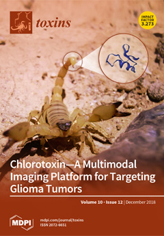
Cover Story (view full-size image): Chlorotoxin is a disulfide-rich stable peptide derived from the venom of the Israeli scorpion, Leiurus quinquestriatus. Many studies demonstrated chlorotoxin could preferentially binds to neuroectodermal tumors, such as glioblastoma, without cross-reactivity to normal brain cells. Because it has the ability to penetrate the blood–brain barrier, and its tyrosine residue allows covalent conjugation with functional moieties, chlorotoxin is an attractive platform to explore development of diagnostic and therapeutic agents for gliomas. In this review, we outline the chlorotoxin structure and its molecular targets, summarize molecular variations of chlorotoxin developed for glioma imaging, and discuss future trends and perspectives for chlorotoxin conjugates as a theranostic agent. View this paper.
  • Issues are regarded as officially published after their release is announced to the table of contents alert mailing list.
  • You may sign up for e-mail alerts to receive table of contents of newly released issues.
  • PDF is the official format for papers published in both, html and pdf forms. To view the papers in pdf format, click on the "PDF Full-text" link, and use the free Adobe Reader to open them.
Order results
Result details
Section
Select all
Export citation of selected articles as:
10 pages, 3139 KiB  
Article
Fibroblast Growth Factor Receptor, a Novel Receptor for Vegetative Insecticidal Protein Vip3Aa
by Kun Jiang, Xiaoyue Hou, Lu Han, Tongtong Tan, Zhanglei Cao and Jun Cai
Toxins 2018, 10(12), 546; https://doi.org/10.3390/toxins10120546 - 18 Dec 2018
Cited by 35 | Viewed by 4461
Abstract
Vegetative insecticidal proteins (Vips), which are secreted by some Bacillus thuringiensis strains during vegetative growth, exhibit high virulence to many pests. Vip3A proteins have been used commercially both in some bio-insecticides and in transgenic crops; however, compared with insecticidal crystal proteins, the mechanism [...] Read more.
Vegetative insecticidal proteins (Vips), which are secreted by some Bacillus thuringiensis strains during vegetative growth, exhibit high virulence to many pests. Vip3A proteins have been used commercially both in some bio-insecticides and in transgenic crops; however, compared with insecticidal crystal proteins, the mechanism of action of Vip3A is still unclear. In this work, we indicated that the fibroblast growth factor receptor-like protein (Sf-FGFR) from the membrane of Sf9 cells could bind to Vip3Aa. The interaction between Vip3Aa and Sf-FGFR was confirmed by pull-down assays and dot blotting experiment in vitro. The binding affinity between Vip3Aa and extracellular regions of Sf-FGFR (GST-FGFR-N) was determined by microscale thermophoresis assay (MST). Moreover, Vip3Aa-Flag could be co-immunoprecipitated with Sf-FGFR-V5 ex vivo. Furthermore, knockdown of Sf-FGFR gene in Sf9 cells resulted in reducing the mortality of those cells to Vip3Aa. In summary, our data indicated that Sf-FGFR is a novel receptor for Vip3Aa. Full article
(This article belongs to the Special Issue Insecticidal Toxins from Bacillus thuringiensis)
Show Figures

Figure 1

10 pages, 4129 KiB  
Communication
Biomarker of Aflatoxin Ingestion: 1H NMR-Based Plasma Metabolomics of Dairy Cows Fed Aflatoxin B1 with or without Sequestering Agents
by Ibukun Ogunade, Yun Jiang, James Adeyemi, Andre Oliveira, Diwakar Vyas and Adegbola Adesogan
Toxins 2018, 10(12), 545; https://doi.org/10.3390/toxins10120545 - 18 Dec 2018
Cited by 14 | Viewed by 4527
Abstract
The study applied 1H NMR-based plasma metabolomics to identify candidate biomarkers of aflatoxin B1 (AFB1) ingestion in dairy cows fed no sequestering agents and evaluate the effect of supplementing clay and/or a Saccharomyces cerevisiae fermentation product (SCFP) on such biomarkers. [...] Read more.
The study applied 1H NMR-based plasma metabolomics to identify candidate biomarkers of aflatoxin B1 (AFB1) ingestion in dairy cows fed no sequestering agents and evaluate the effect of supplementing clay and/or a Saccharomyces cerevisiae fermentation product (SCFP) on such biomarkers. Eight lactating cows were randomly assigned to 1 of 4 treatments in a balanced 4 × 4 Latin square design with 2 squares. Treatments were: control, toxin (T; 1725 µg AFB1/head/day), T with clay (CL; 200 g/head/day), and CL with SCFP (CL + SCFP; 35 g of SCFP/head/day). Cows in T, CL, and CL + SCFP were dosed with AFB1 from d 26 to 30. The sequestering agents were top-dressed from d 1 to 33. On d 30 of each period, 15 mL of blood was taken from the coccygeal vessels and plasma samples were prepared by centrifugation. Compared to the control, T decreased plasma concentrations of alanine, acetic acid, leucine, arginine and valine. In contrast, T increased plasma ethanol concentration 3.56-fold compared to control. Treatment with CL tended to reduce sarcosine concentration, whereas treatment with CL + SCFP increased concentrations of mannose and 12 amino acids. Based on size of the area under the curve (AUC) of receiver operating characteristic and fold change (FC) analyses, ethanol was the most significantly altered metabolite in T (AUC = 0.88; FC = 3.56); hence, it was chosen as the candidate biomarker of aflatoxin ingestion in dairy cows fed no sequestering agent. Full article
(This article belongs to the Special Issue Metabolomics in Mycotoxin Research)
Show Figures

Figure 1

19 pages, 2690 KiB  
Article
Transcriptomic Characterization of the South American Freshwater Stingray Potamotrygon motoro Venom Apparatus
by Filipe Silva, Yu Huang, Vítor Yang, Xidong Mu, Qiong Shi and Agostinho Antunes
Toxins 2018, 10(12), 544; https://doi.org/10.3390/toxins10120544 - 18 Dec 2018
Cited by 16 | Viewed by 5230
Abstract
Venomous animals are found through a wide taxonomic range including cartilaginous fish such as the freshwater stingray Potamotrygon motoro occurring in South America, which can injure people and cause venom-related symptoms. Ensuring the efficacy of drug development to treat stingray injuries can be [...] Read more.
Venomous animals are found through a wide taxonomic range including cartilaginous fish such as the freshwater stingray Potamotrygon motoro occurring in South America, which can injure people and cause venom-related symptoms. Ensuring the efficacy of drug development to treat stingray injuries can be assisted by the knowledge of the venom composition. Here we performed a detailed transcriptomic characterization of the venom gland of the South American freshwater stingray Potamotrygon motoro. The transcripts retrieved showed 418 hits to venom components (comparably to 426 and 396 hits in other two Potamotrygon species), with high expression levels of hyaluronidase, cystatin and calglandulin along with hits uniquely found in P. motoro such as DELTA-alicitoxin-Pse1b, Augerpeptide hhe53 and PI-actitoxin-Aeq3a. We also identified undescribed molecules with extremely high expression values with sequence similarity to the SE-cephalotoxin and Rapunzel genes. Comparative analyses showed that despite being closely related, there may be significant variation among the venoms of freshwater stingrays, highlighting the importance of considering elicit care in handling different envenomation cases. Since hyaluronidase represents a major component of fish venom, we have performed phylogenetic and selective pressure analyses of this gene/protein across all fish with the available information. Results indicated an independent recruitment of the hyaluronidase into the stingray venom relative to that of venomous bony fish. The hyaluronidase residues were found to be mostly under negative selection, but 18 sites showed evidence of diversifying positive selection (P < 0.05). Our data provides new insight into stingray venom variation, composition, and selective pressure in hyaluronidase. Full article
(This article belongs to the Section Animal Venoms)
Show Figures

Figure 1

16 pages, 1956 KiB  
Article
Occurrence and Levels of Aflatoxins in Fish Feeds and Their Potential Effects on Fish in Nyeri, Kenya
by Evalyn Wanjiru Mwihia, Paul Gichohi Mbuthia, Gunnar Sundstøl Eriksen, James K. Gathumbi, Joyce G. Maina, Stephen Mutoloki, Robert Maina Waruiru, Isaac Rumpel Mulei and Jan Ludvig Lyche
Toxins 2018, 10(12), 543; https://doi.org/10.3390/toxins10120543 - 17 Dec 2018
Cited by 44 | Viewed by 6793
Abstract
Aflatoxins are fungal metabolites that contaminate foods and feeds, causing adverse health effects in humans and animals. This study determined the occurrence of aflatoxins in fish feeds and their potential effects on fish. Eighty-one fish feeds were sampled from 70 farms and 8 [...] Read more.
Aflatoxins are fungal metabolites that contaminate foods and feeds, causing adverse health effects in humans and animals. This study determined the occurrence of aflatoxins in fish feeds and their potential effects on fish. Eighty-one fish feeds were sampled from 70 farms and 8 feed manufacturing plants in Nyeri, Kenya for aflatoxin analysis using competitive enzyme-linked immunosorbent assay. Fish were sampled from 12 farms for gross and microscopic pathological examination. Eighty-four percent of feeds sampled tested positive for aflatoxins, ranging from 1.8 to 39.7 µg/kg with a mean of 7.0 ± 8.3 µg/kg and the median of 3.6 µg/kg. Fifteen feeds (18.5%) had aflatoxins above the maximum allowable level in Kenya of 10 µg/kg. Homemade and tilapia feeds had significantly higher aflatoxin levels than commercial and trout feeds. Feeds containing maize bran and fish meal had significantly higher aflatoxin levels than those without these ingredients. Five trout farms (41.7%) had fish with swollen abdomens, and enlarged livers with white or yellow nodules, which microscopically had large dark basophilic hepatic cells with hyperchromatic nuclei in irregular cords. In conclusion, aflatoxin contamination of fish feeds is prevalent in Nyeri, and may be the cause of adverse health effects in fish in this region. Full article
(This article belongs to the Collection Aflatoxins)
Show Figures

Graphical abstract

19 pages, 693 KiB  
Review
Pharmacological Targeting of Pore-Forming Toxins as Adjunctive Therapy for Invasive Bacterial Infection
by Tamara Escajadillo and Victor Nizet
Toxins 2018, 10(12), 542; https://doi.org/10.3390/toxins10120542 - 17 Dec 2018
Cited by 34 | Viewed by 7614
Abstract
For many of the most important human bacterial infections, invasive disease severity is fueled by the cell damaging and pro-inflammatory effects of secreted pore-forming toxins (PFTs). Isogenic PFT-knockout mutants, e.g., Staphylococcus aureus lacking α-toxin or Streptococcus pneumoniae deficient in pneumolysin, show attenuation in [...] Read more.
For many of the most important human bacterial infections, invasive disease severity is fueled by the cell damaging and pro-inflammatory effects of secreted pore-forming toxins (PFTs). Isogenic PFT-knockout mutants, e.g., Staphylococcus aureus lacking α-toxin or Streptococcus pneumoniae deficient in pneumolysin, show attenuation in animal infection models. This knowledge has inspired multi-model investigations of strategies to neutralize PFTs or counteract their toxicity as a novel pharmacological approach to ameliorate disease pathogenesis in clinical disease. Promising examples of small molecule, antibody or nanotherapeutic drug candidates that directly bind and neutralize PFTs, block their oligomerization or membrane receptor interactions, plug establishment membrane pores, or boost host cell resiliency to withstand PFT action have emerged. The present review highlights these new concepts, with a special focus on β-PFTs produced by leading invasive human Gram-positive bacterial pathogens. Such anti-virulence therapies could be applied as an adjunctive therapy to antibiotic-sensitive and -resistant strains alike, and further could be free of deleterious effects that deplete the normal microflora. Full article
(This article belongs to the Special Issue Bacterial Pore-Forming Toxins)
Show Figures

Figure 1

16 pages, 3651 KiB  
Article
Deoxynivalenol Impairs Porcine Intestinal Host Defense Peptide Expression in Weaned Piglets and IPEC-J2 Cells
by Shuai Wang, Jiacheng Yang, Beiyu Zhang, Kuntan Wu, Ao Yang, Chong Li, Jiacai Zhang, Cong Zhang, Shahid Ali Rajput, Niya Zhang, Lvhui Sun and Desheng Qi
Toxins 2018, 10(12), 541; https://doi.org/10.3390/toxins10120541 - 15 Dec 2018
Cited by 55 | Viewed by 4650
Abstract
Host defense peptides (HDPs) are efficient defense components of the innate immune system, playing critical roles in intestinal homeostasis and protection against pathogens. This study aims to investigate the interference effects of DON on the intestinal porcine HDPs expression in piglets and intestinal [...] Read more.
Host defense peptides (HDPs) are efficient defense components of the innate immune system, playing critical roles in intestinal homeostasis and protection against pathogens. This study aims to investigate the interference effects of DON on the intestinal porcine HDPs expression in piglets and intestinal porcine epithelial cell line (IPEC-J2) cells, and elucidate the underlying mechanisms through which it functions. In an animal experiment, intestinal HDPs were determined in weaned piglets fed control and 1.28 mg/kg or 2.89 mg/kg DON-contaminated diets. Dietary exposure to DON significantly decreased piglet average daily gain, increased intestinal permeability and depressed the expression of porcine β-defensin1 (pBD1), pBD2, pBD3, epididymis protein 2 splicing variant C (pEP2C), PMAP23, and proline/arginine-rich peptide of 39 amino acids (PR39) in the intestine (p < 0.05). In IPEC-J2 cells, DON decreased cell viability and inhibited the expression of pBD1, pBD3, pEP2C, PG1-5, and PR39 (p < 0.05). NOD2, key regulator that is responsible for HDPs production, was markedly downregulated, whereas caspase-12 was activated in the presence of DON. In conclusion, DON induced caspase-12 activation and inhibited the NOD2-mediated HDPs production, which led to an impaired intestinal barrier integrity of weaned piglets. Our study provides a promising target for future therapeutic strategies to prevent the adverse effects of DON. Full article
(This article belongs to the Section Mycotoxins)
Show Figures

Figure 1

9 pages, 1399 KiB  
Article
Alternative to Animal Use for Detecting Biologically Active Staphylococcal Enterotoxin Type A
by Reuven Rasooly, Paula Do, Xiaohua He and Bradley Hernlem
Toxins 2018, 10(12), 540; https://doi.org/10.3390/toxins10120540 - 15 Dec 2018
Cited by 5 | Viewed by 3584
Abstract
Staphylococcal enterotoxins (SEs) are a food safety concern. Existing methods for biologically active SE detection rely on the emetic response in live kittens or monkeys. This method suffers from low sensitivity, poor reproducibility, and causes ethical concerns regarding the use of experimental animals. [...] Read more.
Staphylococcal enterotoxins (SEs) are a food safety concern. Existing methods for biologically active SE detection rely on the emetic response in live kittens or monkeys. This method suffers from low sensitivity, poor reproducibility, and causes ethical concerns regarding the use of experimental animals. The Lautenberg Chemical Safety Act encourages the development and adoption of alternatives to testing on animals for chemical toxicity methodologies. In this study, we utilized the superantigenic effect of SE type A (SEA) and used an ex vivo bioassay as an alternative to live animal testing. We found that interleukin-2 (IL-2) secreted by splenocyte can be utilized for quantifiable detection of SEA in food products. To avoid food matrix interference and attenuation of signal, we separated SEA from spiked food products by employing immunomagnetic beads that were coated with an anti-SEA antibody. This ex vivo method has achieved the detection of 1 ng mL−1 of SEA, which is 107 times more sensitive than the existing live animal testing methods. However, this ex vivo bioassay requires sacrificing of mice. To overcome this limitation, we established a cell based in vitro assay using CCRF-CEM, a human CD4+ T-cell line, for the quantitative detection of SEA. Incubation of SEA with CCRF-CEM human T-cells and Raji cells led to quantifiable and dose dependent secretion of IL-2. This novel cell-based assay is highly specific to biologically active SEA, compared with the related SE toxin subtypes B, D, and E or heat inactivated SEA, which produce no secretion of IL-2. This is the first demonstration of an alternative assay that completely eliminates the use of animals for quantitative detection of active SEA. Full article
(This article belongs to the Section Bacterial Toxins)
Show Figures

Figure 1

12 pages, 2659 KiB  
Article
Impact of Naja nigricollis Venom on the Production of Methaemoglobin
by Harry F. Williams, Paul Hayter, Divyashree Ravishankar, Anthony Baines, Harry J. Layfield, Lorraine Croucher, Catherine Wark, Andrew B. Bicknell, Steven Trim and Sakthivel Vaiyapuri
Toxins 2018, 10(12), 539; https://doi.org/10.3390/toxins10120539 - 15 Dec 2018
Cited by 11 | Viewed by 8708
Abstract
Snakebite envenomation is an affliction currently estimated to be killing upwards of 100,000 people annually. Snakebite is associated with a diverse pathophysiology due to the magnitude of variation in venom composition that is observed worldwide. The haemolytic (i.e., lysis of red blood cells) [...] Read more.
Snakebite envenomation is an affliction currently estimated to be killing upwards of 100,000 people annually. Snakebite is associated with a diverse pathophysiology due to the magnitude of variation in venom composition that is observed worldwide. The haemolytic (i.e., lysis of red blood cells) actions of snake venoms are well documented, although the direct impact of venoms on haemoglobin is not fully understood. Here we report on the varied ability of a multitude of snake venoms to oxidise haemoglobin into methaemoglobin. Moreover, our results demonstrate that the venom of an elapid, the black necked spitting cobra, Naja nigricollis, oxidises oxyhaemoglobin (Fe2+) into methaemoglobin (Fe3+) in a time- and concentration-dependent manner that is unparalleled within the 47 viper and elapid venoms evaluated. The treatment of venom with a reducing agent, dithiothreitol (DTT) is observed to potentiate this effect at higher concentrations, and the use of denatured venom demonstrates that this effect is dependent upon the heat-sensitive proteinaceous elements of the venom. Together, our results suggest that Naja nigricollis venom appears to promote methaemoglobin production to a degree that is rare within the Elapidae family, and this activity appears to be independent of proteolytic activities of venom components on haemoglobin. Full article
(This article belongs to the Section Animal Venoms)
Show Figures

Graphical abstract

8 pages, 2018 KiB  
Article
Complexes of the Mycotoxins Citrinin and Ochratoxin A with Aluminum Ions and their Spectroscopic Properties
by Julia Keller, Daniel Moldenhauer, Liam Byrne, Hajo Haase, Ute Resch-Genger and Matthias Koch
Toxins 2018, 10(12), 538; https://doi.org/10.3390/toxins10120538 - 14 Dec 2018
Cited by 7 | Viewed by 4005
Abstract
The sensitive detection of the mycotoxin citrinin (CIT) utilizing its fluorescence requires approaches to enhance the emission. In this respect, we studied the complexation of CIT and ochratoxin A (OTA) with Al3+ in methanol using absorption and fluorescence spectroscopy. In this context, [...] Read more.
The sensitive detection of the mycotoxin citrinin (CIT) utilizing its fluorescence requires approaches to enhance the emission. In this respect, we studied the complexation of CIT and ochratoxin A (OTA) with Al3+ in methanol using absorption and fluorescence spectroscopy. In this context, an isocratic high performance liquid chromatography (HPLC) method using a polymer column and a fluorescence detector was also developed that enables the separation of the metal ion complexes from the free ligands and non-complexed Al3+. CIT and OTA showed distinct changes in their absorption and fluorescence properties upon Al3+-coordination, and the fluorescence of CIT was considerably enhanced. Analysis of the photometrically assessed titration of CIT and OTA with Al3+ using the Job plot method revealed 1:2 and 1:1 stoichiometries for the Al3+ complexes of CIT (Al:CIT) and OTA (Al:OTA), respectively. In the case of CIT, only one β-diketone moiety participates in Al3+ coordination. These findings can be elegantly exploited for signal amplification and provide the base to reduce the limit of detection for CIT quantification by about an order of magnitude, as revealed by HPLC measurements using a fluorescence detector. Full article
(This article belongs to the Section Mycotoxins)
Show Figures

Figure 1

11 pages, 1746 KiB  
Article
A Sensitive LC-MS/MS Method for Palytoxin Using Lithium Cationization
by Mirjam D. Klijnstra and Arjen Gerssen
Toxins 2018, 10(12), 537; https://doi.org/10.3390/toxins10120537 - 14 Dec 2018
Cited by 13 | Viewed by 5205
Abstract
Palytoxin (PlTX) and analogues are produced by certain dinoflagellates, sea anemones, corals and cyanobacteria. PlTX can accumulate in the food chain and when consumed it may cause intoxication with symptoms like myalgia, weakness, fever, nausea, and vomiting. The analysis of PlTXs is challenging, [...] Read more.
Palytoxin (PlTX) and analogues are produced by certain dinoflagellates, sea anemones, corals and cyanobacteria. PlTX can accumulate in the food chain and when consumed it may cause intoxication with symptoms like myalgia, weakness, fever, nausea, and vomiting. The analysis of PlTXs is challenging, and because of the large molecular structure, it is difficult to develop a sensitive and selective liquid chromatography-mass spectrometry (LC-MS/MS) method. In this work, an LC-MS/MS method was developed to analyse PlTXs with use of lithium iodine and formic acid as additives in the mobile phase. For method development, initially, LC-hrMS was used to accurately determine the elemental composition of the precursor and product ions. The main adduct formed was [M + H + 2Li]3+. Fragments were identified with LC-hrMS and these were incorporated in the LC-MS/MS method. A method of 10 min was developed and a solid phase extraction clean-up procedure was optimised for shellfish matrix. The determined limits of detection were respectively 8 and 22 µg PlTX kg−1 for mussel and oyster matrix. Oysters gave a low recovery of approximately 50% for PlTX during extraction. The method was successfully in-house validated, repeatability had a relative standard deviation less than 20% (n = 5) at 30 µg PlTX kg−1 in mussel, cockle, and ensis, and at 60 µg PlTX kg−1 in oyster. Full article
(This article belongs to the Special Issue Emerging Marine Biotoxins)
Show Figures

Figure 1

15 pages, 2074 KiB  
Article
Further Understanding of Degradation Pathways of Microcystin-LR by an Indigenous Sphingopyxis sp. in Environmentally Relevant Pollution Concentrations
by Qin Ding, Kaiyan Liu, Kai Xu, Rongli Sun, Juan Zhang, Lihong Yin and Yuepu Pu
Toxins 2018, 10(12), 536; https://doi.org/10.3390/toxins10120536 - 14 Dec 2018
Cited by 32 | Viewed by 4447
Abstract
Microcystin-LR (MC-LR) is the most widely distributed microcystin (MC) that is hazardous to environmental safety and public health, due to high toxicity. Microbial degradation is regarded as an effective and environment-friendly method to remove it, however, the performance of MC-degrading bacteria in environmentally [...] Read more.
Microcystin-LR (MC-LR) is the most widely distributed microcystin (MC) that is hazardous to environmental safety and public health, due to high toxicity. Microbial degradation is regarded as an effective and environment-friendly method to remove it, however, the performance of MC-degrading bacteria in environmentally relevant pollution concentrations of MC-LR and the degradation pathways remain unclear. In this study, one autochthonous bacterium, Sphingopyxis sp. m6 which exhibited high MC-LR degradation ability, was isolated from Lake Taihu, and the degrading characteristics in environmentally relevant pollution concentrations were demonstrated. In addition, degradation products were identified by utilizing the full scan mode of UPLC-MS/MS. The data illustrated that strain m6 could decompose MC-LR (1–50 μg/L) completely within 4 h. The degradation rates were significantly affected by temperatures, pH and MC-LR concentrations. Moreover, except for the typical degradation products of MC-LR (linearized MC-LR, tetrapeptide, and Adda), there were 8 different products identified, namely, three tripeptides (Adda-Glu-Mdha, Glu-Mdha-Ala, and Leu-MeAsp-Arg), three dipeptides (Glu-Mdha, Mdha-Ala, and MeAsp-Arg) and two amino acids (Leu, and Arg). To our knowledge, this is the first report of Mdha-Ala, MeAsp-Arg, and Leu as MC-LR metabolites. This study expanded microbial degradation pathways of MC-LR, which lays a foundation for exploring degradation mechanisms and eliminating the pollution of microcystins (MCs). Full article
(This article belongs to the Special Issue Harmful Algal Bloom Dynamics)
Show Figures

Figure 1

14 pages, 415 KiB  
Article
AbobotulinumtoxinA (Dysport®), OnabotulinumtoxinA (Botox®), and IncobotulinumtoxinA (Xeomin®) Neurotoxin Content and Potential Implications for Duration of Response in Patients
by Malgorzata Field, Andrew Splevins, Philippe Picaut, Marcel Van der Schans, Jan Langenberg, Daan Noort and Keith Foster
Toxins 2018, 10(12), 535; https://doi.org/10.3390/toxins10120535 - 13 Dec 2018
Cited by 105 | Viewed by 17252 | Correction
Abstract
Botulinum neurotoxin type-A (BoNT-A) blocks the release of acetylcholine from peripheral cholinergic nerve terminals and is an important option for the treatment of disorders characterised by excessive cholinergic neuronal activity. Several BoNT-A products are currently marketed, each with unique manufacturing processes, excipients, formulation, [...] Read more.
Botulinum neurotoxin type-A (BoNT-A) blocks the release of acetylcholine from peripheral cholinergic nerve terminals and is an important option for the treatment of disorders characterised by excessive cholinergic neuronal activity. Several BoNT-A products are currently marketed, each with unique manufacturing processes, excipients, formulation, and non-interchangeable potency units. Nevertheless, the effects of all the products are mediated by the 150 kDa BoNT-A neurotoxin. We assessed the quantity and light chain (LC) activity of BoNT-A in three commercial BoNT-A products (Dysport®; Botox®; Xeomin®). We quantified 150 kDa BoNT-A by sandwich ELISA and assessed LC activity by EndoPep assay. In both assays, we assessed the results for the commercial products against recombinant 150 kDa BoNT-A. The mean 150 kDa BoNT-A content per vial measured by ELISA was 2.69 ng/500 U vial Dysport®, 0.90 ng/100 U vial Botox®, and 0.40 ng/100 U vial Xeomin®. To present clinically relevant results, we calculated the 150 kDa BoNT-A/US Food and Drug Administration (FDA)-approved dose in adult upper limb spasticity: 5.38 ng Dysport® (1000 U; 2 × 500 U vials), 3.60 ng Botox® (400 U; 4 × 100 U vials), and 1.61 ng Xeomin® (400 U; 4 × 100 U vials). EndoPep assay showed similar LC activity among BoNT-A products. Thus, greater amounts of active neurotoxin are injected with Dysport®, at FDA-approved doses, than with other products. This fact might explain the long duration of action reported across multiple indications, which benefits patients, caregivers, clinicians, and healthcare systems. Full article
Show Figures

Figure 1

10 pages, 1036 KiB  
Concept Paper
Biosynthetic Oligoclonal Antivenom (BOA) for Snakebite and Next-Generation Treatments for Snakebite Victims
by R. Manjunatha Kini, Sachdev S. Sidhu and Andreas Hougaard Laustsen
Toxins 2018, 10(12), 534; https://doi.org/10.3390/toxins10120534 - 13 Dec 2018
Cited by 64 | Viewed by 6973
Abstract
Snakebite envenoming is a neglected tropical disease that each year claims the lives of 80,000–140,000 victims worldwide. The only effective treatment against envenoming involves intravenous administration of antivenoms that comprise antibodies that have been isolated from the plasma of immunized animals, typically horses. [...] Read more.
Snakebite envenoming is a neglected tropical disease that each year claims the lives of 80,000–140,000 victims worldwide. The only effective treatment against envenoming involves intravenous administration of antivenoms that comprise antibodies that have been isolated from the plasma of immunized animals, typically horses. The drawbacks of such conventional horse-derived antivenoms include their propensity for causing allergenic adverse reactions due to their heterologous and foreign nature, an inability to effectively neutralize toxins in distal tissue, a low content of toxin-neutralizing antibodies, and a complex manufacturing process that is dependent on husbandry and procurement of snake venoms. In recent years, an opportunity to develop a fundamentally novel type of antivenom has presented itself. By using modern antibody discovery strategies, such as phage display selection, and repurposing small molecule enzyme inhibitors, next-generation antivenoms that obviate the drawbacks of existing plasma-derived antivenoms could be developed. This article describes the conceptualization of a novel therapeutic development strategy for biosynthetic oligoclonal antivenom (BOA) for snakebites based on recombinantly expressed oligoclonal mixtures of human monoclonal antibodies, possibly combined with repurposed small molecule enzyme inhibitors. Full article
Show Figures

Figure 1

17 pages, 2602 KiB  
Article
Post-Harvest Contamination with Mycotoxins in the Context of the Geographic and Agroclimatic Conditions in Romania
by Valeria Gagiu, Elena Mateescu, Ileana Armeanu, Alina Alexandra Dobre, Irina Smeu, Mirela Elena Cucu, Oana Alexandra Oprea, Enuta Iorga and Nastasia Belc
Toxins 2018, 10(12), 533; https://doi.org/10.3390/toxins10120533 - 13 Dec 2018
Cited by 25 | Viewed by 6278
Abstract
This study aimed to assess post-harvest contamination with mycotoxins in the context of the geographic and agroclimatic conditions in Romania in 2012–2015, a period that was characterized by extreme meteorological events and the effects of climate change. The samples were randomly sampled from [...] Read more.
This study aimed to assess post-harvest contamination with mycotoxins in the context of the geographic and agroclimatic conditions in Romania in 2012–2015, a period that was characterized by extreme meteorological events and the effects of climate change. The samples were randomly sampled from five agricultural regions of Romania and analyzed for mycotoxins by enzyme-linked immunosorbent assay. An SPSS analysis was done to explore correlations between mycotoxins (deoxynivalenol—DON, aflatoxins—AF, ochratoxin A—OTA, zearalenone—ZEA), product types (raw cereal, processed cereal, cereal-based food), geographic coordinates (latitude, longitude, agricultural region), and agroclimatic factors (air temperature, precipitation, soil moisture reserve, aridity index, soil type). In the southeast part of the Southern Plain and Dobrogea (Baragan Plain, located at 44–45° N, 26–27° E), contamination with AF and OTA was higher in raw and processed cereals (maize in silo, silo corn germs) in the dry years (2012 and 2013), and contamination with DON was high in processed cereals (wheat flour type 450) in the rainy year (2014). DON and OTA contamination were significantly correlated with cumulative precipitation in all years, while AF and ZEA contamination were non-significantly correlated with climatic factors and aridity indices. The distribution of mycotoxins by product type and the non-robust correlations between post-harvest mycotoxins and agrometeorological factors could be explained by the use of quality management systems that control cereal at warehouse receptions, performant processing technologies, and the quality of storage spaces of agri-food companies. The Baragan Plain is Romania’s most sensitive area to the predicted climate change in southeast Europe, which may be associated with its increased cereal contamination with AF and OTA. Full article
(This article belongs to the Special Issue Fungal Growth and Mycotoxins: Challenges for developing countries)
Show Figures

Figure 1

9 pages, 2099 KiB  
Article
Spatiotemporal Gait Analysis and Lower Limb Functioning in Foot Dystonia Treated with Botulinum Toxin
by Anupam Datta Gupta, Graeme Tucker, Simon Koblar, Renuka Visvanathan and Ian D. Cameron
Toxins 2018, 10(12), 532; https://doi.org/10.3390/toxins10120532 - 12 Dec 2018
Cited by 15 | Viewed by 4906
Abstract
Foot dystonia (FD) is a disabling condition causing pain, spasm and difficulty in walking. We treated fourteen (14) adult patients experiencing FD with onabotulinum toxin A injection into the dystonic foot muscles. We analyzed the spatiotemporal gait utilizing the GaitRite system pre- and [...] Read more.
Foot dystonia (FD) is a disabling condition causing pain, spasm and difficulty in walking. We treated fourteen (14) adult patients experiencing FD with onabotulinum toxin A injection into the dystonic foot muscles. We analyzed the spatiotemporal gait utilizing the GaitRite system pre- and 3 weeks post-botulinum toxin injection along with measuring dystonia by the Fahn–Marsden Dystonia Scale (FMDS), pain by the Visual Analog Scale (VAS) and other lower limb functional outcomes such as gait velocity, the Berg Balance Scale (BBS), the Unified Parkinson’s Disease Rating Scale–Lower Limb Score (UPDRS–LL), the Timed Up and Go (TUG) test and the Goal Attainment Scale (GAS). We found that stride length increased significantly in both the affected (p = 0.02) and unaffected leg (p = 0.01) after treatment, and the improvement in stride length was roughly the same in each leg. Similar results were found for step length (p = 0.02) with improvement in the step length differential (p = 0.01). The improvements in the lower limb functional outcomes were also significant—FMDS, VAS, TUG, and UPDRS–LL decreased significantly after treatment (all p < 0.001), and BBS (p = 0.001), GAS (p < 0.001) except cadence (p = 0.37). BT injection improved walking in foot dystonia as evidenced through gait analysis, pain and lower limb functional outcomes. Main study limitations were small sample size and lack of control. Full article
(This article belongs to the Special Issue Botulinum Toxin Treatment of Movement Disorders)
Show Figures

Figure 1

11 pages, 3034 KiB  
Article
Acute Toxicity of the Recombinant and Native Phα1β Toxin: New Analgesic from Phoneutria nigriventer Spider Venom
by Eliane Dallegrave, Eliane Taschetto, Mirna Bainy Leal, Flavia Tasmim Techera Antunes, Marcus Vinicius Gomez and Alessandra Hubner de Souza
Toxins 2018, 10(12), 531; https://doi.org/10.3390/toxins10120531 - 12 Dec 2018
Cited by 11 | Viewed by 3662
Abstract
Phα1β, a purified peptide from the venom of the spider Phoneutria nigriventer, and its recombinant form CTK 01512-2 are voltage-dependent calcium channel (CaV) blockers of types N, R, P/Q, and L with a preference for type N. These peptides [...] Read more.
Phα1β, a purified peptide from the venom of the spider Phoneutria nigriventer, and its recombinant form CTK 01512-2 are voltage-dependent calcium channel (CaV) blockers of types N, R, P/Q, and L with a preference for type N. These peptides show analgesic action in different pain models in rats. The aim of this study was to evaluate the acute intrathecal toxicity of the native and recombinant Phα1β toxin in Wistar rats. Clinical signs, serum biochemistry, organ weight, and histopathological alterations were evaluated in male and/or female rats. Dyspnea was observed in males, hyporesponsiveness in females, and Straub tail and tremors in both genders. There were no significant differences in male organ weight, although significant differences in the female relative weight of the adrenal glands and spleen have been observed; these values are within the normal range. Serum biochemical data revealed a significant reduction within the physiological limits of species related to urea, ALT, AST, and FA. Hepatic and renal congestion were observed for toxin groups. In renal tissue, glomerular infiltrates were observed with increased glomerular space. These histological alterations were presented in focal areas and in mild degree. Therefore, Phα1β and CTK 01512-2 presented a good safety profile with transient toxicity clinical signals in doses higher than used to obtain the analgesic effect. Full article
Show Figures

Figure 1

11 pages, 1041 KiB  
Article
Chestnut Drying Is Critical in Determining Aspergillus flavus Growth and Aflatoxin Contamination
by Simona Prencipe, Ilenia Siciliano, Carlotta Gatti, Maria Lodovica Gullino, Angelo Garibaldi and Davide Spadaro
Toxins 2018, 10(12), 530; https://doi.org/10.3390/toxins10120530 - 11 Dec 2018
Cited by 11 | Viewed by 4055
Abstract
Chestnut drying is used to prevent postharvest losses and microorganism contamination during storage. Several studies reported the contamination by aflatoxins (AFs) produced by Aspergillus spp. in chestnuts. The effect of drying temperatures (from 30 to 50 °C) was evaluated on the growth of [...] Read more.
Chestnut drying is used to prevent postharvest losses and microorganism contamination during storage. Several studies reported the contamination by aflatoxins (AFs) produced by Aspergillus spp. in chestnuts. The effect of drying temperatures (from 30 to 50 °C) was evaluated on the growth of A. flavus and the production of aflatoxins in chestnuts. The influence of the treatment on the proximate composition, the total phenol content and antioxidant activity of chestnuts was considered. Fungal colonization was observed on the nuts dried at 30, 35, and 40 °C; the incidence was lower at 40 °C. The highest concentrations of AFB1 and AFB2 were produced at 40 °C. No aflatoxins were detected at 45 or 50 °C. At 40 °C A. flavus was under suboptimal conditions for growth (aw 0.78), but the fungus was able to synthesize aflatoxins. As the temperatures applied increased, the total phenol content increased, while the antioxidant activity decreased. A drying treatment at 45 °C for seven days (aw 0.64) could be a promising method to effectively control both the growth of aflatoxigenic fungi and the production of aflatoxins. This study provides preliminary data useful to improve the current drying conditions used in chestnut mills, to reduce both fungal growth and aflatoxin production. Full article
(This article belongs to the Collection Aflatoxins)
Show Figures

Figure 1

12 pages, 1874 KiB  
Article
Response Surface Methodology for the Optimization of Ultrasound-Assisted Extraction of Tetrodotoxin from the Liver of Takifugu pseudommus
by Xiaojun Zhang, Chengcheng Han, Si Chen, Le Li, Jingjing Zong, Junjie Zeng and Guangming Mei
Toxins 2018, 10(12), 529; https://doi.org/10.3390/toxins10120529 - 10 Dec 2018
Cited by 8 | Viewed by 4472
Abstract
Tetrodotoxin (TTX) is a marine biotoxin that has high scientific value. However, the lack of efficient TTX extraction and preparation methods has led to a scarcity of TTX samples for clinical application. In this study, TTX from the liver of Takifugu pseudommus was [...] Read more.
Tetrodotoxin (TTX) is a marine biotoxin that has high scientific value. However, the lack of efficient TTX extraction and preparation methods has led to a scarcity of TTX samples for clinical application. In this study, TTX from the liver of Takifugu pseudommus was ultrasound-assisted extracted with acidified organic solvents. The extraction process was analyzed and optimized by single factor method and response surface methodology (RSM). The optimal extraction conditions predicted by a response surface model were as follows: liquid:material ratio, 2.8:1; extraction temperature, 60 °C; extraction time, 23.3 min. Under these conditions, the extraction of TTX had a yield of 89.65%, and the results were further verified by experimental extraction, and analyzed by ultra performance liquid chromatography–tandem mass spectrometry (UPLC–MS/MS). It was found that the extracts of T. pseudommus liver contained TTX and its four analogues at certain proportions (TTX: 10.4%; 5,6,11-trideoxyTTX: 83.3%; 5,11-dideoxyTTX:2.4%; 4,9-anhydro TTX:2.6%; 5-deoxyTTX:1.3%). This study demonstrates a stable and efficient extraction process of TTX from pufferfish liver, which can be helpful for further research and analysis, as well as the utilization of TTX from pufferfish. Full article
(This article belongs to the Special Issue Tetrodotoxins)
Show Figures

Figure 1

14 pages, 3770 KiB  
Article
Aspergillus flavus NRRL 3251 Growth, Oxidative Status, and Aflatoxins Production Ability In Vitro under Different Illumination Regimes
by Tihomir Kovač, Bojan Šarkanj, Biljana Crevar, Marija Kovač, Ante Lončarić, Ivica Strelec, Chibundu N. Ezekiel, Michael Sulyok and Rudolf Krska
Toxins 2018, 10(12), 528; https://doi.org/10.3390/toxins10120528 - 10 Dec 2018
Cited by 16 | Viewed by 4070
Abstract
Aspergillus flavus is the most important mycotoxin-producing fungus involved in the global episodes of aflatoxin B1 contamination of crops at both the pre-harvest and post-harvest stages. However, in order to effectively control aflatoxin contamination in crops using antiaflatoxigenic and/or antifungal compounds, some [...] Read more.
Aspergillus flavus is the most important mycotoxin-producing fungus involved in the global episodes of aflatoxin B1 contamination of crops at both the pre-harvest and post-harvest stages. However, in order to effectively control aflatoxin contamination in crops using antiaflatoxigenic and/or antifungal compounds, some of which are photosensitive, a proper understanding of the photo-sensitive physiology of potential experimental strains need to be documented. The purpose of the study is therefore to evaluate the effect of visible (VIS) light illumination on growth and conidiation, aflatoxin production ability and modulation of A. flavus oxidative status during in vitro experiment. Aflatoxigenic A. flavus strain was inoculated in aflatoxin-inducing YES media and incubated under three different VIS illumination regimes during a 168 h growth period at 29 °C. VIS illumination reduced A. flavus mycelia biomass yield, both during growth on plates and in liquid media, promoted conidiation and increased the aflatoxin production. Furthermore, aflatoxin production increased with increased reactive oxidative species (ROS) levels at 96 h of growth, confirming illumination-driven oxidative stress modulation activity on A. flavus cells. Full article
(This article belongs to the Collection Aflatoxins)
Show Figures

Figure 1

17 pages, 3545 KiB  
Article
Cyanobacterial Neurotoxin Beta-Methyl-Amino-l-Alanine Affects Dopaminergic Neurons in Optic Ganglia and Brain of Daphnia magna
by Megan Brooke-Jones, Martina Gáliková and Heinrich Dircksen
Toxins 2018, 10(12), 527; https://doi.org/10.3390/toxins10120527 - 8 Dec 2018
Cited by 12 | Viewed by 4500
Abstract
The non-proteinogenic amino acid beta-methyl-amino-l-alanine (BMAA) is a neurotoxin produced by cyanobacteria. BMAA accumulation in the brain of animals via biomagnification along the food web can contribute to the development of neurodegenerative diseases such as Amyotrophic lateral sclerosis/Parkinsonism dementia complex (ALS/PDC), [...] Read more.
The non-proteinogenic amino acid beta-methyl-amino-l-alanine (BMAA) is a neurotoxin produced by cyanobacteria. BMAA accumulation in the brain of animals via biomagnification along the food web can contribute to the development of neurodegenerative diseases such as Amyotrophic lateral sclerosis/Parkinsonism dementia complex (ALS/PDC), the latter being associated with a loss of dopaminergic neurons. Daphnia magna is an important microcrustacean zooplankton species that plays a key role in aquatic food webs, and BMAA-producing cyanobacteria often form part of their diet. Here, we tested the effects of BMAA on putative neurodegeneration of newly identified specific dopaminergic neurons in the optic ganglia/brain complex of D. magna using quantitative tyrosine-hydroxylase immunohistochemistry and fluorescence cytometry. The dopaminergic system was analysed in fed and starved isogenic D. magna adults incubated under different BMAA concentrations over 4 days. Increased BMAA concentration showed significant decrease in the stainability of dopaminergic neurons of D. magna, with fed animals showing a more extreme loss. Furthermore, higher BMAA concentrations tended to increase offspring mortality during incubation. These results are indicative of ingested BMAA causing neurodegeneration of dopaminergic neurons in D. magna and adversely affecting reproduction. This may imply similar effects of BMAA on known human neurodegenerative diseases involving dopaminergic neurons. Full article
(This article belongs to the Special Issue Emerging Marine Biotoxins)
Show Figures

Graphical abstract

14 pages, 1522 KiB  
Article
Combined Cytotoxicity of the Phycotoxin Okadaic Acid and Mycotoxins on Intestinal and Neuroblastoma Human Cell Models
by Aiko Hayashi, Juan José Dorantes-Aranda, John P. Bowman and Gustaaf Hallegraeff
Toxins 2018, 10(12), 526; https://doi.org/10.3390/toxins10120526 - 8 Dec 2018
Cited by 18 | Viewed by 5700
Abstract
Mycotoxins are emerging toxins in the marine environment, which can co-occur with algal toxins to exert synergistic or antagonistic effects for human seafood consumption. The current study assesses the cytotoxicity of the algal toxin okadaic acid, shellfish, and dust storm-associated mycotoxins alone or [...] Read more.
Mycotoxins are emerging toxins in the marine environment, which can co-occur with algal toxins to exert synergistic or antagonistic effects for human seafood consumption. The current study assesses the cytotoxicity of the algal toxin okadaic acid, shellfish, and dust storm-associated mycotoxins alone or in combination on human intestinal (HT-29) and neuroblastoma (SH-SY5Y) cell lines. Based on calculated IC50 (inhibitory concentration 50%) values, mycotoxins and the algal toxin on their own exhibited increased cytotoxicity in the order of sydowinin A < sydowinin B << patulin < alamethicin < sydowinol << gliotoxin ≈ okadaic acid against the HT-29 cell line, and sydowinin B < sydowinin A << alamethicin ≈ sydowinol < patulin, << gliotoxin < okadaic acid against the SH-SY5Y cell line. Combinations of okadaic acid–sydowinin A, –alamethicin, –patulin, and –gliotoxin exhibited antagonistic effects at low-moderate cytotoxicity, but became synergistic at high cytotoxicity, while okadaic acid–sydowinol displayed an antagonistic relationship against HT-29 cells. Furthermore, only okadaic acid–sydowinin A showed synergism, while okadaic acid–sydowinol, –alamethicin, –patulin, and –gliotoxin combinations demonstrated antagonism against SH-SY5Y. While diarrhetic shellfish poisoning (DSP) from okadaic acid and analogues in many parts of the world is considered to be a comparatively minor seafood toxin syndrome, our human cell model studies suggest that synergisms with certain mycotoxins may aggravate human health impacts, depending on the concentrations. These findings highlight the issues of the shortcomings of current regulatory approaches, which do not regulate for mycotoxins in shellfish and treat seafood toxins as if they occur as single toxins. Full article
(This article belongs to the Special Issue Marine Biotoxins and Seafood Poisoning)
Show Figures

Graphical abstract

18 pages, 4452 KiB  
Article
Occurrence and Identification of Aspergillus Section Flavi in the Context of the Emergence of Aflatoxins in French Maize
by Sylviane Bailly, Anwar El Mahgubi, Amaranta Carvajal-Campos, Sophie Lorber, Olivier Puel, Isabelle P. Oswald, Jean-Denis Bailly and Béatrice Orlando
Toxins 2018, 10(12), 525; https://doi.org/10.3390/toxins10120525 - 7 Dec 2018
Cited by 40 | Viewed by 4371
Abstract
Aflatoxins (AFs) are secondary metabolites produced by Aspergillus section Flavi during their development, particularly in maize. It is widely accepted that AFB1 is a major contaminant in regions where hot climate conditions favor the development of aflatoxigenic species. Global warming could lead to [...] Read more.
Aflatoxins (AFs) are secondary metabolites produced by Aspergillus section Flavi during their development, particularly in maize. It is widely accepted that AFB1 is a major contaminant in regions where hot climate conditions favor the development of aflatoxigenic species. Global warming could lead to the appearance of AFs in maize produced in Europe. This was the case in 2015, in France, when the exceptionally hot and dry climatic conditions were favorable for AF production. Our survey revealed AF contamination of 6% (n = 114) of maize field samples and of 15% (n = 81) of maize silo samples analyzed. To understand the origin of the contamination, we characterized the mycoflora in contaminated samples and in samples produced in the same geographic and climatic conditions but with no AFs. A special focus was placed on Aspergillus section Flavi. A total of 67 strains of Aspergillus section Flavi were isolated from the samples. As expected, the strains were observed in all AF+ samples and, remarkably, also in almost 40% of AF− samples, demonstrating the presence of these potent toxin producers in fields in France. A. flavus was the most frequent species of the section Flavi (69% of the strains). But surprisingly, A. parasiticus was also a frequent contaminant (28% of the strains), mostly isolated from AF+ samples. This finding is in agreement with the presence of AFG in most of those samples. Full article
(This article belongs to the Section Mycotoxins)
Show Figures

Figure 1

10 pages, 1034 KiB  
Article
New Insights into the Occurrence and Toxin Profile of Ciguatoxins in Selvagens Islands (Madeira, Portugal)
by Pedro Reis Costa, Pablo Estevez, David Castro, Lucía Soliño, Neide Gouveia, Carolina Santos, Susana Margarida Rodrigues, José Manuel Leao and Ana Gago-Martínez
Toxins 2018, 10(12), 524; https://doi.org/10.3390/toxins10120524 - 7 Dec 2018
Cited by 42 | Viewed by 5358
Abstract
Ciguatoxins (CTXs), endemic from tropical and subtropical regions of the Pacific and Indian Ocean and the Caribbean Sea, have caused several human poisonings during the last decade in Europe. Ciguatera fish poisonings (CFP) in Madeira and Canary Islands appear to be particularly related [...] Read more.
Ciguatoxins (CTXs), endemic from tropical and subtropical regions of the Pacific and Indian Ocean and the Caribbean Sea, have caused several human poisonings during the last decade in Europe. Ciguatera fish poisonings (CFP) in Madeira and Canary Islands appear to be particularly related with consumption of fish caught close to Selvagens Islands, a Portuguese natural reserve composed of three small islands that harbor high fish biomass. In this study, fish specimens considered as potential vectors of CTXs were caught in Madeira and Selvagens archipelagos for toxins determination via sensitive liquid chromatography with tandem mass spectrometry detection (LC–MS/MS). CTXs were found in most of the fish samples from Selvagens and none from Madeira. Caribbean ciguatoxin-1 (C-CTX1) was the only toxin congener determined, reaching the highest value of 0.25 µg C-CTX1 kg−1 in a 4.6 kg island grouper (Mycteroperca fusca). This study indicates that a diversity of fish from different trophic levels contains CTXs, Selvagens appear to be one of the most favorable locations for CTXs food web transfer and finally, this study highlights the need of further research based on intensive environmental and biological sampling on these remote islands. Full article
(This article belongs to the Special Issue Emerging Marine Biotoxins)
Show Figures

Figure 1

11 pages, 2432 KiB  
Article
Characterization of A Staphylococcal Food Poisoning Outbreak in A Workplace Canteen during the Post-Earthquake Reconstruction of Central Italy
by Fabrizia Guidi, Anna Duranti, Silvia Gallina, Yacine Nia, Annalisa Petruzzelli, Angelo Romano, Valeria Travaglini, Alberto Olivastri, Vincenzo Calvaresi, Lucia Decastelli and Giuliana Blasi
Toxins 2018, 10(12), 523; https://doi.org/10.3390/toxins10120523 - 6 Dec 2018
Cited by 25 | Viewed by 5621
Abstract
In summer 2017, a foodborne outbreak occurred in Central Italy, involving 26 workers employed in the post-earthquake reconstruction. After eating a meal provided by a catering service, they manifested gastrointestinal symptoms; 23 of them were hospitalized. The retrospective cohort study indicated the pasta [...] Read more.
In summer 2017, a foodborne outbreak occurred in Central Italy, involving 26 workers employed in the post-earthquake reconstruction. After eating a meal provided by a catering service, they manifested gastrointestinal symptoms; 23 of them were hospitalized. The retrospective cohort study indicated the pasta salad as the most likely vehicle of poisoning. Foods, environmental samples, and food handlers’ nasal swabs were collected. Bacillus cereus (Bc) and coagulase-positive staphylococci (CPS) including S. aureus, together with their toxins, were the targets of the analysis. CPS, detected in all the leftovers, exceeded 105 CFU/g in the pasta salad, in which we found Staphylococcal Enterotoxins (SEs) (0.033 ng SEA/g; 0.052 ng SED/g). None of the environmental and human swabs showed contamination. We characterized 23 S. aureus from foods. They all belonged to the human biotype, showed the same toxigenic profile (sea, sed, sej, and ser genes), and had the same Pulsed Field Gel Electrophoresis (PFGE) pattern; none of them harbored mecA or mupA genes. We also detected Bc contamination in the pasta salad but none of the isolates harbored the ces gene for the emetic toxin cereulide. The EU Reference Laboratory for CPS confirmed the case as a strong-evidence outbreak caused by the ingestion of SEs produced by a single strain of S. aureus carried by the same human source. This outbreak was successfully investigated despite the emergency situation in which it occurred. Full article
(This article belongs to the Collection Bacterial Enterotoxins)
Show Figures

Figure 1

19 pages, 3465 KiB  
Article
Loxosceles gaucho Spider Venom: An Untapped Source of Antimicrobial Agents
by Paula J Segura-Ramírez and Pedro I Silva Júnior
Toxins 2018, 10(12), 522; https://doi.org/10.3390/toxins10120522 - 6 Dec 2018
Cited by 19 | Viewed by 5607
Abstract
The remarkable ability of microorganisms to develop resistance to conventional antibiotics is one of the biggest challenges that the pharmaceutical industry currently faces. Recent studies suggest that antimicrobial peptides discovered in spider venoms may be useful resources for the design of structurally new [...] Read more.
The remarkable ability of microorganisms to develop resistance to conventional antibiotics is one of the biggest challenges that the pharmaceutical industry currently faces. Recent studies suggest that antimicrobial peptides discovered in spider venoms may be useful resources for the design of structurally new anti-infective agents effective against drug-resistant microorganisms. In this work, we found an anionic antibacterial peptide named U1-SCRTX-Lg1a in the venom of the spider Loxosceles gaucho. The peptide was purified using high-performance liquid chromatography (HPLC), its antimicrobial activity was tested through liquid growth inhibition assays, and its chemical properties were characterized using mass spectrometry. U1-SCRTX-Lg1a was found to show a monoisotopic mass of 1695.75 Da, activity against Gram-negative bacteria, a lack of hemolytic effects against human red blood cells, and a lack of cytotoxicity against human cervical carcinoma cells (HeLa). Besides this, the sequence of the peptide exhibited great similarity to specific regions of phospholipases D from different species of Loxosceles spiders, leading to the hypothesis that U1-SCRTX-Lg1a may have originated from a limited proteolytic cleavage. Our data suggest that U1-SCRTX-Lg1a is a promising candidate for the development of new antibiotics that could help fight bacterial infections and represents an exciting discovery for Loxosceles spiders. Full article
(This article belongs to the Section Animal Venoms)
Show Figures

Figure 1

19 pages, 2752 KiB  
Article
Alternative Splicing of the Aflatoxin-Associated Baeyer–Villiger Monooxygenase from Aspergillus flavus: Characterisation of MoxY Isoforms
by Carmien Tolmie, Martha S. Smit and Diederik J. Opperman
Toxins 2018, 10(12), 521; https://doi.org/10.3390/toxins10120521 - 5 Dec 2018
Cited by 3 | Viewed by 3646
Abstract
Aflatoxins are carcinogenic mycotoxins that are produced by the filamentous fungus Aspergillus flavus, a contaminant of numerous food crops. Aflatoxins are synthesised via the aflatoxin biosynthesis pathway, with the enzymes involved encoded by the aflatoxin biosynthesis gene cluster. MoxY is a type [...] Read more.
Aflatoxins are carcinogenic mycotoxins that are produced by the filamentous fungus Aspergillus flavus, a contaminant of numerous food crops. Aflatoxins are synthesised via the aflatoxin biosynthesis pathway, with the enzymes involved encoded by the aflatoxin biosynthesis gene cluster. MoxY is a type I Baeyer–Villiger monooxygenase (BVMO), responsible for the conversion of hydroxyversicolorone (HVN) and versicolorone (VN) to versiconal hemiacetal acetate (VHA) and versiconol acetate (VOAc), respectively. Using mRNA data, an intron near the C-terminus was identified that is alternatively spliced, creating two possible MoxY isoforms which exist in vivo, while analysis of the genomic DNA suggests an alternative start codon leading to possible elongation of the N-terminus. These four variants of the moxY gene were recombinantly expressed in Escherichia coli, and their activity evaluated with respect to their natural substrates HVN and VN, as well as surrogate ketone substrates. Activity of the enzyme is absolutely dependent on the additional 22 amino acid residues at the N-terminus. Two MoxY isoforms with alternative C-termini, MoxYAltN and MoxYAltNC, converted HVN and VN, in addition to a range of ketone substrates. Stability and flavin-binding data suggest that MoxYAltN is, most likely, the dominant isoform. MoxYAltNC is generated by intron splicing, in contrast to intron retention, which is the most prevalent type of alternative splicing in ascomycetes. The alternative C-termini did not alter the substrate acceptance profile, or regio- or enantioselectivity of the enzyme, but did significantly affect the solubility and stability. Full article
(This article belongs to the Collection Aflatoxins)
Show Figures

Figure 1

13 pages, 2038 KiB  
Article
Association between Protein-Bound Uremic Toxins and Asymptomatic Cardiac Dysfunction in Patients with Chronic Kidney Disease
by Shanmugakumar Chinnappa, Yu-Kang Tu, Yi Chun Yeh, Griet Glorieux, Raymond Vanholder and Andrew Mooney
Toxins 2018, 10(12), 520; https://doi.org/10.3390/toxins10120520 - 5 Dec 2018
Cited by 23 | Viewed by 4028
Abstract
Although the relationship between protein-bound uremic toxins (PBUTs) and cardiac structure and cardiac mortality in chronic kidney disease (CKD) has been studied in the past, the association between cardiac dysfunction and PBUTs has not yet been studied. We therefore evaluated the association between [...] Read more.
Although the relationship between protein-bound uremic toxins (PBUTs) and cardiac structure and cardiac mortality in chronic kidney disease (CKD) has been studied in the past, the association between cardiac dysfunction and PBUTs has not yet been studied. We therefore evaluated the association between impaired peak cardiac performance and the serum free and total concentrations of potentially cardiotoxic PBUTs. In a cross-sectional study of 56 male CKD patients (stages 2–5 (pre-dialysis)) who were asymptomatic with no known cardiac diseases or diabetes we measured peak cardiac power (CPOmax), aerobic exercise capacity (VO2max), and echocardiographic parameters of cardiac morphology and evaluated their association with PBUTs. The serum total and free concentrations of indoxyl sulfate (IXS), p-cresyl sulfate (PCS), p-cresyl glucuronide, indole acetic acid, and hippuric acid showed significant negative correlation with CPOmax and VO2max. IXS and PCS were independently associated with CPOmax and VO2max even after controlling for eGFR. No correlation between left ventricular mass index (LVMI) and PBUTs was seen. The present study for the first time has demonstrated the association between subclinical cardiac dysfunction in CKD and serum levels of a panel of PBUTs. Further studies are required to evaluate the mechanism of cardiotoxicity of the individual uremic toxins. Full article
(This article belongs to the Special Issue Uremia and Cardiovascular Disease)
Show Figures

Figure 1

14 pages, 3651 KiB  
Article
Improvement of the Culture Medium for the Dichlorvos-Ammonia (DV-AM) Method to Selectively Detect Aflatoxigenic Fungi in Soil
by Kimiko Yabe, Haruna Ozaki, Takuya Maruyama, Keisuke Hayashi, Yuki Matto, Marika Ishizaka, Takeru Makita, Syun-ya Noma, Kousuke Fujiwara and Masayo Kushiro
Toxins 2018, 10(12), 519; https://doi.org/10.3390/toxins10120519 - 5 Dec 2018
Cited by 5 | Viewed by 3799
Abstract
The dichlorvos-ammonia (DV-AM) method is a simple but sensitive visual method for detecting aflatoxigenic fungi. Here we sought to develop a selective medium that is appropriate for the growth of aflatoxigenic fungi among soil mycoflora. We examined the effects of different concentrations of [...] Read more.
The dichlorvos-ammonia (DV-AM) method is a simple but sensitive visual method for detecting aflatoxigenic fungi. Here we sought to develop a selective medium that is appropriate for the growth of aflatoxigenic fungi among soil mycoflora. We examined the effects of different concentrations of carbon sources (sucrose and glucose) and detergents (deoxycholate (DOC), Triton X-100, and Tween 80) on microorganisms in soils, using agar medium supplemented with chloramphenicol. The results demonstrated that 5–10% sucrose concentrations and 0.1–0.15% DOC concentrations were appropriate for the selective detection of aflatoxigenic fungi in soil. We also identified the optimal constituents of the medium on which the normal rapid growth of Rhizopus sp. was completely inhibited. By using the new medium along with the DV-AM method, we succeeded in the isolation of aflatoxigenic fungi from non-agricultural fields in Fukui city, Japan. The fungi were identified as Aspergillus nomius based on their calmodulin gene sequences. These results indicate that the new medium will be useful in practice for the detection of aflatoxigenic fungi in soil samples including those from non-agricultural environments. Full article
(This article belongs to the Section Mycotoxins)
Show Figures

Figure 1

20 pages, 638 KiB  
Review
Phylogenetic Comparative Methods can Provide Important Insights into the Evolution of Toxic Weaponry
by Kevin Arbuckle
Toxins 2018, 10(12), 518; https://doi.org/10.3390/toxins10120518 - 5 Dec 2018
Cited by 9 | Viewed by 5734
Abstract
The literature on chemical weaponry of organisms is vast and provides a rich understanding of the composition and mechanisms of the toxins and other components involved. However, an ecological or evolutionary perspective has often been lacking and is largely limited to (1) molecular [...] Read more.
The literature on chemical weaponry of organisms is vast and provides a rich understanding of the composition and mechanisms of the toxins and other components involved. However, an ecological or evolutionary perspective has often been lacking and is largely limited to (1) molecular evolutionary studies of particular toxins (lacking an ecological view); (2) comparisons across different species that ignore phylogenetic relatedness (lacking an evolutionary view); or (3) descriptive studies of venom composition and toxicology that contain post hoc and untested ecological or evolutionary interpretations (a common event but essentially uninformative speculation). Conveniently, comparative biologists have prolifically been developing and using a wide range of phylogenetic comparative methods that allow us to explicitly address many ecological and evolutionary questions relating to venoms and poisons. Nevertheless, these analytical tools and approaches are rarely used and poorly known by biological toxinologists and toxicologists. In this review I aim to (1) introduce phylogenetic comparative methods to the latter audience; (2) highlight the range of questions that can be addressed using them; and (3) encourage biological toxinologists and toxicologists to either seek out adequate training in comparative biology or seek collaboration with comparative biologists to reap the fruits of a powerful interdisciplinary approach to the field. Full article
(This article belongs to the Special Issue Toxins:10th Anniversary)
Show Figures

Figure 1

21 pages, 2030 KiB  
Article
Transfer of Deoxynivalenol (DON) through Placenta, Colostrum and Milk from Sows to Their Offspring during Late Gestation and Lactation
by Amin Sayyari, Silvio Uhlig, Christiane Kruse Fæste, Tore Framstad and Tore Sivertsen
Toxins 2018, 10(12), 517; https://doi.org/10.3390/toxins10120517 - 4 Dec 2018
Cited by 13 | Viewed by 4227
Abstract
Deoxynivalenol (DON) contamination of feed may result in reduced growth, feed refusal, immunosuppression, and health problems in swine. Piglets can be exposed to DON via placenta before birth and via milk during lactation. The extent of early-life exposure of piglets to DON is, [...] Read more.
Deoxynivalenol (DON) contamination of feed may result in reduced growth, feed refusal, immunosuppression, and health problems in swine. Piglets can be exposed to DON via placenta before birth and via milk during lactation. The extent of early-life exposure of piglets to DON is, however, not fully known. This study was therefore aimed at investigating DON uptake in sows fed with naturally contaminated diets, DON transfer across placenta during late gestation, and transfer of DON to piglets via colostrum and milk. Forty-four crossbred sows were evaluated from day 93 ± 1 of gestation until weaning of piglets and fed with feed made from naturally DON-contaminated oats at three concentration levels: (1) control (DON < 0.2 mg/kg), (2) DON level 1 (1.4 mg DON/kg), and (3) DON level 2 (1.7 mg DON/kg). The transfer of DON to the piglets was evaluated in 15 sows, with repeated sampling of blood and milk from the sows and blood samples from five piglets of each litter. The piglet/sow plasma DON ratio and milk/plasma (M/P) DON ratio in sows were calculated to estimate the degree of transfer. Piglet/sow plasma ratios were 2.14 at birth, 2.30 within 12–36 h after parturition, 0.08 on day 7, 0.16 on day 21, and 0.20 at weaning. M/P ratios were 0.92, 1.11, 0.94, 1.21, and 0.90, respectively. The results indicate that DON is efficiently transferred across placenta and into milk. However, the low piglet/sow plasma ratios at mid-lactation to weaning indicate that the piglets were most strongly exposed to DON in early life, despite the high M/P ratios and efficient secretion of DON in milk throughout the entire lactation. Full article
(This article belongs to the Special Issue Recent Advances in Fusarium Research)
Show Figures

Figure 1

Previous Issue
Next Issue
Back to TopTop