Next Issue
Volume 10, September
Previous Issue
Volume 10, July


Toxins, Volume 10, Issue 8 (August 2018) – 32 articles

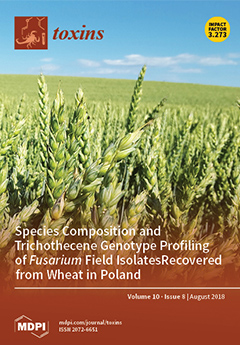
Cover Story (view full-size image): Fusarium head blight (FHB, syn. scab) is one of the most severe wheat diseases worldwide. The disease is caused mainly by fungi of the genus Fusarium. Warm and humid weather conditions at the flowering stage are conducive to disease development. Symptoms may occur over the entire head or on just a few spikelets (Figure 1A), which leads to the formation of Fusarium-damaged kernels (FDK) that are shriveled and appear chalky white and/or pink (Figure 1B). Diseased kernels can contain high amounts of mycotoxins, which pose a significant risk to food and feed safety. View the paper here.
  • Issues are regarded as officially published after their release is announced to the table of contents alert mailing list.
  • You may sign up for e-mail alerts to receive table of contents of newly released issues.
  • PDF is the official format for papers published in both, html and pdf forms. To view the papers in pdf format, click on the "PDF Full-text" link, and use the free Adobe Reader to open them.
Order results
Result details
Section
Select all
Export citation of selected articles as:
14 pages, 920 KiB  
Review
The Development of Toad Toxins as Potential Therapeutic Agents
by Ji Qi, Abu Hasanat Md Zulfiker, Chun Li, David Good and Ming Q. Wei
Toxins 2018, 10(8), 336; https://doi.org/10.3390/toxins10080336 - 20 Aug 2018
Cited by 56 | Viewed by 8375
Abstract
Toxins from toads have long been known to contain rich chemicals with great pharmaceutical potential. Recent studies have shown more than 100 such chemical components, including peptides, steroids, indole alkaloids, bufogargarizanines, organic acids, and others, in the parotoid and skins gland secretions from [...] Read more.
Toxins from toads have long been known to contain rich chemicals with great pharmaceutical potential. Recent studies have shown more than 100 such chemical components, including peptides, steroids, indole alkaloids, bufogargarizanines, organic acids, and others, in the parotoid and skins gland secretions from different species of toads. In traditional Chinese medicine (TCM), processed toad toxins have been used for treating various diseases for hundreds of years. Modern studies, including both experimental and clinical trials, have also revealed the molecular mechanisms that support the development of these components into medicines for the treatment of inflammatory diseases and cancers. More recently, there have been studies that demonstrated the therapeutic potential of toxins from other species of toads, such as Australian cane toads. Previous reviews mostly focused on the pharmaceutical effects of the whole extracts from parotoid glands or skins of toads. However, to fully understand the molecular basis of toad toxins in their use for therapy, a comprehensive understanding of the individual compound contained in toad toxins is necessary; thus, this paper seeks to review the recent studies of some typical compounds frequently identified in toad secretions. Full article
(This article belongs to the Special Issue Venom and Toxin as Targeted Therapy)
Show Figures

Figure 1

15 pages, 3285 KiB  
Review
Structural and Functional Investigation and Pharmacological Mechanism of Trichosanthin, a Type 1 Ribosome-Inactivating Protein
by Wei-Wei Shi, Kam-Bo Wong and Pang-Chui Shaw
Toxins 2018, 10(8), 335; https://doi.org/10.3390/toxins10080335 - 20 Aug 2018
Cited by 17 | Viewed by 5802
Abstract
Trichosanthin (TCS) is an RNA N-glycosidase that depurinates adenine-4324 in the conserved α-sarcin/ricin loop (α-SRL) of rat 28 S ribosomal RNA (rRNA). TCS has only one chain, and is classified as type 1 ribosome-inactivating protein (RIP). Our structural studies revealed that TCS [...] Read more.
Trichosanthin (TCS) is an RNA N-glycosidase that depurinates adenine-4324 in the conserved α-sarcin/ricin loop (α-SRL) of rat 28 S ribosomal RNA (rRNA). TCS has only one chain, and is classified as type 1 ribosome-inactivating protein (RIP). Our structural studies revealed that TCS consists of two domains, with five conserved catalytic residues Tyr70, Tyr111, Glu160, Arg163 and Phe192 at the active cleft formed between them. We also found that the structural requirements of TCS to interact with the ribosomal stalk protein P2 C-terminal tail. The structural analyses suggest TCS attacks ribosomes by first binding to the C-terminal domain of ribosomal P protein. TCS exhibits a broad spectrum of biological and pharmacological activities including anti-tumor, anti-virus, and immune regulatory activities. This review summarizes an updated knowledge in the structural and functional studies and the mechanism of its multiple pharmacological effects. Full article
(This article belongs to the Special Issue Toxicity of Plant Toxins in Medical Herbs)
Show Figures

Figure 1

14 pages, 4166 KiB  
Article
Insights into the Voltage Regulation Mechanism of the Pore-Forming Toxin Lysenin
by Sheenah Lynn Bryant, Tyler Clark, Christopher Alex Thomas, Kaitlyn Summer Ware, Andrew Bogard, Colleen Calzacorta, Daniel Prather and Daniel Fologea
Toxins 2018, 10(8), 334; https://doi.org/10.3390/toxins10080334 - 17 Aug 2018
Cited by 3 | Viewed by 3502
Abstract
Lysenin, a pore forming toxin (PFT) extracted from Eisenia fetida, inserts voltage-regulated channels into artificial lipid membranes containing sphingomyelin. The voltage-induced gating leads to a strong static hysteresis in conductance, which endows lysenin with molecular memory capabilities. To explain this history-dependent behavior, [...] Read more.
Lysenin, a pore forming toxin (PFT) extracted from Eisenia fetida, inserts voltage-regulated channels into artificial lipid membranes containing sphingomyelin. The voltage-induced gating leads to a strong static hysteresis in conductance, which endows lysenin with molecular memory capabilities. To explain this history-dependent behavior, we hypothesized a gating mechanism that implies the movement of a voltage domain sensor from an aqueous environment into the hydrophobic core of the membrane under the influence of an external electric field. In this work, we employed electrophysiology approaches to investigate the effects of ionic screening elicited by metal cations on the voltage-induced gating and hysteresis in conductance of lysenin channels exposed to oscillatory voltage stimuli. Our experimental data show that screening of the voltage sensor domain strongly affects the voltage regulation only during inactivation (channel closing). In contrast, channel reactivation (reopening) presents a more stable, almost invariant voltage dependency. Additionally, in the presence of anionic Adenosine 5′-triphosphate (ATP), which binds at a different site in the channel’s structure and occludes the conducting pathway, both inactivation and reactivation pathways are significantly affected. Therefore, the movement of the voltage domain sensor into a physically different environment that precludes electrostatically bound ions may be an integral part of the gating mechanism. Full article
(This article belongs to the Section Bacterial Toxins)
Show Figures

Figure 1

15 pages, 1745 KiB  
Article
Pharmacokinetic Properties of the Nephrotoxin Orellanine in Rats
by Deman Najar, Börje Haraldsson, Annika Thorsell, Carina Sihlbom, Jenny Nyström and Kerstin Ebefors
Toxins 2018, 10(8), 333; https://doi.org/10.3390/toxins10080333 - 17 Aug 2018
Cited by 8 | Viewed by 7373
Abstract
Orellanine is a nephrotoxin found in mushrooms of the Cortinarius family. Accidental intake of this substance may cause renal failure. Orellanine is specific for proximal tubular cells and could, therefore, potentially be used as treatment for metastatic renal cancer, which originates from these [...] Read more.
Orellanine is a nephrotoxin found in mushrooms of the Cortinarius family. Accidental intake of this substance may cause renal failure. Orellanine is specific for proximal tubular cells and could, therefore, potentially be used as treatment for metastatic renal cancer, which originates from these cells. However, more information is needed about the distribution and elimination of orellanine from the body to understand its potential use for therapy. In this study, 5 mg/kg orellanine (unlabeled and 3H-labeled) was injected intravenously in rats (Wistar and Sprague Dawley). Distribution was measured (Wistar rats, n = 10, n = 12) using radioluminography and the highest amount of orellanine was found in the kidney cortex and bladder at all time-points investigated. The pharmacokinetic properties of orellanine was investigated using LC-MS/MS and β-scintillation to measure the amount of orellanine in plasma. Three groups of rats were investigated: control rats with intact kidneys (n = 10) and two groups with bilateral renal artery ligation (n = 7) where animals in one of these groups were treated with peritoneal dialysis (n = 8). Using LC-MS/MS, the half-life of orellanine was found to be 109 ± 6 min in the controls. In the groups with ligated renal arteries, orellanine had a half-life of 756 ± 98 min without and 238 ± 28 min with dialysis. Thus, orellanine was almost exclusively eliminated by glomerular filtration as well as by peritoneal dialysis. Full article
(This article belongs to the Special Issue Venom and Toxin as Targeted Therapy)
Show Figures

Figure 1

10 pages, 1557 KiB  
Article
Purification and Characterization of a Novel Antiplatelet Peptide from Deinagkistrodon acutus Venom
by Yi Kong, Qing Sun, Qi Zhao and Yaqiong Zhang
Toxins 2018, 10(8), 332; https://doi.org/10.3390/toxins10080332 - 16 Aug 2018
Cited by 7 | Viewed by 3535
Abstract
Animal venoms are considered as one of the most important sources for drug development. Deinagkistrodon acutus is famous for its toxicity to the human hematological system and envenomed patients develop a coagulation disorder with the symptoms of hemorrhage and microthrombi formation. The purpose [...] Read more.
Animal venoms are considered as one of the most important sources for drug development. Deinagkistrodon acutus is famous for its toxicity to the human hematological system and envenomed patients develop a coagulation disorder with the symptoms of hemorrhage and microthrombi formation. The purpose of this study was to separate antiplatelet peptides from D. acutus venom using a combination of an ultrafiltration technique and reversed-phase high performance liquid chromatography (HPLC), which was guided by monitoring antiplatelet aggregation bioactivity. A novel octa-peptide named DAA-8 was found. This peptide inhibited protease-activated receptor1 (PAR-1) agonist (SFLLRN-NH2) induced platelet aggregation and it also inhibited platelet aggregation induced by thrombin, ADP, and collagen. Furthermore, DAA-8 showed significant antithrombotic activity and resulted in a slightly increased bleeding risk in vivo. This is the first report of a peptide derived from snake venom, which inhibited PAR-1 agonist-induced platelet aggregation. This peptide may provide a template to design a new PAR-1 inhibitor. Full article
(This article belongs to the Section Animal Venoms)
Show Figures

Figure 1

9 pages, 1031 KiB  
Review
Endotoxins from a Pharmacopoeial Point of View
by Elvira Franco, Verónica Garcia-Recio, Pilar Jiménez, Manuel Garrosa, Tomás Girbés, Manuel Cordoba-Diaz and Damián Cordoba-Diaz
Toxins 2018, 10(8), 331; https://doi.org/10.3390/toxins10080331 - 16 Aug 2018
Cited by 25 | Viewed by 10133
Abstract
A pyrogen is a substance that causes fever after intravenous administration or inhalation. Gram negative endotoxins are the most important pyrogens to pharmaceutical laboratories. In the International, United States, Japanese and European Pharmacopoeias, there are two official methods to evaluate pyrogenicity—that is, the [...] Read more.
A pyrogen is a substance that causes fever after intravenous administration or inhalation. Gram negative endotoxins are the most important pyrogens to pharmaceutical laboratories. In the International, United States, Japanese and European Pharmacopoeias, there are two official methods to evaluate pyrogenicity—that is, the bacterial endotoxin test, and the pyrogen test. The main objective of this review is to compare the monographs of each test among the different Pharmacopeias, to detect similarities and differences. The former can be considered fully harmonized, and only non-significant differences were detected. The latter, which is the only available assay for some products and formulations to demonstrate apyrogenicity, shows large differences, which should be considered. Full article
(This article belongs to the Special Issue Lipopolysaccharide: Bacterial Endotoxin)
Show Figures

Figure 1

15 pages, 2339 KiB  
Article
Biological Degradation of Aflatoxin B1 by Cell-Free Extracts of Bacillus velezensis DY3108 with Broad PH Stability and Excellent Thermostability
by Xian Shu, Yuting Wang, Qing Zhou, Minghao Li, Hao Hu, Yuhan Ma, Xue Chen, Jun Ni, Weiwei Zhao, Shengwei Huang and Lifang Wu
Toxins 2018, 10(8), 330; https://doi.org/10.3390/toxins10080330 - 14 Aug 2018
Cited by 73 | Viewed by 6996
Abstract
(1) Background: Aflatoxin contamination in food and grain poses serious problems both for economic development and public health protection, thus leading to a focus on an effective approach to control it; (2) Methods: Aflatoxin B1 (AFB1) degrading bacteria were isolated [...] Read more.
(1) Background: Aflatoxin contamination in food and grain poses serious problems both for economic development and public health protection, thus leading to a focus on an effective approach to control it; (2) Methods: Aflatoxin B1 (AFB1) degrading bacteria were isolated using a medium containing coumarin as the sole carbon source, and the biodegradation of AFB1 by the isolate was examined by high performance liquid chromatography, and liquid chromatography mass spectrometry; (3) Results: a bacterial strain exhibiting strong AFB1 degradation activity (91.5%) was isolated and identified as Bacillus velezensis DY3108. The AFB1 degrading activity was predominantly attributed to the cell-free supernatant of strain DY3108. Besides, it was heat-stable and resistant to proteinase K treatment but sensitive to sodium dodecyl sulfate treatment. The optimal temperature for the maximal degradation of AFB1 was 80 °C. Even more notable, the supernatant showed a high level of activity over a broad pH (4.0 to 11.0) and exhibited the highest degradation (94.70%) at pH 8.0. Cytotoxicity assays indicated that the degradation products displayed significantly (p < 0.05) lower cytotoxic effects than the parent AFB1; (4) Conclusions: B. velezensis DY3108 might be a promising candidate for exploitation in AFB1 detoxification and bioremediation in food and feed matrices. Full article
Show Figures

Figure 1

16 pages, 2112 KiB  
Article
A Novel Sensitive Cell-Based Immunoenzymatic Assay for Palytoxin Quantitation in Mussels
by Marco Pelin, Silvio Sosa, Valentina Brovedani, Laura Fusco, Mark Poli and Aurelia Tubaro
Toxins 2018, 10(8), 329; https://doi.org/10.3390/toxins10080329 - 14 Aug 2018
Cited by 11 | Viewed by 4374
Abstract
The marine algal toxin palytoxin (PLTX) and its analogues are some of the most toxic marine compounds. Their accumulation in edible marine organisms and entrance into the food chain represent their main concerns for human health. Indeed, several fatal human poisonings attributed to [...] Read more.
The marine algal toxin palytoxin (PLTX) and its analogues are some of the most toxic marine compounds. Their accumulation in edible marine organisms and entrance into the food chain represent their main concerns for human health. Indeed, several fatal human poisonings attributed to these compounds have been recorded in tropical and subtropical areas. Due to the increasing occurrence of PLTX in temperate areas such as the Mediterranean Sea, the European Food Safety Authority (EFSA) has suggested a maximum limit of 30 µg PLTX/kg in shellfish meat, and has recommended the development of rapid, specific, and sensitive methods for detection and quantitation of PLTX in seafood. Thus, a novel, sensitive cell-based ELISA was developed and characterized for PLTX quantitation in mussels. The estimated limits of detection (LOD) and quantitation (LOQ) were 1.2 × 10⁻¹¹ M (32.2 pg/mL) and 2.8 × 10⁻¹¹ M (75.0 pg/mL), respectively, with good accuracy (bias = 2.5%) and repeatability (15% and 9% interday and intraday relative standard deviation of repeatability (RSDr), respectively). Minimal interference of 80% aqueous methanol extract allows PLTX quantitation in mussels at concentrations lower than the maximum limit suggested by EFSA, with an LOQ of 9.1 µg PLTX equivalent/kg mussel meat. Given its high sensitivity and specificity, the cell-based ELISA should be considered a suitable method for PLTX quantitation. Full article
(This article belongs to the Section Marine and Freshwater Toxins)
Show Figures

Graphical abstract

15 pages, 2898 KiB  
Article
Involvement of Osteocytes in the Action of Pasteurella multocida Toxin
by Hannah Heni, Julia K. Ebner, Gudula Schmidt, Klaus Aktories and Joachim H. C. Orth
Toxins 2018, 10(8), 328; https://doi.org/10.3390/toxins10080328 - 13 Aug 2018
Cited by 8 | Viewed by 3724
Abstract
Pasteurella multocida toxin (PMT) causes progressive atrophic rhinitis with severe turbinate bone degradation in pigs. It has been reported that the toxin deamidates and activates heterotrimeric G proteins, resulting in increased differentiation of osteoclasts and blockade of osteoblast differentiation. So far, the action [...] Read more.
Pasteurella multocida toxin (PMT) causes progressive atrophic rhinitis with severe turbinate bone degradation in pigs. It has been reported that the toxin deamidates and activates heterotrimeric G proteins, resulting in increased differentiation of osteoclasts and blockade of osteoblast differentiation. So far, the action of PMT on osteocytes, which is the most abundant cell type in bone tissue, is not known. In MLO-Y4 osteocytes, PMT deamidated heterotrimeric G proteins, resulting in loss of osteocyte dendritic processes, stress fiber formation, cell spreading and activation of RhoC but not of RhoA. Moreover, the toxin caused processing of membrane-bound receptor activator of NF-κB ligand (RANKL) to release soluble RANKL and enhanced the secretion of osteoclastogenic TNF-α. In a co-culture model of osteocytes and bone marrow cells, PMT-induced osteoclastogenesis was largely increased as compared to the mono-culture model. The enhancement of osteoclastogenesis observed in the co-culture was blocked by sequestering RANKL with osteoprotegerin and by an antibody against TNF-α indicating involvement of release of the osteoclastogenic factors from osteocytes. Data support the crucial role of osteocytes in bone metabolism and osteoclastogenesis and identify osteocytes as important target cells of PMT in progressive atrophic rhinitis. Full article
(This article belongs to the Section Bacterial Toxins)
Show Figures

Figure 1

12 pages, 2285 KiB  
Article
Feasibility of A Novel On-Site Detection Method for Aflatoxin in Maize Flour from Markets and Selected Households in Kampala, Uganda
by Alex Paul Wacoo, Deborah Wendiro, Sarah Nanyonga, Joseph F. Hawumba, Wilbert Sybesma and Remco Kort
Toxins 2018, 10(8), 327; https://doi.org/10.3390/toxins10080327 - 11 Aug 2018
Cited by 16 | Viewed by 5067
Abstract
In sub-Saharan Africa, there is a high demand for affordable and accessible methods for on-site detection of aflatoxins for appropriate food safety management. In this study, we validated an electrochemical immunosensor device by the on-site detection of 60 maize flour samples from six [...] Read more.
In sub-Saharan Africa, there is a high demand for affordable and accessible methods for on-site detection of aflatoxins for appropriate food safety management. In this study, we validated an electrochemical immunosensor device by the on-site detection of 60 maize flour samples from six markets and 72 samples from households in Kampala. The immunosensor was successfully validated with a linear range from 0.7 ± 0.1 to 11 ± 0.3 µg/kg and limit of detection (LOD) of 0.7 µg/kg. The maize flour samples from the markets had a mean total aflatoxin concentration of 7.6 ± 2.3 µg/kg with approximately 20% of the samples higher than 10 µg/kg, which is the maximum acceptable level in East Africa. Further down the distribution chain, at the household level, approximately 45% of the total number contained total aflatoxin levels higher than the acceptable limit. The on-site detection method correlated well with the established laboratory-based HPLC and ELISA-detection methods for aflatoxin B1 with the correlation coefficients of 0.94 and 0.98, respectively. This study shows the feasibility of a novel on-site detection method and articulates the severity of aflatoxin contamination in Uganda. Full article
(This article belongs to the Special Issue Advanced Methods for Mycotoxins Detection)
Show Figures

Graphical abstract

19 pages, 6201 KiB  
Review
TRP Channels as Sensors of Bacterial Endotoxins
by Brett Boonen, Yeranddy A. Alpizar, Victor M. Meseguer and Karel Talavera
Toxins 2018, 10(8), 326; https://doi.org/10.3390/toxins10080326 - 11 Aug 2018
Cited by 52 | Viewed by 8964
Abstract
The cellular and systemic effects induced by bacterial lipopolysaccharides (LPS) have been solely attributed to the activation of the Toll-like receptor 4 (TLR4) signalling cascade. However, recent studies have shown that LPS activates several members of the Transient Receptor Potential (TRP) family of [...] Read more.
The cellular and systemic effects induced by bacterial lipopolysaccharides (LPS) have been solely attributed to the activation of the Toll-like receptor 4 (TLR4) signalling cascade. However, recent studies have shown that LPS activates several members of the Transient Receptor Potential (TRP) family of cation channels. Indeed, LPS induces activation of the broadly-tuned chemosensor TRPA1 in sensory neurons in a TLR4-independent manner, and genetic ablation of this channel reduced mouse pain and inflammatory responses triggered by LPS and the gustatory-mediated avoidance to LPS in fruit flies. LPS was also shown to activate TRPV4 channels in airway epithelial cells, an effect leading to an immediate production of bactericidal nitric oxide and to an increase in ciliary beat frequency. In this review, we discuss the role of TRP channels as sensors of bacterial endotoxins, and therefore, as crucial players in the timely detection of invading gram-negative bacteria. Full article
(This article belongs to the Special Issue Lipopolysaccharide: Bacterial Endotoxin)
Show Figures

Figure 1

14 pages, 1020 KiB  
Article
Species Composition and Trichothecene Genotype Profiling of Fusarium Field Isolates Recovered from Wheat in Poland
by Katarzyna Bilska, Sebastian Jurczak, Tomasz Kulik, Ewa Ropelewska, Jacek Olszewski, Maciej Żelechowski and Piotr Zapotoczny
Toxins 2018, 10(8), 325; https://doi.org/10.3390/toxins10080325 - 10 Aug 2018
Cited by 29 | Viewed by 5385
Abstract
Fusarium head blight (FHB) of cereals is the major head disease negatively affecting grain production worldwide. In 2016 and 2017, serious outbreaks of FHB occurred in wheat crops in Poland. In this study, we characterized the diversity of Fusaria responsible for these epidemics [...] Read more.
Fusarium head blight (FHB) of cereals is the major head disease negatively affecting grain production worldwide. In 2016 and 2017, serious outbreaks of FHB occurred in wheat crops in Poland. In this study, we characterized the diversity of Fusaria responsible for these epidemics using TaqMan assays. From a panel of 463 field isolates collected from wheat, four Fusarium species were identified. The predominant species were F. graminearum s.s. (81%) and, to a lesser extent, F. avenaceum (15%). The emergence of the 15ADON genotype was found ranging from 83% to 87% of the total trichothecene genotypes isolated in 2016 and 2017, respectively. Our results indicate two dramatic shifts within fungal field populations in Poland. The first shift is associated with the displacement of F. culmorum by F. graminearum s.s. The second shift resulted from a loss of nivalenol genotypes. We suggest that an emerging prevalence of F. graminearum s.s. may be linked to boosted maize production, which has increased substantially over the last decade in Poland. To detect variation within Tri core clusters, we compared sequence data from randomly selected field isolates with a panel of strains from geographically diverse origins. We found that the newly emerged 15ADON genotypes do not exhibit a specific pattern of polymorphism enabling their clear differentiation from the other European strains. Full article
(This article belongs to the Special Issue Recent Advances in Fusarium Research)
Show Figures

Figure 1

38 pages, 1310 KiB  
Review
Human Poisoning from Marine Toxins: Unknowns for Optimal Consumer Protection
by Natalia Vilariño, M. Carmen Louzao, Paula Abal, Eva Cagide, Cristina Carrera, Mercedes R. Vieytes and Luis M. Botana
Toxins 2018, 10(8), 324; https://doi.org/10.3390/toxins10080324 - 9 Aug 2018
Cited by 117 | Viewed by 13211
Abstract
Marine biotoxins are produced by aquatic microorganisms and accumulate in shellfish or finfish following the food web. These toxins usually reach human consumers by ingestion of contaminated seafood, although other exposure routes like inhalation or contact have also been reported and may cause [...] Read more.
Marine biotoxins are produced by aquatic microorganisms and accumulate in shellfish or finfish following the food web. These toxins usually reach human consumers by ingestion of contaminated seafood, although other exposure routes like inhalation or contact have also been reported and may cause serious illness. This review shows the current data regarding the symptoms of acute intoxication for several toxin classes, including paralytic toxins, amnesic toxins, ciguatoxins, brevetoxins, tetrodotoxins, diarrheic toxins, azaspiracids and palytoxins. The information available about chronic toxicity and relative potency of different analogs within a toxin class are also reported. The gaps of toxicological knowledge that should be studied to improve human health protection are discussed. In general, gathering of epidemiological data in humans, chronic toxicity studies and exploring relative potency by oral administration are critical to minimize human health risks related to these toxin classes in the near future. Full article
(This article belongs to the Special Issue Public Health Outreach to Prevention of Aquatic Toxin Exposure)
Show Figures

Figure 1

10 pages, 942 KiB  
Review
Bouganin, an Attractive Weapon for Immunotoxins
by Massimo Bortolotti, Andrea Bolognesi and Letizia Polito
Toxins 2018, 10(8), 323; https://doi.org/10.3390/toxins10080323 - 8 Aug 2018
Cited by 15 | Viewed by 4481
Abstract
Bougainvillea (Bougainvillea spectabilis Willd.) is a plant widely used in folk medicine and many extracts from different tissues of this plant have been employed against several pathologies. The observation that leaf extracts of Bougainvillea possess antiviral properties led to the purification and [...] Read more.
Bougainvillea (Bougainvillea spectabilis Willd.) is a plant widely used in folk medicine and many extracts from different tissues of this plant have been employed against several pathologies. The observation that leaf extracts of Bougainvillea possess antiviral properties led to the purification and characterization of a protein, named bouganin, which exhibits typical characteristics of type 1 ribosome-inactivating proteins (RIPs). Beyond that, bouganin has some peculiarities, such as a higher activity on DNA with respect to ribosomal RNA, low systemic toxicity, and immunological properties quite different than other RIPs. The sequencing of bouganin and the knowledge of its three-dimensional structure allowed to obtain a not immunogenic mutant of bouganin. These features make bouganin a very attractive tool as a component of immunotoxins (ITs), chimeric proteins obtained by linking a toxin to a carrier molecule. Bouganin-containing ITs showed very promising results in the experimental treatment of both hematological and solid tumors, and one bouganin-containing IT has entered Phase I clinical trial. In this review, we summarize the milestones of the research on bouganin such as bouganin chemico-physical characteristics, the structural properties and de-immunization studies. In addition, the in vitro and in vivo results obtained with bouganin-containing ITs are summarized. Full article
(This article belongs to the Special Issue Venom and Toxin as Targeted Therapy)
Show Figures

Figure 1

11 pages, 690 KiB  
Article
Effects of Heme Modulation on Ovophis and Trimeresurus Venom Activity in Human Plasma
by Vance G. Nielsen, Nathaniel Frank and Ryan W. Matika
Toxins 2018, 10(8), 322; https://doi.org/10.3390/toxins10080322 - 8 Aug 2018
Cited by 6 | Viewed by 3421
Abstract
Geographic isolation and other factors result in evolution-driven diversity of the enzymatic composition of venom of pit vipers in the same genus. The present investigation sought to characterize venoms obtained from such genetically diverse Ovophis and Trimeresurus pit vipers utilizing thrombelastographic coagulation kinetic [...] Read more.
Geographic isolation and other factors result in evolution-driven diversity of the enzymatic composition of venom of pit vipers in the same genus. The present investigation sought to characterize venoms obtained from such genetically diverse Ovophis and Trimeresurus pit vipers utilizing thrombelastographic coagulation kinetic analyses. The coagulation kinetics of human plasma were assessed after exposure to venom obtained from two Ovophis and three Trimeresurus species. The potency of each venom was defined (µg/mL required to equivalently change coagulation); additionally, venoms were exposed to carbon monoxide (CO) or a metheme-inducing agent to modulate any enzyme-associated heme. All venoms had fibrinogenolytic activity, with four being CO-inhibitable. While Ovophis venoms had similar potency, one demonstrated the presence of a thrombin-like activity, whereas the other demonstrated a thrombin-generating activity. There was a 10-fold difference in potency and 10-fold different vulnerability to CO inhibition between the Trimeresurus species. Metheme formation enhanced fibrinogenolytic-like activity in both Ovophis species venoms, whereas the three Trimeresurus species venoms had fibrinogenolytic-like activity enhanced, inhibited, or not changed. This novel “venom kinetomic” approach has potential to identify clinically relevant enzymatic activity and assess efficacy of antivenoms between genetically and geographically diverse species. Full article
(This article belongs to the Section Animal Venoms)
Show Figures

Figure 1

24 pages, 1157 KiB  
Review
Recombinant and Chimeric Disintegrins in Preclinical Research
by Victor David, Barbara Barbosa Succar, João Alfredo De Moraes, Roberta Ferreira Gomes Saldanha-Gama, Christina Barja-Fidalgo and Russolina Benedeta Zingali
Toxins 2018, 10(8), 321; https://doi.org/10.3390/toxins10080321 - 7 Aug 2018
Cited by 21 | Viewed by 5464
Abstract
Disintegrins are a family of small cysteine-rich peptides, found in a wide variety of snake venoms of different phylogenetic origin. These peptides selectively bind to integrins, which are heterodimeric adhesion receptors that play a fundamental role in the regulation of many physiological and [...] Read more.
Disintegrins are a family of small cysteine-rich peptides, found in a wide variety of snake venoms of different phylogenetic origin. These peptides selectively bind to integrins, which are heterodimeric adhesion receptors that play a fundamental role in the regulation of many physiological and pathological processes, such as hemostasis and tumor metastasis. Most disintegrins interact with integrins through the RGD (Arg-Gly-Asp) sequence loop, resulting in an active site that modulates the integrin activity. Some variations in the tripeptide sequence and the variability in its neighborhood result in a different specificity or affinity toward integrin receptors from platelets, tumor cells or neutrophils. Recombinant forms of these proteins are obtained mainly through Escherichia coli, which is the most common host used for heterologous expression. Advances in the study of the structure-activity relationship and importance of some regions of the molecule, especially the hairpin loop and the C-terminus, rely on approaches such as site-directed mutagenesis and the design and expression of chimeric peptides. This review provides highlights of the biological relevance and contribution of recombinant disintegrins to the understanding of their binding specificity, biological activities and therapeutic potential. The biological and pharmacological relevance on the newest discoveries about this family of integrin-binding proteins are discussed. Full article
(This article belongs to the Special Issue From Toxins to Drugs)
Show Figures

Figure 1

11 pages, 1522 KiB  
Article
Biological Activities of Cationicity-Enhanced and Hydrophobicity-Optimized Analogues of an Antimicrobial Peptide, Dermaseptin-PS3, from the Skin Secretion of Phyllomedusa sauvagii
by Yining Tan, Xiaoling Chen, Chengbang Ma, Xinping Xi, Lei Wang, Mei Zhou, James F. Burrows, Hang Fai Kwok and Tianbao Chen
Toxins 2018, 10(8), 320; https://doi.org/10.3390/toxins10080320 - 7 Aug 2018
Cited by 21 | Viewed by 4408
Abstract
The skin secretions of the subfamily Phyllomedusinae have long been known to contain a number of compounds with antimicrobial potential. Herein, a biosynthetic dermaseptin-precursor cDNA was obtained from a Phyllomedusa sauvagii skin secretion-derived cDNA library, and thereafter, the presence of the mature peptide, [...] Read more.
The skin secretions of the subfamily Phyllomedusinae have long been known to contain a number of compounds with antimicrobial potential. Herein, a biosynthetic dermaseptin-precursor cDNA was obtained from a Phyllomedusa sauvagii skin secretion-derived cDNA library, and thereafter, the presence of the mature peptide, namely dermaseptin-PS3 (DPS3), was confirmed by LC–MS/MS. Moreover, this naturally occurring peptide was utilized to design two analogues, K5, 17-DPS3 (introducing two lysine residues at positions 5 and 17 to replace acidic amino acids) and L10, 11-DPS3 (replacing two neutral amino acids with the hydrophobic amino acid, leucine), improving its cationicity on the polar/unipolar face and hydrophobicity in a highly conserved sequence motif, respectively. The results in regard to the two analogues show that either increasing cationicity, or hydrophobicity, enhance the antimicrobial activity. Also, the latter analogue had an enhanced anticancer activity, with pretreatment of H157 cells with 1 µM L10, 11-DPS3 decreasing viability by approximately 78%, even though this concentration of peptide exhibited no haemolytic effect. However, it must be noted that in comparison to the initial peptide, both analogues demonstrate higher membrane-rupturing capacity towards mammalian red blood cells. Full article
(This article belongs to the Special Issue Venom and Toxin as Targeted Therapy)
Show Figures

Figure 1

15 pages, 1937 KiB  
Article
Distribution Analysis of Twelve Mycotoxins in Corn and Corn-Derived Products by LC-MS/MS to Evaluate the Carry-Over Ratio during Wet-Milling
by Juhee Park, Dong-Ho Kim, Ji-Young Moon, Jin-Ah An, Young-Woo Kim, Soo-Hyun Chung and Chan Lee
Toxins 2018, 10(8), 319; https://doi.org/10.3390/toxins10080319 - 6 Aug 2018
Cited by 49 | Viewed by 5439
Abstract
This study investigated the distribution of twelve mycotoxins (aflatoxins B1, B2, G1, and G2; ochratoxin A; fumonisins B1 and B2; deoxynivalenol; nivalenol; zearalenone; T-2 toxin; and HT-2 toxin) in corn and corn [...] Read more.
This study investigated the distribution of twelve mycotoxins (aflatoxins B1, B2, G1, and G2; ochratoxin A; fumonisins B1 and B2; deoxynivalenol; nivalenol; zearalenone; T-2 toxin; and HT-2 toxin) in corn and corn by-products (corn bran, cornstarch, corn gluten, corn gluten feed, corn germ, light steep water, and corn steep liquor) produced by wet-milling in Korea. Fifty-two samples were collected from three factories producing cornstarch and other corn by-products. The samples were pretreated on an immunoaffinity column (IAC), and then the levels of the 12 mycotoxins were analyzed simultaneously by liquid chromatography-coupled triple-quadrupole mass spectrometry (LC-MS/MS). Fusarium mycotoxins were mainly found in raw corn and corn gluten feed samples. Other mycotoxins—such as aflatoxins, ochratoxin A, and HT-2 toxin—were detected in tiny amounts below the limit of quantification (LOQ) in cornstarch, corn germ, and corn bran. Ochratoxin A and nivalenol were mainly carried over into cornstarch. Aflatoxin B1, deoxynivalenol, T-2 toxin, HT-2 toxin, and the fumonisins were concentrated in corn gluten feed. Zearalenone was evenly distributed in all corn by-products except cornstarch during the milling process. Full article
(This article belongs to the Section Mycotoxins)
Show Figures

Figure 1

12 pages, 2697 KiB  
Article
Histopathological Evaluation of the Exposure by Cyanobacteria Cultive Containing [d-Leu1]Microcystin-LR on Lithobates catesbeianus Tadpoles
by Osmindo Rodrigues Pires Júnior, Natiela Beatriz De Oliveira, Renan J. Bosque, Maria Fernanda Nice Ferreira, Veronica Morais Aurélio da Silva, Ana Carolina Martins Magalhães, Carlos José Correia de Santana and Mariana De Souza Castro
Toxins 2018, 10(8), 318; https://doi.org/10.3390/toxins10080318 - 6 Aug 2018
Cited by 5 | Viewed by 4065
Abstract
This study evaluated the effects of [d-Leu1]Microcystin-LR variant by the exposure of Lithobates catesbeianus tadpole to unialgal culture Microcystis aeruginosa NPLJ-4 strain. The Tadpole was placed in aquariums and exposed to Microcystis aeruginosa culture or disrupted cells. For 16 [...] Read more.
This study evaluated the effects of [d-Leu1]Microcystin-LR variant by the exposure of Lithobates catesbeianus tadpole to unialgal culture Microcystis aeruginosa NPLJ-4 strain. The Tadpole was placed in aquariums and exposed to Microcystis aeruginosa culture or disrupted cells. For 16 days, 5 individuals were removed every 2 days, and tissue samples of liver, skeletal muscle, and intestinal tract were collected for histopathology and bioaccumulation analyses. After exposure, those surviving tadpoles were placed in clean water for 15 days to evaluate their recovery. A control without algae and toxins was maintained in the same conditions and exhibited normal histology and no tissue damage. In exposed tadpoles, samples were characterized by serious damages that similarly affected the different organs, such as loss of adhesion between cells, nucleus fragmentation, necrosis, and hemorrhage. Samples showed signs of recovery but severe damages were still observed. Neither HPLC-PDA nor mass spectrometry analysis showed any evidence of free Microcystins bioaccumulation. Full article
(This article belongs to the Special Issue Cyanobacteria and Cyanotoxins: New Advances and Future Challenges)
Show Figures

Figure 1

12 pages, 1564 KiB  
Article
Development of a Magnetic Nanoparticles-Based Screen-Printed Electrodes (MNPs-SPEs) Biosensor for the Quantification of Ochratoxin A in Cereal and Feed Samples
by Xian Zhang, Zuohuan Wang, Hui Xie, Renjie Sun, Tong Cao, Narayan Paudyal, Weihuan Fang and Houhui Song
Toxins 2018, 10(8), 317; https://doi.org/10.3390/toxins10080317 - 6 Aug 2018
Cited by 26 | Viewed by 4566
Abstract
A rapid and sensitive electrochemical biosensor based on magnetic nanoparticles and screen-printed electrodes (MNPs-SPEs sensor) was developed for the detection of ochratoxin A (OTA) in cereal and feed samples. Different types of magnetic nanoparticles-based ELISA (MNPs-ELISA) were optimized, and the signal detection, as [...] Read more.
A rapid and sensitive electrochemical biosensor based on magnetic nanoparticles and screen-printed electrodes (MNPs-SPEs sensor) was developed for the detection of ochratoxin A (OTA) in cereal and feed samples. Different types of magnetic nanoparticles-based ELISA (MNPs-ELISA) were optimized, and the signal detection, as well as sensitivity, was enhanced by the combined use of screen-printed electrodes (SPEs). Under the optimized conditions, the calibration curve of the MNPs-SPEs sensor was y = 0.3372x + 0.8324 (R2 = 0.9805). The linear range of detection and the detection limit were 0.01–0.82 ng/mL and 0.007 ng/mL, respectively. In addition, 50% inhibition (IC50) was detectable at 0.10 ng/mL. The limit of detection (LOD) of this MNPs-SPEs sensor in cereal and feed samples was 0.28 μg/kg. The recovery rates in spiked samples were between 78.7% and 113.5%, and the relative standard deviations (RSDs) were 3.6–9.8%, with the coefficient of variation lower than 15%. Parallel analysis of commercial samples (corn, wheat, and feedstuff) showed a good correlation between MNPs-SPEs sensor and liquid chromatography tandem mass spectrometry (LC/MS-MS). This new method provides a rapid, highly sensitive, and less time-consuming method to determine levels of ochratoxin A in cereal and feedstuff samples. Full article
(This article belongs to the Collection Biorecognition Assays for Mycotoxins)
Show Figures

Graphical abstract

9 pages, 1399 KiB  
Article
Low Doses of Ochratoxin-A Decrease IgY and IgA Production in Broiler Chicks
by Shahzad A. Khan, Emerson J. Venancio, Eduardo V. Fernandes, Elisa Y. Hirooka, Alexandre Oba, Karina K. M. C. Flaiban and Eiko N. Itano
Toxins 2018, 10(8), 316; https://doi.org/10.3390/toxins10080316 - 6 Aug 2018
Cited by 13 | Viewed by 4666
Abstract
The mycotoxin, ochratoxin-A (OTA), produced by some fungi, and is a natural contaminant of many foods and animal feeds worldwide. Due to its toxic effects, the recommended maximum daily intake of OTA for poultry feeds is 0.1 mg OTA/kg (ECR2006/575/EC); this dose does [...] Read more.
The mycotoxin, ochratoxin-A (OTA), produced by some fungi, and is a natural contaminant of many foods and animal feeds worldwide. Due to its toxic effects, the recommended maximum daily intake of OTA for poultry feeds is 0.1 mg OTA/kg (ECR2006/575/EC); this dose does not induce changes in hepatic/renal parameters, but decreases thymus size and serum globulin concentrations. Accordingly, in this study, we assessed quantitatively the total circulating IgY and IgA serum levels, in chicks consuming a 0.1 mg OTA/kg diet (limit) and higher doses (0.3–1.1 mg OTA/kg diet) for 14 or 21 days. We also evaluated other immunological parameters (thymus, bursa of Fabricius, and spleen weights and leukocyte profiles) at day 21. Decreased IgY serum levels were observed in all OTA-treated groups (p < 0.05). In the low-dose group, IgA levels were decreased on day 21, but not on day 14. The size of the thymus and the bursa of Fabricius was decreased in all OTA-treated groups (p < 0.05), whereas reduced spleen size and altered leukocyte profiles were detected only in the high-dose group (p < 0.05). We concluded that chronic exposure to OTA, even at the recommended highest dose, affected IgY and IgA production in chicks. Full article
(This article belongs to the Collection Ochratoxins-Collection)
Show Figures

Figure 1

22 pages, 4686 KiB  
Article
Variations of Bacterial Community Composition and Functions in an Estuary Reservoir during Spring and Summer Alternation
by Zheng Xu, Shu Harn Te, Cong Xu, Yiliang He and Karina Yew-Hoong Gin
Toxins 2018, 10(8), 315; https://doi.org/10.3390/toxins10080315 - 6 Aug 2018
Cited by 10 | Viewed by 4089
Abstract
In this study, we focused on the dynamics of bacterial community composition in a large reservoir in the Yangtze estuary during spring and summer seasons, especially the variations of functional mechanisms of microbial community during the seasonal alternation between spring and summer. Both [...] Read more.
In this study, we focused on the dynamics of bacterial community composition in a large reservoir in the Yangtze estuary during spring and summer seasons, especially the variations of functional mechanisms of microbial community during the seasonal alternation between spring and summer. Both 16S rRNA gene sequencing and shotgun metagenomic sequencing technology were used for these purposes. The results indicated that obvious variations of bacterial community structures were found at different sites. Particle-associated bacterial taxa exhibited higher abundance at the inlet site, which was closer to the Yangtze River with a high level of turbidity. In other sites, Synechococcus, as the most dominant cyanobacterial species, revealed high abundance driven by increased temperature. Moreover, some heterotrophic bacterial taxa revealed high abundance following the increased Synechococcus in summer, which indicated potential correlations about carbon source utilization between these microorganisms. In addition, the shotgun metagenomic data indicated during the period of seasonal alternation between spring and summer, the carbohydrate transport and metabolism, energy production and conversion, translation/ribosomal biogenesis, and cell wall/membrane/envelope biogenesis were significantly enhanced at the exit site. However, the course of cell cycle control/division was more active at the internal site. Full article
(This article belongs to the Special Issue Cyanobacteria and Cyanotoxins: New Advances and Future Challenges)
Show Figures

Figure 1

13 pages, 2573 KiB  
Article
Accumulation and Biotransformation of Dinophysis Toxins by the Surf Clam Mesodesma donacium
by Juan Blanco, Gonzalo Álvarez, José Rengel, Rosario Díaz, Carmen Mariño, Helena Martín and Eduardo Uribe
Toxins 2018, 10(8), 314; https://doi.org/10.3390/toxins10080314 - 4 Aug 2018
Cited by 27 | Viewed by 4806
Abstract
Surf clams, Mesodesma donacium, were shown to accumulate toxins from Dinophysis acuminata blooms. Only pectenotoxin 2 (PTX2) and some of its derivatives were found, and no toxins from the okadaic acid group were detected. PTX2 seems to be transformed to PTX2 seco-acid [...] Read more.
Surf clams, Mesodesma donacium, were shown to accumulate toxins from Dinophysis acuminata blooms. Only pectenotoxin 2 (PTX2) and some of its derivatives were found, and no toxins from the okadaic acid group were detected. PTX2 seems to be transformed to PTX2 seco-acid (PTX2sa), which was found in concentrations more than ten-fold those of PTX2. The seco-acid was transformed to acyl-derivatives by esterification with different fatty acids. The estimated amount of these derivatives in the mollusks was much higher than that of PTX2. Most esters were originated by even carbon chain fatty acids, but some originated by odd carbon number were also found in noticeable concentrations. Some peaks of toxin in the bivalves did not coincide with those of Dinophysis abundance, suggesting that there were large differences in toxin content per cell among the populations that developed throughout the year. The observed depuration (from the digestive gland) was fast (more than 0.2 day−1), and was faster for PTX2 than for PTX2sa, which in turn was faster than that of esters of PTX2sa. PTX2 and PTX2sa were distributed nearly equally between the digestive gland and the remaining tissues, but less than 5% of the palmytoyl-esters were found outside the digestive gland. Full article
(This article belongs to the Special Issue Dinophysis Toxins: Distribution, Fate in Shellfish and Impacts)
Show Figures

Figure 1

11 pages, 264 KiB  
Review
Botulinum Toxin Induced Atrophy: An Uncharted Territory
by Mehri Salari, Soumya Sharma and Mandar S. Jog
Toxins 2018, 10(8), 313; https://doi.org/10.3390/toxins10080313 - 2 Aug 2018
Cited by 27 | Viewed by 7461
Abstract
Botulinum neurotoxins (BoNTs) produce local chemo-denervation by cleaving soluble N-ethylmaleimide-sensitive factor activating protein receptor (SNARE) proteins. Botulinum neurotoxins are therapeutically indicated in several neurological disorders and have been in use for three decades. The long-term efficacy, safety, and side effects of BoNTs have [...] Read more.
Botulinum neurotoxins (BoNTs) produce local chemo-denervation by cleaving soluble N-ethylmaleimide-sensitive factor activating protein receptor (SNARE) proteins. Botulinum neurotoxins are therapeutically indicated in several neurological disorders and have been in use for three decades. The long-term efficacy, safety, and side effects of BoNTs have been well documented in the literature. However, the development of muscle atrophy following chronic exposure to BoNTs has not received sufficient attention. Muscle atrophy is not only cosmetically distressing, but also has an impact on future injections. An extensive literature search was conducted on atrophy and mechanisms of atrophy. Five hundred and four relevant articles in the English language were reviewed. This review revealed the surprising lack of documentation of atrophy within the literature. In addition, as demonstrated in this review, the mechanisms and the clinical factors that may lead to atrophy have also been poorly studied. However, even with this limited information it is possible to indicate factors that could modify the clinical approach to botulinum toxin injections. This review highlights the need for further study of atrophy following BoNT injections. Full article
(This article belongs to the Special Issue Botulinum Toxin Treatment of Movement Disorders)
10 pages, 274 KiB  
Article
Effects of Feeding a Mycotoxin Binder on Nutrient Digestibility, Alkaloid Recovery in Feces, and Performance of Lambs Fed Diets Contaminated with Cereal Ergot
by Kim Stanford, Mary Lou Swift, Yuxi Wang, Tim A. McAllister, John McKinnon, Barry Blakley and Alex V. Chaves
Toxins 2018, 10(8), 312; https://doi.org/10.3390/toxins10080312 - 1 Aug 2018
Cited by 12 | Viewed by 4520
Abstract
As contamination with cereal ergot has been increasing in western Canada, this study evaluated impacts of feeding a mycotoxin binder (Biomin® II; BB) on nutrient digestibility, alkaloid recovery in feces, and lamb growth performance. Forty-eight ram lambs (25.9 ± 1.4 kg) were [...] Read more.
As contamination with cereal ergot has been increasing in western Canada, this study evaluated impacts of feeding a mycotoxin binder (Biomin® II; BB) on nutrient digestibility, alkaloid recovery in feces, and lamb growth performance. Forty-eight ram lambs (25.9 ± 1.4 kg) were randomly assigned to one of four barley-based diets: Control (C), no added alkaloids, Control + BB fed at 30 g/head per day (CBB); Ergot, 2564 ppb total R + S epimers (E); Ergot + BB, 2534 ppb R + S epimers (EBB). Lambs were fed ab libitum for up to 11 weeks until slaughter at >46 kg live weight. Both average daily gain (ADG) and gain/feed ratio were greater (p < 0.01) for lambs fed C and CBB diets as compared with those containing added ergot, although dry matter intake was not affected by dietary ergot or BB. Serum prolactin concentrations were two times higher in EBB- compared with E-fed lambs (p < 0.05), although both were lower than in C or CBB (p < 0.001) lambs. Rectal temperatures were greater in lambs receiving dietary ergot (p ≤ 0.001) than in C- and CBB-fed lambs. In a digestibility study using eight ram lambs, treatment with BB increased neutral detergent fiber (NDF) digestibility (p = 0.01). Nitrogen retention (g) was greater (p < 0.05) for lambs receiving C or CBB compared with ergot-contaminated diets. Feces of EBB lambs had 38.5% greater (p < 0.001) recovery of alkaloids compared with those fed E. Based on sparing of prolactin, BB may reduce impacts of ergot alkaloids by increasing their excretion in feces. Accordingly, concentrations of dietary alkaloids, which would not harm sheep, would be increased by feeding BB. Full article
Show Figures

Graphical abstract

16 pages, 3258 KiB  
Article
Botulinum Neurotoxin F Subtypes Cleaving the VAMP-2 Q58–K59 Peptide Bond Exhibit Unique Catalytic Properties and Substrate Specificities
by Stefan Sikorra, Martin Skiba, Martin B. Dorner, Jasmin Weisemann, Mirjam Weil, Sylvia Valdezate, Bazbek Davletov, Andreas Rummel, Brigitte G. Dorner and Thomas Binz
Toxins 2018, 10(8), 311; https://doi.org/10.3390/toxins10080311 - 1 Aug 2018
Cited by 8 | Viewed by 4757
Abstract
In the recent past, about 40 botulinum neurotoxin (BoNT) subtypes belonging to serotypes A, B, E, and F pathogenic to humans were identified among hundreds of independent isolates. BoNTs are the etiological factors of botulism and represent potential bioweapons; however, they are also [...] Read more.
In the recent past, about 40 botulinum neurotoxin (BoNT) subtypes belonging to serotypes A, B, E, and F pathogenic to humans were identified among hundreds of independent isolates. BoNTs are the etiological factors of botulism and represent potential bioweapons; however, they are also recognized pharmaceuticals for the efficient counteraction of hyperactive nerve terminals in a variety of human diseases. The detailed biochemical characterization of subtypes as the basis for development of suitable countermeasures and possible novel therapeutic applications is lagging behind the increase in new subtypes. Here, we report the primary structure of a ninth subtype of BoNT/F. Its amino-acid sequence diverges by at least 8.4% at the holotoxin and 13.4% at the enzymatic domain level from all other known BoNT/F subtypes. We found that BoNT/F9 shares the scissile Q58/K59 bond in its substrate vesicle associated membrane protein 2 with the prototype BoNT/F1. Comparative biochemical analyses of four BoNT/F enzymatic domains showed that the catalytic efficiencies decrease in the order F1 > F7 > F9 > F6, and vary by up to a factor of eight. KM values increase in the order F1 > F9 > F6 ≈ F7, whereas kcat decreases in the order F7 > F1 > F9 > F6. Comparative substrate scanning mutagenesis studies revealed a unique pattern of crucial substrate residues for each subtype. Based upon structural coordinates of F1 bound to an inhibitor polypeptide, the mutational analyses suggest different substrate interactions in the substrate binding channel of each subtype. Full article
(This article belongs to the Special Issue Novel BoNTs and Toxin Engineering)
Show Figures

Figure 1

14 pages, 1269 KiB  
Opinion
Botulinum Neurotoxin Diversity from a Gene-Centered View
by Roger M. Benoit
Toxins 2018, 10(8), 310; https://doi.org/10.3390/toxins10080310 - 1 Aug 2018
Cited by 9 | Viewed by 6146
Abstract
Botulinum neurotoxins (BoNTs) rank amongst the most potent toxins known. The factors responsible for the emergence of the many known and yet unknown BoNT variants remain elusive. It also remains unclear why anaerobic bacteria that are widely distributed in our environment and normally [...] Read more.
Botulinum neurotoxins (BoNTs) rank amongst the most potent toxins known. The factors responsible for the emergence of the many known and yet unknown BoNT variants remain elusive. It also remains unclear why anaerobic bacteria that are widely distributed in our environment and normally do not pose a threat to humans, produce such deadly toxins. Even the possibility of accidental toxicity to humans has not been excluded. Here, I review the notion that BoNTs may have specifically evolved to target vertebrates. Considering the extremely complex molecular architecture of the toxins, which enables them to reach the bloodstream, to recognize and enter neurons, and to block neurotransmitter release, it seems highly unlikely that BoNT toxicity to vertebrates is a coincidence. The carcass–maggot cycle provides a plausible explanation for a natural role of the toxins: to enable mass reproduction of bacteria, spores, and toxins, using toxin-unaffected invertebrates, such as fly maggots, as the vectors. There is no clear correlation between toxigenicity and a selective advantage of clostridia in their natural habitat. Possibly, non-toxigenic strains profit from carcasses resulting from the action of toxigenic strains. Alternatively, a gene-centered view of toxin evolution would also explain this observation. Toxin-coding mobile genetic elements may have evolved as selfish genes, promoting their own propagation, similar to commensal viruses, using clostridia and other bacteria as the host. Research addressing the role of BoNTs in nature and the origin of toxin variability goes hand in hand with the identification of new toxin variants and the design of improved toxin variants for medical applications. These research directions may also reveal yet unknown natural antidotes against these extremely potent neurotoxins. Full article
(This article belongs to the Special Issue Novel BoNTs and Toxin Engineering)
Show Figures

Figure 1

8 pages, 627 KiB  
Commentary
Integrating Engineering, Manufacturing, and Regulatory Considerations in the Development of Novel Antivenoms
by Andreas Hougaard Laustsen and Netty Dorrestijn
Toxins 2018, 10(8), 309; https://doi.org/10.3390/toxins10080309 - 31 Jul 2018
Cited by 24 | Viewed by 5041
Abstract
Snakebite envenoming is a neglected tropical disease that requires immediate attention. Conventional plasma-derived snakebite antivenoms have existed for more than 120 years and have been instrumental in saving thousands of lives. However, both a need and an opportunity exist for harnessing biotechnology and [...] Read more.
Snakebite envenoming is a neglected tropical disease that requires immediate attention. Conventional plasma-derived snakebite antivenoms have existed for more than 120 years and have been instrumental in saving thousands of lives. However, both a need and an opportunity exist for harnessing biotechnology and modern drug development approaches to develop novel snakebite antivenoms with better efficacy, safety, and affordability. For this to be realized, though, development approaches, clinical testing, and manufacturing must be feasible for any novel treatment modality to be brought to the clinic. Here, we present engineering, manufacturing, and regulatory considerations that need to be taken into account for any development process for a novel antivenom product, with a particular emphasis on novel antivenoms based on mixtures of monoclonal antibodies. We highlight key drug development challenges that must be addressed, and we attempt to outline some of the important shifts that may have to occur in the ways snakebite antivenoms are designed and evaluated. Full article
(This article belongs to the Special Issue Snakebite – From Science to Society. Selected papers)
Show Figures

Figure 1

17 pages, 4761 KiB  
Article
Detoxification- and Immune-Related Transcriptomic Analysis of Gills from Bay Scallops (Argopecten irradians) in Response to Algal Toxin Okadaic Acid
by Cheng Chi, Sib Sankar Giri, Jin Woo Jun, Sang Wha Kim, Hyoun Joong Kim, Jeong Woo Kang and Se Chang Park
Toxins 2018, 10(8), 308; https://doi.org/10.3390/toxins10080308 - 28 Jul 2018
Cited by 30 | Viewed by 4834
Abstract
To reveal the molecular mechanisms triggered by okadaic acid (OA)-exposure in the detoxification and immune system of bay scallops, we studied differentially-expressed genes (DEGs) and the transcriptomic profile in bay scallop gill tissue after 48 h exposure to 500 nM of OA using [...] Read more.
To reveal the molecular mechanisms triggered by okadaic acid (OA)-exposure in the detoxification and immune system of bay scallops, we studied differentially-expressed genes (DEGs) and the transcriptomic profile in bay scallop gill tissue after 48 h exposure to 500 nM of OA using the Illumina HiSeq 4000 deep-sequencing platform. De novo assembly of paired-end reads yielded 55,876 unigenes, of which 3204 and 2620 genes were found to be significantly up- or down-regulated, respectively. Gene ontology classification and enrichment analysis of the DEGs detected in bay scallops exposed to OA revealed four ontologies with particularly high functional enrichment, which were ‘cellular process’ (cellular component), ‘metabolic process’ (biological process), ‘immune system process’ (biological process), and ‘catalytic process’ (molecular function). The DEGs revealed that cyclic AMP-responsive element-binding proteins, acid phosphatase, toll-like receptors, nuclear erythroid 2-related factor, and the NADPH2 quinone reductase-related gene were upregulated. In contrast, the expression of some genes related to glutathione S-transferase 1, C-type lectin, complement C1q tumor necrosis factor-related protein, Superoxide dismutase 2 and fibrinogen C domain-containing protein, decreased. The outcomes of this study will be a valuable resource for the study of gene expression induced by marine toxins, and will help understanding of the molecular mechanisms underlying the scallops’ response to OA exposure. Full article
(This article belongs to the Special Issue Dinophysis Toxins: Distribution, Fate in Shellfish and Impacts)
Show Figures

Figure 1

6 pages, 411 KiB  
Editorial
An Interview with Cesare Montecucco
by Mandë Holford and Andreas Rummel
Toxins 2018, 10(8), 307; https://doi.org/10.3390/toxins10080307 - 24 Jul 2018
Viewed by 4017
Abstract
Cesare Montecucco is a member of the board of various leading scientific journals. His research focus is on the regeneration of the peripheral nervous system and on the mechanisms of action of toxins and related diseases, including tetanus, botulism, anthrax and Helicobacter pylori-associated [...] Read more.
Cesare Montecucco is a member of the board of various leading scientific journals. His research focus is on the regeneration of the peripheral nervous system and on the mechanisms of action of toxins and related diseases, including tetanus, botulism, anthrax and Helicobacter pylori-associated diseases. In this interview, we talked to Cesare Montecucco about his career, learning and culture. Full article
Show Figures

Figure 1

Previous Issue
Back to TopTop