Next Issue
Volume 16, February
Previous Issue
Volume 15, December


Toxins, Volume 16, Issue 1 (January 2024) – 61 articles

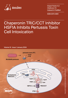
Cover Story (view full-size image): This study investigates the protective effect of the chaperonin TRiC/CCT inhibitor, HSF1A, against pertussis toxin (PT) produced by Bordetella pertussis, the causative agent of whooping cough. TRiC/CCT is a cytosolic protein-folding complex. Previous research highlighted its significance in the functionality of related bacterial toxins. Our study reveals a novel interaction between PTS1 and CCT5, a subunit of the TRiC/CCT chaperonin complex. HSF1A effectively reduces PTS1-mediated ADP-ribosylation of Gαi, the specific intracellular substrate of PT. HSF1A does not impact enzyme activity in vitro or PT cell binding but reduces cytosolic PTS1 levels. Accordingly, HSF1A mitigates adverse effects of PT on inhibitory GPCR/cAMP signaling, suggesting therapeutic potential for countering PT-mediated pathogenicity. View this paper
  • Issues are regarded as officially published after their release is announced to the table of contents alert mailing list.
  • You may sign up for e-mail alerts to receive table of contents of newly released issues.
  • PDF is the official format for papers published in both, html and pdf forms. To view the papers in pdf format, click on the "PDF Full-text" link, and use the free Adobe Reader to open them.
Order results
Result details
Section
Select all
Export citation of selected articles as:
13 pages, 1938 KiB  
Article
Unraveling Hematotoxicity of α-Amanitin in Cultured Hematopoietic Cells
by Willemien F. J. Hof, Miranda Visser, Joyce J. de Jong, Marian N. Rajasekar, Jan Jacob Schuringa, Inge A. M. de Graaf, Daan J. Touw and Bart G. J. Dekkers
Toxins 2024, 16(1), 61; https://doi.org/10.3390/toxins16010061 - 22 Jan 2024
Viewed by 2149
Abstract
Amanita phalloides poisonings account for the majority of fatal mushroom poisonings. Recently, we identified hematotoxicity as a relevant aspect of Amanita poisonings. In this study, we investigated the effects of the main toxins of Amanita phalloides, α- and β-amanitin, on hematopoietic cell [...] Read more.
Amanita phalloides poisonings account for the majority of fatal mushroom poisonings. Recently, we identified hematotoxicity as a relevant aspect of Amanita poisonings. In this study, we investigated the effects of the main toxins of Amanita phalloides, α- and β-amanitin, on hematopoietic cell viability in vitro. Hematopoietic cell lines were exposed to α-amanitin or β-amanitin for up to 72 h with or without the pan-caspase inhibitor Z-VAD(OH)-FMK, antidotes N-acetylcysteine, silibinin, and benzylpenicillin, and organic anion-transporting polypeptide 1B3 (OATP1B3) inhibitors rifampicin and cyclosporin. Cell viability was established by trypan blue exclusion, annexin V staining, and a MTS assay. Caspase-3/7 activity was determined with Caspase-Glo assay, and cleaved caspase-3 was quantified by Western analysis. Cell number and colony-forming units were quantified after exposure to α-amanitin in primary CD34+ hematopoietic stem cells. In all cell lines, α-amanitin concentration-dependently decreased viability and mitochondrial activity. β-Amanitin was less toxic, but still significantly reduced viability. α-Amanitin increased caspase-3/7 activity by 2.8-fold and cleaved caspase-3 by 2.3-fold. Z-VAD(OH)-FMK significantly reduced α-amanitin-induced toxicity. In CD34+ stem cells, α-amanitin decreased the number of colonies and cells. The antidotes and OATP1B3 inhibitors did not reverse α-amanitin-induced toxicity. In conclusion, α-amanitin induces apoptosis in hematopoietic cells via a caspase-dependent mechanism. Full article
(This article belongs to the Topic Toxins in Medical Toxicology)
Show Figures

Figure 1

13 pages, 1325 KiB  
Article
Establishing a Receptor Binding Assay for Ciguatoxins: Challenges, Assay Performance and Application
by Lisbet Díaz-Asencio, Donaida Chamero-Lago, Gabriel L. Rojas-Abrahantes, Carlos M. Alonso-Hernández and Marie-Yasmine Dechraoui Bottein
Toxins 2024, 16(1), 60; https://doi.org/10.3390/toxins16010060 - 22 Jan 2024
Cited by 1 | Viewed by 1780
Abstract
Ciguatera, a global issue, lacks adequate capacity for ciguatoxin analysis in most affected countries. The Caribbean region, known for its endemic ciguatera and being home to a majority of the global small island developing states, particularly needs established methods for ciguatoxin detection in [...] Read more.
Ciguatera, a global issue, lacks adequate capacity for ciguatoxin analysis in most affected countries. The Caribbean region, known for its endemic ciguatera and being home to a majority of the global small island developing states, particularly needs established methods for ciguatoxin detection in seafood and the environment. The radioligand receptor binding assay (r-RBA) is among the in vitro bioassays currently used for ciguatoxin analysis; however, similarly to the other chemical-based or bioassays that have been developed, it faces challenges due to limited standards and interlaboratory comparisons. This work presents a single laboratory validation of an r-RBA developed in a Cuban laboratory while characterizing the performance of the liquid scintillation counter instrument as a key external parameter. The results obtained show the assay is precise, accurate and robust, confirming its potential as a routine screening method for the detection and quantification of ciguatoxins. The new method will aid in identifying high-risk ciguatoxic fish in Cuba and the Caribbean region, supporting monitoring and scientific management of ciguatera and the development of early warning systems to enhance food safety and food security, and promote fair trade fisheries. Full article
(This article belongs to the Section Marine and Freshwater Toxins)
Show Figures

Figure 1

14 pages, 859 KiB  
Article
Longitudinal Assessment of Facial Hyperhidrosis Management: Evaluating the Utility and Quality of Life Improvements following Botulinum Toxin Injection
by Catalin Prodan-Barbulescu, Luca Castiglione, Sonia Roxana Burtic, Marius Murariu, Shruta Reddy, Ovidiu Rosca, Felix Bratosin, Camelia Melania Fizedean, Pavel Krupyshev and Ileana Enatescu
Toxins 2024, 16(1), 59; https://doi.org/10.3390/toxins16010059 - 21 Jan 2024
Cited by 1 | Viewed by 1639
Abstract
Facial hyperhidrosis is a debilitating condition that can severely impact the quality of life. This study aimed to assess the long-term utility of Botulinum toxin type A therapy (BTA) for facial hyperhidrosis and its impact on quality of life over a one-year period. [...] Read more.
Facial hyperhidrosis is a debilitating condition that can severely impact the quality of life. This study aimed to assess the long-term utility of Botulinum toxin type A therapy (BTA) for facial hyperhidrosis and its impact on quality of life over a one-year period. Conducted at the Pius Brinzeu Clinical Emergency Hospital in Timisoara, Romania, this longitudinal observational study involved 77 adult patients with primary facial hyperhidrosis. Participants received two sessions of Botulinum toxin injections (50 U IncoBTX-A each) and were evaluated at baseline, 6 months, and 12 months using the Hyperhidrosis Disease Severity Scale (HDSS), WHOQOL-BREF, Dermatology Life Quality Index (DLQI), and a bespoke survey. The study demonstrated significant reductions in HDSS scores from 3.6 ± 0.5 to 1.2 ± 0.8 post-treatment, sustained at 1.3 ± 0.6 at 12 months (p-value < 0.001). DLQI scores markedly decreased from 24.8 ± 4.2 to 6.2 ± 2.1 post-treatment, stabilizing at 6.5 ± 2.5 at 12 months (p-value < 0.001). Sweat production significantly dropped from 0.75 g ± 0.15 to 0.18 g ± 0.07 per 15 min (p-value < 0.001). WHOQOL-BREF scores improved notably in the mental domain from 66.7 ± 6.1 to 70.8 ± 5.2 at 12 months (p-value < 0.001), with physical and social domains also showing significant improvements. Correlation analysis revealed strong negative correlations between DLQI total score and HDSS (rho = −0.72, p-value < 0.001) and sweat production (rho = −0.68, p-value < 0.001). Regression analysis indicated significant predictors for DLQI total score, including HDSS (B Coefficient = −3.8, p-value < 0.001) and sweat production (B Coefficient = −2.2, p-value < 0.001). BTA therapy significantly improved the quality of life in facial hyperhidrosis patients, with lasting effects on symptom severity, sweat production, and quality of life domains. The correlation and regression analyses further substantiated the treatment’s impact on both physical and psychological aspects. These findings advocate Botulinum toxin as a viable long-term treatment for facial hyperhidrosis. Full article
(This article belongs to the Special Issue Uses of Botulinum Toxin Injection in Medicine)
Show Figures

Figure 1

27 pages, 789 KiB  
Review
Practical Strategies to Reduce Ochratoxin A in Foods
by Hyun Jung Lee, Hae Dun Kim and Dojin Ryu
Toxins 2024, 16(1), 58; https://doi.org/10.3390/toxins16010058 - 20 Jan 2024
Cited by 5 | Viewed by 2895
Abstract
Ochratoxin A (OTA), a potent nephrotoxin, is one of the most deleterious mycotoxins, with its prevalence in agricultural crops and their processed foods around the world. OTA is a major concern to food safety, as OTA exposure through dietary intake may lead to [...] Read more.
Ochratoxin A (OTA), a potent nephrotoxin, is one of the most deleterious mycotoxins, with its prevalence in agricultural crops and their processed foods around the world. OTA is a major concern to food safety, as OTA exposure through dietary intake may lead to a significant level of accumulation in the body as a result of its long half-life (about 35 days). Its potent renal toxicity and high risk of exposure as well as the difficulty in controlling environmental factors OTA production has prompted the need for timely information on practical strategies for the food industry to effectively manage OTA contamination during food processing. The effects of various food processes, including both nonthermal and thermal methods, on the reduction in OTA were summarized in this review, with emphasis on the toxicity of residual OTA as well as its known and unknown degradation products. Since complete removal of OTA from foodstuffs is not feasible, additional strategies that may facilitate the reduction in OTA in food, such as adding baking soda and sugars, was also discussed, so that the industry may understand and apply practical measures to ensure the safety of its products destined for human consumption. Full article
(This article belongs to the Section Mycotoxins)
Show Figures

Figure 1

17 pages, 1801 KiB  
Article
A Magnetic Reduced Graphene Oxide Nanocomposite: Synthesis, Characterization, and Application for High-Efficiency Detoxification of Aflatoxin B1
by Chushu Zhang, Haixiang Zhou, Shining Cao, Jing Chen, Chunjuan Qu, Yueyi Tang, Mian Wang, Lifei Zhu, Xiaoyue Liu and Jiancheng Zhang
Toxins 2024, 16(1), 57; https://doi.org/10.3390/toxins16010057 - 19 Jan 2024
Cited by 2 | Viewed by 2139
Abstract
(1) Background: Safety problems associated with aflatoxin B1 (AFB1) contamination have always been a major threat to human health. Removing AFB1 through adsorption is considered an attractive remediation technique. (2) Methods: To produce an adsorbent with a high AFB [...] Read more.
(1) Background: Safety problems associated with aflatoxin B1 (AFB1) contamination have always been a major threat to human health. Removing AFB1 through adsorption is considered an attractive remediation technique. (2) Methods: To produce an adsorbent with a high AFB1 adsorption efficiency, a magnetic reduced graphene oxide composite (Fe3O4@rGO) was synthesized using one-step hydrothermal fabrication. Then, the adsorbent was characterized using a series of techniques, such as SEM, TEM, XRD, FT-IR, VSM, and nitrogen adsorption–desorption analysis. Finally, the effects of this nanocomposite on the nutritional components of treated foods, such as vegetable oil and peanut milk, were also examined. (3) Results: The optimal synthesis conditions for Fe3O4@rGO were determined to be 200 °C for 6 h. The synthesis temperature significantly affected the adsorption properties of the prepared material due to its effect on the layered structure of graphene and the loading of Fe3O4 nanoparticles. The results of various characterizations illustrated that the surface of Fe3O4@rGO had a two-dimensional layered nanostructure with many folds and that Fe3O4 nanoparticles were distributed uniformly on the surface of the composite material. Moreover, the results of isotherm, kinetic, and thermodynamic analyses indicated that the adsorption of AFB1 by Fe3O4@rGO conformed to the Langmuir model, with a maximum adsorption capacity of 82.64 mg·g−1; the rapid and efficient adsorption of AFB1 occurred mainly through chemical adsorption via a spontaneous endothermic process. When applied to treat vegetable oil and peanut milk, the prepared material minimized the loss of nutrients and thus preserved food quality. (4) Conclusions: The above findings reveal a promising adsorbent, Fe3O4@rGO, with favorable properties for AFB1 adsorption and potential for food safety applications. Full article
(This article belongs to the Special Issue Advances in Rapid Detection and Reduction of Aflatoxins)
Show Figures

Figure 1

14 pages, 2148 KiB  
Article
Development of a Biosensor to Detect Venom of Malayan Krait (Bungarus candidus)
by Kiattawee Choowongkomon, Janeyuth Chaisakul, Supaphorn Seetaha, Taksa Vasaruchapong, Wayne C. Hodgson, Natchaya Rasri, Katechawin Chaeksin, Sattawat Boonchaleaw and Nattapon Sookprasert
Toxins 2024, 16(1), 56; https://doi.org/10.3390/toxins16010056 - 19 Jan 2024
Cited by 1 | Viewed by 2391
Abstract
Malayan krait (Bungarus candidus) envenoming is a cause of significant morbidity and mortality in many Southeast Asian countries. If intubation and specific antivenom administration are delayed, the most significant life-threatening outcome may be the inhibition of neuromuscular transmission and subsequent respiratory [...] Read more.
Malayan krait (Bungarus candidus) envenoming is a cause of significant morbidity and mortality in many Southeast Asian countries. If intubation and specific antivenom administration are delayed, the most significant life-threatening outcome may be the inhibition of neuromuscular transmission and subsequent respiratory failure. It is recommended that krait-envenomed victims without indications of neurotoxicity, e.g., skeletal muscle weakness or ptosis, immediately receive 10 vials of antivenom. However, the administration of excess antivenom may lead to hypersensitivity or serum sickness. Therefore, monitoring venom concentrations in patients could be used as an indicator for snake antivenom treatment. In this study, we aimed to develop a screen-printed gold electrode (SPGE) biosensor to detect B. candidus venom in experimentally envenomed rats. The gold electrodes were coated with monovalent Malayan krait IgG antivenom and used as venom detection biosensors. Electrochemical impedance spectrometry (EIS) and square wave voltammetry (SWV) measurements were performed to detect the electrical characterization between B. candidus venom and monovalent IgG antivenom in the biosensor. The EIS measurements showed increases in charge transfer resistance (Rct) following IgG immobilization and incubation with B. candidus venom solution (0.1–0.4 mg/mL); thus, the antibody was immobilized on the electrode surface and venom was successfully detected. The lowest current signal was detected by SWV measurement in rat plasma collected 30 min following B. candidus experimental envenoming, indicating the highest level of venom concentration in blood circulation (4.3 ± 0.7 µg/mL). The present study demonstrates the ability of the SPGE biosensor to detect B. candidus venom in plasma from experimentally envenomed rats. The technology obtained in this work may be developed as a detection tool for use along with the standard treatment of Malayan krait envenoming. Full article
(This article belongs to the Special Issue Pre-clinical and Clinical Management of Snakebite Envenomation)
Show Figures

Graphical abstract

17 pages, 2798 KiB  
Review
Voltage-Gated Sodium Channel Inhibition by µ-Conotoxins
by Kirsten L. McMahon, Irina Vetter and Christina I. Schroeder
Toxins 2024, 16(1), 55; https://doi.org/10.3390/toxins16010055 - 18 Jan 2024
Viewed by 2154
Abstract
µ-Conotoxins are small, potent pore-blocker inhibitors of voltage-gated sodium (NaV) channels, which have been identified as pharmacological probes and putative leads for analgesic development. A limiting factor in their therapeutic development has been their promiscuity for different NaV channel subtypes, [...] Read more.
µ-Conotoxins are small, potent pore-blocker inhibitors of voltage-gated sodium (NaV) channels, which have been identified as pharmacological probes and putative leads for analgesic development. A limiting factor in their therapeutic development has been their promiscuity for different NaV channel subtypes, which can lead to undesirable side-effects. This review will focus on four areas of µ-conotoxin research: (1) mapping the interactions of µ-conotoxins with different NaV channel subtypes, (2) µ-conotoxin structure–activity relationship studies, (3) observed species selectivity of µ-conotoxins and (4) the effects of µ-conotoxin disulfide connectivity on activity. Our aim is to provide a clear overview of the current status of µ-conotoxin research. Full article
(This article belongs to the Special Issue Conotoxins: Evolution, Classifications and Targets)
Show Figures

Figure 1

22 pages, 3553 KiB  
Article
Cytotoxic Effects of Major and Emerging Mycotoxins on HepaRG Cells and Transcriptomic Response after Exposure of Spheroids to Enniatins B and B1
by France Coulet, Monika Coton, Cristian Iperi, Marine Belinger Podevin, Emmanuel Coton and Nolwenn Hymery
Toxins 2024, 16(1), 54; https://doi.org/10.3390/toxins16010054 - 18 Jan 2024
Cited by 1 | Viewed by 2162
Abstract
Mycotoxins, produced by fungi, frequently occur at different stages in the food supply chain between pre- and postharvest. Globally produced cereal crops are known to be highly susceptible to contamination, thus constituting a major public health concern. Among the encountered mycotoxigenic fungi in [...] Read more.
Mycotoxins, produced by fungi, frequently occur at different stages in the food supply chain between pre- and postharvest. Globally produced cereal crops are known to be highly susceptible to contamination, thus constituting a major public health concern. Among the encountered mycotoxigenic fungi in cereals, Fusarium spp. are the most frequent and produce both regulated (i.e., T-2 toxin, deoxynivalenol -DON-, zearalenone -ZEA-) and emerging (i.e., enniatins -ENNs-, beauvericin -BEA-) mycotoxins. In this study, we investigated the in vitro cytotoxic effects of regulated and emerging fusariotoxins on HepaRG cells in 2D and 3D models using undifferentiated and differentiated cells. We also studied the impact of ENN B1 and ENN B exposure on gene expression of HepaRG spheroids. Gene expression profiling pinpointed the differentially expressed genes (DEGs) and overall similar pathways were involved in responses to mycotoxin exposure. Complement cascades, metabolism, steroid hormones, bile secretion, and cholesterol pathways were all negatively impacted by both ENNs. For cholesterol biosynthesis, 23/27 genes were significantly down-regulated and could be correlated to a 30% reduction in cholesterol levels. Our results show the impact of ENNs on the cholesterol biosynthesis pathway for the first time. This finding suggests a potential negative effect on human health due to the essential role this pathway plays. Full article
(This article belongs to the Special Issue Mycotoxin Health Exposure: Molecular Interactions and Cytotoxicity)
Show Figures

Graphical abstract

18 pages, 1968 KiB  
Article
An Occurrence Study of Mycotoxins in Plant-Based Beverages Using Liquid Chromatography–Mass Spectrometry
by Romans Pavlenko, Zane Berzina, Ingars Reinholds, Elena Bartkiene and Vadims Bartkevics
Toxins 2024, 16(1), 53; https://doi.org/10.3390/toxins16010053 - 17 Jan 2024
Cited by 4 | Viewed by 2356
Abstract
Mycotoxins are toxic mold metabolites that can adversely affect human and animal health. More than 400 mycotoxins have been identified so far. Cereals and nuts are the predominant mycotoxin-contaminated foodstuffs. Plant-based drinks produced from cereals, nuts, and legumes have grown in popularity. The [...] Read more.
Mycotoxins are toxic mold metabolites that can adversely affect human and animal health. More than 400 mycotoxins have been identified so far. Cereals and nuts are the predominant mycotoxin-contaminated foodstuffs. Plant-based drinks produced from cereals, nuts, and legumes have grown in popularity. The mycotoxins accumulated in these crops may transfer to these beverages. A liquid chromatography–tandem mass spectrometry method was developed and optimized for the assessment of 22 mycotoxins in commercially available plant-based drinks in Latvia and Lithuania. A total of 64% of the seventy-two analyzed beverages were positive for one to sixteen mycotoxins, with deoxynivalenol, beauvericin, and enniatins A, B, B1, T-2, and HT-2 toxins detected most frequently. The European Commission has not yet set guidelines for the maximum mycotoxin concentrations in plant-based beverages, nor has the European Food Safety Authority conducted a risk assessment. Therefore, acute exposure studies were provided for the Latvian population based on the assumed replacement of dairy milk with plant-based beverages to ascertain the safety of plant-based milk substitutes. Based on the observed levels of mycotoxin prevalence and contamination levels and assumed exposure, it can be concluded that tested plant-based beverages may be relatively safe. However, exposure to emerging mycotoxins should be considered. Full article
(This article belongs to the Special Issue Detection, Control and Contamination of Mycotoxins (Volume II))
Show Figures

Graphical abstract

16 pages, 5874 KiB  
Article
Exploring the Biocontrol Capability of Non-Mycotoxigenic Strains of Penicillium expansum
by Belén Llobregat, Luis González-Candelas and Ana-Rosa Ballester
Toxins 2024, 16(1), 52; https://doi.org/10.3390/toxins16010052 - 17 Jan 2024
Cited by 2 | Viewed by 2056
Abstract
Penicillium expansum is one the major postharvest pathogens of pome fruit during postharvest handling and storage. This fungus also produces patulin, which is a highly toxic mycotoxin that can contaminate infected fruits and their derived products and whose levels are regulated in many [...] Read more.
Penicillium expansum is one the major postharvest pathogens of pome fruit during postharvest handling and storage. This fungus also produces patulin, which is a highly toxic mycotoxin that can contaminate infected fruits and their derived products and whose levels are regulated in many countries. In this study, we investigated the biocontrol potential of non-mycotoxigenic strains of Penicillium expansum against a mycotoxigenic strain. We analyzed the competitive behavior of two knockout mutants that were unable to produce patulin. The first mutant (∆patK) involved the deletion of the patK gene, which is the initial gene in patulin biosynthesis. The second mutant (∆veA) involved the deletion of veA, which is a global regulator of primary and secondary metabolism. At the phenotypic level, the ∆patK mutant exhibited similar phenotypic characteristics to the wild-type strain. In contrast, the ∆veA mutant displayed altered growth characteristics compared with the wild type, including reduced conidiation and abnormal conidiophores. Neither mutant produced patulin under the tested conditions. Under various stress conditions, the ∆veA mutants exhibited reduced growth and conidiation when exposed to stressors, including cell membrane stress, oxidative stress, osmotic stress, and different pH values. However, no significant changes were observed in the ∆patK mutant. In competitive growth experiments, the presence of non-mycotoxigenic strains reduced the population of the wild-type strain during in vitro growth. Furthermore, the addition of either of the non-mycotoxigenic strains resulted in a significant decrease in patulin levels. Overall, our results suggest the potential use of non-mycotoxigenic mutants, particularly ∆patK mutants, as biocontrol agents to reduce patulin contamination in food and feed. Full article
(This article belongs to the Special Issue Effect of Mycotoxin on Crop and Methods of Prevention and Degradation)
Show Figures

Figure 1

12 pages, 4511 KiB  
Article
Toxicokinetics of Zearalenone following Oral Administration in Female Dezhou Donkeys
by Honglei Qu, Yunduo Zheng, Ruifen Kang, Yulong Feng, Pengshuai Li, Yantao Wang, Jie Cheng, Cheng Ji, Wenqiong Chai and Qiugang Ma
Toxins 2024, 16(1), 51; https://doi.org/10.3390/toxins16010051 - 17 Jan 2024
Cited by 1 | Viewed by 1537
Abstract
Zearalenone (ZEN) is a mycotoxin produced by various Fusarium strains, that is present in food and feed raw materials worldwide, causing toxicity effects in animals and humans. This research aimed to explore the toxicokinetics of ZEN on female Dezhou donkeys following a single [...] Read more.
Zearalenone (ZEN) is a mycotoxin produced by various Fusarium strains, that is present in food and feed raw materials worldwide, causing toxicity effects in animals and humans. This research aimed to explore the toxicokinetics of ZEN on female Dezhou donkeys following a single oral exposure dosage of 2 mg/kg BW (body weight). The sample collection of donkeys plasma was carried out at 0, 5, 10, 15, 20, 30, 45, 60, 90 min, 2 h, 2.5 h, 3 h, 3.5 h, 4 h, 4.5 h, 6 h, 9 h, 12 h, 24 h, 48 h, 72 h, 96 h and 120 h via intravenous catheter, and fecal and urinary samples were severally collected at 0 h and every 6 h until 120 h. The concentrations of ZEN, α-zearalenol (α-ZOL), β-zearalenol (β-ZOL), α-zearalanol (α-ZAL), β-zearalanol (β-ZAL), zearalanone (ZAN) in plasma, urine, and feces were detected by UPLC-MS/MS. Only ZEN was detected in plasma, and the maximum was 15.34 ± 5.12 µg/L occurred at 0.48 h after gavage. The total plasma clearance (Cl) of ZEN was 95.20 ± 8.01 L·kg·BW−1·h−1. In addition, the volume of distribution (Vd) was up to 216.17 ± 58.71 L/kg. The percentage of total ZEN (ZEN plus the main metabolites) excretion in feces and urine was 2.49% and 2.10%, respectively. In summary, ZEN was fast absorbed and relatively slowly excreted in female donkeys during 120 h after a single gavage, indicating a trend of wider tissue distribution and longer tissue persistence. Full article
(This article belongs to the Special Issue Metabolism and Toxicology of Mycotoxins and Their Masked Forms)
Show Figures

Figure 1

12 pages, 1451 KiB  
Article
Assessing the Risk of Exposure to Aflatoxin B1 through the Consumption of Peanuts among Children Aged 6–59 Months in the Lusaka District, Zambia
by Grace Musawa, Flavien Nsoni Bumbangi, Chisoni Mumba, Branly Kilola Mbunga, Gladys Phiri, Vistorina Benhard, Henson Kainga, Mkuzi Banda, Enock Ndaki, Ethel Mkandawire and John Bwalya Muma
Toxins 2024, 16(1), 50; https://doi.org/10.3390/toxins16010050 - 17 Jan 2024
Cited by 1 | Viewed by 2525
Abstract
Aflatoxins B1 (AFB1) are fungi-produced toxins found in crops like peanuts, maize, and tree nuts. They constitute a public health concern due to their genotoxic and carcinogenic effects. A deterministic exposure risk assessment to AFB1 through the consumption of peanuts was conducted on [...] Read more.
Aflatoxins B1 (AFB1) are fungi-produced toxins found in crops like peanuts, maize, and tree nuts. They constitute a public health concern due to their genotoxic and carcinogenic effects. A deterministic exposure risk assessment to AFB1 through the consumption of peanuts was conducted on children using the Margin of Exposure (MOE) and the liver cancer risk approaches. Data on AFB1 concentrations in peanuts, quantities of peanut consumption, and the weights of the children were obtained from the literature. Generally, MOE values were below the safe margin of 10,000, ranging between 3.68 and 0.14, 754.34 and 27.33, and 11,428.57 and 419.05 for the high (0.0466 ng/kg), median (0.00023 ng/kg), and low (0.000015 ng/kg) AFB1 concentration levels, respectively. The liver cancer risk upon lifetime exposure to highly AFB1-contaminated peanuts (0.0466 ng/kg) ranged between 1 and 23 (95% lower bound) and 2 and 50 (95% upper bound) cases in a million individuals: a public health concern. A low liver cancer risk (≤1 case in a billion individuals upon lifetime exposure) was shown at median and low AFB1 concentrations. However, the risk of AFB1 should be a priority for risk management since its harmful effects could be potentiated by poor diet, high malnutrition levels, and other disease burdens in Zambia’s children. Full article
(This article belongs to the Special Issue Mycotoxins: Risk Assessment, Biomonitoring and Toxicology)
Show Figures

Figure 1

36 pages, 8285 KiB  
Article
Molecular Phylogeny, Morphology, Growth and Toxicity of Three Benthic Dinoflagellates Ostreopsis sp. 9, Prorocentrum lima and Coolia monotis Developing in Strait of Gibraltar, Southwestern Mediterranean
by Mustapha Ibghi, Benlahcen Rijal Leblad, Mohammed L’Bachir El Kbiach, Hicham Aboualaalaa, Mouna Daoudi, Estelle Masseret, Emilie Le Floc’h, Fabienne Hervé, Gwenael Bilien, Nicolas Chomerat, Zouher Amzil and Mohamed Laabir
Toxins 2024, 16(1), 49; https://doi.org/10.3390/toxins16010049 - 16 Jan 2024
Viewed by 2600
Abstract
Few works have been carried out on benthic harmful algal blooms (BHAB) species in the southern Mediterranean and no data are available for the highly dynamic Strait of Gibraltar (western Mediterranean waters). For the first time, Ostreopsis sp. 9, Prorocentrum lima and Coolia [...] Read more.
Few works have been carried out on benthic harmful algal blooms (BHAB) species in the southern Mediterranean and no data are available for the highly dynamic Strait of Gibraltar (western Mediterranean waters). For the first time, Ostreopsis sp. 9, Prorocentrum lima and Coolia monotis were isolated in this key region in terms of exchanges between the Atlantic Ocean and the Mediterranean and subject to intense maritime traffic. Ribotyping confirmed the morphological identification of these three dinoflagellates species. Monoclonal cultures were established and the maximum growth rate and cell yield were measured at a temperature of 24 °C and an irradiance of 90 µmol photons m−2 s−1, for each species: 0.26 ± 0.02 d−1 (8.75 × 10³ cell mL−1 after 28 days) for Ostreopsis sp. 9, 0.21 ± 0.01 d−1 (49 × 103 cell mL−1 after 145 days) for P. lima and 0.21 ± 0.01 d−1 (10.02 × 10³ cell mL−1 after 28 days) for C. monotis. Only P. lima was toxic with concentrations of okadaic acid and dinophysistoxin-1 measured in optimal growth conditions ranging from 6.4 pg cell−1 to 26.97 pg cell−1 and from 5.19 to 25.27 pg cell−1, respectively. The toxin content of this species varied in function of the growth phase. Temperature influenced the growth and toxin content of P. lima. Results suggest that future warming of Mediterranean coastal waters may lead to higher growth rates and to increases in cellular toxin levels in P. lima. Nitrate and ammonia affected the toxin content of P. lima but no clear trend was noted. In further studies, we have to isolate other BHAB species and strains from Strait of Gibraltar waters to obtain more insight into their diversity and toxicity. Full article
Show Figures

Figure 1

20 pages, 421 KiB  
Review
Zootoxins and Domestic Animals: A European View
by Andras-Laszlo Nagy, Sabrina Ardelean, Ronan J. J. Chapuis, Juliette Bouillon, Dalma Pivariu, Beatrice De Felice, Mirko Bertazzo, Paola Fossati, Leon J. Spicer, Alexandra Iulia Dreanca and Francesca Caloni
Toxins 2024, 16(1), 48; https://doi.org/10.3390/toxins16010048 - 16 Jan 2024
Viewed by 2387
Abstract
Zootoxins are produced by venomous and poisonous species and are an important cause of poisoning in companion animals and livestock in Europe. Little information about the incidence of zootoxin poisoning is available in Europe, with only a few case reports and review papers [...] Read more.
Zootoxins are produced by venomous and poisonous species and are an important cause of poisoning in companion animals and livestock in Europe. Little information about the incidence of zootoxin poisoning is available in Europe, with only a few case reports and review papers being published. This review presents the most important zootoxins produced by European venomous and poisonous animal species responsible for poisoning episodes in companion animals and livestock. The main zootoxin-producing animal species, components of the toxins/venoms and their clinical effects are presented. The most common zootoxicoses involve terrestrial zootoxins excreted by the common toad, the fire salamander, the pine processionary caterpillar, and vipers. The lack of a centralized reporting/poison control system in Europe makes the evaluation of the epidemiology of zootoxin-induced poisonings extremely difficult. Even if there are many anecdotal reports in the veterinary community about the exposure of domestic animals to terrestrial and marine zootoxins, the number of published papers regarding these toxicoses is low. Climate change and its consequences regarding species distribution and human-mediated transportation are responsible for the emerging nature of some intoxications in which zootoxins are involved. Although new venomous or poisonous animal species have emerged in regions where they were previously unreported, zootoxins produced by native species remain the main concern in Europe. The diversity of poisonous and venomous animal species and the emerging nature of certain poisonings warrant the continuous update to such knowledge by veterinary professionals and animal owners. This review offers an overview about zootoxin-related poisonings in domestic animals in Europe and also provides important information from a health perspective. Full article
22 pages, 4550 KiB  
Review
Mycotoxins-Imprinted Polymers: A State-of-the-Art Review
by Simone Cavalera, Laura Anfossi, Fabio Di Nardo and Claudio Baggiani
Toxins 2024, 16(1), 47; https://doi.org/10.3390/toxins16010047 - 15 Jan 2024
Cited by 7 | Viewed by 2269
Abstract
Mycotoxins are toxic metabolites of molds which can contaminate food and beverages. Because of their acute and chronic toxicity, they can have harmful effects when ingested or inhaled, posing severe risks to human health. Contemporary analytical methods have the sensitivity required for contamination [...] Read more.
Mycotoxins are toxic metabolites of molds which can contaminate food and beverages. Because of their acute and chronic toxicity, they can have harmful effects when ingested or inhaled, posing severe risks to human health. Contemporary analytical methods have the sensitivity required for contamination detection and quantification, but the direct application of these methods on real samples is not straightforward because of matrix complexity, and clean-up and preconcentration steps are needed, more and more requiring the application of highly selective solid-phase extraction materials. Molecularly imprinted polymers (MIPs) are artificial receptors mimicking the natural antibodies that are increasingly being used as a solid phase in extraction methods where selectivity towards target analytes is mandatory. In this review, the state-of-the-art about molecularly imprinted polymers as solid-phase extraction materials in mycotoxin contamination analysis will be discussed, with particular attention paid to the use of mimic molecules in the synthesis of mycotoxin-imprinted materials, to the application of these materials to food real samples, and to the development of advanced extraction methods involving molecular imprinting technology. Full article
(This article belongs to the Special Issue Toxins: 15th Anniversary)
Show Figures

Graphical abstract

16 pages, 10078 KiB  
Article
Modulation of Broiler Intestinal Changes Induced by Clostridium perfringens and Deoxynivalenol through Probiotic, Paraprobiotic, and Postbiotic Supplementation
by Marielen de Souza, Ana Angelita Sampaio Baptista, Maísa Fabiana Menck-Costa, Larissa Justino, Eduardo Micotti da Glória, Gabriel Danilo Shimizu, Camila Rodrigues Ferraz, Waldiceu A. Verri, Filip Van Immerseel and Ana Paula Frederico Rodrigues Loureiro Bracarense
Toxins 2024, 16(1), 46; https://doi.org/10.3390/toxins16010046 - 14 Jan 2024
Cited by 2 | Viewed by 2198
Abstract
Deoxynivalenol (DON) is a predisposing factor for necrotic enteritis. This study aimed to investigate the effects of a DON and Clostridium perfringens (CP) challenge on the intestinal morphology, morphometry, oxidative stress, and immune response of broilers. Additionally, we evaluated the potential of a [...] Read more.
Deoxynivalenol (DON) is a predisposing factor for necrotic enteritis. This study aimed to investigate the effects of a DON and Clostridium perfringens (CP) challenge on the intestinal morphology, morphometry, oxidative stress, and immune response of broilers. Additionally, we evaluated the potential of a Lactobacillus spp. mixture as an approach to mitigate the damage induced by the challenge. One-day-old broiler chickens (n = 252) were divided into seven treatment groups: Control, DON, CP, CP + DON, VL (DON + CP + viable Lactobacillus spp. mixture), HIL (DON + CP + heat-inactivated Lactobacillus spp. mixture), and LCS (DON + CP + Lactobacillus spp. mixture culture supernatant). Macroscopic evaluation of the intestines revealed that the CP + DON group exhibited the highest lesion score, while the VL and HIL groups showed the lowest scores. Microscopically, all Lactobacillus spp. treatments mitigated the morphological changes induced by the challenge. DON increased levels of reactive oxygen species (ROS) in the jejunum, and CP increased ROS levels in the jejunum and ileum. Notably, the Lactobacillus spp. treatments did not improve the antioxidant defense against CP-induced oxidative stress. In summary, a Lactobacillus spp. mixture, whether used as a probiotic, paraprobiotic, or postbiotic, exerted a partially protective effect in mitigating most of the intestinal damage induced by DON and CP challenges. Full article
Show Figures

Figure 1

11 pages, 502 KiB  
Article
Aflatoxin M1 Analysis in Urine of Mill Workers in Bangladesh: A Pilot Study
by Nurshad Ali, Ahsan Habib, Firoz Mahmud, Humaira Rashid Tuba and Gisela H. Degen
Toxins 2024, 16(1), 45; https://doi.org/10.3390/toxins16010045 - 14 Jan 2024
Cited by 2 | Viewed by 1808
Abstract
Presence of aflatoxin B1 (AFB1) in food and feed is a serious problem, especially in developing countries. Human exposure to this carcinogenic mycotoxin can occur through dietary intake, but also through inhalation or dermal contact when handling and processing AFB [...] Read more.
Presence of aflatoxin B1 (AFB1) in food and feed is a serious problem, especially in developing countries. Human exposure to this carcinogenic mycotoxin can occur through dietary intake, but also through inhalation or dermal contact when handling and processing AFB1-contaminated crops. A suitable biomarker of AFB1 exposure by all routes is the occurrence of its hydroxylated metabolite aflatoxin M1 (AFM1) in urine. To assess mycotoxin exposure in mill workers in Bangladesh, we analyzed AFM1 levels in urine samples of this population group who may encounter both dietary and occupational AFB1 exposure. In this pilot study, a total of 76 participants (51 mill workers and 25 controls) were enrolled from the Sylhet region of Bangladesh. Urine samples were collected from people who worked in rice, wheat, maize and spice mills and from controls with no occupational contact to these materials. A questionnaire was used to collect information on basic characteristics and normal food habits of all participants. Levels of AFM1 in the urine samples were determined by a competitive enzyme linked immunosorbent assay. AFM1 was detected in 96.1% of mill workers’ urine samples with a range of LOD (40) of 217.7 pg/mL and also in 92% of control subject’s urine samples with a range of LOD of 307.0 pg/mL). The mean level of AFM1 in mill workers’ urine (106.5 ± 35.0 pg/mL) was slightly lower than that of the control group (123.3 ± 52.4 pg/mL), whilst the mean AFM1 urinary level adjusted for creatinine was higher in mill workers (142.1 ± 126.1 pg/mg crea) than in the control group (98.5 ± 71.2 pg/mg crea). Yet, these differences in biomarker levels were not statistically significant. Slightly different mean urinary AFM1 levels were observed between maize mill, spice mill, rice mill, and wheat mill workers, yet biomarker values are based on a small number of individuals in these subgroups. No significant correlations were found between the study subjects’ urine AFM1 levels and their consumption of some staple food items, except for a significant correlation observed between urinary biomarker levels and consumption of groundnuts. In conclusion, this pilot study revealed the frequent presence of AFM1 in the urine of mill workers in Bangladesh and those of concurrent controls with dietary AFB1 exposure only. The absence of a statistical difference in mean biomarker levels for workers and controls suggests that in the specific setting, no extra occupational exposure occurred. Yet, the high prevalence of non-negligible AFM1 levels in the collected urines encourage further studies in Bangladesh regarding aflatoxin exposure. Full article
(This article belongs to the Special Issue Mycotoxins: Risk Assessment, Biomonitoring and Toxicology)
Show Figures

Figure 1

21 pages, 6949 KiB  
Article
Bacillus velezensis A2 Can Protect against Damage to IPEC-J2 Cells Induced by Zearalenone via the Wnt/FRZB/β-Catenin Signaling Pathway
by Jing Cai, Xuanshuai Yuan, Yuhang Sun, Jia Chen, Peng Li, Shuhua Yang and Miao Long
Toxins 2024, 16(1), 44; https://doi.org/10.3390/toxins16010044 - 13 Jan 2024
Cited by 1 | Viewed by 1927
Abstract
Zearalenone (ZEA) has adverse effects on human and animal health, and finding effective strategies to combat its toxicity is essential. The probiotic Bacillus velezensis A2 shows various beneficial physiological functions, including the potential to combat fungal toxins. However, the detailed mechanism by which [...] Read more.
Zearalenone (ZEA) has adverse effects on human and animal health, and finding effective strategies to combat its toxicity is essential. The probiotic Bacillus velezensis A2 shows various beneficial physiological functions, including the potential to combat fungal toxins. However, the detailed mechanism by which the Bacillus velezensis A2 strain achieves this protective effect is not yet fully revealed. This experiment was based on transcriptome data to study the protective mechanism of Bacillus velezensis A2 against ZEA-induced damage to IPEC-J2 cells. The experiment was divided into CON, A2, ZEA, and A2+ZEA groups. This research used an oxidation kit to measure oxidative damage indicators, the terminal deoxynucleotidyl transferase-mediated nick end labeling (TUNEL) method to detect cell apoptosis, flow cytometry to determine the cell cycle, and transcriptome sequencing to screen and identify differentially expressed genes. In addition, gene ontology (GO) and the Kyoto Encyclopedia of Genes and Genomes (KEGG) were adopted to screen out relevant signaling pathways. Finally, to determine whether A2 can alleviate the damage caused by ZEA to cells, the genes and proteins involved in inflammation, cell apoptosis, cell cycles, and related pathways were validated using a quantitative reverse transcription polymerase chain reaction (qRT-PCR) and Western blot methods. Compared with the CON group, the levels of reactive oxygen species (ROS) and malondialdehyde (MDA) in the ZEA group increased significantly (p < 0.01), while the levels of antioxidant enzyme activity, total superoxide dismutase (T-SOD), glutathione peroxidase (GSH-PX), total antioxidant capacity (T-AOC), and catalase (CAT) decreased significantly (p < 0.01). Compared with the ZEA group, the A2+ZEA group showed a significant decrease in ROS and MDA levels (p < 0.01), while the levels of T-SOD, GSH-PX, T-AOC, and CAT increased significantly (p < 0.01). TUNEL and cell cycle results indicated that compared with the ZEA group, the A2+ZEA group demonstrated a significant decrease in the cell apoptosis rate (p < 0.01), and the cell cycle was restored. Combining transcriptome data, qRT-PCR, and Western blot, the results showed that compared with the CON group, the mRNA and protein expression levels of Wnt10 and β-catenin increased significantly (p < 0.01), while the expression level of FRZB decreased significantly (p < 0.01); compared with the ZEA group, the expression levels of these mRNA and proteins were reversed. Bacillus velezensis A2 can increase the antioxidant level, reduce inflammatory damage, decrease cell apoptosis, and correct the cell cycle when that damage is being caused by ZEA. The protective mechanism may be related to the regulation of the Wnt/FRZB cell/β-catenin signaling pathway. Full article
(This article belongs to the Special Issue Metabolism and Toxicology of Mycotoxins and Their Masked Forms)
Show Figures

Figure 1

16 pages, 2938 KiB  
Communication
Tetrodotoxin and Its Analogues (TTXs) in the Food-Capture and Defense Organs of the Palaeonemertean Cephalothrix cf. simula
by Grigorii V. Malykin, Peter V. Velansky and Timur Yu. Magarlamov
Toxins 2024, 16(1), 43; https://doi.org/10.3390/toxins16010043 - 12 Jan 2024
Cited by 2 | Viewed by 1752
Abstract
Tetrodotoxin (TTX), an extremely potent low-molecular-weight neurotoxin, is widespread among marine animals including ribbon worms (Nemertea). Previously, studies on the highly toxic palaeonemertean Cephalothrix cf. simula showed that toxin-positive structures are present all over its body and are mainly associated with glandular cells [...] Read more.
Tetrodotoxin (TTX), an extremely potent low-molecular-weight neurotoxin, is widespread among marine animals including ribbon worms (Nemertea). Previously, studies on the highly toxic palaeonemertean Cephalothrix cf. simula showed that toxin-positive structures are present all over its body and are mainly associated with glandular cells and epithelial tissues. The highest TTXs concentrations were detected in a total extract from the intestine of the anterior part of the body and also in a total extract from the proboscis. However, many questions as to the TTXs distribution in the organs of the anterior part of the worm’s body and the functions of the toxins in these organs are still unanswered. In the present report, we provide additional results of a detailed and comprehensive analysis of TTXs distribution in the nemertean’s proboscis, buccal cavity, and cephalic gland using an integrated approach including high-performance liquid chromatography–tandem mass spectrometry (HPLC–MS/MS), confocal laser scanning microscopy with anti-TTX antibodies, light and electron microscopies, and observations of feeding behavior. For the proboscis, we have found a TTXs profile different from that characteristic of other organs and tissues. We have also shown for the first time that the major amount of TTXs is localized in the anterior part of the proboscis that is mainly involved in hunting. TTX-containing glandular cells, which can be involved in the prey immobilization, have been found in the buccal cavities of the nemerteans. A significant contribution of the cephalic gland to the toxicity of this animal has been shown for the first time, and the role of the gland is hypothesized to be involved not only in protection against potential enemies but also in immobilizing prey. The data obtained have made it possible to extend the understanding of the role and features of the use of TTXs in the organs of the anterior part of nemertean’s body. Full article
(This article belongs to the Section Marine and Freshwater Toxins)
Show Figures

Figure 1

14 pages, 1745 KiB  
Article
TDP-43 and Alzheimer’s Disease Pathology in the Brain of a Harbor Porpoise Exposed to the Cyanobacterial Toxin BMAA
by Susanna P. Garamszegi, Daniel J. Brzostowicki, Thomas M. Coyne, Regina T. Vontell and David A. Davis
Toxins 2024, 16(1), 42; https://doi.org/10.3390/toxins16010042 - 12 Jan 2024
Cited by 7 | Viewed by 2871
Abstract
Cetaceans are well-regarded as sentinels for toxin exposure. Emerging studies suggest that cetaceans can also develop neuropathological changes associated with neurodegenerative disease. The occurrence of neuropathology makes cetaceans an ideal species for examining the impact of marine toxins on the brain across the [...] Read more.
Cetaceans are well-regarded as sentinels for toxin exposure. Emerging studies suggest that cetaceans can also develop neuropathological changes associated with neurodegenerative disease. The occurrence of neuropathology makes cetaceans an ideal species for examining the impact of marine toxins on the brain across the lifespan. Here, we describe TAR DNA-binding protein 43 (TDP-43) proteinopathy and Alzheimer’s disease (AD) neuropathological changes in a beached harbor porpoise (Phocoena phocoena) that was exposed to a toxin produced by cyanobacteria called β-N-methylamino-L-alanine (BMAA). We found pathogenic TDP-43 cytoplasmic inclusions in neurons throughout the cerebral cortex, midbrain and brainstem. P62/sequestosome-1, responsible for the autophagy of misfolded proteins, was observed in the amygdala, hippocampus and frontal cortex. Genes implicated in AD and TDP-43 neuropathology such as APP and TARDBP were expressed in the brain. AD neuropathological changes such as amyloid-β plaques, neurofibrillary tangles, granulovacuolar degeneration and Hirano bodies were present in the hippocampus. These findings further support the development of progressive neurodegenerative disease in cetaceans and a potential causative link to cyanobacterial toxins. Climate change, nutrient pollution and industrial waste are increasing the frequency of harmful cyanobacterial blooms. Cyanotoxins like BMAA that are associated with neurodegenerative disease pose an increasing public health risk. Full article
(This article belongs to the Section Marine and Freshwater Toxins)
Show Figures

Figure 1

16 pages, 805 KiB  
Review
Nanoparticles for Mitigation of Harmful Cyanobacterial Blooms
by Ilana N. Tseytlin, Anna K. Antrim and Ping Gong
Toxins 2024, 16(1), 41; https://doi.org/10.3390/toxins16010041 - 12 Jan 2024
Cited by 1 | Viewed by 1906
Abstract
With the rapid advancement of nanotechnology and its widespread applications, increasing amounts of manufactured and natural nanoparticles (NPs) have been tested for their potential utilization in treating harmful cyanobacterial blooms (HCBs). NPs can be used as a photocatalyst, algaecide, adsorbent, flocculant, or coagulant. [...] Read more.
With the rapid advancement of nanotechnology and its widespread applications, increasing amounts of manufactured and natural nanoparticles (NPs) have been tested for their potential utilization in treating harmful cyanobacterial blooms (HCBs). NPs can be used as a photocatalyst, algaecide, adsorbent, flocculant, or coagulant. The primary mechanisms explored for NPs to mitigate HCBs include photocatalysis, metal ion-induced cytotoxicity, physical disruption of the cell membrane, light-shielding, flocculation/coagulation/sedimentation of cyanobacterial cells, and the removal of phosphorus (P) and cyanotoxins from bloom water by adsorption. As an emerging and promising chemical/physical approach for HCB mitigation, versatile NP-based technologies offer great advantages, such as being environmentally benign, cost-effective, highly efficient, recyclable, and adaptable. The challenges we face include cost reduction, scalability, and impacts on non-target species co-inhabiting in the same environment. Further efforts are required to scale up to real-world operations through developing more efficient, recoverable, reusable, and deployable NP-based lattices or materials that are adaptable to bloom events in different water bodies of different sizes, such as reservoirs, lakes, rivers, and marine environments. Full article
(This article belongs to the Special Issue Monitoring and Management of Algal and Cyanobacterial Blooms)
Show Figures

Figure 1

20 pages, 3448 KiB  
Article
Analysis of Pyrrolizidine Alkaloids in Stingless Bee Honey and Identification of a Botanical Source as Ageratum conyzoides
by Natasha L. Hungerford, Norhasnida Zawawi, Tianqi (Evonne) Zhu, Steve J. Carter, Kevin J. Melksham and Mary T. Fletcher
Toxins 2024, 16(1), 40; https://doi.org/10.3390/toxins16010040 - 12 Jan 2024
Viewed by 1721
Abstract
Stingless bee honeys (SBHs) from Australian and Malaysian species were analysed using ultra-high performance liquid chromatography-tandem mass spectrometry (UHPLC-MS/MS) for the presence of pyrrolizidine alkaloids (PAs) and the corresponding N-oxides (PANOs) due to the potential for such hepatotoxic alkaloids to contaminate honey [...] Read more.
Stingless bee honeys (SBHs) from Australian and Malaysian species were analysed using ultra-high performance liquid chromatography-tandem mass spectrometry (UHPLC-MS/MS) for the presence of pyrrolizidine alkaloids (PAs) and the corresponding N-oxides (PANOs) due to the potential for such hepatotoxic alkaloids to contaminate honey as a result of bees foraging on plants containing these alkaloids. Low levels of alkaloids were found in these SBHs when assessed against certified PA standards in targeted analysis. However, certain isomers were identified using untargeted analysis in a subset of honeys of Heterotrigona itama which resulted in the identification of a PA weed species (Ageratum conyzoides) near the hives. The evaluation of this weed provided a PA profile matching that of the SBH of H. itama produced nearby, and included supinine, supinine N-oxide (or isomers) and acetylated derivatives. These PAs lacking a hydroxyl group at C7 are thought to be less hepatoxic. However, high levels were also observed in SBH (and in A. conyzoides) of a potentially more toxic diester PA corresponding to an echimidine isomer. Intermedine, the C7 hydroxy equivalent of supinine, was also observed. Species differences in nectar collection were evident as the same alkaloids were not identified in SBH of G. thoracica from the same location. This study highlights that not all PAs and PANOs are identified using available standards in targeted analyses and confirms the need for producers of all types of honey to be aware of nearby potential PA sources, particularly weeds. Full article
Show Figures

Graphical abstract

4 pages, 189 KiB  
Editorial
Animal Poisoning: Toxins from Plants or Feed—An Important Chemical Risk for Domestic Animals
by Andras-Laszlo Nagy
Toxins 2024, 16(1), 39; https://doi.org/10.3390/toxins16010039 - 12 Jan 2024
Viewed by 1594
Abstract
Feed-, food-, water- and plant-related toxins are a major threat for animal and human health worldwide [...] Full article
(This article belongs to the Special Issue Animal Poisoning: Toxins from Plants or Feed)
17 pages, 11828 KiB  
Article
A Streamlined Method to Obtain Biologically Active TcdA and TcdB Toxins from Clostridioides difficile
by Diane Sapa, Anaïs Brosse, Héloïse Coullon, Gauthier Péan de Ponfilly, Thomas Candela and Alban Le Monnier
Toxins 2024, 16(1), 38; https://doi.org/10.3390/toxins16010038 - 11 Jan 2024
Cited by 1 | Viewed by 1676
Abstract
The major virulence factors of Clostridioides difficile (C. difficile) are enterotoxins A (TcdA) and B (TcdB). The study of toxins is a crucial step in exploring the virulence of this pathogen. Currently, the toxin purification process is either laborious and time-consuming [...] Read more.
The major virulence factors of Clostridioides difficile (C. difficile) are enterotoxins A (TcdA) and B (TcdB). The study of toxins is a crucial step in exploring the virulence of this pathogen. Currently, the toxin purification process is either laborious and time-consuming in C. difficile or performed in heterologous hosts. Therefore, we propose a streamlined method to obtain functional toxins in C. difficile. Two C. difficile strains were generated, each harboring a sequence encoding a His-tag at the 3′ end of C. difficile 630∆erm tcdA or tcdB genes. Each toxin gene is expressed using the Ptet promoter, which is inducible by anhydro-tetracycline. The obtained purification yields were 0.28 mg and 0.1 mg per liter for rTcdA and rTcdB, respectively. In this study, we successfully developed a simple routine method that allows the production and purification of biologically active rTcdA and rTcdB toxins with similar activities compared to native toxins. Full article
Show Figures

Figure 1

14 pages, 500 KiB  
Review
Use of Antibiotics following Snakebite in the Era of Antimicrobial Stewardship
by Helena Brenes-Chacon, José María Gutiérrez and María L. Avila-Aguero
Toxins 2024, 16(1), 37; https://doi.org/10.3390/toxins16010037 - 11 Jan 2024
Cited by 2 | Viewed by 3926
Abstract
Even though there are guidelines for the management of snakebite envenoming (SBE), the use of antibiotics in this pathology remains controversial. The aim of this study is to provide a narrative review of the literature and recommendations based on the best available evidence [...] Read more.
Even though there are guidelines for the management of snakebite envenoming (SBE), the use of antibiotics in this pathology remains controversial. The aim of this study is to provide a narrative review of the literature and recommendations based on the best available evidence regarding antibiotic use in SBE. We performed a narrative review of relevant literature regarding SBE and antibiotic use as prophylaxis or treatment. A total of 26 articles were included. There is wide use of antibiotics in SBE; nevertheless, infection was not necessarily documented. The antibiotics used varied according to the study, from beta lactams to lincosamide and nitroimidazoles, and from monotherapy to combined antimicrobials. The most common recommendations were to manage skin and soft tissue infections and avoid infectious complications, but these suggestions are not necessarily based on bacteriological findings. Prophylactic use of antibiotics in SBE is discouraged in most studies. Antibiotic prescription in SBE should be based on the susceptibility of microorganisms isolated from the affected tissue or identified in snakes’ oral cavities. Antibiotics should be reserved only for patients with a demonstrated infection, or those at a high risk of developing an infection, i.e., presenting severe local envenoming, local signs of infection, or those with incorrect manipulation of wounds. Prospective studies are needed to correlate microbiological findings at the wound site and the response to antibiotic use. Full article
(This article belongs to the Special Issue Pre-clinical and Clinical Management of Snakebite Envenomation)
Show Figures

Figure 1

16 pages, 2889 KiB  
Article
The Chaperonin TRiC/CCT Inhibitor HSF1A Protects Cells from Intoxication with Pertussis Toxin
by Jinfang Jia, Manuel Zoeschg, Holger Barth, Arto T. Pulliainen and Katharina Ernst
Toxins 2024, 16(1), 36; https://doi.org/10.3390/toxins16010036 - 10 Jan 2024
Cited by 1 | Viewed by 2533
Abstract
Pertussis toxin (PT) is a bacterial AB5-toxin produced by Bordetella pertussis and a major molecular determinant of pertussis, also known as whooping cough, a highly contagious respiratory disease. In this study, we investigate the protective effects of the chaperonin TRiC/CCT inhibitor, [...] Read more.
Pertussis toxin (PT) is a bacterial AB5-toxin produced by Bordetella pertussis and a major molecular determinant of pertussis, also known as whooping cough, a highly contagious respiratory disease. In this study, we investigate the protective effects of the chaperonin TRiC/CCT inhibitor, HSF1A, against PT-induced cell intoxication. TRiC/CCT is a chaperonin complex that facilitates the correct folding of proteins, preventing misfolding and aggregation, and maintaining cellular protein homeostasis. Previous research has demonstrated the significance of TRiC/CCT in the functionality of the Clostridioides difficile TcdB AB-toxin. Our findings reveal that HSF1A effectively reduces the levels of ADP-ribosylated Gαi, the specific substrate of PT, in PT-treated cells, without interfering with enzyme activity in vitro or the cellular binding of PT. Additionally, our study uncovers a novel interaction between PTS1 and the chaperonin complex subunit CCT5, which correlates with reduced PTS1 signaling in cells upon HSF1A treatment. Importantly, HSF1A mitigates the adverse effects of PT on cAMP signaling in cellular systems. These results provide valuable insights into the mechanisms of PT uptake and suggest a promising starting point for the development of innovative therapeutic strategies to counteract pertussis toxin-mediated pathogenicity. Full article
(This article belongs to the Special Issue Toxins: 15th Anniversary)
Show Figures

Figure 1

16 pages, 6031 KiB  
Article
Inhibition of CYP1A1 Alleviates Colchicine-Induced Hepatotoxicity
by Ruoyue Huang, Jingyi Duan, Wen Huang, Yan Cheng, Beiwei Zhu and Fei Li
Toxins 2024, 16(1), 35; https://doi.org/10.3390/toxins16010035 - 9 Jan 2024
Cited by 4 | Viewed by 2657
Abstract
Colchicine, a natural compound extracted from Colchicum autumnale, is a phytotoxin, but interestingly, it also has multiple pharmacological activities. Clinically, colchicine is widely used for the treatment of gouty arthritis, familial Mediterranean fever, cardiovascular dysfunction and new coronary pneumonia. However, overdose intake of [...] Read more.
Colchicine, a natural compound extracted from Colchicum autumnale, is a phytotoxin, but interestingly, it also has multiple pharmacological activities. Clinically, colchicine is widely used for the treatment of gouty arthritis, familial Mediterranean fever, cardiovascular dysfunction and new coronary pneumonia. However, overdose intake of colchicine could cause lethal liver damage, which is a limitation of its application. Therefore, exploring the potential mechanism of colchicine-induced hepatotoxicity is meaningful. Interestingly, it was found that CYP1A1 played an important role in the hepatotoxicity of colchicine, while it might also participate in its metabolism. Inhibition of CYP1A1 could alleviate oxidative stress and pyroptosis in the liver upon colchicine treatment. By regulating CYP1A1 through the CASPASE-1-GSDMD pathway, colchicine-induced liver injury was effectively relieved in a mouse model. In summary, we concluded that CYP1A1 may be a potential target, and the inhibition of CYP1A1 alleviates colchicine-induced liver injury through pyroptosis regulated by the CASPASE-1-GSDMD pathway. Full article
Show Figures

Figure 1

14 pages, 4214 KiB  
Review
Diversity and Evolutionary Analysis of Venom Insulin Derived from Cone Snails
by Qiqi Guo, Meiling Huang, Ming Li, Jiao Chen, Shuanghuai Cheng, Linlin Ma and Bingmiao Gao
Toxins 2024, 16(1), 34; https://doi.org/10.3390/toxins16010034 - 9 Jan 2024
Cited by 2 | Viewed by 2494
Abstract
Cone snails possess a diverse array of novel peptide toxins, which selectively target ion channels and receptors in the nervous and cardiovascular systems. These numerous novel peptide toxins are a valuable resource for future marine drug development. In this review, we compared and [...] Read more.
Cone snails possess a diverse array of novel peptide toxins, which selectively target ion channels and receptors in the nervous and cardiovascular systems. These numerous novel peptide toxins are a valuable resource for future marine drug development. In this review, we compared and analyzed the sequence diversity, three-dimensional structural variations, and evolutionary aspects of venom insulin derived from different cone snail species. The comparative analysis reveals that there are significant variations in the sequences and three-dimensional structures of venom insulins from cone snails with different feeding habits. Notably, the venom insulin of some piscivorous cone snails exhibits a greater similarity to humans and zebrafish insulins. It is important to emphasize that these venom insulins play a crucial role in the predatory strategies of these cone snails. Furthermore, a phylogenetic tree was constructed to trace the lineage of venom insulin sequences, shedding light on the evolutionary interconnections among cone snails with diverse diets. Full article
(This article belongs to the Special Issue Conotoxins: Evolution, Classifications and Targets)
Show Figures

Figure 1

16 pages, 1318 KiB  
Editorial
Gaston Ramon’s Big Four
by Jean-Philippe Chippaux
Toxins 2024, 16(1), 33; https://doi.org/10.3390/toxins16010033 - 9 Jan 2024
Viewed by 1716
Abstract
When immunology was still in its infancy, Gaston Ramon made several major contributions to humoral immunology [...] Full article
Show Figures

Figure 1

15 pages, 1692 KiB  
Article
Effect of Temperature, pH, and aw on Cereulide Synthesis and Regulator Genes Transcription with Respect to Bacillus cereus Growth and Cereulide Production
by Yating Wang, Yangtai Liu, Shuo Yang, Yuhang Chen, Yang Liu, Dasheng Lu, Hongmei Niu, Fanchong Ren, Anning Xu and Qingli Dong
Toxins 2024, 16(1), 32; https://doi.org/10.3390/toxins16010032 - 8 Jan 2024
Cited by 2 | Viewed by 2258
Abstract
Bacillus cereus is a food-borne pathogen that can produce cereulide in the growth period, which causes food poisoning symptoms. Due to its resistance to heat, extreme pH, and proteolytic enzymes, cereulide poses a serious threat to food safety. Temperature, pH, and aw [...] Read more.
Bacillus cereus is a food-borne pathogen that can produce cereulide in the growth period, which causes food poisoning symptoms. Due to its resistance to heat, extreme pH, and proteolytic enzymes, cereulide poses a serious threat to food safety. Temperature, pH, and aw can influence cereulide production, but there is still a lack of research with multi-environmental impacts. In this study, the effects of temperature (15~45 °C), pH (5~8), and aw (0.945~0.996) on the emetic reference strain B. cereus F4810/72 growth, cereulide production, relevant ces genes (cesA, cesB, cesP), and transcription regulators genes (codY and abrB) expression at transcription level were studied. B. cereus survived for 4~53 h or grew to 6.85~8.15 log10 CFU/mL in environmental combinations. Cereulide accumulation was higher in mid-temperature, acidic, or high aw environments. Increased temperature resulted in a lower cereulide concentration at pH 8 or aw of 0.970. The lowest cereulide concentration was found at pH 6.5 with an increased aw from 0.970 to 0.996. Water activity had a strong effect on transcriptional regulator genes as well as the cesB gene, and temperature was the main effect factor of cesP gene expression. Moreover, environmental factors also impact cereulide synthesis at transcriptional levels thereby altering the cereulide concentrations. The interaction of environmental factors may result in the survival of B. cereus without growth for a period. Gene expression is affected by environmental factors, and temperature and pH may be the main factors influencing the correlation between B. cereus growth and cereulide formation. This study contributed to an initial understanding of the intrinsic link between the impact of environmental factors and cereulide formation and provided valuable information for clarifying the mechanism of cereulide synthesis in combined environmental conditions. Full article
(This article belongs to the Section Bacterial Toxins)
Show Figures

Figure 1

Previous Issue
Next Issue
Back to TopTop