Next Issue
Volume 10, January
Previous Issue
Volume 9, November


Toxins, Volume 9, Issue 12 (December 2017) – 31 articles

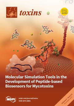
Cover Story (view full-size image): Computer-aided design of the bioreceptor for ochratoxin monitoring: In solution, the unmodified bioreceptor (blue) binds to ochratoxin (magenta) with its intrinsic affinity. However, on the biotransducer surface (cyan), the bioreceptor becomes sterically hindered. A hexa-histidine tag (His-tag ) (green) immobilizes the bioreceptor and recovers some of the configurational freedom. However, linking via the amine or acid end alters the mechanism of binding and the efficacy. The configurational freedom is improved by placing a polyethylene glycol (PEG) spacer (red) between the His-tag and bioreceptor. While experimental techniques are limited, computer-aided technologies are promising in a rationalized route to bioreceptor design. View this paper
  • Issues are regarded as officially published after their release is announced to the table of contents alert mailing list.
  • You may sign up for e-mail alerts to receive table of contents of newly released issues.
  • PDF is the official format for papers published in both, html and pdf forms. To view the papers in pdf format, click on the "PDF Full-text" link, and use the free Adobe Reader to open them.
Order results
Result details
Section
Select all
Export citation of selected articles as:
9000 KiB  
Review
Microbial Inhibition of Fusarium Pathogens and Biological Modification of Trichothecenes in Cereal Grains
by Urszula Wachowska, Danuta Packa and Marian Wiwart
Toxins 2017, 9(12), 408; https://doi.org/10.3390/toxins9120408 - 20 Dec 2017
Cited by 33 | Viewed by 6901
Abstract
Fungi of the genus Fusarium infect cereal crops during the growing season and cause head blight and other diseases. Their toxic secondary metabolites (mycotoxins) contaminate grains. Several dozen toxic compounds produced by fungal pathogens have been identified to date. Type B trichothecenes—deoxynivalenol, its [...] Read more.
Fungi of the genus Fusarium infect cereal crops during the growing season and cause head blight and other diseases. Their toxic secondary metabolites (mycotoxins) contaminate grains. Several dozen toxic compounds produced by fungal pathogens have been identified to date. Type B trichothecenes—deoxynivalenol, its acetyl derivatives and nivalenol (produced mainly by F. graminearum and F. culmorum)—are most commonly detected in cereal grains. “T-2 toxin” (produced by, among others, F. sporotrichioides) belongs to type-A trichothecenes which are more toxic than other trichothecenes. Antagonistic bacteria and fungi can affect pathogens of the genus Fusarium via different modes of action: direct (mycoparasitism or hyperparasitism), mixed-path (antibiotic secretion, production of lytic enzymes) and indirect (induction of host defense responses). Microbial modification of trichothecenes involves acetylation, deacetylation, oxidation, de-epoxidation, and epimerization, and it lowers the pathogenic potential of fungi of the genus Fusarium. Other modifing mechanisms described in the paper involve the physical adsorption of mycotoxins in bacterial cells and the conjugation of mycotoxins to glucose and other compounds in plant and fungal cells. The development of several patents supports the commercialization and wider application of microorganisms biodegrading mycotoxins in grains and, consequently, in feed additives. Full article
(This article belongs to the Collection Understanding Mycotoxin Occurrence in Food and Feed Chains)
Show Figures

Graphical abstract

671 KiB  
Article
Food-Borne Outbreak Investigation and Molecular Typing: High Diversity of Staphylococcus aureus Strains and Importance of Toxin Detection
by Sarah Denayer, Laurence Delbrassinne, Yacine Nia and Nadine Botteldoorn
Toxins 2017, 9(12), 407; https://doi.org/10.3390/toxins9120407 - 20 Dec 2017
Cited by 84 | Viewed by 8586
Abstract
Staphylococcus aureus is an important aetiological agent of food intoxications in the European Union as it can cause gastro-enteritis through the production of various staphylococcal enterotoxins (SEs) in foods. Reported enterotoxin dose levels causing food-borne illness are scarce and varying. Three food poisoning [...] Read more.
Staphylococcus aureus is an important aetiological agent of food intoxications in the European Union as it can cause gastro-enteritis through the production of various staphylococcal enterotoxins (SEs) in foods. Reported enterotoxin dose levels causing food-borne illness are scarce and varying. Three food poisoning outbreaks due to enterotoxin-producing S. aureus strains which occurred in 2013 in Belgium are described. The outbreaks occurred in an elderly home, at a barbecue event and in a kindergarten and involved 28, 18, and six cases, respectively. Various food leftovers contained coagulase positive staphylococci (CPS). Low levels of staphylococcal enterotoxins ranging between 0.015 ng/g and 0.019 ng/g for enterotoxin A (SEA), and corresponding to 0.132 ng/g for SEC were quantified in the food leftovers for two of the reported outbreaks. Molecular typing of human and food isolates using pulsed-field gel electrophoresis (PFGE) and enterotoxin gene typing, confirmed the link between patients and the suspected foodstuffs. This also demonstrated the high diversity of CPS isolates both in the cases and in healthy persons carrying enterotoxin genes encoding emetic SEs for which no detection methods currently exist. For one outbreak, the investigation pointed out to the food handler who transmitted the outbreak strain to the food. Tools to improve staphylococcal food poisoning (SFP) investigations are presented. Full article
(This article belongs to the Collection Staphylococcus aureus Toxins)
Show Figures

Graphical abstract

1525 KiB  
Review
Secreted Phospholipases A2 from Animal Venoms in Pain and Analgesia
by Vanessa O. Zambelli, Gisele Picolo, Carlos A. H. Fernandes, Marcos R. M. Fontes and Yara Cury
Toxins 2017, 9(12), 406; https://doi.org/10.3390/toxins9120406 - 19 Dec 2017
Cited by 60 | Viewed by 7217
Abstract
Animal venoms comprise a complex mixture of components that affect several biological systems. Based on the high selectivity for their molecular targets, these components are also a rich source of potential therapeutic agents. Among the main components of animal venoms are the secreted [...] Read more.
Animal venoms comprise a complex mixture of components that affect several biological systems. Based on the high selectivity for their molecular targets, these components are also a rich source of potential therapeutic agents. Among the main components of animal venoms are the secreted phospholipases A2 (sPLA2s). These PLA2 belong to distinct PLA2s groups. For example, snake venom sPLA2s from Elapidae and Viperidae families, the most important families when considering envenomation, belong, respectively, to the IA and IIA/IIB groups, whereas bee venom PLA2 belongs to group III of sPLA2s. It is well known that PLA2, due to its hydrolytic activity on phospholipids, takes part in many pathophysiological processes, including inflammation and pain. Therefore, secreted PLA2s obtained from animal venoms have been widely used as tools to (a) modulate inflammation and pain, uncovering molecular targets that are implicated in the control of inflammatory (including painful) and neurodegenerative diseases; (b) shed light on the pathophysiology of inflammation and pain observed in human envenomation by poisonous animals; and, (c) characterize molecular mechanisms involved in inflammatory diseases. The present review summarizes the knowledge on the nociceptive and antinociceptive actions of sPLA2s from animal venoms, particularly snake venoms. Full article
(This article belongs to the Special Issue Animal Venoms and Pain)
Show Figures

Figure 1

282 KiB  
Article
Effects of Continuously Feeding Diets Containing Cereal Ergot Alkaloids on Nutrient Digestibility, Alkaloid Recovery in Feces, and Performance Traits of Ram Lambs
by Stephanie Coufal-Majewski, Kim Stanford, Tim McAllister, Yuxi Wang, Barry Blakley, John McKinnon, Mary Lou Swift and Alexandre V. Chaves
Toxins 2017, 9(12), 405; https://doi.org/10.3390/toxins9120405 - 19 Dec 2017
Cited by 14 | Viewed by 4853
Abstract
Allowable limits for cereal ergot alkaloids in livestock feeds are being re-examined, and the objective of this study was to compare nutrient digestibility, growth performance and carcass characteristics of ram lambs fed a range of alkaloid concentrations, including the maximum currently allowed in [...] Read more.
Allowable limits for cereal ergot alkaloids in livestock feeds are being re-examined, and the objective of this study was to compare nutrient digestibility, growth performance and carcass characteristics of ram lambs fed a range of alkaloid concentrations, including the maximum currently allowed in Canada (2 to 3 ppm). Four pelleted diets were fed: control, with no added alkaloids; 930; 1402; and 2447 ppb alkaloids based on total R and S epimers. Eight ram lambs (30.0 ± 3.1 kg) were used to examine the impacts of dietary treatments on nutrient digestibility and alkaloid recovery from feces. Concentrations of dietary alkaloids evaluated did not affect nutrient digestibility or N metabolism. Excepting ergocornine and ergocryptine, recovery of alkaloids in feces varied among periods, suggesting that individual lambs may differ in their ability to metabolize ergocristine, ergometrine, ergosine, ergotamine and their S epimers. In a second experiment, ram lambs (n = 47, 30 ± 8 kg) were randomly assigned to a diet and weighed weekly until they achieved a slaughter weight of ≥ 45 kg (average 9 weeks; range 6 to 13 weeks). Intake of DM did not differ (p = 0.91) among diets, although lambs fed 2447 ppb alkaloids had a lower (p < 0.01) ADG than did lambs receiving other treatments. The concentration of serum prolactin linearly declined (p < 0.01) with increasing alkaloids. Feeding 2447 ppb total alkaloids negatively impacted growth, while feeding 1402 ppb did not harm growth performance, but reduced carcass dressing percentage. Due to different concentrations of alkaloids affecting growth and carcass characteristics in the present study, determining allowable limits for total dietary alkaloids will require a better understanding of impacts of alkaloid profiles and interactions among individual alkaloids. Full article
(This article belongs to the Collection Understanding Mycotoxin Occurrence in Food and Feed Chains)
Show Figures

Graphical abstract

2672 KiB  
Communication
High Conservation of Tetanus and Botulinum Neurotoxins Cleavage Sites on Human SNARE Proteins Suggests That These Pathogens Exerted Little or No Evolutionary Pressure on Humans
by Stefan Carle, Marco Pirazzini, Ornella Rossetto, Holger Barth and Cesare Montecucco
Toxins 2017, 9(12), 404; https://doi.org/10.3390/toxins9120404 - 19 Dec 2017
Cited by 9 | Viewed by 5388
Abstract
The Genome Aggregation Database presently contains >120,000 human genomes. We searched in this database for the presence of mutations at the sites of tetanus (TeNT) and botulinum neurotoxins (BoNTs) cleavages of the three SNARE proteins: VAMP, SNAP-25 and Syntaxin. These mutations could account [...] Read more.
The Genome Aggregation Database presently contains >120,000 human genomes. We searched in this database for the presence of mutations at the sites of tetanus (TeNT) and botulinum neurotoxins (BoNTs) cleavages of the three SNARE proteins: VAMP, SNAP-25 and Syntaxin. These mutations could account for some of the BoNT/A resistant patients. At the same time, this approach was aimed at testing the possibility that TeNT and BoNT may have acted as selective agents in the development of resistance to tetanus or botulism. We found that mutations of the SNARE proteins are very rare and concentrated outside the SNARE motif required for the formation of the SNARE complex involved in neuroexocytosis. No changes were found at the BoNT cleavage sites of VAMP and syntaxins and only one very rare mutation was found in the essential C-terminus region of SNAP-25, where Arg198 was replaced with a Cys residue. This is the P1’ cleavage site for BoNT/A and the P1 cleavage site for BoNT/C. We found that the Arg198Cys mutation renders SNAP-25 resistant to BoNT/A. Nonetheless, its low frequency (1.8 × 10⁻⁵) indicates that mutations of SNAP-25 at the BoNT/A cleavage site are unlikely to account for the existence of BoNT/A resistant patients. More in general, the present findings indicate that tetanus and botulinum neurotoxins have not acted as selective agents during human evolution as it appears to have been the case for tetanus in rats and chicken. Full article
(This article belongs to the Section Bacterial Toxins)
Show Figures

Figure 1

251 KiB  
Review
Botulinum Toxin in the Field of Dermatology: Novel Indications
by Yoon Seob Kim, Eun Sun Hong and Hei Sung Kim
Toxins 2017, 9(12), 403; https://doi.org/10.3390/toxins9120403 - 16 Dec 2017
Cited by 40 | Viewed by 6621
Abstract
Since its approval by the US Food and Drug Administration in 2002 for glabellar wrinkles, botulinum toxin (BTX) has been widely used to correct facial wrinkles. As a result, many consider BTX synonymous with cosmetic dermatology. Recent studies indicate that BTX elicits biological [...] Read more.
Since its approval by the US Food and Drug Administration in 2002 for glabellar wrinkles, botulinum toxin (BTX) has been widely used to correct facial wrinkles. As a result, many consider BTX synonymous with cosmetic dermatology. Recent studies indicate that BTX elicits biological effects on various skin cell types via the modulation of neurotransmitter release, and it seems that BTX has a wider zone of dermatologic influence than originally understood. Clinicians and researchers are now beginning to explore the potential of BTX beyond the amelioration of facial lines and encouraging results are seen with BTX in a variety of skin conditions. In this paper, we review novel dermatological indications of BTX which includes (but not limited to) scar prevention, facial flushing, post-herpetic neuralgia and itch. These areas show great promise, but there is definite need for larger, double-blinded, randomized control trials against established treatments before BTX becomes a clinical reality. Full article
371 KiB  
Review
In Vitro Toxicological Assessment of Cylindrospermopsin: A Review
by Silvia Pichardo, Ana M. Cameán and Angeles Jos
Toxins 2017, 9(12), 402; https://doi.org/10.3390/toxins9120402 - 16 Dec 2017
Cited by 75 | Viewed by 5826
Abstract
Cylindrospermopsin (CYN) is a cyanobacterial toxin that is gaining importance, owing to its increasing expansion worldwide and the increased frequency of its blooms. CYN mainly targets the liver, but also involves other organs. Various mechanisms have been associated with its toxicity, such as [...] Read more.
Cylindrospermopsin (CYN) is a cyanobacterial toxin that is gaining importance, owing to its increasing expansion worldwide and the increased frequency of its blooms. CYN mainly targets the liver, but also involves other organs. Various mechanisms have been associated with its toxicity, such as protein synthesis inhibition, oxidative stress, etc. However, its toxic effects are not yet fully elucidated and additional data for hazard characterization purposes are required. In this regard, in vitro methods can play an important role, owing to their advantages in comparison to in vivo trials. The aim of this work was to compile and evaluate the in vitro data dealing with CYN available in the scientific literature, focusing on its toxicokinetics and its main toxicity mechanisms. This analysis would be useful to identify research needs and data gaps in order to complete knowledge about the toxicity profile of CYN. For example, it has been shown that research on various aspects, such as new emerging toxicity effects, the toxicity of analogs, or the potential interaction of CYN with other cyanotoxins, among others, is still very scarce. New in vitro studies are therefore welcome. Full article
(This article belongs to the Special Issue Cyanobacteria and Cyanotoxins: New Advances and Future Challenges)
263 KiB  
Review
Expression of Staphylococcal Enterotoxins under Stress Encountered during Food Production and Preservation
by Jenny Schelin, Yusak Budi Susilo and Sophia Johler
Toxins 2017, 9(12), 401; https://doi.org/10.3390/toxins9120401 - 15 Dec 2017
Cited by 41 | Viewed by 5292
Abstract
Staphylococcal food poisoning (SFP) is the most prevalent cause of food-borne intoxications worldwide. Consumption of enterotoxins preformed in food causes violent vomiting and can be fatal in children and the elderly. While being repressed by competing bacteria in most matrices, Staphylococcus aureus benefits [...] Read more.
Staphylococcal food poisoning (SFP) is the most prevalent cause of food-borne intoxications worldwide. Consumption of enterotoxins preformed in food causes violent vomiting and can be fatal in children and the elderly. While being repressed by competing bacteria in most matrices, Staphylococcus aureus benefits from crucial competitive advantages in foods with high osmolarity or low pH. During recent years, the long-standing belief in the feasibility of assessing SFP risk based on colony-forming units of S. aureus present in food products has been disproven. Instead, researchers and food business operators are acutely aware of the imminent threat arising from unforeseeable enterotoxin production under stress conditions. This paradigm shift led to a variety of new publications enabling an improved understanding of enterotoxin expression under stress conditions encountered in food. The wealth of data provided by these studies is extremely diverse, as it is based on different methodological approaches, staphylococcal strains, stressors, and enterotoxins. Therefore, in this review, we aggregated and critically evaluated the complex findings of these studies, to provide readers with a current overview of the state of research in the field. Full article
(This article belongs to the Special Issue Heat-Stable Enterotoxins)
3932 KiB  
Article
Investigating β-N-Methylamino-l-alanine Misincorporation in Human Cell Cultures: A Comparative Study with Known Amino Acid Analogues
by Rianita Van Onselen, Simoné Downing, Gabré Kemp and Tim Downing
Toxins 2017, 9(12), 400; https://doi.org/10.3390/toxins9120400 - 14 Dec 2017
Cited by 22 | Viewed by 5396
Abstract
Misincorporation of β-N-methylamino-l-alanine (BMAA) into proteins has been proposed to be a mechanism of toxicity to explain the role of BMAA in neurodegenerative disease development. However, studies have shown that all detectable BMAA can be removed from proteins by [...] Read more.
Misincorporation of β-N-methylamino-l-alanine (BMAA) into proteins has been proposed to be a mechanism of toxicity to explain the role of BMAA in neurodegenerative disease development. However, studies have shown that all detectable BMAA can be removed from proteins by SDS-PAGE purification and that the toxicity of l-canavanine cannot be reproduced in prokaryotes or in a rat pheochromocytoma cell line, strongly indicating that the misincorporation hypothesis of BMAA should be re-investigated. The aim of this study was therefore to determine if BMAA misincorporates into proteins in cells of human origin with subsequent misincorporation-type toxicity. Almost complete loss of viability in response to exposure to l-4-fluorophenylalanine and l-m-tyrosine was observed in all of the cell lines, corresponding to a concentration-dependent increase of the analogues in protein extracts from exposed cells. In contrast, BMAA exposure resulted in slight toxicity in one of the cell lines but the observed toxicity was not the result of misincorporation of BMAA into proteins, as no BMAA was detected in any of the SDS-PAGE purified protein extracts that were obtained from the cells following BMAA exposure. The results show that BMAA is not misincorporated into human proteins and that misincorporation is not a valid mechanism of toxicity. Full article
(This article belongs to the Special Issue The Cyanobacterial Neurotoxin BMAA)
Show Figures

Figure 1

2939 KiB  
Article
A Deeper Examination of Thorellius atrox Scorpion Venom Components with Omic Technologies
by Teresa Romero-Gutierrez, Esteban Peguero-Sanchez, Miguel A. Cevallos, Cesar V. F. Batista, Ernesto Ortiz and Lourival D. Possani
Toxins 2017, 9(12), 399; https://doi.org/10.3390/toxins9120399 - 12 Dec 2017
Cited by 34 | Viewed by 6645
Abstract
This communication reports a further examination of venom gland transcripts and venom composition of the Mexican scorpion Thorellius atrox using RNA-seq and tandem mass spectrometry. The RNA-seq, which was performed with the Illumina protocol, yielded more than 20,000 assembled transcripts. Following a database [...] Read more.
This communication reports a further examination of venom gland transcripts and venom composition of the Mexican scorpion Thorellius atrox using RNA-seq and tandem mass spectrometry. The RNA-seq, which was performed with the Illumina protocol, yielded more than 20,000 assembled transcripts. Following a database search and annotation strategy, 160 transcripts were identified, potentially coding for venom components. A novel sequence was identified that potentially codes for a peptide with similarity to spider ω-agatoxins, which act on voltage-gated calcium channels, not known before to exist in scorpion venoms. Analogous transcripts were found in other scorpion species. They could represent members of a new scorpion toxin family, here named omegascorpins. The mass fingerprint by LC-MS identified 135 individual venom components, five of which matched with the theoretical masses of putative peptides translated from the transcriptome. The LC-MS/MS de novo sequencing allowed to reconstruct and identify 42 proteins encoded by assembled transcripts, thus validating the transcriptome analysis. Earlier studies conducted with this scorpion venom permitted the identification of only twenty putative venom components. The present work performed with more powerful and modern omic technologies demonstrates the capacity of accomplishing a deeper characterization of scorpion venom components and the identification of novel molecules with potential applications in biomedicine and the study of ion channel physiology. Full article
(This article belongs to the Special Issue Scorpion Toxins)
Show Figures

Figure 1

751 KiB  
Review
Innate Lymphoid Cells (ILCs) as Mediators of Inflammation, Release of Cytokines and Lytic Molecules
by Noha Mousaad Elemam, Suad Hannawi and Azzam A. Maghazachi
Toxins 2017, 9(12), 398; https://doi.org/10.3390/toxins9120398 - 10 Dec 2017
Cited by 35 | Viewed by 8757
Abstract
Innate lymphoid cells (ILCs) are an emerging group of immune cells that provide the first line of defense against various pathogens as well as contributing to tissue repair and inflammation. ILCs have been classically divided into three subgroups based on their cytokine secretion [...] Read more.
Innate lymphoid cells (ILCs) are an emerging group of immune cells that provide the first line of defense against various pathogens as well as contributing to tissue repair and inflammation. ILCs have been classically divided into three subgroups based on their cytokine secretion and transcription factor profiles. ILC nomenclature is analogous to that of T helper cells. Group 1 ILCs composed of natural killer (NK) cells as well as IFN-γ secreting ILC1s. ILC2s have the capability to produce TH2 cytokines while ILC3s and lymphoid tissue inducer (LTis) are subsets of cells that are able to secrete IL-17 and/or IL-22. A recent subset of ILC known as ILC4 was discovered, and the cells of this subset were designated as NK17/NK1 due to their release of IL-17 and IFN-γ. In this review, we sought to explain the subclasses of ILCs and their roles as mediators of lytic enzymes and inflammation. Full article
Show Figures

Figure 1

210 KiB  
Editorial
Editorial for Special Issue: The Insecticidal Bacterial Toxins in Modern Agriculture
by Juan Ferré and Baltasar Escriche
Toxins 2017, 9(12), 396; https://doi.org/10.3390/toxins9120396 - 9 Dec 2017
Cited by 1 | Viewed by 3877
(This article belongs to the Special Issue The Insecticidal Bacterial Toxins in Modern Agriculture)
5169 KiB  
Review
Cone Snails: A Big Store of Conotoxins for Novel Drug Discovery
by Bingmiao Gao, Chao Peng, Jiaan Yang, Yunhai Yi, Junqing Zhang and Qiong Shi
Toxins 2017, 9(12), 397; https://doi.org/10.3390/toxins9120397 - 7 Dec 2017
Cited by 102 | Viewed by 24770
Abstract
Marine drugs have developed rapidly in recent decades. Cone snails, a group of more than 700 species, have always been one of the focuses for new drug discovery. These venomous snails capture prey using a diverse array of unique bioactive neurotoxins, usually named [...] Read more.
Marine drugs have developed rapidly in recent decades. Cone snails, a group of more than 700 species, have always been one of the focuses for new drug discovery. These venomous snails capture prey using a diverse array of unique bioactive neurotoxins, usually named as conotoxins or conopeptides. These conotoxins have proven to be valuable pharmacological probes and potential drugs due to their high specificity and affinity to ion channels, receptors, and transporters in the nervous systems of target prey and humans. Several research groups, including ours, have examined the venom gland of cone snails using a combination of transcriptomic and proteomic sequencing, and revealed the existence of hundreds of conotoxin transcripts and thousands of conopeptides in each Conus species. Over 2000 nucleotide and 8000 peptide sequences of conotoxins have been published, and the number is still increasing quickly. However, more than 98% of these sequences still lack 3D structural and functional information. With the rapid development of genomics and bioinformatics in recent years, functional predictions and investigations on conotoxins are making great progress in promoting the discovery of novel drugs. For example, ω-MVIIA was approved by the U.S. Food and Drug Administration in 2004 to treat chronic pain, and nine more conotoxins are at various stages of preclinical or clinical evaluation. In short, the genus Conus, the big family of cone snails, has become an important genetic resource for conotoxin identification and drug development. Full article
(This article belongs to the Special Issue Toxins in Drug Discovery and Pharmacology)
Show Figures

Figure 1

2541 KiB  
Perspective
Molecular Modeling and Simulation Tools in the Development of Peptide-Based Biosensors for Mycotoxin Detection: Example of Ochratoxin
by Aby A. Thyparambil, Ingrid Bazin and Anthony Guiseppi-Elie
Toxins 2017, 9(12), 395; https://doi.org/10.3390/toxins9120395 - 6 Dec 2017
Cited by 11 | Viewed by 6883
Abstract
Mycotoxin contamination of food and feed is now ubiquitous. Exposures to mycotoxin via contact or ingestion can potentially induce adverse health outcomes. Affordable mycotoxin-monitoring systems are highly desired but are limited by (a) the reliance on technically challenging and costly molecular recognition by [...] Read more.
Mycotoxin contamination of food and feed is now ubiquitous. Exposures to mycotoxin via contact or ingestion can potentially induce adverse health outcomes. Affordable mycotoxin-monitoring systems are highly desired but are limited by (a) the reliance on technically challenging and costly molecular recognition by immuno-capture technologies; and (b) the lack of predictive tools for directing the optimization of alternative molecular recognition modalities. Our group has been exploring the development of ochratoxin detection and monitoring systems using the peptide NFO4 as the molecular recognition receptor in fluorescence, electrochemical and multimodal biosensors. Using ochratoxin as the model mycotoxin, we share our perspective on addressing the technical challenges involved in biosensor fabrication, namely: (a) peptide receptor design; and (b) performance evaluation. Subsequently, the scope and utility of molecular modeling and simulation (MMS) approaches to address the above challenges are described. Informed and enabled by phage display, the subsequent application of MMS approaches can rationally guide subsequent biomolecular engineering of peptide receptors, including bioconjugation and bioimmobilization approaches to be used in the fabrication of peptide biosensors. MMS approaches thus have the potential to reduce biosensor development cost, extend product life cycle, and facilitate multi-analyte detection of mycotoxins, each of which positively contributes to the overall affordability of mycotoxin biosensor monitoring systems. Full article
(This article belongs to the Special Issue Advanced Sensors for Toxins)
Show Figures

Figure 1

3492 KiB  
Review
The Potential Pathogenic Contributions of Endothelial Barrier and Arterial Contractile Dysfunction to Shock Due to B. anthracis Lethal and Edema Toxins
by Dante A. Suffredini, Xizhong Cui, Wanying Xu, Yan Li and Peter Q. Eichacker
Toxins 2017, 9(12), 394; https://doi.org/10.3390/toxins9120394 - 6 Dec 2017
Cited by 6 | Viewed by 4579
Abstract
Shock with B. anthracis infection is particularly resistant to conventional cardiovascular support and its mortality rate appears higher than with more common bacterial pathogens. As opposed to many bacteria that lack exotoxins directly depressing hemodynamic function, lethal and edema toxin (LT and ET [...] Read more.
Shock with B. anthracis infection is particularly resistant to conventional cardiovascular support and its mortality rate appears higher than with more common bacterial pathogens. As opposed to many bacteria that lack exotoxins directly depressing hemodynamic function, lethal and edema toxin (LT and ET respectively) both cause shock and likely contribute to the high lethality rate with B. anthracis. Selective inhibition of the toxins is protective in infection models, and administration of either toxin alone in animals produces hypotension with accompanying organ injury and lethality. Shock during infection is typically due to one of two mechanisms: (i) intravascular volume depletion related to disruption of endothelial barrier function; and (ii) extravasation of fluid and/or maladaptive dilation of peripheral resistance arteries. Although some data suggests that LT can produce myocardial dysfunction, growing evidence demonstrates that it may also interfere with endothelial integrity thereby contributing to the extravasation of fluid that helps characterize severe B. anthracis infection. Edema toxin, on the other hand, while known to produce localized tissue edema when injected subcutaneously, has potent vascular relaxant effects that could lead to pathologic arterial dilation. This review will examine recent data supporting a role for these two pathophysiologic mechanisms underlying the shock LT and ET produce. Further research and a better understanding of these mechanisms may lead to improved management of B. anthracis in patients. Full article
(This article belongs to the Section Bacterial Toxins)
Show Figures

Figure 1

1541 KiB  
Article
Insight into the Mode of Action of Celangulin V on the Transmembrane Potential of Midgut Cells in Lepidopteran Larvae
by Yingying Wang, Jiwen Zhang, Mingxing Feng, Wenjun Wu and Zhaonong Hu
Toxins 2017, 9(12), 393; https://doi.org/10.3390/toxins9120393 - 6 Dec 2017
Cited by 11 | Viewed by 4685
Abstract
Celangulin V (CV) is the main insecticidal constituent of Celastrus angulatus. The V-ATPase H subunit of the midgut cells of lepidopteran larvae is the putative target protein of CV. Here, we compared the effects of CV on the midgut membrane potentials of [...] Read more.
Celangulin V (CV) is the main insecticidal constituent of Celastrus angulatus. The V-ATPase H subunit of the midgut cells of lepidopteran larvae is the putative target protein of CV. Here, we compared the effects of CV on the midgut membrane potentials of Mythimna separata and Agrotis ipsilon larvae with those of the Cry1Ab toxin from Bacillus thuringiensis and with those of inactive CV-MIA, a synthetic derivative of CV. We investigated the changes in the apical membrane potentials (Vam) and basolateral membrane potentials (Vbm) of the midguts of sixth-instar larvae force-fed with the test toxins. We also measured the Vam and Vbm of larval midguts that were directly incubated with the test toxins. Similar to the effect of Cry1Ab, the Vam of CV-treated midguts rapidly decayed over time in a dose-dependent manner. By contrast, CV-MIA did not influence Vam. Meanwhile, the Vam of A. ipsilon larval midguts directly incubated with CV decayed less than that of M. separata larval midguts, whereas that of larvae force-fed with CV did not significantly change. Similar to Cry1Ab, CV did not affect the Vbm of isolated midguts. CV significantly inhibited V-ATPase activity in a dose-dependent manner. Therefore, CV initially inhibits V-ATPase in the apical membrane and affects intracellular pH, homeostasis, and nutrient transport mechanisms in lepidopteran midgut cells. Full article
(This article belongs to the Section Plant Toxins)
Show Figures

Graphical abstract

6059 KiB  
Review
Direct Fibrinolytic Snake Venom Metalloproteinases Affecting Hemostasis: Structural, Biochemical Features and Therapeutic Potential
by Eladio F. Sanchez, Renzo J. Flores-Ortiz, Valeria G. Alvarenga and Johannes A. Eble
Toxins 2017, 9(12), 392; https://doi.org/10.3390/toxins9120392 - 5 Dec 2017
Cited by 47 | Viewed by 8994
Abstract
Snake venom metalloproteinases (SVMPs) are predominant in viperid venoms, which provoke hemorrhage and affect hemostasis and thrombosis. P-I class enzymes consist only of a single metalloproteinase domain. Despite sharing high sequence homology, only some of them induce hemorrhage. They have direct fibrin(ogen)olytic activity. [...] Read more.
Snake venom metalloproteinases (SVMPs) are predominant in viperid venoms, which provoke hemorrhage and affect hemostasis and thrombosis. P-I class enzymes consist only of a single metalloproteinase domain. Despite sharing high sequence homology, only some of them induce hemorrhage. They have direct fibrin(ogen)olytic activity. Their main biological substrate is fibrin(ogen), whose Aα-chain is degraded rapidly and independently of activation of plasminogen. It is important to understand their biochemical and physiological mechanisms, as well as their applications, to study the etiology of some human diseases and to identify sites of potential intervention. As compared to all current antiplatelet therapies to treat cardiovascular events, the SVMPs have outstanding biochemical attributes: (a) they are insensitive to plasma serine proteinase inhibitors; (b) they have the potential to avoid bleeding risk; (c) mechanistically, they are inactivated/cleared by α2-macroglobulin that limits their range of action in circulation; and (d) few of them also impair platelet aggregation that represent an important target for therapeutic intervention. This review will briefly highlight the structure–function relationships of these few direct-acting fibrinolytic agents, including, barnettlysin-I, isolated from Bothrops barnetti venom, that could be considered as potential agent to treat major thrombotic disorders. Some of their pharmacological advantages are compared with plasmin. Full article
(This article belongs to the Special Issue Toxins in Drug Discovery and Pharmacology)
Show Figures

Graphical abstract

3439 KiB  
Article
Detection of a Planktothrix agardhii Bloom in Portuguese Marine Coastal Waters
by Catarina Churro, Joana Azevedo, Vitor Vasconcelos and Alexandra Silva
Toxins 2017, 9(12), 391; https://doi.org/10.3390/toxins9120391 - 3 Dec 2017
Cited by 15 | Viewed by 8852
Abstract
Cyanobacteria blooms are frequent in freshwaters and are responsible for water quality deterioration and human intoxication. Although, not a new phenomenon, concern exists on the increasing persistence, scale, and toxicity of these blooms. There is evidence, in recent years, of the transfer of [...] Read more.
Cyanobacteria blooms are frequent in freshwaters and are responsible for water quality deterioration and human intoxication. Although, not a new phenomenon, concern exists on the increasing persistence, scale, and toxicity of these blooms. There is evidence, in recent years, of the transfer of these toxins from inland to marine waters through freshwater outflow. However, the true impact of these blooms in marine habitats has been overlooked. In the present work, we describe the detection of Planktothrix agardhii, which is a common microcystin producer, in the Portuguese marine coastal waters nearby a river outfall in an area used for shellfish harvesting and recreational activities. P. agardhii was first observed in November of 2016 in seawater samples that are in the scope of the national shellfish monitoring system. This occurrence was followed closely between November and December of 2016 by a weekly sampling of mussels and water from the sea pier and adjacent river mouth with salinity ranging from 35 to 3. High cell densities were found in the water from both sea pier and river outfall, reaching concentrations of 4,960,608 cells·L−1 and 6810.3 × 106 cells·L−1 respectively. Cultures were also established with success from the environment and microplate salinity growth assays showed that the isolates grew at salinity 10. HPLC-PDA analysis of total microcystin content in mussel tissue, water biomass, and P. agardhii cultures did not retrieve a positive result. In addition, microcystin related genes were not detected in the water nor cultures. So, the P. agardhii present in the environment was probably a non-toxic strain. This is, to our knowledge, the first report on a P. agardhii bloom reaching the sea and points to the relevance to also monitoring freshwater harmful phytoplankton and related toxins in seafood harvesting and recreational coastal areas, particularly under the influence of river plumes. Full article
(This article belongs to the Special Issue Selected Papers from the 5th Iberoamerican Cyanotoxins Meeting)
Show Figures

Figure 1

685 KiB  
Review
Targeting Metastasis with Snake Toxins: Molecular Mechanisms
by Félix A. Urra and Ramiro Araya-Maturana
Toxins 2017, 9(12), 390; https://doi.org/10.3390/toxins9120390 - 30 Nov 2017
Cited by 26 | Viewed by 6817
Abstract
Metastasis involves the migration of cancer cells from a primary tumor to invade and establish secondary tumors in distant organs, and it is the main cause for cancer-related deaths. Currently, the conventional cytostatic drugs target the proliferation of malignant cells, being ineffective in [...] Read more.
Metastasis involves the migration of cancer cells from a primary tumor to invade and establish secondary tumors in distant organs, and it is the main cause for cancer-related deaths. Currently, the conventional cytostatic drugs target the proliferation of malignant cells, being ineffective in metastatic disease. This highlights the need to find new anti-metastatic drugs. Toxins isolated from snake venoms are a natural source of potentially useful molecular scaffolds to obtain agents with anti-migratory and anti-invasive effects in cancer cells. While there is greater evidence concerning the mechanisms of cell death induction of several snake toxin classes on cancer cells; only a reduced number of toxin classes have been reported on (i.e., disintegrins/disintegrin-like proteins, C-type lectin-like proteins, C-type lectins, serinproteases, cardiotoxins, snake venom cystatins) as inhibitors of adhesion, migration, and invasion of cancer cells. Here, we discuss the anti-metastatic mechanisms of snake toxins, distinguishing three targets, which involve (1) inhibition of extracellular matrix components-dependent adhesion and migration, (2) inhibition of epithelial-mesenchymal transition, and (3) inhibition of migration by alterations in the actin/cytoskeleton network. Full article
(This article belongs to the Special Issue Toxins in Drug Discovery and Pharmacology)
Show Figures

Figure 1

1381 KiB  
Communication
Site I Inactivation Impacts Calmodulin Calcium Binding and Activation of Bordetella pertussis Adenylate Cyclase Toxin
by Christian W. Johns and Natosha L. Finley
Toxins 2017, 9(12), 389; https://doi.org/10.3390/toxins9120389 - 30 Nov 2017
Cited by 2 | Viewed by 4653
Abstract
Site I inactivation of calmodulin (CaM) was used to examine the importance of aspartic acid 22 at position 3 in CaM calcium binding, protein folding, and activation of the Bordetella pertussis adenylate cyclase toxin domain (CyaA-ACD). NMR calcium titration experiments showed that site [...] Read more.
Site I inactivation of calmodulin (CaM) was used to examine the importance of aspartic acid 22 at position 3 in CaM calcium binding, protein folding, and activation of the Bordetella pertussis adenylate cyclase toxin domain (CyaA-ACD). NMR calcium titration experiments showed that site I in the CaM mutant (D22A) remained largely unperturbed, while sites II, III, and IV exhibited calcium-induced conformational changes similar to wild-type CaM (CaMWt). Circular dichroism analyses revealed that D22A had comparable α-helical content to CaMWt, and only modest differences in α-helical composition were detected between CaMWt-CyaA-ACD and D22A-CyaA-ACD complexes. However, the thermal stability of the D22A-CyaA-ACD complex was reduced, as compared to the CaMWt-CyaA-ACD complex. Moreover, CaM-dependent activity of CyaA-ACD decreased 87% in the presence of D22A. Taken together, our findings provide evidence that D22A engages CyaA-ACD, likely through C-terminal mediated binding, and that site I inactivation exerts functional effects through the modification of stabilizing interactions that occur between N-terminal CaM and CyaA-ACD. Full article
(This article belongs to the Special Issue Adenylate Cyclase (CyaA) Toxin)
Show Figures

Figure 1

712 KiB  
Article
T-2 Toxin/HT-2 Toxin and Ochratoxin A ELISAs Development and In-House Validation in Food in Accordance with Commission Regulation (EU) No 519/2014
by Michalina Oplatowska-Stachowiak, Tim Kleintjens, Nermin Sajic, Willem Haasnoot, Katrina Campbell, Christopher T. Elliott and Martin Salden
Toxins 2017, 9(12), 388; https://doi.org/10.3390/toxins9120388 - 30 Nov 2017
Cited by 16 | Viewed by 7654
Abstract
T-2 toxin/HT-2 toxin (T-2/HT-2) and ochratoxin A (OTA) are mycotoxins that can contaminate a variety of agricultural commodities. To protect consumers’ health, indicative limits for T-2/HT-2 and maximum limits for OTA have been set by the European Commission, requiring food business operators and [...] Read more.
T-2 toxin/HT-2 toxin (T-2/HT-2) and ochratoxin A (OTA) are mycotoxins that can contaminate a variety of agricultural commodities. To protect consumers’ health, indicative limits for T-2/HT-2 and maximum limits for OTA have been set by the European Commission, requiring food business operators and controlling agencies to conduct routine checks for the presence of these harmful contaminants. Screening methods are increasingly used for monitoring purposes. Due to the demand for new and improved screening tools, two individual detection methods, T-2/HT-2 and OTA enzyme-linked immunosorbent assays (ELISAs), were developed in this study. The T-2/HT-2 ELISA was based on a T-2 monoclonal antibody with an IC50 (50% inhibitory concentration) of 0.28 ng/mL and 125% cross-reactivity with HT-2. As regards the OTA ELISA, a new sensitive monoclonal antibody specific to OTA with an IC50 of 0.13 ng/mL was produced. Both developed ELISA tests were then validated in agricultural commodities in accordance with the new performance criteria guidelines for the validation of screening methods for mycotoxins included in Commission Regulation (EU) No 519/2014. The T-2/HT-2 ELISA was demonstrated to be suitable for the detection of T-2/HT-2 in cereals and baby food at and above the screening target concentration (STC) of 12.5 μg/kg and 7.5 μg/kg, respectively. The OTA ELISA was shown to be applicable for the detection of OTA in cereals, coffee, cocoa and wine at and above the STC of 2 μg/kg, 2.5 μg/kg, 2.5 μg/kg and 0.4 ng/mL, respectively. The accuracy of both ELISAs was further confirmed by analysing proficiency test and reference samples. The developed methods can be used for sensitive and high-throughput screening for the presence of T-2/HT-2 and OTA in agricultural commodities. Full article
(This article belongs to the Collection Biorecognition Assays for Mycotoxins)
Show Figures

Figure 1

7204 KiB  
Article
Investigation of Binding Modes and Functional Surface of Scorpion Toxins ANEP to Sodium Channels 1.7
by Yongbo Song, Zeyu Liu, Qi Zhang, Chunming Li, Wei Jin, Lili Liu, Jianye Zhang and Jinghai Zhang
Toxins 2017, 9(12), 387; https://doi.org/10.3390/toxins9120387 - 29 Nov 2017
Cited by 11 | Viewed by 5002
Abstract
The depressant β toxin anti-neuroexcitation peptide (ANEP) from the Chinese scorpion Buthus martensii Karsch has analgesic activity by interacting with receptor site 4 of the voltage-gated sodium channels (VGSCs). Here, with molecular dynamics simulations, we examined the binding modes between ANEP and the [...] Read more.
The depressant β toxin anti-neuroexcitation peptide (ANEP) from the Chinese scorpion Buthus martensii Karsch has analgesic activity by interacting with receptor site 4 of the voltage-gated sodium channels (VGSCs). Here, with molecular dynamics simulations, we examined the binding modes between ANEP and the site 4 of mice sodium channel 1.7 (mNav1.7), a subtype of VGSCs related to peripheral pain. Homology modeling, molecular mechanics, and molecular dynamics in the biomembrane environment were adopted. The results suggested that ANEP bound to the resting site 4 mainly by amino acid residues in the β2–β3 loop and the ‘NC’ domains, and the activate site 4 mainly by amino acid residues in the hydrophobic domain of N-groove and residues in the ‘pharmacophore’. Effects analysis of 14 mutants in the predicted functional domains of ANEP on mouse twisting models showed that the analgesic activity of mutants L15 and E24 of the ‘pharmacophore’, W36, T37, W38, and T39 forming the loop between the β2- and β3-strands and N8, V12, C60, and K64 in the NC domain increased distinctly after these residues were substituted for Ala, respectively. The binding modes and the active sites predicted were consistent with available mutagenesis data, and which is meaningful to understand the related mechanisms of ANEP for Nav1.7. Full article
(This article belongs to the Special Issue Scorpion Toxins)
Show Figures

Figure 1

5682 KiB  
Article
Detection of Abrin Holotoxin Using Novel Monoclonal Antibodies
by Xiaohua He, Stephanie Patfield, Luisa W. Cheng, Larry H. Stanker, Reuven Rasooly, Thomas A. McKeon, Yuzhu Zhang and David L. Brandon
Toxins 2017, 9(12), 386; https://doi.org/10.3390/toxins9120386 - 28 Nov 2017
Cited by 16 | Viewed by 6742
Abstract
Abrin, a member of the ribosome-inactivating protein family, is produced by the Abrus precatorius plant. Having the potential to pose a severe threat to both human and animal health, abrin is classified as a Select Agent by the U.S. Department of Health and [...] Read more.
Abrin, a member of the ribosome-inactivating protein family, is produced by the Abrus precatorius plant. Having the potential to pose a severe threat to both human and animal health, abrin is classified as a Select Agent by the U.S. Department of Health and Human Services. However, an immunoassay that is specific for intact abrin holotoxin has not yet been reported. In this study, seven new monoclonal antibodies (mAbs), designated as Abrin-1 through Abrin-7 have been developed. Isotyping analyses indicate these mAbs have IgG1, IgG2a, or IgG2b heavy-chains and kappa light-chains. Western blot analyses identified two abrin A-chain specific mAbs, Abrin-1 and Abrin-2, and four B-chain specific mAbs (Abrin-3, -5, -6, and -7). A sandwich enzyme-linked immunosorbent assay (ELISA), capable of detecting a mixture of abrin isoforms and agglutinins was developed using B-chain specific Abrin-3 for capture and A-chain specific Abrin-2 as detector. The ELISA is highly sensitive and detects 1 ng/mL of the abrin holotoxin in phosphate-buffered saline, nonfat milk, and whole milk, significantly below concentrations that would pose a health concern for consumers. This ELISA also detects native abrin in plant extracts with a very low background signal. The new abrin mAbs and ELISA should be useful for detecting this potent toxin in the milk supply chain and other complex matrices. Full article
(This article belongs to the Section Plant Toxins)
Show Figures

Figure 1

2417 KiB  
Article
Are We Underestimating Benthic Cyanotoxins? Extensive Sampling Results from Spain
by Enrique A. Cantoral Uriza, Antonia D. Asencio and Marina Aboal
Toxins 2017, 9(12), 385; https://doi.org/10.3390/toxins9120385 - 28 Nov 2017
Cited by 36 | Viewed by 6274
Abstract
Microcystins (MCs) are potent hepatotoxins, and their presence in water bodies poses a threat to wildlife and human populations. Most of the available information refers to plankton, and much less is known about microcystins in other habitats. To broaden our understanding of the [...] Read more.
Microcystins (MCs) are potent hepatotoxins, and their presence in water bodies poses a threat to wildlife and human populations. Most of the available information refers to plankton, and much less is known about microcystins in other habitats. To broaden our understanding of the presence and environmental distribution of this group of toxins, we conducted extensive sampling throughout Spain, under a range of conditions and in distinct aquatic and terrestrial habitats. More than half of the tested strains were toxic; concentrations of the hepatotoxin were low compared with planktic communities, and the number of toxic variants identified in each sample of the Spanish strains ranged from 1–3. The presence of microcystins LF and LY (MC-LF and MC-LY) in the tested samples was significant, and ranged from 21.4% to 100% of the total microcystins per strain. These strains were only detected in cyanobacteria Oscillatoriales and Nostocales. We can report, for the first time, seven new species of microcystin producers in high mountain rivers and chasmoendolithic communities. This is the first report of these species in Geitlerinema and the confirmation of Anatoxin-a in Phormidium uncinatum. Our findings show that microcystins are widespread in all habitat types, including both aerophytic and endolithic peat bogs and that it is necessary to identify all the variants of microcystins in aquatic bodies as the commonest toxins sometimes represent a very low proportion of the total. Full article
(This article belongs to the Special Issue Selected Papers from the 5th Iberoamerican Cyanotoxins Meeting)
Show Figures

Figure 1

22953 KiB  
Article
Immunohistochemical Analysis of Rat Renal Tumours Caused by Ochratoxin A
by Diana Herman and Peter Mantle
Toxins 2017, 9(12), 384; https://doi.org/10.3390/toxins9120384 - 28 Nov 2017
Cited by 14 | Viewed by 6385
Abstract
Experimental renal cancer caused by ochratoxin A (OTA) in rats was first defined in the US National Toxicology Program (1989) and raised questions about any aetiological role in human urinary tract tumours. A review of histopathology in several rat kidney tumours from dietary [...] Read more.
Experimental renal cancer caused by ochratoxin A (OTA) in rats was first defined in the US National Toxicology Program (1989) and raised questions about any aetiological role in human urinary tract tumours. A review of histopathology in several rat kidney tumours from dietary OTA in recently described London studies, augmented by clinical immunohistochemistry for the first time for this mycotoxin, establishes their renal tubular cell origin. It had been assumed that the toxin might cause the human urothelial tumours associated with Balkan endemic nephropathy, but the present study could not support this. Comparison with a similar review of a metastasising renal tumour from a female rat of the NTP study consistently shows the kidney as the primary carcinogenic site for OTA. Morphological heterogeneity of these kidney tumours as epithelioid and/or sarcomatoid is revealed. Leiomyosarcoma was also diagnosed, and rhabdomyosarcoma differentiation was observed in the exceptionally aggressive NTP female tumour. The present pilot study involving immunohistochemistry indicates need for wider review of archived tumours for experimental evidence before formulating any epidemiological basis from a rat model for OTA’s relevance to idiopathic human renal cell carcinoma. Although the NTP study concluded that females are less sensitive to OTA than males, some female tumours still had heterogeneous morphology. Full article
(This article belongs to the Collection Ochratoxins-Collection)
Show Figures

Figure 1

5187 KiB  
Article
Effects of Adding Clostridium sp. WJ06 on Intestinal Morphology and Microbial Diversity of Growing Pigs Fed with Natural Deoxynivalenol Contaminated Wheat
by FuChang Li, JinQuan Wang, LiBo Huang, HongJu Chen and ChunYang Wang
Toxins 2017, 9(12), 383; https://doi.org/10.3390/toxins9120383 - 27 Nov 2017
Cited by 45 | Viewed by 5894
Abstract
Deoxynivalenol (DON) is commonly detected in cereals, and is a threat to human and animal health. The effects of microbiological detoxification are now being widely studied. A total of 24 pigs (over four months) were randomly divided into three treatments. Treatment A was [...] Read more.
Deoxynivalenol (DON) is commonly detected in cereals, and is a threat to human and animal health. The effects of microbiological detoxification are now being widely studied. A total of 24 pigs (over four months) were randomly divided into three treatments. Treatment A was fed with a basal diet as the control group. Treatment B was fed with naturally DON-contaminated wheat as a negative control group. Treatment C was fed with a contaminated diet that also had Clostridium sp. WJ06, which was used as a detoxicant. Growth performance, relative organ weight, intestinal morphology, and the intestinal flora of bacteria and fungi were examined. The results showed that after consuming a DON-contaminated diet, the growth performance of the pigs decreased significantly (p < 0.05), the relative organ weight of the liver and kidney increased significantly (p < 0.05), and the integrity of the intestinal barrier was also impaired, though the toxic effects of the contaminated diets on growing pigs were relieved after adding Clostridium sp. WJ06. The data from MiSeq sequencing of the 16S ribosomal ribonucleic acid (rRNA) gene and internal transcribed spacer 1 (ITS1) gene suggested that the abundance of intestinal flora was significantly different across the three treatments. In conclusion, the application of Clostridium sp. WJ06 can reduce the toxic effects of DON and adjust the intestinal microecosystem of growing pigs. Full article
(This article belongs to the Special Issue Effects of Mycotoxins on the Intestine)
Show Figures

Figure 1

4045 KiB  
Article
Multipurpose HTS Coagulation Analysis: Assay Development and Assessment of Coagulopathic Snake Venoms
by Kristina B. M. Still, Randjana S. S. Nandlal, Julien Slagboom, Govert W. Somsen, Nicholas R. Casewell and Jeroen Kool
Toxins 2017, 9(12), 382; https://doi.org/10.3390/toxins9120382 - 25 Nov 2017
Cited by 34 | Viewed by 7833
Abstract
Coagulation assays currently employed are often low throughput, require specialized equipment and/or require large blood/plasma samples. This study describes the development, optimization and early application of a generic low-volume and high-throughput screening (HTS) assay for coagulation activity. The assay is a time-course spectrophotometric [...] Read more.
Coagulation assays currently employed are often low throughput, require specialized equipment and/or require large blood/plasma samples. This study describes the development, optimization and early application of a generic low-volume and high-throughput screening (HTS) assay for coagulation activity. The assay is a time-course spectrophotometric measurement which kinetically measures the clotting profile of bovine or human plasma incubated with Ca2+ and a test compound. The HTS assay can be a valuable new tool for coagulation diagnostics in hospitals, for research in coagulation disorders, for drug discovery and for venom research. A major effect following envenomation by many venomous snakes is perturbation of blood coagulation caused by haemotoxic compounds present in the venom. These compounds, such as anticoagulants, are potential leads in drug discovery for cardiovascular diseases. The assay was implemented in an integrated analytical approach consisting of reversed-phase liquid chromatography (LC) for separation of crude venom components in combination with parallel post-column coagulation screening and mass spectrometry (MS). The approach was applied for the rapid assessment and identification of profiles of haemotoxic compounds in snake venoms. Procoagulant and anticoagulant activities were correlated with accurate masses from the parallel MS measurements, facilitating the detection of peptides showing strong anticoagulant activity. Full article
(This article belongs to the Section Animal Venoms)
Show Figures

Figure 1

7279 KiB  
Article
Interaction of Cholesterol with Perfringolysin O: What Have We Learned from Functional Analysis?
by Sergey N. Savinov and Alejandro P. Heuck
Toxins 2017, 9(12), 381; https://doi.org/10.3390/toxins9120381 - 23 Nov 2017
Cited by 31 | Viewed by 8786
Abstract
Cholesterol-dependent cytolysins (CDCs) constitute a family of pore-forming toxins secreted by Gram-positive bacteria. These toxins form transmembrane pores by inserting a large β-barrel into cholesterol-containing membranes. Cholesterol is absolutely required for pore-formation. For most CDCs, binding to cholesterol triggers conformational changes that lead [...] Read more.
Cholesterol-dependent cytolysins (CDCs) constitute a family of pore-forming toxins secreted by Gram-positive bacteria. These toxins form transmembrane pores by inserting a large β-barrel into cholesterol-containing membranes. Cholesterol is absolutely required for pore-formation. For most CDCs, binding to cholesterol triggers conformational changes that lead to oligomerization and end in pore-formation. Perfringolysin O (PFO), secreted by Clostridium perfringens, is the prototype for the CDCs. The molecular mechanisms by which cholesterol regulates the cytolytic activity of the CDCs are not fully understood. In particular, the location of the binding site for cholesterol has remained elusive. We have summarized here the current body of knowledge on the CDCs-cholesterol interaction, with focus on PFO. We have employed sterols in aqueous solution to identify structural elements in the cholesterol molecule that are critical for its interaction with PFO. In the absence of high-resolution structural information, site-directed mutagenesis data combined with binding studies performed with different sterols, and molecular modeling are beginning to shed light on this interaction. Full article
(This article belongs to the Special Issue Cellular Entry of Binary and Pore-Forming Bacterial Toxins)
Show Figures

Figure 1

1908 KiB  
Article
Tityus serrulatus Scorpion Venom: In Vitro Tests and Their Correlation with In Vivo Lethal Dose Assay
by Daniela Cajado-Carvalho, Juliana Galvão, Alexandre K. Kuniyoshi, Patrícia Dos Santos Carneiro, Adriana Franco Paes Leme, Bianca Alves Pauletti, Eliana Blini Marengo and Fernanda V. Portaro
Toxins 2017, 9(12), 380; https://doi.org/10.3390/toxins9120380 - 23 Nov 2017
Cited by 6 | Viewed by 6607
Abstract
Scorpion stings are the main cause of human envenomation in Brazil and, for the treatment of victims, the World Health Organization (WHO) recommends the use of antivenoms. The first step to achieve effective antivenom is to use a good quality venom pool and [...] Read more.
Scorpion stings are the main cause of human envenomation in Brazil and, for the treatment of victims, the World Health Organization (WHO) recommends the use of antivenoms. The first step to achieve effective antivenom is to use a good quality venom pool and to evaluate it, with LD50 determination as the most accepted procedure. It is, however, time-consuming and requires advanced technical training. Further, there are significant ethical concerns regarding the number of animals required for testing. Hence, we investigated the correspondence between LD50 results, in vitro assays, and a strong correlation with proteolytic activity levels was observed, showing, remarkably, that proteases are potential toxicity markers for Tityus serrulatus venom. The comparison of reversed-phase chromatographic profiles also has a potential application in venoms’ quality control, as there were fewer neurotoxins detected in the venom with high LD50 value. These results were confirmed by mass spectrometry analysis. Therefore, these methods could precede the LD50 assay to evaluate the venom excellence by discriminating—and discarding—poor-quality batches, and, consequently, with a positive impact on the number of animals used. Notably, proposed assays are fast and inexpensive, being technically and economically feasible in Tityus serrulatus venom quality control to produce effective antivenoms. Full article
(This article belongs to the Special Issue Scorpion Toxins)
Show Figures

Figure 1

547 KiB  
Review
Therapeutic Potential of Cholera Toxin B Subunit for the Treatment of Inflammatory Diseases of the Mucosa
by Joshua M. Royal and Nobuyuki Matoba
Toxins 2017, 9(12), 379; https://doi.org/10.3390/toxins9120379 - 23 Nov 2017
Cited by 24 | Viewed by 8811
Abstract
Cholera toxin B subunit (CTB) is a mucosal immunomodulatory protein that induces robust mucosal and systemic antibody responses. This well-known biological activity has been exploited in cholera prevention (as a component of Dukoral® vaccine) and vaccine development for decades. On the other [...] Read more.
Cholera toxin B subunit (CTB) is a mucosal immunomodulatory protein that induces robust mucosal and systemic antibody responses. This well-known biological activity has been exploited in cholera prevention (as a component of Dukoral® vaccine) and vaccine development for decades. On the other hand, several studies have investigated CTB’s immunotherapeutic potential in the treatment of inflammatory diseases such as Crohn’s disease and asthma. Furthermore, we recently found that a variant of CTB could induce colon epithelial wound healing in mouse colitis models. This review summarizes the possible mechanisms behind CTB’s anti-inflammatory activity and discuss how the protein could impact mucosal inflammatory disease treatment. Full article
(This article belongs to the Special Issue Toxins in Drug Discovery and Pharmacology)
Show Figures

Figure 1

Previous Issue
Next Issue
Back to TopTop