Next Issue
Volume 14, May-1
Previous Issue
Volume 14, April-1


Cancers, Volume 14, Issue 8 (April-2 2022) – 194 articles

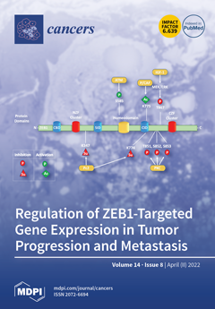
Cover Story (view full-size image): Given the importance of the epithelial-to-mesenchymal transition in tumor progression and the pivotal role of ZEB1 as a transcriptional repressor during this process, the regulation of ZEB1-targeted gene expression is an active area of investigation. Diverse signaling pathways converge to induce ZEB1 activity, but few studies have focused on the enzyme-dependent modifications of ZEB1 that occur after its translation from mRNA (i.e., post-translational modifications, PTM). In addition to outlining the current knowledge in the field, we outline several questions regarding the PTM-mediated regulation of ZEB1 that remain unanswered. Thus, the areas of research covered in this review paper will enhance the development of treatment strategies by identifying knowledge gaps in ZEB1′s regulatory mechanisms that could potentially be targeted to prevent and treat metastasis in cancer patients. View this paper
  • Issues are regarded as officially published after their release is announced to the table of contents alert mailing list.
  • You may sign up for e-mail alerts to receive table of contents of newly released issues.
  • PDF is the official format for papers published in both, html and pdf forms. To view the papers in pdf format, click on the "PDF Full-text" link, and use the free Adobe Reader to open them.
Order results
Result details
Section
Select all
Export citation of selected articles as:
11 pages, 1022 KiB  
Article
Novel Intraoperative Navigation Using Ultra-High-Resolution CT in Robot-Assisted Partial Nephrectomy
by Kiyoshi Takahara, Yoshiharu Ohno, Kosuke Fukaya, Ryo Matsukiyo, Takuhisa Nukaya, Masashi Takenaka, Kenji Zennami, Manabu Ichino, Naohiko Fukami, Hitomi Sasaki, Mamoru Kusaka, Hiroshi Toyama, Makoto Sumitomo and Ryoichi Shiroki
Cancers 2022, 14(8), 2047; https://doi.org/10.3390/cancers14082047 - 18 Apr 2022
Cited by 4 | Viewed by 1929
Abstract
To assess the perioperative and short-term functional outcomes of robot-assisted partial nephrectomy (RAPN) with intraoperative navigation using an ultra-high-resolution computed tomography (UHR-CT) scanner, we retrospectively analyzed 323 patients who underwent RAPN using an UHR-CT or area-detector CT (ADCT). Perioperative outcomes and the postoperative [...] Read more.
To assess the perioperative and short-term functional outcomes of robot-assisted partial nephrectomy (RAPN) with intraoperative navigation using an ultra-high-resolution computed tomography (UHR-CT) scanner, we retrospectively analyzed 323 patients who underwent RAPN using an UHR-CT or area-detector CT (ADCT). Perioperative outcomes and the postoperative preservation ratio of estimated glomerular filtration rate (eGFR) were compared. After the propensity score matching, we evaluated 99 patients in each group. Although the median warm ischemia time (WIT) was less than 25 min in both groups, it was significantly shorter in the UHR-CT group than in the ADCT group (15 min vs. 17 min, p = 0.032). Moreover, the estimated blood loss (EBL) was significantly lower in the UHR-CT group than in the ADCT group (33 mL vs. 50 mL, p = 0.028). However, there were no significant intergroup differences in the postoperative preservation ratio of eGFR at 3 or 6 months of follow-up (ADCT 91.8% vs. UHR-CT 93.5%, p = 0.195; and ADCT 91.7% vs. UHR-CT 94.0%, p = 0.160, respectively). Although no differences in short-term renal function were observed in intraoperative navigation for RAPN in this propensity score–matched cohort, this study is the first to demonstrate that UHR-CT resulted in a shorter WIT and lower EBL than ADCT. Full article
Show Figures

Figure 1

12 pages, 2917 KiB  
Review
The Road to Dissemination: The Concept of Oligometastases and the Barriers for Widespread Disease
by Hamza AlGhamdi, Jennifer Dhont, Mohammad Krayem, Pauline De Bruyn, Benedikt Engels, Dirk Van Gestel and Robbe Van den Begin
Cancers 2022, 14(8), 2046; https://doi.org/10.3390/cancers14082046 - 18 Apr 2022
Cited by 7 | Viewed by 3461
Abstract
Over the last years, the oligometastatic disease state has gained more and more interest, and randomized trials are now suggesting an added value of stereotactic radiotherapy on all macroscopic disease in oligometastatic patients; but what barriers could impede widespread disease in some patients? [...] Read more.
Over the last years, the oligometastatic disease state has gained more and more interest, and randomized trials are now suggesting an added value of stereotactic radiotherapy on all macroscopic disease in oligometastatic patients; but what barriers could impede widespread disease in some patients? In this review, we first discuss the concept of oligometastatic disease and some examples of clinical evidence. We then explore the route to dissemination: the hurdles a tumoral clone has to overtake before it can produce efficient and widespread dissemination. The spectrum theory argues that the range of metastatic patterns encountered in the clinic is the consequence of gradually obtained metastatic abilities of the tumor cells. Tumor clones can obtain these capabilities by Darwinian evolution, hence early in their genetic progression tumors might produce only a limited number of metastases. We illustrate selective dissemination by discussing organ tropism, the preference of different cancer (sub)types to metastasize to certain organs. Finally we discuss biomarkers that may help to distinguish the oligometastatic state. Full article
(This article belongs to the Special Issue Oligometastatic Disease)
Show Figures

Figure 1

18 pages, 3391 KiB  
Article
Gene-Mutation-Based Algorithm for Prediction of Treatment Response in Colorectal Cancer Patients
by Heather Johnson, Zahra El-Schich, Amjad Ali, Xuhui Zhang, Athanasios Simoulis, Anette Gjörloff Wingren and Jenny L. Persson
Cancers 2022, 14(8), 2045; https://doi.org/10.3390/cancers14082045 - 18 Apr 2022
Cited by 5 | Viewed by 2732
Abstract
Purpose: Despite the high mortality of metastatic colorectal cancer (mCRC), no new biomarker tools are available for predicting treatment response. We developed gene-mutation-based algorithms as a biomarker classifier to predict treatment response with better precision than the current predictive factors. Methods: Random forest [...] Read more.
Purpose: Despite the high mortality of metastatic colorectal cancer (mCRC), no new biomarker tools are available for predicting treatment response. We developed gene-mutation-based algorithms as a biomarker classifier to predict treatment response with better precision than the current predictive factors. Methods: Random forest machine learning (ML) was applied to identify the candidate algorithms using the MSK Cohort (n = 471) as a training set and validated in the TCGA Cohort (n = 221). Logistic regression, progression-free survival (PFS), and univariate/multivariate Cox proportional hazard analyses were performed and the performance of the candidate algorithms was compared with the established risk parameters. Results: A novel 7-Gene Algorithm based on mutation profiles of seven KRAS-associated genes was identified. The algorithm was able to distinguish non-progressed (responder) vs. progressed (non-responder) patients with AUC of 0.97 and had predictive power for PFS with a hazard ratio (HR) of 16.9 (p < 0.001) in the MSK cohort. The predictive power of this algorithm for PFS was more pronounced in mCRC (HR = 16.9, p < 0.001, n = 388). Similarly, in the TCGA validation cohort, the algorithm had AUC of 0.98 and a significant predictive power for PFS (p < 0.001). Conclusion: The novel 7-Gene Algorithm can be further developed as a biomarker model for prediction of treatment response in mCRC patients to improve personalized therapies. Full article
Show Figures

Graphical abstract

20 pages, 3959 KiB  
Review
How Did Conventional Nanoparticle-Mediated Photothermal Therapy Become “Hot” in Combination with Cancer Immunotherapy?
by Wan Su Yun, Ji-Ho Park, Dong-Kwon Lim, Cheol-Hee Ahn, In-Cheol Sun and Kwangmeyung Kim
Cancers 2022, 14(8), 2044; https://doi.org/10.3390/cancers14082044 - 18 Apr 2022
Cited by 24 | Viewed by 3559
Abstract
One of the promising cancer treatment methods is photothermal therapy (PTT), which has achieved good therapeutic efficiency through nanoparticle-based photoabsorbers. Because of the various functions of nanoparticles, such as targeting properties, high light-to-heat conversion, and photostability, nanoparticle-mediated PTT successfully induces photothermal damage in [...] Read more.
One of the promising cancer treatment methods is photothermal therapy (PTT), which has achieved good therapeutic efficiency through nanoparticle-based photoabsorbers. Because of the various functions of nanoparticles, such as targeting properties, high light-to-heat conversion, and photostability, nanoparticle-mediated PTT successfully induces photothermal damage in tumor tissues with minimal side effects on surrounding healthy tissues. The therapeutic efficacy of PTT originates from cell membrane disruption, protein denaturation, and DNA damage by light-induced heat, but these biological impacts only influence localized tumor areas. This conventional nanoparticle-mediated PTT still attracts attention as a novel cancer immunotherapy, because PTT causes immune responses against cancer. PTT-induced immunogenic cell death activates immune cells for systemic anti-cancer effect. Additionally, the excellent compatibility of PTT with other treatment methods (e.g., chemotherapy and immune checkpoint blockade therapy) reinforces the therapeutic efficacy of PTT as combined immunotherapy. In this review, we investigate various PTT agents of nanoparticles and compare their applications to reveal how nanoparticle-mediated PTT undergoes a transition from thermotherapy to immunotherapy. Full article
(This article belongs to the Section Cancer Immunology and Immunotherapy)
Show Figures

Graphical abstract

16 pages, 2995 KiB  
Article
Integrated Network Pharmacology Approach for Drug Combination Discovery: A Multi-Cancer Case Study
by Antonio Federico, Michele Fratello, Giovanni Scala, Lena Möbus, Alisa Pavel, Giusy del Giudice, Michele Ceccarelli, Valerio Costa, Alfredo Ciccodicola, Vittorio Fortino, Angela Serra and Dario Greco
Cancers 2022, 14(8), 2043; https://doi.org/10.3390/cancers14082043 - 18 Apr 2022
Cited by 14 | Viewed by 4526
Abstract
Despite remarkable efforts of computational and predictive pharmacology to improve therapeutic strategies for complex diseases, only in a few cases have the predictions been eventually employed in the clinics. One of the reasons behind this drawback is that current predictive approaches are based [...] Read more.
Despite remarkable efforts of computational and predictive pharmacology to improve therapeutic strategies for complex diseases, only in a few cases have the predictions been eventually employed in the clinics. One of the reasons behind this drawback is that current predictive approaches are based only on the integration of molecular perturbation of a certain disease with drug sensitivity signatures, neglecting intrinsic properties of the drugs. Here we integrate mechanistic and chemocentric approaches to drug repositioning by developing an innovative network pharmacology strategy. We developed a multilayer network-based computational framework integrating perturbational signatures of the disease as well as intrinsic characteristics of the drugs, such as their mechanism of action and chemical structure. We present five case studies carried out on public data from The Cancer Genome Atlas, including invasive breast cancer, colon adenocarcinoma, lung squamous cell carcinoma, hepatocellular carcinoma and prostate adenocarcinoma. Our results highlight paclitaxel as a suitable drug for combination therapy for many of the considered cancer types. In addition, several non-cancer-related genes representing unusual drug targets were identified as potential candidates for pharmacological treatment of cancer. Full article
(This article belongs to the Special Issue Targeting the (Un)Usual Suspects in Cancer)
Show Figures

Figure 1

11 pages, 2253 KiB  
Article
Breast Tumor Identification in Ultrafast MRI Using Temporal and Spatial Information
by Xueping Jing, Monique D. Dorrius, Mirjam Wielema, Paul E. Sijens, Matthijs Oudkerk and Peter van Ooijen
Cancers 2022, 14(8), 2042; https://doi.org/10.3390/cancers14082042 - 18 Apr 2022
Cited by 6 | Viewed by 2152
Abstract
Purpose: To investigate the feasibility of using deep learning methods to differentiate benign from malignant breast lesions in ultrafast MRI with both temporal and spatial information. Methods: A total of 173 single breasts of 122 women (151 examinations) with lesions above 5 mm [...] Read more.
Purpose: To investigate the feasibility of using deep learning methods to differentiate benign from malignant breast lesions in ultrafast MRI with both temporal and spatial information. Methods: A total of 173 single breasts of 122 women (151 examinations) with lesions above 5 mm were retrospectively included. A total of 109 out of 173 lesions were benign. Maximum intensity projection (MIP) images were generated from each of the 14 contrast-enhanced T1-weighted acquisitions in the ultrafast MRI scan. A 2D convolutional neural network (CNN) and a long short-term memory (LSTM) network were employed to extract morphological and temporal features, respectively. The 2D CNN model was trained with the MIPs from the last four acquisitions to ensure the visibility of the lesions, while the LSTM model took MIPs of an entire scan as input. The performance of each model and their combination were evaluated with 100-times repeated stratified four-fold cross-validation. Those models were then compared with models developed with standard DCE-MRI which followed the same data split. Results: In the differentiation between benign and malignant lesions, the ultrafast MRI-based 2D CNN achieved a mean AUC of 0.81 ± 0.06, and the LSTM network achieved a mean AUC of 0.78 ± 0.07; their combination showed a mean AUC of 0.83 ± 0.06 in the cross-validation. The mean AUC values were significantly higher for ultrafast MRI-based models than standard DCE-MRI-based models. Conclusion: Deep learning models developed with ultrafast breast MRI achieved higher performances than standard DCE-MRI for malignancy discrimination. The improved AUC values of the combined models indicate an added value of temporal information extracted by the LSTM model in breast lesion characterization. Full article
(This article belongs to the Section Cancer Causes, Screening and Diagnosis)
Show Figures

Figure 1

15 pages, 2653 KiB  
Article
Multi-Omics Approach Points to the Importance of Oxylipins Metabolism in Early-Stage Breast Cancer
by Dmitry V. Chistyakov, Mariia V. Guryleva, Elena S. Stepanova, Lyubov M. Makarenkova, Elena V. Ptitsyna, Sergei V. Goriainov, Arina I. Nikolskaya, Alina A. Astakhova, Anna S. Klimenko, Olga A. Bezborodova, Elena A. Rasskazova, Olga G. Potanina, Rimma A. Abramovich, Elena R. Nemtsova and Marina G. Sergeeva
Cancers 2022, 14(8), 2041; https://doi.org/10.3390/cancers14082041 - 18 Apr 2022
Cited by 16 | Viewed by 3230
Abstract
The involvement of oxylipins, metabolites of polyunsaturated fatty acids, in cancer pathogenesis was known long ago, but only the development of the high-throughput methods get the opportunity to study oxylipins on a system level. The study aimed to elucidate alterations in oxylipin metabolism [...] Read more.
The involvement of oxylipins, metabolites of polyunsaturated fatty acids, in cancer pathogenesis was known long ago, but only the development of the high-throughput methods get the opportunity to study oxylipins on a system level. The study aimed to elucidate alterations in oxylipin metabolism as characteristics of breast cancer patients. We compared the ultra-high-performance liquid chromatography-mass spectrometry (UPLC-MS/MS) oxylipin profile signatures in the blood plasma of 152 healthy volunteers (HC) and 169 patients with different stages of breast cancer (BC). To integrate lipidomics, transcriptomics, and genomics data, we analyzed a transcriptome of 10 open database datasets obtained from tissues and blood cells of BC patients and SNP data for 33 genes related to oxylipin metabolism. We identified 18 oxylipins, metabolites of omega-3 or omega-6 polyunsaturated fatty acids, that were differentially expressed between BCvsHC patients, including anandamide, prostaglandins and hydroxydocosahexaenoic acids. DEGs analysis of tissue and blood samples from BC patients revealed that 19 genes for oxylipin biosynthesis change their expression level, with CYP2C19, PTGS2, HPGD, and FAAH included in the list of DEGs in the analysis of transcriptomes and the list of SNPs associated with BC. Results allow us to suppose that oxylipin signatures reflect the organism’s level of response to the disease. Our data regarding changes in oxylipins at the system level show that oxylipin profiles can be used to evaluate the early stages of breast cancer. Full article
(This article belongs to the Special Issue Advanced Breast Cancer: From Biology to Cure)
Show Figures

Figure 1

24 pages, 1349 KiB  
Review
Head and Neck Cancer Susceptibility and Metabolism in Fanconi Anemia
by Tafadzwa Chihanga, Sara Vicente-Muñoz, Sonya Ruiz-Torres, Bidisha Pal, Mathieu Sertorio, Paul R. Andreassen, Ruby Khoury, Parinda Mehta, Stella M. Davies, Andrew N. Lane, Lindsey E. Romick-Rosendale and Susanne I. Wells
Cancers 2022, 14(8), 2040; https://doi.org/10.3390/cancers14082040 - 18 Apr 2022
Cited by 4 | Viewed by 3607
Abstract
Fanconi anemia (FA) is a rare inherited, generally autosomal recessive syndrome, but it displays X-linked or dominant negative inheritance for certain genes. FA is characterized by a deficiency in DNA damage repair that results in bone marrow failure, and in an increased risk [...] Read more.
Fanconi anemia (FA) is a rare inherited, generally autosomal recessive syndrome, but it displays X-linked or dominant negative inheritance for certain genes. FA is characterized by a deficiency in DNA damage repair that results in bone marrow failure, and in an increased risk for various epithelial tumors, most commonly squamous cell carcinomas of the head and neck (HNSCC) and of the esophagus, anogenital tract and skin. Individuals with FA exhibit increased human papilloma virus (HPV) prevalence. Furthermore, a subset of anogenital squamous cell carcinomas (SCCs) in FA harbor HPV sequences and FA-deficient laboratory models reveal molecular crosstalk between HPV and FA proteins. However, a definitive role for HPV in HNSCC development in the FA patient population is unproven. Cellular metabolism plays an integral role in tissue homeostasis, and metabolic deregulation is a known hallmark of cancer progression that supports uncontrolled proliferation, tumor development and metastatic dissemination. The metabolic consequences of FA deficiency in keratinocytes and associated impact on the development of SCC in the FA population is poorly understood. Herein, we review the current literature on the metabolic consequences of FA deficiency and potential effects of resulting metabolic reprogramming on FA cancer phenotypes. Full article
Show Figures

Figure 1

17 pages, 890 KiB  
Review
The Implication of Gastric Microbiome in the Treatment of Gastric Cancer
by George Pappas-Gogos, Kostas Tepelenis, Fotis Fousekis, Konstantinos Katsanos, Michail Pitiakoudis and Konstantinos Vlachos
Cancers 2022, 14(8), 2039; https://doi.org/10.3390/cancers14082039 - 18 Apr 2022
Cited by 14 | Viewed by 3631
Abstract
Gastric cancer (GC) is one of the most common and deadly malignancies worldwide. Helicobacter pylori have been documented as a risk factor for GC. The development of sequencing technology has broadened the knowledge of the gastric microbiome, which is essential in maintaining homeostasis. [...] Read more.
Gastric cancer (GC) is one of the most common and deadly malignancies worldwide. Helicobacter pylori have been documented as a risk factor for GC. The development of sequencing technology has broadened the knowledge of the gastric microbiome, which is essential in maintaining homeostasis. Recent studies have demonstrated the involvement of the gastric microbiome in the development of GC. Therefore, the elucidation of the mechanism by which the gastric microbiome contributes to the development and progression of GC may improve GC’s prevention, diagnosis, and treatment. In this review, we discuss the current knowledge about changes in gastric microbial composition in GC patients, their role in carcinogenesis, the possible therapeutic role of the gastric microbiome, and its implications for current GC therapy. Full article
(This article belongs to the Special Issue Treatment of Gastric Cancer)
Show Figures

Graphical abstract

13 pages, 1651 KiB  
Article
Implication of COPB2 Expression on Cutaneous Squamous Cell Carcinoma Pathogenesis
by Taiqin Chen, Ki-Yeol Kim, Yeongjoo Oh, Hei Cheul Jeung, Kee Yang Chung, Mi Ryung Roh and Xianglan Zhang
Cancers 2022, 14(8), 2038; https://doi.org/10.3390/cancers14082038 - 18 Apr 2022
Cited by 3 | Viewed by 2268
Abstract
The underlying molecular mechanisms of cutaneous squamous cell carcinoma (cSCC) pathogenesis are largely unknown. In the present study, we aimed to evaluate the effect of coatomer protein complex subunit beta 2 (COPB2) expression on cSCC pathogenesis. Clinicopathological significance of COPB2 in cSCC was [...] Read more.
The underlying molecular mechanisms of cutaneous squamous cell carcinoma (cSCC) pathogenesis are largely unknown. In the present study, we aimed to evaluate the effect of coatomer protein complex subunit beta 2 (COPB2) expression on cSCC pathogenesis. Clinicopathological significance of COPB2 in cSCC was investigated by analyzing the Gene Expression Omnibus (GEO) database and through a retrospective cohort study of 95 cSCC patients. The effect of COPB2 expression on the biological behavior of cSCC cells was investigated both in vitro and in vivo. We found that COPB2 expression was significantly higher in cSCC samples than in normal skin samples. In our cohort, a considerable association was found between COPB2 expression and indicators of tumor immune microenvironment (TIME), such as histocompatibility complex class (MHC) I, and MHC II, CD4+/ CD8+ tumor-infiltrating lymphocytes. Additionally, COPB2 expression had an independent impact on worsened recurrence-free survival in our cohort. Furthermore, decreased proliferation, invasion, tumorigenic activities, and increased apoptosis were observed after COPB2 knockdown in cSCC cells. COPB2 may act as a potential oncogene and candidate modulator of the TIME in cSCC. Therefore, it can serve as a novel predictive prognostic biomarker and candidate immunotherapeutic target in cSCC patients. Full article
(This article belongs to the Collection Cancer Biomarkers)
Show Figures

Figure 1

19 pages, 1367 KiB  
Review
The MST1R/RON Tyrosine Kinase in Cancer: Oncogenic Functions and Therapeutic Strategies
by Alex Cazes, Betzaira G. Childers, Edgar Esparza and Andrew M. Lowy
Cancers 2022, 14(8), 2037; https://doi.org/10.3390/cancers14082037 - 18 Apr 2022
Cited by 13 | Viewed by 4017
Abstract
The MST1R/RON receptor tyrosine kinase is a homologue of the more well-known MET receptor. Like MET, RON orchestrates cell signaling pathways that promote oncogenesis and enable cancer cell survival; however, it has a more unique role in the regulation of inflammation. RON was [...] Read more.
The MST1R/RON receptor tyrosine kinase is a homologue of the more well-known MET receptor. Like MET, RON orchestrates cell signaling pathways that promote oncogenesis and enable cancer cell survival; however, it has a more unique role in the regulation of inflammation. RON was originally described as a transmembrane receptor expressed on tissue resident macrophages and various epithelial cells. RON is overexpressed in a variety of cancers and its activation modifies multiple signaling pathways with resultant changes in epithelial and immune cells which together modulate oncogenic phenotypes. While several RON isoforms have been identified with differences in structure, activation, and pathway regulation, increased RON expression and/or activation is consistently associated with worse outcomes. Tyrosine kinase inhibitors targeting RON have been developed, making RON an actionable therapeutic target. Full article
(This article belongs to the Section Cancer Therapy)
Show Figures

Figure 1

23 pages, 32888 KiB  
Article
Contribution of Oxidative Stress Induced by Sonodynamic Therapy to the Calcium Homeostasis Imbalance Enhances Macrophage Infiltration in Glioma Cells
by Lei Chen, Yang Yan, Fangen Kong, Jikai Wang, Jia Zeng, Zhen Fang, Zheyan Wang, Zhigang Liu and Fei Liu
Cancers 2022, 14(8), 2036; https://doi.org/10.3390/cancers14082036 - 18 Apr 2022
Cited by 11 | Viewed by 3292
Abstract
Background: To better understand the Ca2+ overload mechanism of SDT killing gliomas, we examined the hypothesis that the early application of the mechanosensitive Ca2+ channel Piezo1 antagonist (GsMTx4) could have a better anti-tumor effect. Methods: The in vitro effect of low-energy [...] Read more.
Background: To better understand the Ca2+ overload mechanism of SDT killing gliomas, we examined the hypothesis that the early application of the mechanosensitive Ca2+ channel Piezo1 antagonist (GsMTx4) could have a better anti-tumor effect. Methods: The in vitro effect of low-energy SDT combined with GsMTx4 or agonist Yoda 1 on both the ROS-induced distribution of Ca2+ as well as on the opening of Piezo1 and the dissociation and polymerization of the Ca2+ lipid complex were assessed. The same groups were also studied to determine their effects on both tumor-bearing BALB/c-nude and C57BL/6 intracranial tumors, and their effects on the tumor-infiltrating macrophages were studied as well. Results: It was determined that ultrasound-activated Piezo1 contributes to the course of intracellular Ca2+ overload, which mediates macrophages (M1 and M2) infiltrating under the oxidative stress caused by SDT. Moreover, we explored the effects of SDT based on the dissociation of the Ca2+ lipid complex by inhibiting the expression of fatty acid binding protein 4 (FABP4). The Piezo1 channel was blocked early and combined with SDT treatment, recruited macrophages in the orthotopic transplantation glioma model. Conclusions: SDT regulates intracellular Ca2+ signals by upregulating Piezo1 leading to the inhibition of the energy supply from lipid and recruitment of macrophages. Therefore, intervening with the function of the Ca2+ channel on the glioma cell membrane in advance is likely to be the key factor to obtain a better effect combined with SDT treatment. Full article
Show Figures

Figure 1

13 pages, 2277 KiB  
Article
Radiosensitization of PC3 Prostate Cancer Cells by 5-Thiocyanato-2′-deoxyuridine
by Magdalena Zdrowowicz, Magdalena Datta, Michał Rychłowski and Janusz Rak
Cancers 2022, 14(8), 2035; https://doi.org/10.3390/cancers14082035 - 18 Apr 2022
Cited by 5 | Viewed by 2506
Abstract
Purpose: The radiosensitizing properties of uracil analogs modified in the C5 position are very interesting in the context of their effectiveness and safety in radiation therapy. Recently, radiation chemical studies have confirmed that 5-thiocyanato-2′-deoxyuridine (SCNdU) undergoes dissociation induced by an excess electron attachment [...] Read more.
Purpose: The radiosensitizing properties of uracil analogs modified in the C5 position are very interesting in the context of their effectiveness and safety in radiation therapy. Recently, radiation chemical studies have confirmed that 5-thiocyanato-2′-deoxyuridine (SCNdU) undergoes dissociation induced by an excess electron attachment and established this nucleoside as a potential radiosensitizer. In this paper, we verify the sensitizing properties of SCNdU at the cellular level and prove that it can effectively enhance ionizing radiation-induced cellular death. Methods and Materials: Prostate cancer cells were treated with SCNdU and irradiated with X rays. The cytotoxicity of SCNdU was determined by MTT test. Cell proliferation was assessed using a clonogenic assay. Cell cycle analyses, DNA damage, and cell death analyses were performed by flow cytometry. Results: SCNdU treatment significantly suppressed the proliferation and increased the radiosensitivity of prostate cancer cells. The radiosensitizing effect expressed by the dose enhancement factor is equal to 1.69. Simultaneous exposure of cells to SCNdU and radiation causes an increase in the fraction of the most radiosensitive G2/M phase, enhancement of the histone H2A.X phosphorylation level, and apoptosis induction. Finally, SCNdU turned out to be marginally cytotoxic in the absence of ionizing radiation. Conclusions: Our findings indicate that SCNdU treatment enhances the radiosensitivity of prostate cancer cells in a manner associated with the cell cycle regulation, double strand formation, and a slight induction of apoptosis. Full article
(This article belongs to the Special Issue Prostate Cancer Radiotherapy: Recent Advances and Challenges)
Show Figures

Graphical abstract

22 pages, 5557 KiB  
Article
A Bioinformatics Evaluation of the Role of Dual-Specificity Tyrosine-Regulated Kinases in Colorectal Cancer
by Amina Jamal Laham, Raafat El-Awady, Jean-Jacques Lebrun and Maha Saber Ayad
Cancers 2022, 14(8), 2034; https://doi.org/10.3390/cancers14082034 - 18 Apr 2022
Cited by 8 | Viewed by 3551
Abstract
Colorectal cancer (CRC) is the third most common cancer worldwide and has an increasing incidence in younger populations. The dual-specificity tyrosine-regulated kinase (DYRK) family has been implicated in various diseases, including cancer. However, the role and contribution of the distinct family members in [...] Read more.
Colorectal cancer (CRC) is the third most common cancer worldwide and has an increasing incidence in younger populations. The dual-specificity tyrosine-regulated kinase (DYRK) family has been implicated in various diseases, including cancer. However, the role and contribution of the distinct family members in regulating CRC tumorigenesis has not been addressed yet. Herein, we used publicly available CRC patient datasets (TCGA RNA sequence) and several bioinformatics webtools to perform in silico analysis (GTEx, GENT2, GEPIA2, cBioPortal, GSCALite, TIMER2, and UALCAN). We aimed to investigate the DYRK family member expression pattern, prognostic value, and oncological roles in CRC. This study shed light on the role of distinct DYRK family members in CRC and their potential outcome predictive value. Based on mRNA level, DYRK1A is upregulated in late tumor stages, with lymph node and distant metastasis. All DYRKs were found to be implicated in cancer-associated pathways, indicating their key role in CRC pathogenesis. No significant DYRK mutations were identified, suggesting that DYRK expression variation in normal vs. tumor samples is likely linked to epigenetic regulation. The expression of DYRK1A and DYRK3 expression correlated with immune-infiltrating cells in the tumor microenvironment and was upregulated in MSI subtypes, pointing to their potential role as biomarkers for immunotherapy. This comprehensive bioinformatics analysis will set directions for future biological studies to further exploit the molecular basis of these findings and explore the potential of DYRK1A modulation as a novel targeted therapy for CRC. Full article
(This article belongs to the Special Issue Bioinformatics, Big Data and Cancer)
Show Figures

Figure 1

12 pages, 834 KiB  
Article
A Comparative Analysis of Photon versus Proton Beam Therapy in Neoadjuvant Concurrent Chemoradiotherapy for Intrathoracic Squamous Cell Carcinoma of the Esophagus at a Single Institute
by Jin-Ho Choi, Jong Mog Lee, Moon Soo Kim, Youngjoo Lee, Yang-Gun Suh, Sung Uk Lee, Doo Yeul Lee, Eun Sang Oh, Tae hyun Kim and Sung Ho Moon
Cancers 2022, 14(8), 2033; https://doi.org/10.3390/cancers14082033 - 18 Apr 2022
Cited by 2 | Viewed by 2045
Abstract
Background: Proton beam therapy (PBT), as a neoadjuvant chemoradiotherapy (nCRT) modality, is expected to result in better outcomes than photon-based radiotherapy (RT) for esophageal cancer, particularly adenocarcinoma. This study reports the results of nCRT for locally advanced esophageal squamous cell carcinoma (ESCC) using [...] Read more.
Background: Proton beam therapy (PBT), as a neoadjuvant chemoradiotherapy (nCRT) modality, is expected to result in better outcomes than photon-based radiotherapy (RT) for esophageal cancer, particularly adenocarcinoma. This study reports the results of nCRT for locally advanced esophageal squamous cell carcinoma (ESCC) using both modalities. Methods: We retrospectively reviewed the records of patients who underwent nCRT for ESCC between 2001 and 2020. A median of 41.4 Gy or cobalt gray equivalents of radiation was delivered using either photons or protons, with concurrent chemotherapy. Dosimetric and clinical parameters were compared between the two groups. Results: Of the 31 patients, the lungs and heart of the proton group (n = 15) were exposed to significantly less radiation compared to the photon group (n = 16). No significant differences in short-term postoperative outcomes or lymphocyte count were observed between the groups, and there were no significant differences between the photon and proton groups in 2-year overall survival (67.8% vs. 68.6%, p = 0.867) or 2-year disease-free survival (33.3% vs. 34.5%, p = 0.749), with a median follow-up of 17 months. Conclusions: PBT provided a significant dosimetric benefit over photon-based RT during nCRT for ESCC; however, it did not improve clinical outcomes. Full article
(This article belongs to the Topic Cancer Biology and Radiation Therapy)
Show Figures

Figure 1

10 pages, 743 KiB  
Article
Effect of Clinical Parameters on Risk of Death from Cancer after Radical Prostatectomy in Men with Localized and Locally Advanced Prostate Cancer
by Daimantas Milonas, Tomas Ruzgas, Zilvinas Venclovas, Daniele Jonusaite, Aivaras Jonas Matijosaitis, Darius Trumbeckas, Edmundas Varpiotas, Stasys Auskalnis, Darijus Skaudickas, Ramunas Mickevicius, Kestutis Vaiciunas, Jonas Mickevicius and Mindaugas Jievaltas
Cancers 2022, 14(8), 2032; https://doi.org/10.3390/cancers14082032 - 18 Apr 2022
Cited by 1 | Viewed by 1842
Abstract
Background: The study aimed to assess predictors and to identify patients at increased risk of prostate-cancer-specific mortality (CSM) after radical prostatectomy (RP). Methods: A total of 2421 men with localized and locally advanced PCa who underwent RP in 2001–2017 were included in the [...] Read more.
Background: The study aimed to assess predictors and to identify patients at increased risk of prostate-cancer-specific mortality (CSM) after radical prostatectomy (RP). Methods: A total of 2421 men with localized and locally advanced PCa who underwent RP in 2001–2017 were included in the study. CSM predictors were assessed using multivariate competing risk analysis. Death from other causes was considered a competing event. Cumulative CSM and other-cause mortality (OCM) were calculated in various combinations of predictors. Results: During the median 8 years (interquartile range 4.4–11.7) follow-up, 56 (2.3%) of registered deaths were due to PCa. Cumulative 10 years CSM and OCM was 3.6% (95% CI 2.7–4.7) and 15.9% (95% CI 14.2–17.9), respectively. The strongest predictors of CSM were Grade Group 5 (GG5) (hazard ratio (HR) 19.9, p < 0.0001), lymph node invasion (HR 3.4, p = 0.001), stage pT3b-4 (HR 3.1, p = 0.009), and age (HR 1.1, p = 0.0007). In groups created regarding age, stage, and GG, cumulative 10 years CSM ranged from 0.4–84.9%, whereas OCM varied from 0–43.2%. Conclusions: CSM after RP is related to GGs, pathological stage, age, and combinations of these factors, whereas other-cause mortality is only associated with age. Created CSM and OCM plots can help clinicians identify patients with the most aggressive PCa who could benefit from more intensive or novel multimodal treatment strategies. Full article
Show Figures

Figure 1

14 pages, 1856 KiB  
Article
Application of an Ultrasensitive NGS-Based Blood Test for the Diagnosis of Early-Stage Lung Cancer: Sensitivity, a Hurdle Still Difficult to Overcome
by Malaïka Van der Linden, Bram Van Gaever, Lennart Raman, Karim Vermaelen, Ingel Demedts, Veerle Surmont, Ulrike Himpe, Yolande Lievens, Liesbeth Ferdinande, Franceska Dedeurwaerdere, Joni Van der Meulen, Kathleen Claes, Björn Menten and Jo Van Dorpe
Cancers 2022, 14(8), 2031; https://doi.org/10.3390/cancers14082031 - 18 Apr 2022
Cited by 4 | Viewed by 2634
Abstract
Diagnosis of lung cancer requires histological examination of a tissue sample, which in turn requires an invasive procedure that cannot always be obtained. Circulating tumor DNA can be reliably detected in blood samples of advanced-stage lung cancer patients and might also be a [...] Read more.
Diagnosis of lung cancer requires histological examination of a tissue sample, which in turn requires an invasive procedure that cannot always be obtained. Circulating tumor DNA can be reliably detected in blood samples of advanced-stage lung cancer patients and might also be a minimally invasive alternative for early-stage lung cancer detection. We wanted to explore the potential of targeted deep sequencing as a test for the diagnosis of early-stage lung cancer in combination with imaging. Mutation detection on cell-free DNA from pretreatment plasma samples of 51 patients with operable non-small cell lung cancer was performed and results were compared with 12 control patients undergoing surgery for a non-malignant lung lesion. By using a variant allele frequency threshold of 1%, somatic variants were detected in 23.5% of patients with a median variant allele fraction of 3.65%. By using this threshold, we could almost perfectly discriminate early-stage lung cancer patients from controls. Our study results are discussed in the light of those from other studies. Notwithstanding the potential of today’s techniques for the use of liquid biopsy-based cell-free DNA analysis, sensitivity of this application for early-stage lung cancer detection is currently limited by a biological background of somatic variants with low variant allele fraction. Full article
(This article belongs to the Special Issue Cell-Free DNA as Prognostic and Predictive Biomarker in Solid Cancers)
Show Figures

Figure 1

17 pages, 2099 KiB  
Systematic Review
Proline Metabolism in Malignant Gliomas: A Systematic Literature Review
by Magdalena M. Sawicka, Karol Sawicki, Tomasz Łysoń, Barbara Polityńska and Wojciech Miltyk
Cancers 2022, 14(8), 2030; https://doi.org/10.3390/cancers14082030 - 17 Apr 2022
Cited by 12 | Viewed by 3709
Abstract
Background: Proline has attracted growing interest because of its diverse influence on tumor metabolism and the discovery of the regulatory mechanisms that appear to be involved. In contrast to general oncology, data on proline metabolism in central nervous system malignancies are limited. Materials [...] Read more.
Background: Proline has attracted growing interest because of its diverse influence on tumor metabolism and the discovery of the regulatory mechanisms that appear to be involved. In contrast to general oncology, data on proline metabolism in central nervous system malignancies are limited. Materials and Methods: We performed a systematic literature review of the MEDLINE and EMBASE databases according to PRISMA guidelines, searching for articles concerning proline metabolism in malignant glial tumors. From 815 search results, we identified 14 studies pertaining to this topic. Results: The role of the proline cycle in maintaining redox balance in IDH-mutated gliomas has been convincingly demonstrated. Proline is involved in restoring levels of glutamate, the main glial excitatory neurotransmitter. Proline oxidase influences two major signaling pathways: p53 and NF- κB. In metabolomics studies, the metabolism of proline and its link to the urea cycle was found to be a prognostic factor for survival and a marker of malignancy. Data on the prolidase concentration in the serum of glioblastoma patients are contradictory. Conclusions: Despite a paucity of studies in the literature, the available data are interesting enough to encourage further research, especially in terms of extrapolating what we have learned of proline functions from other neoplasms to malignant gliomas. Full article
Show Figures

Figure 1

13 pages, 2531 KiB  
Article
Diagnostic Performance of 18F-Choline Positron Emission Tomography/Contrast-Enhanced Computed Tomography in Adenoma Detection in Primary Hyperparathyroidism after Inconclusive Imaging: A Retrospective Study of 215 Patients
by Johan Benjamin, Laure Maillard, Isabelle Morelec, Philippe Got, Françoise Borson-Chazot and Jean-Christophe Lifante
Cancers 2022, 14(8), 2029; https://doi.org/10.3390/cancers14082029 - 17 Apr 2022
Cited by 5 | Viewed by 2235
Abstract
This large, retrospective, single-centre study evaluated the diagnostic performance of 18F-choline positron emission tomography/contrast-enhanced computed tomography (PET/ceCT) in preoperative parathyroid adenoma detection in primary hyperparathyroidism cases after negative/inconclusive ultrasound or other imaging findings. We included patients who underwent surgery and 18F-choline [...] Read more.
This large, retrospective, single-centre study evaluated the diagnostic performance of 18F-choline positron emission tomography/contrast-enhanced computed tomography (PET/ceCT) in preoperative parathyroid adenoma detection in primary hyperparathyroidism cases after negative/inconclusive ultrasound or other imaging findings. We included patients who underwent surgery and 18F-choline PET/ceCT for inconclusive imaging results between 2015 and 2020. We compared the 18F-choline PET/ceCT results with surgical and histopathological findings and identified the variables influencing the correlation between 18F-choline PET/ceCT and surgical findings. Of 215 enrolled patients, 269 glands (mean lesion size, 10.9 ± 8.0 mm) were analysed. There were 165 unilocular and 50 multilocular lesions; the mean preoperative calcium level was 2.18 ± 0.19 mmol/L. Among 860 estimated lesions, 219 were classified as true positive, 21 as false positive, and 28 as false negative. The per-lesion sensitivity was 88.66%; specificity, 96.57%; positive predictive value, 91.40%; and negative predictive value, 95.39%. The detection and cure rates were 82.0% and 95.0%, respectively. On univariate and multivariate analyses, the maximum standardised uptake value (SUVmax), lesion size, and unilocularity correlated with the pathologic findings of hyperfunctioning glands. 18F-choline PET/ceCT presents favourable diagnostic performance as a second-line imaging method, with SUVmax, lesion size, and unilocularity predicting a high correlation between the 18F-choline PET/ceCT and surgical findings. Full article
(This article belongs to the Special Issue Management and Treatment of Endocrine Tumors)
Show Figures

Figure 1

27 pages, 425 KiB  
Review
Advances in the Diagnosis and Therapeutic Management of Gastroenteropancreatic Neuroendocrine Neoplasms (GEP-NENs)
by Krzysztof Kaliszewski, Maksymilian Ludwig, Maria Greniuk, Agnieszka Mikuła, Karol Zagórski and Jerzy Rudnicki
Cancers 2022, 14(8), 2028; https://doi.org/10.3390/cancers14082028 - 17 Apr 2022
Cited by 8 | Viewed by 2856
Abstract
Neuroendocrine neoplasms (NENs) are an increasingly common cause of neoplastic diseases. One of the largest groups of NENs are neoplasms localized to the gastroenteropancreatic system, which are known as gastroenteropancreatic NENs (GEP-NENs). Because of nonspecific clinical symptoms, GEP-NEN patient diagnosis and, consequently, their [...] Read more.
Neuroendocrine neoplasms (NENs) are an increasingly common cause of neoplastic diseases. One of the largest groups of NENs are neoplasms localized to the gastroenteropancreatic system, which are known as gastroenteropancreatic NENs (GEP-NENs). Because of nonspecific clinical symptoms, GEP-NEN patient diagnosis and, consequently, their treatment, might be difficult and delayed. This situation has forced researchers all over the world to continue progress in the diagnosis and treatment of patients with GEP-NENs. Our review is designed to present the latest reports on the laboratory diagnostic techniques, imaging tests and surgical and nonsurgical treatment strategies used for patients with these rare neoplasms. We paid particular attention to the nuclear approach, the use of which has been applied to GEP-NEN patient diagnosis, and to nonsurgical and radionuclide treatment strategies. Recent publications were reviewed in search of reports on new strategies for effective disease management. Attention was also paid to those studies still in progress, but with successful results. A total of 248 papers were analyzed, from which 141 papers most relevant to the aim of the study were selected. Using these papers, we highlight the progress in the development of diagnostic and treatment strategies for patients with GEP-NENs. Full article
17 pages, 978 KiB  
Review
Circulating Nucleic Acids as Novel Biomarkers for Pancreatic Ductal Adenocarcinoma
by Ryan McGowan, Áine Sally, Anthony McCabe, Brian Michael Moran and Karen Finn
Cancers 2022, 14(8), 2027; https://doi.org/10.3390/cancers14082027 - 17 Apr 2022
Cited by 4 | Viewed by 2611
Abstract
Despite considerable advancements in the clinical management of PDAC it remains a significant cause of mortality. PDAC is often diagnosed at advanced stages due to vague symptoms associated with early-stage disease and a lack of reliable diagnostic biomarkers. Late diagnosis results in a [...] Read more.
Despite considerable advancements in the clinical management of PDAC it remains a significant cause of mortality. PDAC is often diagnosed at advanced stages due to vague symptoms associated with early-stage disease and a lack of reliable diagnostic biomarkers. Late diagnosis results in a high proportion of cases being ineligible for surgical resection, the only potentially curative therapy for PDAC. Furthermore, a lack of prognostic biomarkers impedes clinician’s ability to properly assess the efficacy of therapeutic interventions. Advances in our ability to detect circulating nucleic acids allows for the advent of novel biomarkers for PDAC. Tumor derived circulating and exosomal nucleic acids allow for the detection of PDAC-specific mutations through a non-invasive blood sample. Such biomarkers could expand upon the currently limited repertoire of tests available. This review outlines recent developments in the use of molecular techniques for the detection of these nucleic acids and their potential roles, alongside current techniques, in the diagnosis, prognosis and therapeutic governance of PDAC. Full article
Show Figures

Figure 1

41 pages, 1696 KiB  
Systematic Review
Psychosocial Determinants of Lifestyle Change after a Cancer Diagnosis: A Systematic Review of the Literature
by Meeke Hoedjes, Inge Nijman and Chris Hinnen
Cancers 2022, 14(8), 2026; https://doi.org/10.3390/cancers14082026 - 16 Apr 2022
Cited by 20 | Viewed by 4273
Abstract
The aim of this study is to provide a systematic overview of the scientific literature on sociodemographic, psychological and social determinants that may facilitate or hamper lifestyle change after the diagnosis cancer. Four databases (PubMed, PsychINFO, Cumulative Index to Nursing and Allied Health [...] Read more.
The aim of this study is to provide a systematic overview of the scientific literature on sociodemographic, psychological and social determinants that may facilitate or hamper lifestyle change after the diagnosis cancer. Four databases (PubMed, PsychINFO, Cumulative Index to Nursing and Allied Health Literature (CINAHL), and Web of Science) were searched for relevant papers up to October 2021. Of the 9586 references yielded by the literature search, 123 papers were included: 71 quantitative and 52 qualitative papers. Findings showed a large variety of determinants influencing lifestyle change after cancer diagnosis, with differences between lifestyle behaviors (physical activity, diet, smoking, alcohol, sun protection, and multiple lifestyle behaviors) and findings from quantitative vs. qualitative studies. Findings demonstrate the important role of oncology healthcare professionals in promoting healthy lifestyle changes in cancer survivors. In addition, findings inform researchers involved in the development of health promotion programs about the methods and strategies they can use to promote healthy lifestyle changes in cancer survivors. Favorable lifestyle changes are expected to have beneficial effects on cancer risk and overall health in cancer survivors. Full article
(This article belongs to the Special Issue The Role of Lifestyle-Related Factors in Cancer Survivorship)
Show Figures

Figure 1

14 pages, 1915 KiB  
Article
Azacitidine Plus Venetoclax for the Treatment of Relapsed and Newly Diagnosed Acute Myeloid Leukemia Patients
by Sylvain Garciaz, Marie-Anne Hospital, Anne-Sophie Alary, Colombe Saillard, Yosr Hicheri, Bilal Mohty, Jérôme Rey, Evelyne D’Incan, Aude Charbonnier, Ferdinand Villetard, Valerio Maisano, Laura Lombardi, Antoine Ittel, Marie-Joelle Mozziconacci, Véronique Gelsi-Boyer and Norbert Vey
Cancers 2022, 14(8), 2025; https://doi.org/10.3390/cancers14082025 - 16 Apr 2022
Cited by 27 | Viewed by 5863
Abstract
Venetoclax (VEN) belongs the BH3-mimetic class that selectively targets BCL-2, activating apoptosis. The combination of VEN and azacitidine (AZA) has changed the paradigm of treatment of newly diagnosed (ND) acute myeloid leukemia (AML) patients ineligible for intensive chemotherapy. There is scarce evidence for [...] Read more.
Venetoclax (VEN) belongs the BH3-mimetic class that selectively targets BCL-2, activating apoptosis. The combination of VEN and azacitidine (AZA) has changed the paradigm of treatment of newly diagnosed (ND) acute myeloid leukemia (AML) patients ineligible for intensive chemotherapy. There is scarce evidence for the use of VEN–AZA for relapsed or refractory (R/R) AML. We compared the outcome of 39 R/R AML and 38 ND AML patients treated between 01/20 and 12/21. The median age was 69 (22–86) and 73 (61–81) in the R/R and ND groups, respectively. Adverse cytogenetics were found in 36% of patients in the R/R group and 59% of patients in the ND group. Overall response rate was 37% in R/R AML, including 13% CR, 8% CRi, 3% PR and 13% MLFS, and 58% in the ND AML, including 32% CR, 13% CRi and 13% MLFS. Adverse cytogenetics was associated with treatment failure in the R/R group (Relative Risk = 0.13, p = 0.005). Median overall survival (OS) was 5.9 months in the R/R group and 9.4 months in the ND group. Median OS was 2.2 months in the adverse cytogenetics group versus 8.7 months in the intermediate cytogenetics group in the R/R group (p = 0.02). Median leukemia-free survival was not different between the two groups (9.4 months and 10.3 months), indicating that VEN–AZA can be an efficient salvage treatment for selected R/R AML patients. In conclusion, VEN–AZA is a promising treatment for ND AML and for selected R/R AML patients. Full article
(This article belongs to the Special Issue New Targets and Therapies of Acute Myeloid Leukemia)
Show Figures

Figure 1

17 pages, 4816 KiB  
Article
Identification of TPM2 and CNN1 as Novel Prognostic Markers in Functionally Characterized Human Colon Cancer-Associated Stromal Cells
by Valentina Mele, Camilla Basso, Valeria Governa, Jesus F. Glaus Garzon, Manuele G. Muraro, Silvio Däster, Christian A. Nebiker, Robert Mechera, Martin Bolli, Alexander Schmidt, Roger Geiger, Giulio C. Spagnoli, Dimitri Christoforidis, Pietro E. Majno, Lubor Borsig and Giandomenica Iezzi
Cancers 2022, 14(8), 2024; https://doi.org/10.3390/cancers14082024 - 16 Apr 2022
Cited by 9 | Viewed by 3184
Abstract
Stromal infiltration is associated with poor prognosis in human colon cancers. However, the high heterogeneity of human tumor-associated stromal cells (TASCs) hampers a clear identification of specific markers of prognostic relevance. To address these issues, we established short-term cultures of TASCs and matched [...] Read more.
Stromal infiltration is associated with poor prognosis in human colon cancers. However, the high heterogeneity of human tumor-associated stromal cells (TASCs) hampers a clear identification of specific markers of prognostic relevance. To address these issues, we established short-term cultures of TASCs and matched healthy mucosa-associated stromal cells (MASCs) from human primary colon cancers and, upon characterization of their phenotypic and functional profiles in vitro and in vivo, we identified differentially expressed markers by proteomic analysis and evaluated their prognostic significance. TASCs were characterized by higher proliferation and differentiation potential, and enhanced expression of mesenchymal stem cell markers, as compared to MASCs. TASC triggered epithelial–mesenchymal transition (EMT) in tumor cells in vitro and promoted their metastatic spread in vivo, as assessed in an orthotopic mouse model. Proteomic analysis of matched TASCs and MASCs identified a panel of markers preferentially expressed in TASCs. The expression of genes encoding two of them, calponin 1 (CNN1) and tropomyosin beta chain isoform 2 (TPM2), was significantly associated with poor outcome in independent databases and outperformed the prognostic significance of currently proposed TASC markers. The newly identified markers may improve prognostication of primary colon cancers and identification of patients at risk. Full article
Show Figures

Figure 1

10 pages, 1135 KiB  
Article
Circulating Let-7 Family Members as Non-Invasive Biomarkers for Predicting Hepatocellular Carcinoma Risk after Antiviral Treatment among Chronic Hepatitis C Patients
by Yi-Shan Tsai, Ching-I Huang, Pei-Chien Tsai, Ming-Lun Yeh, Chung-Feng Huang, Meng-Hsuan Hsieh, Ta-Wei Liu, Yi-Hung Lin, Po-Cheng Liang, Zu-Yau Lin, Shinn-Cherng Chen, Jee-Fu Huang, Wan-Long Chuang, Chia-Yen Dai and Ming-Lung Yu
Cancers 2022, 14(8), 2023; https://doi.org/10.3390/cancers14082023 - 16 Apr 2022
Cited by 6 | Viewed by 2478
Abstract
HCC, a leading cause of cancer-related mortality, is diagnosed at advanced stages. Although antiviral therapy has reduced the risk of HCC among chronic hepatitis C (CHC) patients, the risk of HCC remains, thus, highlighting the unmet need for continuous surveillance. Therefore, stable and [...] Read more.
HCC, a leading cause of cancer-related mortality, is diagnosed at advanced stages. Although antiviral therapy has reduced the risk of HCC among chronic hepatitis C (CHC) patients, the risk of HCC remains, thus, highlighting the unmet need for continuous surveillance. Therefore, stable and cost-effective biomarkers, such as circulating microRNAs, must be identified. We aimed to clarify whether serum levels of the Let-7 family can predict HCC risk in patients with CHC using univariate and multivariate Cox’s proportional hazards model. We analyzed the sera of 54 patients with CHC who developed HCC after antiviral therapy and compared the data with those of 173 patients without HCC development. The Let-7 family (except for let-7c) exhibited significant negative correlations with the fibrosis score (r = −0.2736 to −0.34, p = 0.0002 to <0.0001). After Cox’s regression model was used to adjust for age, sex, HCV genotype, and FIB-4 ≥ 3.25, patients with CHC with let-7i median ≥ −1.696 (adjusted hazard ratio [aHR] = 0.31, 95% CI: 0.08–0.94, p = 0.0372) in the sustained virologic response (SVR) groups and ≥−1.696 (aHR = 0.09, 95% CI: 0.08–0.94, p = 0.0022) in the non-SVR group were less likely to develop HCC. Thus, circulating let-7i can be used for early CHC surveillance in patients with HCC risk after antiviral treatment. Full article
(This article belongs to the Special Issue Circulating Biomarkers in Cancer)
Show Figures

Figure 1

19 pages, 4106 KiB  
Article
Combination of LIGHT (TNFSF14)-Armed Myxoma Virus Pre-Loaded into ADSCs and Gemcitabine in the Treatment of Experimental Orthotopic Murine Pancreatic Adenocarcinoma
by Joanna Jazowiecka-Rakus, Aleksander Sochanik, Agata Hadryś, Wojciech Fidyk, Ewa Chmielik, Masmudur M. Rahman and Grant McFadden
Cancers 2022, 14(8), 2022; https://doi.org/10.3390/cancers14082022 - 16 Apr 2022
Cited by 4 | Viewed by 2690
Abstract
Pancreatic ductal adenocarcinoma (PDAC) is a deadly neoplasm. Oncolytic viruses have tumorolytic and immune response-boosting effects and present great potential for PDAC management. We used LIGHT-armed myxoma virus (vMyx-LIGHT) loaded ex vivo into human adipose-derived mesenchymal stem cells (ADSCs) to evaluate murine PDAC [...] Read more.
Pancreatic ductal adenocarcinoma (PDAC) is a deadly neoplasm. Oncolytic viruses have tumorolytic and immune response-boosting effects and present great potential for PDAC management. We used LIGHT-armed myxoma virus (vMyx-LIGHT) loaded ex vivo into human adipose-derived mesenchymal stem cells (ADSCs) to evaluate murine PDAC treatment in conjunction with gemcitabine (GEM). The cytotoxicity of this treatment was confirmed in vitro using human and murine pancreatic cancer cell cultures, which were more sensitive to the combined approach and largely destroyed. Unlike cancer cells, ADSCs sustain significant viability after infection. The in vivo administration of vMyx-LIGHT-loaded ADSCs and gemcitabine was evaluated using immunocompetent mice with induced orthotopic PDAC lesions. The expression of virus-encoded LIGHT increased the influx of T cells to the tumor site. Shielded virus followed by gemcitabine improved tumor regression and survival. The addition of gemcitabine slightly compromised the adaptive immune response boost obtained with the shielded virus alone, conferring no survival benefit. ADSCs pre-loaded with vMyx-LIGHT allowed the effective transport of the oncolytic construct to PDAC lesions and yielded significant immune response; additional GEM administration failed to improve survival. In view of our results, the delivery of targeted/shielded virus in combination with TGF-β ablation and/or checkpoint inhibitors is a promising option to improve the therapeutic effects of vMyx-LIGHT/ADSCs against PDAC in vivo. Full article
(This article belongs to the Special Issue Oncolytic Viruses as Cancer Immunotherapy Agents)
Show Figures

Figure 1

20 pages, 1043 KiB  
Review
Recent Advances in Treatment Options for Childhood Acute Lymphoblastic Leukemia
by Marta Malczewska, Kamil Kośmider, Kinga Bednarz, Katarzyna Ostapińska, Monika Lejman and Joanna Zawitkowska
Cancers 2022, 14(8), 2021; https://doi.org/10.3390/cancers14082021 - 16 Apr 2022
Cited by 35 | Viewed by 8415
Abstract
Acute lymphoblastic leukemia is the most common blood cancer in pediatric patients. There has been enormous progress in ALL treatment in recent years, which is reflected by the increase in the 5-year OS from 57% in the 1970s to up to 96% in [...] Read more.
Acute lymphoblastic leukemia is the most common blood cancer in pediatric patients. There has been enormous progress in ALL treatment in recent years, which is reflected by the increase in the 5-year OS from 57% in the 1970s to up to 96% in the most recent studies. ALL treatment is based primarily on conventional methods, which include chemotherapy and radiotherapy. Their main weakness is severe toxicity, which prompts dose reduction, decreases the effectiveness of the treatment, and, in some cases, can lead to death. Currently, numerous modifications in treatment regimens are applied in order to limit toxicities emerging from conventional approaches and improve outcomes. Hematological treatment of pediatric patients is reaching for more novel treatment options, such as targeted treatment, CAR-T-cells therapy, and immunotherapy. These methods are currently used in conjunction with chemotherapy. Nevertheless, the swift progress in their development and increasing efficacity can lead to applying those novel therapies as standalone therapeutic options for pediatric ALL. Full article
Show Figures

Figure 1

12 pages, 1405 KiB  
Article
Feasibility of Tumor Treating Fields with Pemetrexed and Platinum-Based Chemotherapy for Unresectable Malignant Pleural Mesothelioma: Single-Center, Real-World Data
by Tugce Kutuk, Haley Appel, Maria Carolina Avendano, Federico Albrecht, Paul Kaywin, Suyen Ramos, Melanie E. Suarez-Murias, Minesh P. Mehta and Rupesh Kotecha
Cancers 2022, 14(8), 2020; https://doi.org/10.3390/cancers14082020 - 16 Apr 2022
Cited by 10 | Viewed by 2604
Abstract
Purpose: The objectives of this study were to evaluate the implementation, device usage rates, clinical outcomes, and treatment-related toxicities associated with TTFields and pemetrexed plus platinum-based chemotherapy in patients with unresectable MPM, outside the initial trial results. Methods: Consecutive patients with unresectable MPM [...] Read more.
Purpose: The objectives of this study were to evaluate the implementation, device usage rates, clinical outcomes, and treatment-related toxicities associated with TTFields and pemetrexed plus platinum-based chemotherapy in patients with unresectable MPM, outside the initial trial results. Methods: Consecutive patients with unresectable MPM were enrolled onto an FDA-required HDE protocol from 2019 to 2021. All patients were treated with a protocol-defined regimen of continuous TTFields (150 kHz) and pemetrexed plus platinum-based chemotherapy. Results: Five patients with unresectable MPM were enrolled. The median number of 4-week TTFields cycles was 5 (range: 2–7 cycles). Median TTFields device usage in the first 3 months was 12.5 h per day (range: 5–16.8 h), representing 52% (21–70%) of the potential daily duration. The median follow-up was 5.4 months (range: 1.1–20.9 months). Treatment-related dermatitis was the only side effect associated with TTFields and was reported as grade 1–2 in all patients; no patient had grade 3+ device-related toxicities. Conclusions: This study represents the first results of real-world implementation of TTFields for MPM. In comparison to the initial clinical trial (STELLAR), compliance rates were lower, although skin-related toxicities appeared similar. Further initiatives and guidelines should be developed to manage treatment-related dermatitis and improve device usage. Full article
Show Figures

Figure 1

13 pages, 1290 KiB  
Article
Multicenter Observational Study on Metastatic Non-Small Cell Lung Cancer Harboring BRAF Mutations: Focus on Clinical Characteristics and Treatment Outcome of V600E and Non-V600E Subgroups
by Fabiana Perrone, Giulia Mazzaschi, Roberta Minari, Michela Verzè, Cinzia Azzoni, Lorena Bottarelli, Rita Nizzoli, Monica Pluchino, Annalisa Altimari, Elisa Gruppioni, Francesca Sperandi, Elisa Andrini, Giorgia Guaitoli, Federica Bertolini, Fausto Barbieri, Stefania Bettelli, Lucia Longo, Maria Pagano, Candida Bonelli, Elena Tagliavini, Davide Nicoli, Alessandro Ubiali, Adriano Zangrandi, Serena Trubini, Manuela Proietto, Letizia Gnetti and Marcello Tiseoadd Show full author list remove Hide full author list
Cancers 2022, 14(8), 2019; https://doi.org/10.3390/cancers14082019 - 16 Apr 2022
Cited by 9 | Viewed by 2682
Abstract
Introduction: BRAF mutation involved 2–4% of lung adenocarcinoma. Differences in clinicopathologic features and patient outcome exist between V600E and non-V600E BRAF mutated NSCLC. Thus, we sought to assess the frequency and clinical relevance of BRAF mutations in a real-life population of advanced-NSCLC, investigating [...] Read more.
Introduction: BRAF mutation involved 2–4% of lung adenocarcinoma. Differences in clinicopathologic features and patient outcome exist between V600E and non-V600E BRAF mutated NSCLC. Thus, we sought to assess the frequency and clinical relevance of BRAF mutations in a real-life population of advanced-NSCLC, investigating the potential prognostic significance of distinct genetic alterations. Materials and Methods: The present multicenter Italian retrospective study involved advanced BRAF mutant NSCLC. Complete clinicopathologic data were evaluated for BRAF V600E and non-V600E patients. Results: A total of 44 BRAFmut NSCLC patients were included (V600E, n = 23; non-V600E, n = 21). No significant differences in survival outcome and treatment response were documented, according to V600E vs. non-V600E mutations, although a trend towards prolonged PFS was observed in the V600E subgroup (median PFS = 11.3 vs. 6.0 months in non-V600E). In the overall population, ECOG PS and age significantly impacted on OS, while bone lesions were associated with shorter PFS. Compared to immunotherapy, first-line chemotherapy was associated with longer OS in the overall population, and especially in the BRAF V600E subtype. Conclusions: Here, we report on real-life data from a retrospective cohort of advanced-NSCLC harboring BRAF alterations. Our study offers relevant clues on survival outcome, therapeutic response, and clinicopathologic correlations of BRAF-mutant NSCLC. Full article
Show Figures

Figure 1

15 pages, 303 KiB  
Review
Current Landscape of Immune Checkpoint Inhibitor Therapy for Hepatocellular Carcinoma
by Nikolaos Machairas, Diamantis I. Tsilimigras and Timothy M. Pawlik
Cancers 2022, 14(8), 2018; https://doi.org/10.3390/cancers14082018 - 16 Apr 2022
Cited by 19 | Viewed by 5854
Abstract
Hepatocellular carcinoma (HCC) is the most frequent primary liver tumor. As a result of advanced disease being often present at diagnosis, only a small percentage of patients are amenable to curative-intent treatment options such as surgical resection and liver transplantation. Systemic therapy consisting [...] Read more.
Hepatocellular carcinoma (HCC) is the most frequent primary liver tumor. As a result of advanced disease being often present at diagnosis, only a small percentage of patients are amenable to curative-intent treatment options such as surgical resection and liver transplantation. Systemic therapy consisting of tyrosine kinase inhibitors such as sorafenib had been used for over a decade with limited efficacy. More recently, treatment with immune checkpoint inhibitors has revolutionized the treatment landscape of various malignant tumors. With this shifting paradigm, recent data have demonstrated encouraging outcomes among patients with HCC. In particular, several trials have investigated the safety and efficacy of various immune checkpoint inhibitors (ICI) either as monotherapy or in the form of combined treatments. We sought to provide an overview of recent clinical trials among patients with advanced HCC as well as to highlight predictors of response and immune-related adverse events and to review the evidence on perioperative administration of ICI in patients with resectable HCC. Full article
Previous Issue
Next Issue
Back to TopTop