Next Issue
Volume 15, April-1
Previous Issue
Volume 15, March-1


Cancers, Volume 15, Issue 6 (March-2 2023) – 290 articles

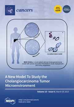
Cover Story (view full-size image): Cholangiocarcinoma (CCA) is a rare and aggressive tumor, arising from the bile ducts. It is a heterogenous disease with a low survival rate, which is also attributed to its therapy resistance. Cancer-associated fibroblasts (CAF) are a major player in resistance formation, as well as tumor progression, invasion, and metastasis. To our knowledge, no human 3D CCA in vitro model that takes CAF into account exists. We isolated and characterized a pCCA and its corresponding CAF cell line in detail. To mimic the in vivo situation as close as possible, we further created a 3D co-culture tumor model (CCTM). In proteomic studies, we were able to confirm typical CCA characteristics, especially in the CCTM group. Our main finding was an increased pro-angiogenic TME, which will be further investigated in ongoing studies. View this paper
  • Issues are regarded as officially published after their release is announced to the table of contents alert mailing list.
  • You may sign up for e-mail alerts to receive table of contents of newly released issues.
  • PDF is the official format for papers published in both, html and pdf forms. To view the papers in pdf format, click on the "PDF Full-text" link, and use the free Adobe Reader to open them.
Order results
Result details
Section
Select all
Export citation of selected articles as:
14 pages, 2434 KiB  
Article
Prevention of Inflammation-Driven Colon Carcinogenesis in Human MUC1 Transgenic Mice by Vaccination with MUC1 DNA and Dendritic Cells
by Retno Murwanti, Kaori Denda-Nagai, Daisuke Sugiura, Kaoru Mogushi, Sandra J. Gendler and Tatsuro Irimura
Cancers 2023, 15(6), 1920; https://doi.org/10.3390/cancers15061920 - 22 Mar 2023
Cited by 1 | Viewed by 2423
Abstract
The preventive efficacy of MUC1-specific DNA immunization on inflammation-driven colon carcinogenesis in human MUC1 transgenic (MUC1.Tg) mice was investigated. Mice were vaccinated with MUC1 DNA mixed with autologous bone-marrow-derived dendritic cells (BMDCs), and then colonic tumors were induced by azoxymethane (AOM) injection [...] Read more.
The preventive efficacy of MUC1-specific DNA immunization on inflammation-driven colon carcinogenesis in human MUC1 transgenic (MUC1.Tg) mice was investigated. Mice were vaccinated with MUC1 DNA mixed with autologous bone-marrow-derived dendritic cells (BMDCs), and then colonic tumors were induced by azoxymethane (AOM) injection and oral administration of dextran sulfate sodium (DSS). Two types of tumors, squamous metaplasia and tubular adenoma, were observed. Both expressed high levels of MUC1 as indicated by the binding of anti-MUC1 antibodies with different specificities, whereas MUC1 expression was not detected in normal colonic mucosa. When mice were immunized with MUC1 DNA + BMDCs, tumor incidence, tumor number, and tumor size were significantly reduced. In contrast, vaccination with MUC1 DNA alone or BMDCs alone was ineffective in reducing tumor burden. Inflammation caused by DSS was not suppressed by the MUC1 DNA + BMDCs vaccination. Furthermore, MUC1 protein expression levels, as judged by anti-MUC1 antibody binding in tumors grown after vaccination, did not significantly differ from the control. In conclusion, an inflammation-driven carcinogenesis model was established in MUC1.Tg mice, closely resembling human colon carcinogenesis. In this model, vaccination with MUC1 DNA + BMDCs was effective in overriding MUC1 tolerance and reducing the tumor burden by a mechanism not affecting the level of colonic inflammation. Full article
(This article belongs to the Special Issue Mucins and Cancers)
Show Figures

Graphical abstract

14 pages, 7633 KiB  
Article
Mismatch between Bioluminescence Imaging (BLI) and MRI When Evaluating Glioblastoma Growth: Lessons from a Study Where BLI Suggested “Regression” while MRI Showed “Progression”
by Mathilde Bausart, Elia Bozzato, Nicolas Joudiou, Xanthippi Koutsoumpou, Bella Manshian, Véronique Préat and Bernard Gallez
Cancers 2023, 15(6), 1919; https://doi.org/10.3390/cancers15061919 - 22 Mar 2023
Cited by 9 | Viewed by 2693
Abstract
Orthotopic glioblastoma xenografts are paramount for evaluating the effect of innovative anti-cancer treatments. In longitudinal studies, tumor growth (or regression) of glioblastoma can only be monitored by noninvasive imaging. For this purpose, bioluminescence imaging (BLI) has gained popularity because of its low cost [...] Read more.
Orthotopic glioblastoma xenografts are paramount for evaluating the effect of innovative anti-cancer treatments. In longitudinal studies, tumor growth (or regression) of glioblastoma can only be monitored by noninvasive imaging. For this purpose, bioluminescence imaging (BLI) has gained popularity because of its low cost and easy access. In the context of the development of new nanomedicines for treating glioblastoma, we were using luciferase-expressing GL261 cell lines. Incidentally, using BLI in a specific GL261 glioblastoma model with cells expressing both luciferase and the green fluorescent protein (GL261-luc-GFP), we observed an apparent spontaneous regression. By contrast, the magnetic resonance imaging (MRI) analysis revealed that the tumors were actually growing over time. For other models (GL261 expressing only luciferase and U87 expressing both luciferase and GFP), data from BLI and MRI correlated well. We found that the divergence in results coming from different imaging modalities was not due to the tumor localization nor the penetration depth of light but was rather linked to the instability in luciferase expression in the viral construct used for the GL261-luc-GFP model. In conclusion, the use of multi-modality imaging prevents possible errors in tumor growth evaluation, and checking the stability of luciferase expression is mandatory when using BLI as the sole imaging modality. Full article
(This article belongs to the Special Issue Pre-clinical Models in Glioblastoma)
Show Figures

Figure 1

15 pages, 2865 KiB  
Article
Quantitative MRI to Characterize Hypoxic Tumors in Comparison to FMISO PET/CT for Radiotherapy in Oropharynx Cancers
by Pierrick Gouel, Françoise Callonnec, Franchel-Raïs Obongo-Anga, Pierre Bohn, Emilie Lévêque, David Gensanne, Sébastien Hapdey, Romain Modzelewski, Pierre Vera and Sébastien Thureau
Cancers 2023, 15(6), 1918; https://doi.org/10.3390/cancers15061918 - 22 Mar 2023
Cited by 3 | Viewed by 2373
Abstract
Intratumoral hypoxia is associated with a poor prognosis and poor response to treatment in head and neck cancers. Its identification would allow for increasing the radiation dose to hypoxic tumor subvolumes. 18F-FMISO PET imaging is the gold standard; however, quantitative multiparametric MRI could [...] Read more.
Intratumoral hypoxia is associated with a poor prognosis and poor response to treatment in head and neck cancers. Its identification would allow for increasing the radiation dose to hypoxic tumor subvolumes. 18F-FMISO PET imaging is the gold standard; however, quantitative multiparametric MRI could show the presence of intratumoral hypoxia. Thus, 16 patients were prospectively included and underwent 18F-FDG PET/CT, 18F-FMISO PET/CT, and multiparametric quantitative MRI (DCE, diffusion and relaxometry T1 and T2 techniques) in the same position before treatment. PET and MRI sub-volumes were segmented and classified as hypoxic or non-hypoxic volumes to compare quantitative MRI parameters between normoxic and hypoxic volumes. In total, 13 patients had hypoxic lesions. The Dice, Jaccard, and overlap fraction similarity indices were 0.43, 0.28, and 0.71, respectively, between the FDG PET and MRI-measured lesion volumes, showing that the FDG PET tumor volume is partially contained within the MRI tumor volume. The results showed significant differences in the parameters of SUV in FDG and FMISO PET between patients with and without measurable hypoxic lesions. The quantitative MRI parameters of ADC, T1 max mapping and T2 max mapping were different between hypoxic and normoxic subvolumes. Quantitative MRI, based on free water diffusion and T1 and T2 mapping, seems to be able to identify intra-tumoral hypoxic sub-volumes for additional radiotherapy doses. Full article
(This article belongs to the Collection Imaging Biomarker in Oncology)
Show Figures

Figure 1

53 pages, 2113 KiB  
Review
What Is Known about Breast Cancer in Young Women?
by Jie Wei Zhu, Parsa Charkhchi, Shadia Adekunte and Mohammad R. Akbari
Cancers 2023, 15(6), 1917; https://doi.org/10.3390/cancers15061917 - 22 Mar 2023
Cited by 40 | Viewed by 15377
Abstract
Breast cancer (BC) is the second leading cause of cancer-related death in women under the age of 40 years worldwide. In addition, the incidence of breast cancer in young women (BCYW) has been rising. Young women are not the focus of screening programs [...] Read more.
Breast cancer (BC) is the second leading cause of cancer-related death in women under the age of 40 years worldwide. In addition, the incidence of breast cancer in young women (BCYW) has been rising. Young women are not the focus of screening programs and BC in younger women tends to be diagnosed in more advanced stages. Such patients have worse clinical outcomes and treatment complications compared to older patients. BCYW has been associated with distinct tumour biology that confers a worse prognosis, including poor tumour differentiation, increased Ki-67 expression, and more hormone-receptor negative tumours compared to women >50 years of age. Pathogenic variants in cancer predisposition genes such as BRCA1/2 are more common in early-onset BC compared to late-onset BC. Despite all these differences, BCYW remains poorly understood with a gap in research regarding the risk factors, diagnosis, prognosis, and treatment. Age-specific clinical characteristics or outcomes data for young women are lacking, and most of the standard treatments used in this subpopulation currently are derived from older patients. More age-specific clinical data and treatment options are required. In this review, we discuss the epidemiology, clinicopathologic characteristics, outcomes, treatments, and special considerations of breast cancer in young women. We also underline future directions and highlight areas that require more attention in future studies. Full article
Show Figures

Figure 1

17 pages, 1498 KiB  
Article
Moyamoya Vasculopathy in Neurofibromatosis Type 1 Pediatric Patients: The Role of Rare Variants of RNF213
by Marzia Ognibene, Marcello Scala, Michele Iacomino, Irene Schiavetti, Francesca Madia, Monica Traverso, Sara Guerrisi, Marco Di Duca, Francesco Caroli, Simona Baldassari, Barbara Tappino, Ferruccio Romano, Paolo Uva, Diego Vozzi, Cristina Chelleri, Gianluca Piatelli, Maria Cristina Diana, Federico Zara, Valeria Capra, Marco Pavanello and Patrizia De Marcoadd Show full author list remove Hide full author list
Cancers 2023, 15(6), 1916; https://doi.org/10.3390/cancers15061916 - 22 Mar 2023
Cited by 4 | Viewed by 2305
Abstract
Neurofibromatosis type 1 (NF1) is a neurocutaneous disorder caused by mutations in NF1 gene, coding for neurofibromin 1. NF1 can be associated with Moyamoya disease (MMD), and this association, typical of paediatric patients, is referred to as Moyamoya syndrome (MMS). MMD is a [...] Read more.
Neurofibromatosis type 1 (NF1) is a neurocutaneous disorder caused by mutations in NF1 gene, coding for neurofibromin 1. NF1 can be associated with Moyamoya disease (MMD), and this association, typical of paediatric patients, is referred to as Moyamoya syndrome (MMS). MMD is a cerebral arteriopathy characterized by the occlusion of intracranial arteries and collateral vessel formation, which increase the risk of ischemic and hemorrhagic events. RNF213 gene mutations have been associated with MMD, so we investigated whether rare variants of RNF213 could act as genetic modifiers of MMS phenotype in a pediatric cohort of 20 MMS children, 25 children affected by isolated MMD and 47 affected only by isolated NF1. By next-generation re-sequencing (NGS) of patients’ DNA and gene burden tests, we found that RNF213 seems to play a role only for MMD occurrence, while it does not appear to be involved in the increased risk of Moyamoya for MMS patients. We postulated that the loss of neurofibromin 1 can be enough for the excessive proliferation of vascular smooth muscle cells, causing Moyamoya arteriopathy associated with NF1. Further studies will be crucial to support these findings and to elucidate the possible role of other genes, enhancing our knowledge about pathogenesis and treatment of MMS. Full article
(This article belongs to the Special Issue Frontiers in Neurofibromatosis)
Show Figures

Figure 1

23 pages, 1612 KiB  
Article
Mothers with Cancer: An Intersectional Mixed-Methods Study Investigating Role Demands and Perceived Coping Abilities
by Athina Spiropoulos, Julie Deleemans, Sara Beattie and Linda E. Carlson
Cancers 2023, 15(6), 1915; https://doi.org/10.3390/cancers15061915 - 22 Mar 2023
Cited by 1 | Viewed by 2704
Abstract
Mothers with cancer report guilt associated with failing to successfully balance their parental roles and cancer. This study utilized a cross-sectional mixed-methods design and intersectional framework to investigate the multiple roles that mothers with cancer assume and their perceived coping ability. Participants included [...] Read more.
Mothers with cancer report guilt associated with failing to successfully balance their parental roles and cancer. This study utilized a cross-sectional mixed-methods design and intersectional framework to investigate the multiple roles that mothers with cancer assume and their perceived coping ability. Participants included mothers diagnosed with any type or stage of cancer, in treatment or ≤3 years post-treatment, and experiencing cancer-related disability with a dependent child (<18 years, living at home). Participants completed a questionnaire battery, semi-structured interview, and optional focus group. Descriptive statistics, correlations, and thematic inductive analyses are reported. The participants’ (N = 18) mean age was 45 years (SD = 5.50), and 67% were in active treatment. Their role participation (M = 42.74, ±6.21), role satisfaction (M = 43.32, ±5.61), and self-efficacy (M = 43.34, ±5.62) were lower than the general population score of 50. Greater role participation and higher role satisfaction were positively correlated (r = 0.74, p ≤ 0.001). A qualitative analysis revealed that the mothers retained most roles, and that their quality of life depended on their capacity to balance those roles through emotion-focused and problem-focused coping. We developed the intersectional Role Coping as a Mother with Cancer (RCMC) model, which has potential research and clinical utility. Full article
(This article belongs to the Special Issue Parenting and Cancer)
Show Figures

Figure 1

21 pages, 807 KiB  
Review
The Role of NK Cells in EBV Infection and Related Diseases: Current Understanding and Hints for Novel Therapies
by Maria G. Desimio, Daniela A. Covino, Beatrice Rivalta, Caterina Cancrini and Margherita Doria
Cancers 2023, 15(6), 1914; https://doi.org/10.3390/cancers15061914 - 22 Mar 2023
Cited by 5 | Viewed by 2458
Abstract
The Epstein–Barr virus (EBV) is a ubiquitous herpesvirus most often transmitted during infancy and infecting the vast majority of human beings. Usually, EBV infection is nearly asymptomatic and results in life-long persistency of the virus in a latent state under the control of [...] Read more.
The Epstein–Barr virus (EBV) is a ubiquitous herpesvirus most often transmitted during infancy and infecting the vast majority of human beings. Usually, EBV infection is nearly asymptomatic and results in life-long persistency of the virus in a latent state under the control of the host immune system. Yet EBV can cause an acute infectious mononucleosis (IM), particularly in adolescents, and is associated with several malignancies and severe diseases that pose a serious threat to individuals with specific inborn error of immunity (IEI). While there is a general consensus on the requirement for functional CD8 T cells to control EBV infection, the role of the natural killer (NK) cells of the innate arm of immunity is more enigmatic. Here we provide an overview of the interaction between EBV and NK cells in the immunocompetent host as well as in the context of primary and secondary immunodeficiencies. Moreover, we report in vitro data on the mechanisms that regulate the capacity of NK cells to recognize and kill EBV-infected cell targets and discuss the potential of recently optimized NK cell-based immunotherapies for the treatment of EBV-associated diseases. Full article
(This article belongs to the Special Issue The Role of T/NK Cells in Anti-tumor Immunity)
Show Figures

Figure 1

28 pages, 1914 KiB  
Review
Implication of Obesity and Gut Microbiome Dysbiosis in the Etiology of Colorectal Cancer
by Samradhi Singh, Poonam Sharma, Devojit Kumar Sarma, Manoj Kumawat, Rajnarayan Tiwari, Vinod Verma, Ravinder Nagpal and Manoj Kumar
Cancers 2023, 15(6), 1913; https://doi.org/10.3390/cancers15061913 - 22 Mar 2023
Cited by 19 | Viewed by 5158
Abstract
The complexity and variety of gut microbiomes within and among individuals have been extensively studied in recent years in connection to human health and diseases. Our growing understanding of the bidirectional communication between metabolic diseases and the gut microbiome has also highlighted the [...] Read more.
The complexity and variety of gut microbiomes within and among individuals have been extensively studied in recent years in connection to human health and diseases. Our growing understanding of the bidirectional communication between metabolic diseases and the gut microbiome has also highlighted the significance of gut microbiome dysbiosis in the genesis and development of obesity-related cancers. Therefore, it is crucial to comprehend the possible role of the gut microbiota in the crosstalk between obesity and colorectal cancer (CRC). Through the induction of gut microbial dysbiosis, gut epithelial barrier impairment, metabolomic dysregulation, chronic inflammation, or dysregulation in energy harvesting, obesity may promote the development of colorectal tumors. It is well known that strategies for cancer prevention and treatment are most effective when combined with a healthy diet, physical activity, and active lifestyle choices. Recent studies also suggest that an improved understanding of the complex linkages between the gut microbiome and various cancers as well as metabolic diseases can potentially improve cancer treatments and overall outcomes. In this context, we herein review and summarize the clinical and experimental evidence supporting the functional role of the gut microbiome in the pathogenesis and progression of CRC concerning obesity and its metabolic correlates, which may pave the way for the development of novel prognostic tools for CRC prevention. Therapeutic approaches for restoring the microbiome homeostasis in conjunction with cancer treatments are also discussed herein. Full article
Show Figures

Figure 1

19 pages, 2622 KiB  
Article
A Novel Family of Lysosomotropic Tetracyclic Compounds for Treating Leukemia
by José M. Carbó, Josep M. Cornet-Masana, Laia Cuesta-Casanovas, Jennifer Delgado-Martínez, Antònia Banús-Mulet, Lise Clément-Demange, Carme Serra, Juanlo Catena, Amadeu Llebaria, Jordi Esteve and Ruth M. Risueño
Cancers 2023, 15(6), 1912; https://doi.org/10.3390/cancers15061912 - 22 Mar 2023
Cited by 3 | Viewed by 2425
Abstract
Acute myeloid leukemia (AML) is a heterogeneous hematological cancer characterized by poor prognosis and frequent relapses. Aside from specific mutation-related changes, in AML, the overall function of lysosomes and mitochondria is drastically altered to fulfill the elevated biomass and bioenergetic demands. On the [...] Read more.
Acute myeloid leukemia (AML) is a heterogeneous hematological cancer characterized by poor prognosis and frequent relapses. Aside from specific mutation-related changes, in AML, the overall function of lysosomes and mitochondria is drastically altered to fulfill the elevated biomass and bioenergetic demands. On the basis of previous results, in silico drug discovery screening was used to identify a new family of lysosome-/mitochondria-targeting compounds. These novel tetracyclic hits, with a cationic amphiphilic structure, specifically eradicate leukemic cells by inducing both mitochondrial damage and apoptosis, and simultaneous lysosomal membrane leakiness. Lysosomal leakiness does not only elicit canonical lysosome-dependent cell death, but also activates the terminal differentiation of AML cells through the Ca2+–TFEB–MYC signaling axis. In addition to being an effective monotherapy, its combination with the chemotherapeutic arsenic trioxide (ATO) used in other types of leukemia is highly synergistic in AML cells, widening the therapeutic window of the treatment. Moreover, the compounds are effective in a wide panel of cancer cell lines and possess adequate pharmacological properties rendering them promising drug candidates for the treatment of AML and other neoplasias. Full article
Show Figures

Figure 1

11 pages, 629 KiB  
Review
Treatment Costs and Social Burden of Pancreatic Cancer
by Elżbieta Cipora, Olga Partyka, Monika Pajewska, Aleksandra Czerw, Katarzyna Sygit, Marian Sygit, Mateusz Kaczmarski, Dominika Mękal, Edyta Krzych-Fałta, Anna Jurczak, Katarzyna Karakiewicz-Krawczyk, Sylwia Wieder-Huszla, Tomasz Banaś, Ewa Bandurska, Weronika Ciećko and Andrzej Deptała
Cancers 2023, 15(6), 1911; https://doi.org/10.3390/cancers15061911 - 22 Mar 2023
Cited by 6 | Viewed by 3252
Abstract
(1) Background: Pancreatic cancer is the cancer with the third-highest mortality rate, and forecasts indicate its growing share in morbidity. The basis of treatment is inpatient chemotherapy and there is a strong focus on palliative care. (2) Methods: A literature review was conducted [...] Read more.
(1) Background: Pancreatic cancer is the cancer with the third-highest mortality rate, and forecasts indicate its growing share in morbidity. The basis of treatment is inpatient chemotherapy and there is a strong focus on palliative care. (2) Methods: A literature review was conducted based on the rapid review methodology in PubMed and Cochrane databases. The search was supplemented with publications from the snowball search. Qualitative assessment of included publications was performed using AMSTAR2 modified scheme. (3) Results: The review included 17 publications, of which majority concerned direct costs related to the adopted treatment regimen. Most of the publications focused on comparing the cost-effectiveness of drug therapies and the costs of palliative treatment. Other publications concerned indirect costs generated by pancreatic cancer. They particularly focused on the economic burden of lost productivity due to sickness absence. (4) Conclusion: The increase in the incidence of pancreatic cancer translates into an increase in the costs of the health care system and indirect costs. Due to the significant share of hospitalization in the health care structure, direct costs are increasing. The inpatient treatment regimen and side effects translate into a loss of productivity for patients with pancreatic cancer. Among gastrointestinal cancers, pancreatic cancer generates the second largest indirect costs, although it has a much lower incidence rate than the dominant colorectal cancer. This indicates a significant problem of the economic burden of this cancer. Full article
(This article belongs to the Special Issue Advances in Pancreatic Ductal Adenocarcinoma Diagnosis and Treatment)
Show Figures

Figure 1

14 pages, 1823 KiB  
Article
Areas of Crush Nuclear Streaming Should Be Included as Tumor Content in the Era of Molecular Diagnostics
by Yuri Noda, Ryosuke Yamaka, Naho Atsumi, Koichiro Higasa and Koji Tsuta
Cancers 2023, 15(6), 1910; https://doi.org/10.3390/cancers15061910 - 22 Mar 2023
Cited by 1 | Viewed by 1611
Abstract
Degenerated tissues are frequently observed in malignant tumors, but are not analyzed. We investigated whether nuclear streaming and necrosis samples could be used for genetic analysis to expand the sample pool. A total of 81 samples were extracted from small cell carcinoma and [...] Read more.
Degenerated tissues are frequently observed in malignant tumors, but are not analyzed. We investigated whether nuclear streaming and necrosis samples could be used for genetic analysis to expand the sample pool. A total of 81 samples were extracted from small cell carcinoma and lymphoma FFPE tissue blocks and classified into three histological cohorts: 33 materials with well-preserved tumor morphology, 31 nuclear streaming samples, and 17 necrosis samples. DNA and RNA integrity numbers, percentage of RNA fragments with >200 nucleotides, and next-generation sequencing quality metrics were compared among the cohorts. DNA quality did not significantly differ between nuclear streaming materials and materials with well-preserved morphology, whereas that of the necrosis samples was inferior. RNA quality decreased in the following order: materials with well-preserved morphology > nuclear streaming > necrosis. The sequencing metrics did not differ significantly between the nuclear streaming samples and materials with well-preserved morphology, and reliable variants were detected. The necrosis samples extracted from resections exhibited sequencing failure and showed significantly fewer on-target aligned reads and variants. However, variant allele frequency did not differ among the cohorts. We revelated that DNA in nuclear streaming samples, especially within biopsies, could be used for genetic analysis. Moreover, degenerated non-tumor cells should be counted when evaluating tumor content to avoid misinterpreting the variant allele frequency. Full article
(This article belongs to the Section Methods and Technologies Development)
Show Figures

Figure 1

18 pages, 1795 KiB  
Article
Examination of the Functional Relationship between PD-L1 DNA Methylation and mRNA Expression in Non-Small-Cell Lung Cancer
by Trine V. Larsen, Nina Dybdal, Tina F. Daugaard, Johanne Lade-Keller, Lin Lin, Boe S. Sorensen and Anders L. Nielsen
Cancers 2023, 15(6), 1909; https://doi.org/10.3390/cancers15061909 - 22 Mar 2023
Cited by 6 | Viewed by 2884
Abstract
Immunotherapy targeting the interaction between programmed cell death protein 1 (PD-1) and programmed death-ligand 1 (PD-L1) is a treatment option for patients with non-small-cell lung cancer (NSCLC). The expression of PD-L1 by the NSCLC cells determines treatment effectiveness, but the relationship between PD-L1 [...] Read more.
Immunotherapy targeting the interaction between programmed cell death protein 1 (PD-1) and programmed death-ligand 1 (PD-L1) is a treatment option for patients with non-small-cell lung cancer (NSCLC). The expression of PD-L1 by the NSCLC cells determines treatment effectiveness, but the relationship between PD-L1 DNA methylation and expression has not been clearly described. We investigated PD-L1 DNA methylation, mRNA expression, and protein expression in NSCLC cell lines and tumor biopsies. We used clustered regularly interspaced short palindromic repeats-associated protein 9 (CRISPR-Cas9) to modify PD-L1 genetic contexts and endonuclease deficient Cas9 (dCas9) fusions with ten-eleven translocation methylcytosine dioxygenase 1 (TET1) and DNA (cytosine-5)-methyltransferase 3A (DNMT3A) to manipulate PD-L1 DNA methylation. In NSCLC cell lines, we identified specific PD-L1 CpG sites with methylation levels inversely correlated with PD-L1 mRNA expression. However, inducing PD-L1 mRNA expression with interferon-γ did not decrease the methylation level for these CpG sites, and using CRISPR-Cas9, we found that the CpG sites did not directly confer a negative regulation. dCas9-TET1 and dCas9-DNMT3A could induce PD-L1 hypo- and hyper-methylation, respectively, with the latter conferring a decrease in expression showing the functional impact of methylation. In NSCLC biopsies, the inverse correlation between the methylation and expression of PD-L1 was weak. We conclude that there is a regulatory link between PD-L1 DNA methylation and expression. However, since these measures are weakly associated, this study highlights the need for further research before PD-L1 DNA methylation can be implemented as a biomarker and drug target for measures to improve the effectiveness of PD-1/PD-L1 immunotherapy in NSCLC. Full article
(This article belongs to the Special Issue Cancer Epigenetic Biomarkers)
Show Figures

Figure 1

21 pages, 16212 KiB  
Article
Stromal Senescence following Treatment with the CDK4/6 Inhibitor Palbociclib Alters the Lung Metastatic Niche and Increases Metastasis of Drug-Resistant Mammary Cancer Cells
by Gregory T. Gallanis, Ghada M. Sharif, Marcel O. Schmidt, Benjamin N. Friedland, Rohith Battina, Raneen Rahhal, John E. Davis, Jr., Irfan S. Khan, Anton Wellstein and Anna T. Riegel
Cancers 2023, 15(6), 1908; https://doi.org/10.3390/cancers15061908 - 22 Mar 2023
Cited by 8 | Viewed by 3377
Abstract
Background: CDK4/6 inhibitors (CDKi) have improved disease control in hormone-receptor-positive, HER2-negative metastatic breast cancer, but most patients develop progressive disease. Methods: We asked whether host stromal senescence after CDK4/6 inhibition affects metastatic seeding and growth of CDKi-resistant mammary cancer cells by using the [...] Read more.
Background: CDK4/6 inhibitors (CDKi) have improved disease control in hormone-receptor-positive, HER2-negative metastatic breast cancer, but most patients develop progressive disease. Methods: We asked whether host stromal senescence after CDK4/6 inhibition affects metastatic seeding and growth of CDKi-resistant mammary cancer cells by using the p16-INK-ATTAC mouse model of inducible senolysis. Results: Palbociclib pretreatment of naïve mice increased lung seeding of CDKi-resistant syngeneic mammary cancer cells, and this effect was reversed by depletion of host senescent cells. RNA sequencing analyses of lungs from non-tumor-bearing p16-INK-ATTAC mice identified that palbociclib downregulates immune-related gene sets and gene expression related to leukocyte migration. Concomitant senolysis reversed a portion of these effects, including pathway-level enrichment of TGF-β- and senescence-related signaling. CIBERSORTx analysis revealed that palbociclib alters intra-lung macrophage/monocyte populations. Notably, lung metastases from palbociclib-pretreated mice revealed senescent endothelial cells. Palbociclib-treated endothelial cells exhibit hallmark senescent features in vitro, upregulate genes involved with the senescence-associated secretory phenotype, leukocyte migration, and TGF-β-mediated paracrine senescence and induce tumor cell migration and monocyte trans-endothelial invasion in co-culture. Conclusions: These studies shed light on how stromal senescence induced by palbociclib affects lung metastasis, and they describe palbociclib-induced gene expression changes in the normal lung and endothelial cell models that correlate with changes in the tumor microenvironment in the lung metastatic niche. Full article
(This article belongs to the Section Cancer Metastasis)
Show Figures

Figure 1

13 pages, 9588 KiB  
Article
Deep Learning Approach to Classify Cutaneous Melanoma in a Whole Slide Image
by Meng Li, Makoto Abe, Shigeo Nakano and Masayuki Tsuneki
Cancers 2023, 15(6), 1907; https://doi.org/10.3390/cancers15061907 - 22 Mar 2023
Cited by 5 | Viewed by 2636
Abstract
Although the histopathological diagnosis of cutaneous melanocytic lesions is fairly accurate and reliable among experienced surgical pathologists, it is not perfect in every case (especially melanoma). Microscopic examination–clinicopathological correlation is the gold standard for the definitive diagnosis of melanoma. Pathologists may encounter diagnostic [...] Read more.
Although the histopathological diagnosis of cutaneous melanocytic lesions is fairly accurate and reliable among experienced surgical pathologists, it is not perfect in every case (especially melanoma). Microscopic examination–clinicopathological correlation is the gold standard for the definitive diagnosis of melanoma. Pathologists may encounter diagnostic controversies when melanoma closely mimics Spitz’s nevus or blue nevus, exhibits amelanotic histopathology, or is in situ. It would be beneficial if diagnosing cutaneous melanocytic lesions can be automated by using deep learning, particularly when assisting surgical pathologists with their workloads. In this preliminary study, we investigated the application of deep learning for classifying cutaneous melanoma in whole-slide images (WSIs). We trained models via weakly supervised learning using a dataset of 66 WSIs (33 melanomas and 33 non-melanomas). We evaluated the models on a test set of 90 WSIs (40 melanomas and 50 non-melanomas), achieving ROC–AUC at 0.821 for the WSI level and 0.936 for the tile level by the best model. Full article
(This article belongs to the Section Cancer Informatics and Big Data)
Show Figures

Figure 1

19 pages, 2921 KiB  
Article
Thai Rat-Tailed Radish Prevents Hepatocarcinogenesis in Rats by Blocking Mutagenicity, Inducing Hepatic Phase II Enzyme, and Decreasing Hepatic Pro-Inflammatory Cytokine Gene Expression
by Piman Pocasap, Natthida Weerapreeyakul and Rawiwan Wongpoomchai
Cancers 2023, 15(6), 1906; https://doi.org/10.3390/cancers15061906 - 22 Mar 2023
Cited by 4 | Viewed by 1928
Abstract
Raphanus sativus L. var. caudatus Alef (RS) is an indigenous Thai plant with nutritional and medicinal values such as anticancer activity, but only in vitro. The chemopreventive effects of RS were, therefore, investigated in the initial stage of hepatocarcinogenesis in rats. Diethylnitrosamine (DEN), [...] Read more.
Raphanus sativus L. var. caudatus Alef (RS) is an indigenous Thai plant with nutritional and medicinal values such as anticancer activity, but only in vitro. The chemopreventive effects of RS were, therefore, investigated in the initial stage of hepatocarcinogenesis in rats. Diethylnitrosamine (DEN), a carcinogen, was intraperitoneally injected into rats to induce liver cancer. Along with the DEN injection, either aqueous (RS-H2O) or dichloromethane (RS-DCM) extract was administered orally. Immunohistochemistry was used to detect glutathione S-transferase placental (GST-P) positive foci and apoptotic cells in rat livers as indicators of initial-stage carcinogenesis. The underlying mechanisms of chemoprevention were investigated with (a) antimutagenic activity, (b) hepatic phase II enzyme induction, and (c) hepatic pro-inflammatory cytokine gene expression. The results showed that RS-DCM was more potent than RS-H2O in decreasing GST-P positive foci and apoptotic cells induced by DEN. The mechanisms of RS-DCM (phenolics and sulforaphene contents) against liver carcinogenesis (1) block the activity of carcinogens; (2) elevate phase II detoxifying enzymes; and (3) suppress the pro-inflammatory gene expression. RS-H2O (phenolics contents), in contrast, only decreases pro-inflammatory gene expression. In conclusion, the RS extract consisting of phenolics and isothiocyanates exerted significant chemopreventive activity against DEN-induced liver carcinogenesis. Full article
Show Figures

Graphical abstract

16 pages, 1650 KiB  
Review
Prospects of POLD1 in Human Cancers: A Review
by Michał Gola, Przemysław Stefaniak, Janusz Godlewski, Barbara Alicja Jereczek-Fossa and Anna Starzyńska
Cancers 2023, 15(6), 1905; https://doi.org/10.3390/cancers15061905 - 22 Mar 2023
Cited by 6 | Viewed by 3185
Abstract
Cancer is the second leading cause of death globally, exceeded only by cardiovascular disease. Despite the introduction of several survival-prolonging treatment modalities, including targeted therapy and immunotherapy, the overall prognosis for the metastatic disease remains challenging. Therefore, the identification of new molecular biomarkers [...] Read more.
Cancer is the second leading cause of death globally, exceeded only by cardiovascular disease. Despite the introduction of several survival-prolonging treatment modalities, including targeted therapy and immunotherapy, the overall prognosis for the metastatic disease remains challenging. Therefore, the identification of new molecular biomarkers and therapeutic targets related to cancer diagnosis and prognosis is of paramount importance. DNA polymerase delta 1 (POLD1), a catalytic and proofreading subunit of the DNA polymerase δ complex, performs a crucial role in DNA replication and repair processes. Recently, germline and somatic mutations of the POLD1 gene have been acknowledged in several malignancies. Moreover, diversified POLD1 expression profiles have been reported in association with clinicopathological features in a variety of tumor types. With this review, we aim to summarize the current knowledge on the role of POLD1 in cancers. In addition, we discuss the future prospects and clinical applications of the assessment of POLD1 mutation and expression patterns in tumors. Full article
(This article belongs to the Section Cancer Informatics and Big Data)
Show Figures

Figure 1

29 pages, 4780 KiB  
Review
Prediction of Surgical Outcome in Advanced Ovarian Cancer by Imaging and Laparoscopy: A Narrative Review
by Patrícia Pinto, Andrea Burgetova, David Cibula, Ingfrid S. Haldorsen, Tereza Indrielle-Kelly and Daniela Fischerova
Cancers 2023, 15(6), 1904; https://doi.org/10.3390/cancers15061904 - 22 Mar 2023
Cited by 14 | Viewed by 6541
Abstract
Maximal-effort upfront or interval debulking surgery is the recommended approach for advanced-stage ovarian cancer. The role of diagnostic imaging is to provide a systematic and structured report on tumour dissemination with emphasis on key sites for resectability. Imaging methods, such as pelvic and [...] Read more.
Maximal-effort upfront or interval debulking surgery is the recommended approach for advanced-stage ovarian cancer. The role of diagnostic imaging is to provide a systematic and structured report on tumour dissemination with emphasis on key sites for resectability. Imaging methods, such as pelvic and abdominal ultrasound, contrast-enhanced computed tomography, whole-body diffusion-weighted magnetic resonance imaging and positron emission tomography, yield high diagnostic performance for diagnosing bulky disease, but they are less accurate for depicting small-volume carcinomatosis, which may lead to unnecessary explorative laparotomies. Diagnostic laparoscopy, on the other hand, may directly visualize intraperitoneal involvement but has limitations in detecting tumours beyond the gastrosplenic ligament, in the lesser sac, mesenteric root or in the retroperitoneum. Laparoscopy has its place in combination with imaging in cases where ima-ging results regarding resectability are unclear. Different imaging models predicting tumour resectability have been developed as an adjunctional objective tool. Incorporating results from tumour quantitative analyses (e.g., radiomics), preoperative biopsies and biomarkers into predictive models may allow for more precise selection of patients eligible for extensive surgery. This review will discuss the ability of imaging and laparoscopy to predict non-resectable disease in patients with advanced ovarian cancer. Full article
Show Figures

Figure 1

20 pages, 3126 KiB  
Article
The Pro-Tumorigenic Role of Chemotherapy-Induced Extracellular HSP70 from Breast Cancer Cells via Intratumoral Macrophages
by Mio Yamaguchi-Tanaka, Kiyoshi Takagi, Yasuhiro Miki, Ai Sato, Erina Iwabuchi, Minoru Miyashita and Takashi Suzuki
Cancers 2023, 15(6), 1903; https://doi.org/10.3390/cancers15061903 - 22 Mar 2023
Cited by 8 | Viewed by 2254
Abstract
Tumor-associated macrophages (TAMs) contribute to tumor progression and chemoresistance; it is therefore important to clarify the altered functions of macrophages following chemotherapy. While extracellular heat shock protein (HSP) 70 is associated with therapeutic resistance, the effects of HSP70 on TAMs remain largely unknown. [...] Read more.
Tumor-associated macrophages (TAMs) contribute to tumor progression and chemoresistance; it is therefore important to clarify the altered functions of macrophages following chemotherapy. While extracellular heat shock protein (HSP) 70 is associated with therapeutic resistance, the effects of HSP70 on TAMs remain largely unknown. Here, we conducted in vitro experiments and immunohistochemistry in 116 breast carcinoma specimens to determine whether the secretion of HSP70 from breast cancer cells following chemotherapy affects macrophage function. It was revealed that the interaction of epirubicin (EPI)-exposed breast cancer cells with macrophages enhanced tumor progression, and EPI promoted the secretion of extracellular HSP70 from breast cancer cells. The expression of pro-tumorigenic macrophage marker CD163 was decreased in macrophages treated with a conditioned medium (CM) from HSP70-silenced breast cancer cells. Breast cancer cells treated with CM from HSP70-silenced breast cancer cells showed decreased expression of transforming growth factor (TGF)-β, and the pro-tumorigenic effects of macrophages were impaired when TGF-β signaling was inhibited. Immunohistochemistry demonstrated that HSP70 served as a poor prognostic factor in conjunction with macrophage infiltration. It was therefore concluded that extracellular HSP70 levels increased following chemotherapy and enhanced the pro-tumorigenic effects of TAMs, either directly or indirectly, by regulating TGF-β expression in breast cancer cells. Full article
Show Figures

Figure 1

24 pages, 7156 KiB  
Article
LncRNA ERVH48-1 Contributes to the Drug Resistance of Prostate Cancer and Proliferation through Sponging of miR-4784 to the Activation of the Wnt/β-Catenin Pathway
by Binshen Chen, Kai Xu, Yiming Zhang, Peng Xu, Chaoming Li, Jun Liu and Yawen Xu
Cancers 2023, 15(6), 1902; https://doi.org/10.3390/cancers15061902 - 22 Mar 2023
Cited by 9 | Viewed by 2280
Abstract
Long noncoding RNAs (LncRNAs) are very important in the way that docetaxel resistance (DR) happens in prostate cancer (PCa) patients. ImmuneScore and StromalScore were calculated using PCa-related expression data from TCGA and the ESTIMATE algorithm. We finally found the DEGs that were related [...] Read more.
Long noncoding RNAs (LncRNAs) are very important in the way that docetaxel resistance (DR) happens in prostate cancer (PCa) patients. ImmuneScore and StromalScore were calculated using PCa-related expression data from TCGA and the ESTIMATE algorithm. We finally found the DEGs that were related to the immune system and the stroma of the patients by making profiles of the DEGs in ImmuneScore and StromalScore. The CancerSubtypes algorithm identified prognosis-related PCa subtypes, and the GSVA assessed their pathway activity. A UniCox regression analysis was used to identify a prognosis-related differential gene set. We then used intersection analysis to identify immunological and prognostic (IP)-related genes (IPGs). The coexpression of long noncoding RNAs (lncRNAs) and IPGs was used to identify IP-related lncRNAs (IPLs). Three methods (SVM-RFE, random forest, and LASSO) were used to find genes that overlap in the GEO database. A gene signature was then validated by building an ROC curve. CIBERSORT technology was used to look at the possibility of a link between the gene signature and immune cells. LncRNA–miRNA pairs and miRNA–mRNA pairs from the miRDB and TargetScan databases were used to construct the ERVH48-1-miR-4784-WNT2B ceRNA regulation network. The concentration of docetaxel elevated the expression of ERVH48-1. Overexpression of ERVH48-1 increased PCa-DR cell proliferation, invasion, and migration while inhibiting apoptosis. ERVH48-1 increased the tumorigenicity of PCa-DR cells in nude mice. ERVH48-1, acting as a ceRNA, targeted miR-4784 to increase WNT2B expression. ICG001 therapy increased Wnt/-catenin signaling activity in PCa-DR cells by inhibiting ERVH48-1. Finally, ERVH48-1 increased docetaxel resistance in a WNT2B-dependent manner via the miR-4784/Wnt/-catenin pathway. Full article
(This article belongs to the Special Issue Molecular Immunotherapy of Solid Tumors)
Show Figures

Figure 1

16 pages, 1327 KiB  
Article
FoxP3 Expression in Tumor-Infiltrating Lymphocytes as Potential Predictor of Response to Immune Checkpoint Inhibitors in Patients with Advanced Melanoma and Non-Small Cell Lung Cancer
by Peter Grell, Simona Borilova, Pavel Fabian, Iveta Selingerova, David Novak, Petr Muller, Igor Kiss and Rostislav Vyzula
Cancers 2023, 15(6), 1901; https://doi.org/10.3390/cancers15061901 - 22 Mar 2023
Cited by 5 | Viewed by 2728
Abstract
Immune checkpoint inhibitors (ICI) are the main therapy currently used in advanced malignant melanoma (MM) and non-small cell lung cancer (NSCLC). Despite the wide variety of uses, the possibility of predicting ICI efficacy in these tumor types is scarce. The aim of our [...] Read more.
Immune checkpoint inhibitors (ICI) are the main therapy currently used in advanced malignant melanoma (MM) and non-small cell lung cancer (NSCLC). Despite the wide variety of uses, the possibility of predicting ICI efficacy in these tumor types is scarce. The aim of our study was to find new predictive biomarkers for ICI treatment. We analyzed, by immunohistochemistry, various cell subsets, including CD3+, CD8+, CD68+, CD20+, and FoxP3+ cells, and molecules such as LAG-3, IDO1, and TGFβ. Comprehensive genomic profiles were analyzed. We evaluated 46 patients with advanced MM (31) and NSCLC (15) treated with ICI monotherapy. When analyzing the malignant melanoma group, shorter median progression-free survival (PFS) was found in tumors positive for nuclear FoxP3 in tumor-infiltrating lymphocytes (TILs) (p = 0.048, HR 3.04) and for CD68 expression (p = 0.034, HR 3.2). Longer PFS was achieved in patients with tumors with PD-L1 TPS ≥ 1 (p = 0.005, HR 0.26). In the NSCLC group, only FoxP3 positivity was associated with shorter PFS and OS. We found that FoxP3 negativity was linked with a better response to ICI in both histological groups. Full article
(This article belongs to the Special Issue T Cell and Immune Checkpoint in Cancer)
Show Figures

Figure 1

18 pages, 330 KiB  
Review
Recent Advances in the Systemic Treatment of Localized Gastroesophageal Cancer
by Hannah Christina Puhr, Thorsten J. Reiter, Matthias Preusser, Gerald W. Prager and Aysegül Ilhan-Mutlu
Cancers 2023, 15(6), 1900; https://doi.org/10.3390/cancers15061900 - 22 Mar 2023
Cited by 3 | Viewed by 2243
Abstract
The overall survival expectancy of localized gastroesophageal cancer patients still remains under 5 years despite advances in neoadjuvant and adjuvant treatment strategies in recent years. For almost a decade, immunotherapy has been successfully implemented as a first-line treatment for various oncological diseases in [...] Read more.
The overall survival expectancy of localized gastroesophageal cancer patients still remains under 5 years despite advances in neoadjuvant and adjuvant treatment strategies in recent years. For almost a decade, immunotherapy has been successfully implemented as a first-line treatment for various oncological diseases in advanced stages. In the case of advanced gastroesophageal cancer, 2021 witnessed several approvals of immune checkpoint inhibitor therapies by different authorities. Although it is still a debate whether this treatment should be restricted to a certain subgroup of patients based on biomarker selection, immunotherapy agents are making remarkable steps in resectable settings as well. The Checkmate-577 study demonstrated significant benefits of nivolumab as an adjuvant treatment for resectable esophageal and gastroesophageal junction tumors and thereby obtained approvals both from U.S. American and European authorities. First results of further potential practice-changing clinical trials are expected in 2023, which might change the treatment armamentarium for resectable gastroesophageal cancers significantly. This review aims to demonstrate the advances of immunotherapy and targeted therapies in treatment of localized gastric, gastroesophageal junction and esophageal tumors and gives a short summary on promising ongoing clinical trials. Full article
(This article belongs to the Special Issue Updates on the Treatment of Gastroesophageal Cancer)
14 pages, 588 KiB  
Review
Cancer-Associated Fibroblasts: Master Tumor Microenvironment Modifiers
by Kellen Wright, Thuc Ly, Matthew Kriet, Andras Czirok and Sufi Mary Thomas
Cancers 2023, 15(6), 1899; https://doi.org/10.3390/cancers15061899 - 22 Mar 2023
Cited by 41 | Viewed by 3708
Abstract
Cancer cells rely on the tumor microenvironment (TME), a composite of non-malignant cells, and extracellular matrix (ECM), for survival, growth, and metastasis. The ECM contributes to the biomechanical properties of the surrounding tissue, in addition to providing signals for tissue development. Cancer-associated fibroblasts [...] Read more.
Cancer cells rely on the tumor microenvironment (TME), a composite of non-malignant cells, and extracellular matrix (ECM), for survival, growth, and metastasis. The ECM contributes to the biomechanical properties of the surrounding tissue, in addition to providing signals for tissue development. Cancer-associated fibroblasts (CAFs) are stromal cells in the TME that are integral to cancer progression. Subtypes of CAFs across a variety of cancers have been revealed, and each play a different role in cancer progression or suppression. CAFs secrete signaling molecules and remodel the surrounding ECM by depositing its constituents as well as degrading enzymes. In cancer, a remodeled ECM can lead to tumor-promoting effects. Not only does the remodeled ECM promote growth and allow for easier metastasis, but it can also modulate the immune system. A better understanding of how CAFs remodel the ECM will likely yield novel therapeutic targets. In this review, we summarize the key factors secreted by CAFs that facilitate tumor progression, ECM remodeling, and immune suppression. Full article
(This article belongs to the Special Issue Crosstalk between Cancer-Associated Fibroblasts and Cancer Cells)
Show Figures

Figure 1

16 pages, 1368 KiB  
Article
Comparison of Hormone-Sensitive Oligorecurrent Prostate Cancer Patients Based on Routine Use of Choline and/or PSMA PET/CT to Guide Metastasis-Directed Therapy
by Raphaël Metz, Aurore Rauscher, Loïg Vaugier, Stéphane Supiot, Franck Drouet, Loic Campion and Caroline Rousseau
Cancers 2023, 15(6), 1898; https://doi.org/10.3390/cancers15061898 - 22 Mar 2023
Cited by 3 | Viewed by 2251
Abstract
Background: In hormone-sensitive oligorecurrent prostate cancer (PC), the literature showed [68Ga]Ga-PSMA (PSMA) and [18F]F-choline (FCH) PET/CT can successfully guide metastasis-directed therapies (MDT). This observational retrospective study aimed to explore, in routine use, the impact of FCH or PSMA PET/CT in guiding MDT for [...] Read more.
Background: In hormone-sensitive oligorecurrent prostate cancer (PC), the literature showed [68Ga]Ga-PSMA (PSMA) and [18F]F-choline (FCH) PET/CT can successfully guide metastasis-directed therapies (MDT). This observational retrospective study aimed to explore, in routine use, the impact of FCH or PSMA PET/CT in guiding MDT for hormone-sensitive oligometastatic PC at different recurrences. Methods: In 2017–2020, patients initially treated with radical prostatectomy but, in biochemical recurrence (with PSA ≤ 2 ng/mL), diagnosed as oligometastatic based on FCH or PSMA PET/CT, were identified. MDT was stereotactic body radiotherapy (SBRT), elective nodal or prostate bed radiotherapy ± boost and ± androgen deprivation therapy (ADT). The primary endpoint was biochemical relapse-free survival (BR-FS), defined as a PSA increase ≥ 0.2 ng/mL above the nadir and increasing over two successive samples and the secondaries were ADT-free survival (ADT-FS). Results: 123 patients (70 PSMA and 53 FCH) were included. The median follow-up was 42.2 months. The median BR-FS was 24.7 months in the PSMA group versus 13.0 months in the FCH group (p = 0.008). Similarly, ADT-FS (p = 0.001) was longer in patients in the PSMA group. In multivariate analysis, a short PSA doubling time before imaging (p = 0.005) and MDT with SBRT (p = 0.001) were poor prognostic factors for BR-FS. Conclusions: Routine use of FCH or PSMA PET/CT in hormone-sensitive PC showed an advantage for using PSMA PET/CT to guide MDT in terms of BR-FS and ADT-FS in patients with low PSA value. Prospective studies are needed to confirm these hypotheses. Full article
(This article belongs to the Special Issue Prostate-Specific Membrane Antigen (PSMA))
Show Figures

Figure 1

31 pages, 2223 KiB  
Review
DNA Repair and Therapeutic Strategies in Cancer Stem Cells
by Matthew S. Gillespie, Ciara M. Ward and Clare C. Davies
Cancers 2023, 15(6), 1897; https://doi.org/10.3390/cancers15061897 - 22 Mar 2023
Cited by 15 | Viewed by 4545
Abstract
First-line cancer treatments successfully eradicate the differentiated tumour mass but are comparatively ineffective against cancer stem cells (CSCs), a self-renewing subpopulation thought to be responsible for tumour initiation, metastasis, heterogeneity, and recurrence. CSCs are thus presented as the principal target for elimination during [...] Read more.
First-line cancer treatments successfully eradicate the differentiated tumour mass but are comparatively ineffective against cancer stem cells (CSCs), a self-renewing subpopulation thought to be responsible for tumour initiation, metastasis, heterogeneity, and recurrence. CSCs are thus presented as the principal target for elimination during cancer treatment. However, CSCs are challenging to drug target because of numerous intrinsic and extrinsic mechanisms of drug resistance. One such mechanism that remains relatively understudied is the DNA damage response (DDR). CSCs are presumed to possess properties that enable enhanced DNA repair efficiency relative to their highly proliferative bulk progeny, facilitating improved repair of double-strand breaks induced by radiotherapy and most chemotherapeutics. This can occur through multiple mechanisms, including increased expression and splicing fidelity of DNA repair genes, robust activation of cell cycle checkpoints, and elevated homologous recombination-mediated DNA repair. Herein, we summarise the current knowledge concerning improved genome integrity in non-transformed stem cells and CSCs, discuss therapeutic opportunities within the DDR for re-sensitising CSCs to genotoxic stressors, and consider the challenges posed regarding unbiased identification of novel DDR-directed strategies in CSCs. A better understanding of the DDR mediating chemo/radioresistance mechanisms in CSCs could lead to novel therapeutic approaches, thereby enhancing treatment efficacy in cancer patients. Full article
Show Figures

Figure 1

12 pages, 925 KiB  
Article
Incidence of Concomitant Neoplastic Diseases, Tumor Characteristics, and the Survival of Patients with Lung Adenocarcinoma or Squamous Cell Lung Carcinoma in Tobacco Smokers and Non-Smokers—10-Year Retrospective Single-Centre Cohort Study
by Błażej Ochman, Paweł Kiczmer, Paweł Ziora, Mateusz Rydel, Maciej Borowiecki, Damian Czyżewski and Bogna Drozdzowska
Cancers 2023, 15(6), 1896; https://doi.org/10.3390/cancers15061896 - 22 Mar 2023
Cited by 4 | Viewed by 2000
Abstract
Changes in smoking trends and changes in lifestyle, together with worldwide data regarding the incidence of lung cancer in the group of patients with no previous history of smoking, leads to consideration of the differences in the course of the disease, the time [...] Read more.
Changes in smoking trends and changes in lifestyle, together with worldwide data regarding the incidence of lung cancer in the group of patients with no previous history of smoking, leads to consideration of the differences in the course of the disease, the time of cancer diagnosis, the survival rate, and the occurrence of comorbidities in this group of patients. This study aimed to determine the occurrence of non-smokers among patients undergoing anatomical resection of the lung tissue due to lung carcinoma and to investigate the differences between the course of lung cancer, survival, and the comorbidities in the groups of patients with lung cancer depending on the history of tobacco smoking. The study included a cohort of 923 patients who underwent radical anatomical resection of the lung tissue with lung primary adenocarcinoma or squamous cell carcinoma. The Chi2 Pearson’s test, the t-test, the Mann–Whitney U test, the Kaplan–Meier method, the Log-rank test with Mantel correction, and the Cox proportional hazard model were used for data analysis. We observed a significantly higher mean age of smoking patients compared to the mean age of non-smoking patients. The coexistence of former neoplastic diseases was significantly more frequent in the group of non-smokers compared to the group of smoking patients. We did not observe differences depending on smoking status in the tumor stage, grade, vascular and pleural involvement status in the diagnostic reports. We did not observe differences in the survival between smokers vs. non-smokers, however, we revealed better survival in the non-smoker women group compared to the non-smoker men group. In conclusion, 22.11% of the patients undergoing radical anatomical resection of the lung tissue due to lung cancers were non-smokers. More research on survival depending on genetic differences and postoperative treatment between smokers and non-smokers is necessary. Full article
(This article belongs to the Special Issue Lung Adenocarcinoma: Screening and Surgical Treatment)
Show Figures

Figure 1

11 pages, 931 KiB  
Article
Spectrum of High-Risk Mutations among Breast Cancer Patients Referred for Multigene Panel Testing in a Romanian Population
by Iulian Gabriel Goidescu, Georgiana Nemeti, Mihai Surcel, Gabriela Caracostea, Andreea Roxana Florian, Gheorghe Cruciat, Adelina Staicu, Daniel Muresan, Cerasela Goidescu, Roxana Pintican and Dan Tudor Eniu
Cancers 2023, 15(6), 1895; https://doi.org/10.3390/cancers15061895 - 22 Mar 2023
Viewed by 2196
Abstract
(1) Background: Multigene panel testing for Hereditary Breast and Ovarian Cancer (HBOC) using next generation sequencing (NGS) is becoming a standard in medical care. There are insufficient genetic studies reported on breast cancer (BC) patients from Romania and most of them are focused [...] Read more.
(1) Background: Multigene panel testing for Hereditary Breast and Ovarian Cancer (HBOC) using next generation sequencing (NGS) is becoming a standard in medical care. There are insufficient genetic studies reported on breast cancer (BC) patients from Romania and most of them are focused only on BRCA 1/2 genes (Breast cancer 1/2). (2) Methods: NGS was performed in 255 consecutive cases of BC referred for management in our clinic between 2015–2019. (3) Results: From the 171 mutations identified, 85 were in the high-penetrance BC susceptibility genes category, 72 were pathogenic genes, and 13 genes were in the (variants of uncertain significance) VUS genes category. Almost half of the mutations were in the BRCA 1 gene. The most frequent BRCA1 variant was c.3607C>T (14 cases), followed by c.5266dupC (11 cases). Regarding BRCA-2 mutations we identified c.9371A>T (nine cases), followed by c.8755-1G>A in three cases, and we diagnosed VUS mutations in three cases. We also identified six pathogenic variants in the PALB2 gene and two pathogenic variants in (tumor protein P 53) TP53. (4) Conclusions: The majority of pathogenic mutations in the Romanian population with BC were in the BRCA 1/ 2 genes, followed by PALB2 (partner and localizer of BRCA2) and TP53, while in the CDH1 (cadherin 1) and STK11 (Serine/Threonine-Protein Kinase) genes we only identified VUS mutations. Full article
(This article belongs to the Special Issue Inherited Breast Cancer Risk: BRCA Mutations and Beyond)
Show Figures

Figure 1

15 pages, 12650 KiB  
Article
Predicting Regions of Local Recurrence in Glioblastomas Using Voxel-Based Radiomic Features of Multiparametric Postoperative MRI
by Santiago Cepeda, Luigi Tommaso Luppino, Angel Pérez-Núñez, Ole Solheim, Sergio García-García, María Velasco-Casares, Anna Karlberg, Live Eikenes, Rosario Sarabia, Ignacio Arrese, Tomás Zamora, Pedro Gonzalez, Luis Jiménez-Roldán and Samuel Kuttner
Cancers 2023, 15(6), 1894; https://doi.org/10.3390/cancers15061894 - 22 Mar 2023
Cited by 13 | Viewed by 3472
Abstract
The globally accepted surgical strategy in glioblastomas is removing the enhancing tumor. However, the peritumoral region harbors infiltration areas responsible for future tumor recurrence. This study aimed to evaluate a predictive model that identifies areas of future recurrence using a voxel-based radiomics analysis [...] Read more.
The globally accepted surgical strategy in glioblastomas is removing the enhancing tumor. However, the peritumoral region harbors infiltration areas responsible for future tumor recurrence. This study aimed to evaluate a predictive model that identifies areas of future recurrence using a voxel-based radiomics analysis of magnetic resonance imaging (MRI) data. This multi-institutional study included a retrospective analysis of patients diagnosed with glioblastoma who underwent surgery with complete resection of the enhancing tumor. Fifty-five patients met the selection criteria. The study sample was split into training (N = 40) and testing (N = 15) datasets. Follow-up MRI was used for ground truth definition, and postoperative structural multiparametric MRI was used to extract voxel-based radiomic features. Deformable coregistration was used to register the MRI sequences for each patient, followed by segmentation of the peritumoral region in the postoperative scan and the enhancing tumor in the follow-up scan. Peritumoral voxels overlapping with enhancing tumor voxels were labeled as recurrence, while non-overlapping voxels were labeled as nonrecurrence. Voxel-based radiomic features were extracted from the peritumoral region. Four machine learning-based classifiers were trained for recurrence prediction. A region-based evaluation approach was used for model evaluation. The Categorical Boosting (CatBoost) classifier obtained the best performance on the testing dataset with an average area under the curve (AUC) of 0.81 ± 0.09 and an accuracy of 0.84 ± 0.06, using region-based evaluation. There was a clear visual correspondence between predicted and actual recurrence regions. We have developed a method that accurately predicts the region of future tumor recurrence in MRI scans of glioblastoma patients. This could enable the adaptation of surgical and radiotherapy treatment to these areas to potentially prolong the survival of these patients. Full article
Show Figures

Graphical abstract

15 pages, 3852 KiB  
Review
Current Understanding of Microbiomes in Cancer Metastasis
by Jiaqi Liu, Feiyang Luo, Liyan Wen, Zhanyi Zhao and Haitao Sun
Cancers 2023, 15(6), 1893; https://doi.org/10.3390/cancers15061893 - 22 Mar 2023
Cited by 17 | Viewed by 3012 | Correction
Abstract
Cancer has been the first killer that threatens people’s lives and health. Despite recent improvements in cancer treatment, metastasis continues to be the main reason for death from cancer. The functions of microbiome in cancer metastasis have been studied recently, and it is [...] Read more.
Cancer has been the first killer that threatens people’s lives and health. Despite recent improvements in cancer treatment, metastasis continues to be the main reason for death from cancer. The functions of microbiome in cancer metastasis have been studied recently, and it is proved that microbiome can influence tumor metastasis, as well as positive or negative responses to therapy. Here, we summarize the mechanisms of microorganisms affecting cancer metastasis, which include epithelial-mesenchymal transition (EMT), immunity, fluid shear stress (FSS), and matrix metalloproteinases (MMPs). This review will not only give a further understanding of relationship between microbiome and cancer metastasis, but also provide a new perspective for the microbiome’s application in cancer metastasis prevention, early detection, and treatment. Full article
(This article belongs to the Section Cancer Metastasis)
Show Figures

Figure 1

25 pages, 1617 KiB  
Review
The Promise of Nanoparticles-Based Radiotherapy in Cancer Treatment
by Munima Haque, Md Salman Shakil and Kazi Mustafa Mahmud
Cancers 2023, 15(6), 1892; https://doi.org/10.3390/cancers15061892 - 22 Mar 2023
Cited by 21 | Viewed by 3851
Abstract
Radiation has been utilized for a long time for the treatment of cancer patients. However, radiotherapy (RT) has many constraints, among which non-selectivity is the primary one. The implementation of nanoparticles (NPs) with RT not only localizes radiation in targeted tissue but also [...] Read more.
Radiation has been utilized for a long time for the treatment of cancer patients. However, radiotherapy (RT) has many constraints, among which non-selectivity is the primary one. The implementation of nanoparticles (NPs) with RT not only localizes radiation in targeted tissue but also provides significant tumoricidal effect(s) compared to radiation alone. NPs can be functionalized with both biomolecules and therapeutic agents, and their combination significantly reduces the side effects of RT. NP-based RT destroys cancer cells through multiple mechanisms, including ROS generation, which in turn damages DNA and other cellular organelles, inhibiting of the DNA double-strand damage-repair system, obstructing of the cell cycle, regulating of the tumor microenvironment, and killing of cancer stem cells. Furthermore, such combined treatments overcome radioresistance and drug resistance to chemotherapy. Additionally, NP-based RT in combined treatments have shown synergistic therapeutic benefit(s) and enhanced the therapeutic window. Furthermore, a combination of phototherapy, i.e., photodynamic therapy and photothermal therapy with NP-based RT, not only reduces phototoxicity but also offers excellent therapeutic benefits. Moreover, using NPs with RT has shown promise in cancer treatment and shown excellent therapeutic outcomes in clinical trials. Therefore, extensive research in this field will pave the way toward improved RT in cancer treatment. Full article
(This article belongs to the Special Issue Preoperative Radiotherapy in Cancers)
Show Figures

Figure 1

17 pages, 2160 KiB  
Review
Immune Checkpoint Inhibitors and the Kidney: A Focus on Diagnosis and Management for Personalised Medicine
by Elisa Longhitano, Paola Muscolino, Claudia Lo Re, Serena Ausilia Ferrara, Valeria Cernaro, Guido Gembillo, Dalila Tessitore, Desirèe Speranza, Francesco Figura, Mariacarmela Santarpia, Nicola Silvestris, Domenico Santoro and Tindara Franchina
Cancers 2023, 15(6), 1891; https://doi.org/10.3390/cancers15061891 - 21 Mar 2023
Cited by 7 | Viewed by 2736
Abstract
Immunity plays a crucial role in fighting cancer, but tumours can evade the immune system and proliferate and metastasize. Enhancing immune responses is a new challenge in anticancer therapies. In this context, efficacy data are accumulating on immune checkpoint inhibitors and adjuvant therapies [...] Read more.
Immunity plays a crucial role in fighting cancer, but tumours can evade the immune system and proliferate and metastasize. Enhancing immune responses is a new challenge in anticancer therapies. In this context, efficacy data are accumulating on immune checkpoint inhibitors and adjuvant therapies for various types of advanced-stage solid tumours. Unfortunately, immune-related adverse events are common. Although infrequent, renal toxicity may occur via several mechanisms and may require temporary or permanent drug suspension, renal biopsy, and/or immunosuppressive treatment. This short review aims to provide a practical approach to the multidisciplinary management of cancer patients with renal toxicity during treatment with immune checkpoint inhibitors. Full article
Show Figures

Figure 1

Previous Issue
Next Issue
Back to TopTop