Next Issue
Volume 10, March
Previous Issue
Volume 10, January


Cells, Volume 10, Issue 2 (February 2021) – 287 articles

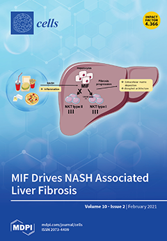
Cover Story (view full-size image): Macrophage migration inhibitory factor (MIF) is a pleiotropic inflammatory cytokine, found to exert an anti-fibrotic effect in toxic liver injury by inhibiting hepatic stellate cell activation. Using genetically modified mouse models, we explored an unexpected pro-fibrogenic role of MIF in non-alcoholic steatohepatitis (NASH) and identified hepatocytes as a functional relevant source of MIF during NASH. Via further in vivo and in vitro analysis, we could evidence that MIF directly contributes to NKT polarization during NASH, shifting the balance toward the pro-fibrotic type I subtype. Complementing expression analysis studies in human liver samples with different NAFLD stages indicates that the role of MIF and the identified mechanisms are conserved between men and mice. The study adds an unidentified layer to the understanding of the complex role of MIF during chronic liver injury. View this paper
  • Issues are regarded as officially published after their release is announced to the table of contents alert mailing list.
  • You may sign up for e-mail alerts to receive table of contents of newly released issues.
  • PDF is the official format for papers published in both, html and pdf forms. To view the papers in pdf format, click on the "PDF Full-text" link, and use the free Adobe Reader to open them.
Order results
Result details
Section
Select all
Export citation of selected articles as:
18 pages, 8361 KiB  
Article
Influence of Mesenchymal Stem Cell Sources on Their Regenerative Capacities on Different Surfaces
by Arkaitz Mucientes, Eva Herranz, Enrique Moro, Aranzazu González-Corchón, María Jesús Peña-Soria, Lydia Abasolo, Luis Rodriguez-Rodriguez, Jose Ramon Lamas and Benjamín Fernández-Gutiérrez
Cells 2021, 10(2), 481; https://doi.org/10.3390/cells10020481 - 23 Feb 2021
Cited by 10 | Viewed by 3267
Abstract
Current gold-standard strategies for bone regeneration do not achieve the optimal recovery of bone biomechanical properties. To bypass these limitations, tissue engineering techniques based on hybrid materials made up of osteoprogenitor cells—such as mesenchymal stem cells (MSCs)—and bioactive ceramic scaffolds—such as calcium phosphate-based [...] Read more.
Current gold-standard strategies for bone regeneration do not achieve the optimal recovery of bone biomechanical properties. To bypass these limitations, tissue engineering techniques based on hybrid materials made up of osteoprogenitor cells—such as mesenchymal stem cells (MSCs)—and bioactive ceramic scaffolds—such as calcium phosphate-based (CaPs) bioceramics—seem promising. The biological properties of MSCs are influenced by the tissue source. This study aims to define the optimal MSC source and construct (i.e., the MSC–CaP combination) for clinical application in bone regeneration. A previous iTRAQ analysis generated the hypothesis that anatomical proximity to bone has a direct effect on MSC phenotype. MSCs were isolated from adipose tissue, bone marrow, and dental pulp, then cultured both on a plastic surface and on CaPs (hydroxyapatite and β-tricalcium phosphate), to compare their biological features. On plastic, MSCs isolated from dental pulp (DPSCs) presented the highest proliferation capacity and the greatest osteogenic potential. On both CaPs, DPSCs demonstrated the greatest capacity to colonise the bioceramics. Furthermore, the results demonstrated a trend that DPSCs had the most robust increase in ALP activity. Regarding CaPs, β-tricalcium phosphate obtained the best viability results, while hydroxyapatite had the highest ALP activity values. Therefore, we propose DPSCs as suitable MSCs for cell-based bone regeneration strategies. Full article
(This article belongs to the Special Issue Oral Stem Cells in Tissue Engineering and Regenerative Medicine)
Show Figures

Figure 1

12 pages, 2976 KiB  
Article
A LRRK2 GTP Binding Inhibitor, 68, Reduces LPS-Induced Signaling Events and TNF-α Release in Human Lymphoblasts
by Tianxia Li, Bo Ning, Lingbo Kong, Bingling Dai, Xiaofei He, Joseph M. Thomas, Akira Sawa, Christopher A. Ross and Wanli W. Smith
Cells 2021, 10(2), 480; https://doi.org/10.3390/cells10020480 - 23 Feb 2021
Cited by 17 | Viewed by 3820
Abstract
Mutations in the leucine-rich repeat kinase-2 (LRRK2) gene cause autosomal-dominant Parkinson’s disease (PD) and contribute to sporadic PD. Common genetic variation in LRRK2 modifies susceptibility to immunological disorders including Crohn’s disease and leprosy. Previous studies have reported that LRRK2 is expressed in B [...] Read more.
Mutations in the leucine-rich repeat kinase-2 (LRRK2) gene cause autosomal-dominant Parkinson’s disease (PD) and contribute to sporadic PD. Common genetic variation in LRRK2 modifies susceptibility to immunological disorders including Crohn’s disease and leprosy. Previous studies have reported that LRRK2 is expressed in B lymphocytes and macrophages, suggesting a role for LRRK2 in immunological functions. In this study, we characterized the LRRK2 protein expression and phosphorylation using human lymphoblasts. Lipopolysaccharide (LPS), a proinflammatory agent, induced the increase of LRRK2 expression and kinase activities in human lymphoblasts in a time-dependent manner. Moreover, LPS activated the Toll-like receptor (TLR) signaling pathway, increased TRAF6/LRRK2 interaction, and elevated the phosphorylation levels of MAPK (JNK1/2, p38, and ERK1/2) and IkBα. Treatment with LRRK2 inhibitor 68 reduced LPS-induced TRAF6/LRRK2 interaction and MAPK and IkBα phosphorylation, thereby reducing TNF-α secretion. These results indicate that LRRK2 is actively involved in proinflammatory responses in human lymphoblasts, and inhibition of GTP binding by 68 results in an anti-inflammation effect against proinflammatory stimuli. These findings not only provide novel insights into the mechanisms of LRRK2-linked immune and inflammatory responses in B-cell-like lymphoblasts, but also suggest that 68 may also have potential therapeutic value for LRRK2-linked immunological disorders. Full article
(This article belongs to the Collection LRRK2-Dependent Neurodegeneration in Parkinson’s Disease)
Show Figures

Figure 1

29 pages, 4739 KiB  
Review
Unraveling Cell Death Pathways during Malaria Infection: What Do We Know So Far?
by Camille Sena-dos-Santos, Cíntia Braga-da-Silva, Diego Marques, Jhully Azevedo dos Santos Pinheiro, Ândrea Ribeiro-dos-Santos and Giovanna C. Cavalcante
Cells 2021, 10(2), 479; https://doi.org/10.3390/cells10020479 - 23 Feb 2021
Cited by 23 | Viewed by 6096
Abstract
Malaria is a parasitic disease (caused by different Plasmodium species) that affects millions of people worldwide. The lack of effective malaria drugs and a vaccine contributes to this disease, continuing to cause major public health and socioeconomic problems, especially in low-income countries. Cell [...] Read more.
Malaria is a parasitic disease (caused by different Plasmodium species) that affects millions of people worldwide. The lack of effective malaria drugs and a vaccine contributes to this disease, continuing to cause major public health and socioeconomic problems, especially in low-income countries. Cell death is implicated in malaria immune responses by eliminating infected cells, but it can also provoke an intense inflammatory response and lead to severe malaria outcomes. The study of the pathophysiological role of cell death in malaria in mammalians is key to understanding the parasite–host interactions and design prophylactic and therapeutic strategies for malaria. In this work, we review malaria-triggered cell death pathways (apoptosis, autophagy, necrosis, pyroptosis, NETosis, and ferroptosis) and we discuss their potential role in the development of new approaches for human malaria therapies. Full article
(This article belongs to the Special Issue Programmed Cell Death in Health and Disease)
Show Figures

Figure 1

18 pages, 3426 KiB  
Article
IQGAP1 Is a Scaffold of the Core Proteins of the Hippo Pathway and Negatively Regulates the Pro-Apoptotic Signal Mediated by This Pathway
by Niall P. Quinn, Lucía García-Gutiérrez, Carolanne Doherty, Alexander von Kriegsheim, Emma Fallahi, David B. Sacks and David Matallanas
Cells 2021, 10(2), 478; https://doi.org/10.3390/cells10020478 - 23 Feb 2021
Cited by 13 | Viewed by 4225
Abstract
The Hippo pathway regulates a complex signalling network which mediates several biological functions including cell proliferation, organ size and apoptosis. Several scaffold proteins regulate the crosstalk of the members of the pathway with other signalling pathways and play an important role in the [...] Read more.
The Hippo pathway regulates a complex signalling network which mediates several biological functions including cell proliferation, organ size and apoptosis. Several scaffold proteins regulate the crosstalk of the members of the pathway with other signalling pathways and play an important role in the diverse output controlled by this pathway. In this study we have identified the scaffold protein IQGAP1 as a novel interactor of the core kinases of the Hippo pathway, MST2 and LATS1. Our results indicate that IQGAP1 scaffolds MST2 and LATS1 supresses their kinase activity and YAP1-dependent transcription. Additionally, we show that IQGAP1 is a negative regulator of the non-canonical pro-apoptotic pathway and may enable the crosstalk between this pathway and the ERK and AKT signalling modules. Our data also show that bile acids regulate the IQGAP1-MST2-LATS1 signalling module in hepatocellular carcinoma cells, which could be necessary for the inhibition of MST2-dependent apoptosis and hepatocyte transformation. Full article
(This article belongs to the Section Cell Signaling)
Show Figures

Graphical abstract

22 pages, 3018 KiB  
Review
Iron Deficiency in Pulmonary Arterial Hypertension: A Deep Dive into the Mechanisms
by Marceau Quatredeniers, Pedro Mendes-Ferreira, Diana Santos-Ribeiro, Morad K. Nakhleh, Maria-Rosa Ghigna, Sylvia Cohen-Kaminsky and Frédéric Perros
Cells 2021, 10(2), 477; https://doi.org/10.3390/cells10020477 - 23 Feb 2021
Cited by 28 | Viewed by 5563
Abstract
Pulmonary arterial hypertension (PAH) is a severe cardiovascular disease that is caused by the progressive occlusion of the distal pulmonary arteries, eventually leading to right heart failure and death. Almost 40% of patients with PAH are iron deficient. Although widely studied, the mechanisms [...] Read more.
Pulmonary arterial hypertension (PAH) is a severe cardiovascular disease that is caused by the progressive occlusion of the distal pulmonary arteries, eventually leading to right heart failure and death. Almost 40% of patients with PAH are iron deficient. Although widely studied, the mechanisms linking between PAH and iron deficiency remain unclear. Here we review the mechanisms regulating iron homeostasis and the preclinical and clinical data available on iron deficiency in PAH. Then we discuss the potential implications of iron deficiency on the development and management of PAH. Full article
(This article belongs to the Special Issue Cellular Pathogenesis of Pulmonary Arterial Hypertension)
Show Figures

Figure 1

12 pages, 1969 KiB  
Article
Establishment of Human Leukocyte Antigen-Mismatched Immune Responses after Transplantation of Human Liver Bud in Humanized Mouse Models
by Akihiro Mori, Soichiro Murata, Nao Tashiro, Tomomi Tadokoro, Satoshi Okamoto, Ryo Otsuka, Haruka Wada, Tomoki Murata, Takeshi Takahashi, Ken-ichiro Seino and Hideki Taniguchi
Cells 2021, 10(2), 476; https://doi.org/10.3390/cells10020476 - 23 Feb 2021
Cited by 8 | Viewed by 3795
Abstract
Humanized mouse models have contributed significantly to human immunology research. In transplant immunity, human immune cell responses to donor grafts have not been reproduced in a humanized animal model. To elicit human T-cell immune responses, we generated immune-compromised nonobese diabetic/Shi-scid, IL-2RγKO Jic (NOG) [...] Read more.
Humanized mouse models have contributed significantly to human immunology research. In transplant immunity, human immune cell responses to donor grafts have not been reproduced in a humanized animal model. To elicit human T-cell immune responses, we generated immune-compromised nonobese diabetic/Shi-scid, IL-2RγKO Jic (NOG) with a homozygous expression of human leukocyte antigen (HLA) class I heavy chain (NOG-HLA-A2Tg) mice. After the transplantation of HLA-A2 human hematopoietic stem cells into NOG-HLA-A2Tg, we succeeded in achieving alloimmune responses after the HLA-mismatched human-induced pluripotent stem cell (hiPSC)-derived liver-like tissue transplantation. This immune response was inhibited by administering tacrolimus. In this model, we reproduced allograft rejection after the human iPSC-derived liver-like tissue transplantation. Human tissue transplantation on the humanized mouse liver surface is a good model that can predict T-cell-mediated cellular rejection that may occur when organ transplantation is performed. Full article
(This article belongs to the Special Issue Current Progress in Organ Regeneration: Cells, Organoids and Organs)
Show Figures

Figure 1

23 pages, 1464 KiB  
Review
HIV-1 Latency and Viral Reservoirs: Existing Reversal Approaches and Potential Technologies, Targets, and Pathways Involved in HIV Latency Studies
by Sushant Khanal, Madison Schank, Mohamed El Gazzar, Jonathan P. Moorman and Zhi Q. Yao
Cells 2021, 10(2), 475; https://doi.org/10.3390/cells10020475 - 23 Feb 2021
Cited by 25 | Viewed by 8701
Abstract
Eradication of latent human immunodeficiency virus (HIV) infection is a global health challenge. Reactivation of HIV latency and killing of virus-infected cells, the so-called “kick and kill” or “shock and kill” approaches, are a popular strategy for HIV cure. While antiretroviral therapy (ART) [...] Read more.
Eradication of latent human immunodeficiency virus (HIV) infection is a global health challenge. Reactivation of HIV latency and killing of virus-infected cells, the so-called “kick and kill” or “shock and kill” approaches, are a popular strategy for HIV cure. While antiretroviral therapy (ART) halts HIV replication by targeting multiple steps in the HIV life cycle, including viral entry, integration, replication, and production, it cannot get rid of the occult provirus incorporated into the host-cell genome. These latent proviruses are replication-competent and can rebound in cases of ART interruption or cessation. In general, a very small population of cells harbor provirus, serve as reservoirs in ART-controlled HIV subjects, and are capable of expressing little to no HIV RNA or proteins. Beyond the canonical resting memory CD4+ T cells, HIV reservoirs also exist within tissue macrophages, myeloid cells, brain microglial cells, gut epithelial cells, and hematopoietic stem cells (HSCs). Despite a lack of active viral production, latently HIV-infected subjects continue to exhibit aberrant cellular signaling and metabolic dysfunction, leading to minor to major cellular and systemic complications or comorbidities. These include genomic DNA damage; telomere attrition; mitochondrial dysfunction; premature aging; and lymphocytic, cardiac, renal, hepatic, or pulmonary dysfunctions. Therefore, the arcane machineries involved in HIV latency and its reversal warrant further studies to identify the cryptic mechanisms of HIV reservoir formation and clearance. In this review, we discuss several molecules and signaling pathways, some of which have dual roles in maintaining or reversing HIV latency and reservoirs, and describe some evolving strategies and possible approaches to eliminate viral reservoirs and, ultimately, cure/eradicate HIV infection. Full article
(This article belongs to the Section Cellular Immunology)
Show Figures

Figure 1

7 pages, 234 KiB  
Editorial
Molecular and Cellular Basis of Autoimmune Diseases
by Yasmina Juarranz
Cells 2021, 10(2), 474; https://doi.org/10.3390/cells10020474 - 23 Feb 2021
Cited by 10 | Viewed by 3037
Abstract
The defense organization of our organism is found in the immune system, which has two important components, the innate and the adaptive immunity, where different molecules, cells, and organs are involved and coordinated to protect us from external and internal damage [...] Full article
(This article belongs to the Special Issue Molecular and Cellular Basis of Autoimmune Diseases)
14 pages, 648 KiB  
Review
Regulation of Mitochondrial Homeostasis by sAC-Derived cAMP Pool: Basic and Translational Aspects
by Muhammad Aslam and Yury Ladilov
Cells 2021, 10(2), 473; https://doi.org/10.3390/cells10020473 - 22 Feb 2021
Cited by 6 | Viewed by 3743
Abstract
In contrast to the traditional view of mitochondria being solely a source of cellular energy, e.g., the “powerhouse” of the cell, mitochondria are now known to be key regulators of numerous cellular processes. Accordingly, disturbance of mitochondrial homeostasis is a basic mechanism in [...] Read more.
In contrast to the traditional view of mitochondria being solely a source of cellular energy, e.g., the “powerhouse” of the cell, mitochondria are now known to be key regulators of numerous cellular processes. Accordingly, disturbance of mitochondrial homeostasis is a basic mechanism in several pathologies. Emerging data demonstrate that 3′–5′-cyclic adenosine monophosphate (cAMP) signalling plays a key role in mitochondrial biology and homeostasis. Mitochondria are equipped with an endogenous cAMP synthesis system involving soluble adenylyl cyclase (sAC), which localizes in the mitochondrial matrix and regulates mitochondrial function. Furthermore, sAC localized at the outer mitochondrial membrane contributes significantly to mitochondrial biology. Disturbance of the sAC-dependent cAMP pools within mitochondria leads to mitochondrial dysfunction and pathology. In this review, we discuss the available data concerning the role of sAC in regulating mitochondrial biology in relation to diseases. Full article
(This article belongs to the Section Mitochondria)
Show Figures

Figure 1

26 pages, 9388 KiB  
Article
Lipid Droplets Are a Physiological Nucleoporin Reservoir
by Sylvain Kumanski, Benjamin T. Viart, Sofia Kossida and María Moriel-Carretero
Cells 2021, 10(2), 472; https://doi.org/10.3390/cells10020472 - 22 Feb 2021
Cited by 12 | Viewed by 6130
Abstract
Lipid Droplets (LD) are dynamic organelles that originate in the Endoplasmic Reticulum and mostly bud off toward the cytoplasm, where they store neutral lipids for energy and protection purposes. LD also have diverse proteins on their surface, many of which are necessary for [...] Read more.
Lipid Droplets (LD) are dynamic organelles that originate in the Endoplasmic Reticulum and mostly bud off toward the cytoplasm, where they store neutral lipids for energy and protection purposes. LD also have diverse proteins on their surface, many of which are necessary for the their correct homeostasis. However, these organelles also act as reservoirs of proteins that can be made available elsewhere in the cell. In this sense, they act as sinks that titrate key regulators of many cellular processes. Among the specialized factors that reside on cytoplasmic LD are proteins destined for functions in the nucleus, but little is known about them and their impact on nuclear processes. By screening for nuclear proteins in publicly available LD proteomes, we found that they contain a subset of nucleoporins from the Nuclear Pore Complex (NPC). Exploring this, we demonstrate that LD act as a physiological reservoir, for nucleoporins, that impacts the conformation of NPCs and hence their function in nucleo-cytoplasmic transport, chromatin configuration, and genome stability. Furthermore, our in silico modeling predicts a role for LD-released fatty acids in regulating the transit of nucleoporins from LD through the cytoplasm and to nuclear pores. Full article
(This article belongs to the Section Cell Nuclei: Function, Transport and Receptors)
Show Figures

Graphical abstract

20 pages, 1727 KiB  
Article
Hyperglycemia Potentiates Prothrombotic Effect of Aldosterone in a Rat Arterial Thrombosis Model
by Anna Gromotowicz-Poplawska, Piotr Szoka, Agnieszka Zakrzeska, Patrycjusz Kolodziejczyk, Natalia Marcinczyk, Janusz Szemraj, Piotr Tutka and Ewa Chabielska
Cells 2021, 10(2), 471; https://doi.org/10.3390/cells10020471 - 22 Feb 2021
Cited by 2 | Viewed by 2921
Abstract
We investigated the role of aldosterone (ALDO) in the development of arterial thrombosis in streptozotocin-induced diabetic rats. To evaluate the effect of endogenous ALDO, the rats underwent adrenalectomy (ADX). ADX reduced the development of arterial thrombosis. A 1 h infusion of ALDO (30 [...] Read more.
We investigated the role of aldosterone (ALDO) in the development of arterial thrombosis in streptozotocin-induced diabetic rats. To evaluate the effect of endogenous ALDO, the rats underwent adrenalectomy (ADX). ADX reduced the development of arterial thrombosis. A 1 h infusion of ALDO (30 μg/kg/h) enhanced thrombosis in adrenalectomized rats, while this effect was potentiated in diabetic rats. ALDO shortened bleeding time, increased plasma levels of tissue factor (TF) and plasminogen activator inhibitor, decreased plasma level of nitric oxide (NO) metabolites, and increased oxidative stress. Moreover, 2 h incubation of human umbilical vein endothelial cells (HUVECs) with ALDO (10−7 M) disrupted hemostatic balance in endothelial cells in normoglycemia (glucose 5.5 mM), and this effect was more pronounced in hyperglycemia (glucose 30 mM). We demonstrated that the acute ALDO infusion enhances arterial thrombosis in rats and hyperglycemia potentiates this prothrombotic effect. The mechanism of ALDO action was partially mediated by mineralocorticoid (MR) and glucocorticoid (GR) receptors and related to impact of the hormone on primary hemostasis, TF-dependent coagulation cascade, fibrinolysis, NO bioavailability, and oxidative stress balance. Our in vitro study confirmed that ALDO induces prothrombotic phenotype in the endothelium, particularly under hyperglycemic conditions. Full article
(This article belongs to the Special Issue Renin–Angiotensin–Aldosterone System)
Show Figures

Figure 1

25 pages, 2107 KiB  
Review
Cytochrome c Oxidase at Full Thrust: Regulation and Biological Consequences to Flying Insects
by Rafael D. Mesquita, Alessandro Gaviraghi, Renata L.S. Gonçalves, Marcos A. Vannier-Santos, Julio A. Mignaco, Carlos Frederico L. Fontes, Luciana E.S.F. Machado and Marcus F. Oliveira
Cells 2021, 10(2), 470; https://doi.org/10.3390/cells10020470 - 22 Feb 2021
Cited by 8 | Viewed by 4711
Abstract
Flight dispersal represents a key aspect of the evolutionary and ecological success of insects, allowing escape from predators, mating, and colonization of new niches. The huge energy demand posed by flight activity is essentially met by oxidative phosphorylation (OXPHOS) in flight muscle mitochondria. [...] Read more.
Flight dispersal represents a key aspect of the evolutionary and ecological success of insects, allowing escape from predators, mating, and colonization of new niches. The huge energy demand posed by flight activity is essentially met by oxidative phosphorylation (OXPHOS) in flight muscle mitochondria. In insects, mitochondrial ATP supply and oxidant production are regulated by several factors, including the energy demand exerted by changes in adenylate balance. Indeed, adenylate directly regulates OXPHOS by targeting both chemiosmotic ATP production and the activities of specific mitochondrial enzymes. In several organisms, cytochrome c oxidase (COX) is regulated at transcriptional, post-translational, and allosteric levels, impacting mitochondrial energy metabolism, and redox balance. This review will present the concepts on how COX function contributes to flying insect biology, focusing on the existing examples in the literature where its structure and activity are regulated not only by physiological and environmental factors but also how changes in its activity impacts insect biology. We also performed in silico sequence analyses and determined the structure models of three COX subunits (IV, VIa, and VIc) from different insect species to compare with mammalian orthologs. We observed that the sequences and structure models of COXIV, COXVIa, and COXVIc were quite similar to their mammalian counterparts. Remarkably, specific substitutions to phosphomimetic amino acids at critical phosphorylation sites emerge as hallmarks on insect COX sequences, suggesting a new regulatory mechanism of COX activity. Therefore, by providing a physiological and bioenergetic framework of COX regulation in such metabolically extreme models, we hope to expand the knowledge of this critical enzyme complex and the potential consequences for insect dispersal. Full article
(This article belongs to the Collection Regulation of Eukaryotic Cytochrome c Oxidase)
Show Figures

Graphical abstract

18 pages, 6008 KiB  
Article
CellMAPtracer: A User-Friendly Tracking Tool for Long-Term Migratory and Proliferating Cells Associated with FUCCI Systems
by Salim Ghannoum, Kamil Antos, Waldir Leoncio Netto, Cecil Gomes, Alvaro Köhn-Luque and Hesso Farhan
Cells 2021, 10(2), 469; https://doi.org/10.3390/cells10020469 - 22 Feb 2021
Cited by 6 | Viewed by 8660
Abstract
Cell migration is a fundamental biological process of key importance in health and disease. Advances in imaging techniques have paved the way to monitor cell motility. An ever-growing collection of computational tools to track cells has improved our ability to analyze moving cells. [...] Read more.
Cell migration is a fundamental biological process of key importance in health and disease. Advances in imaging techniques have paved the way to monitor cell motility. An ever-growing collection of computational tools to track cells has improved our ability to analyze moving cells. One renowned goal in the field is to provide tools that track cell movement as comprehensively and automatically as possible. However, fully automated tracking over long intervals of time is challenged by dividing cells, thus calling for a combination of automated and supervised tracking. Furthermore, after the emergence of various experimental tools to monitor cell-cycle phases, it is of relevance to integrate the monitoring of cell-cycle phases and motility. We developed CellMAPtracer, a multiplatform tracking system that achieves that goal. It can be operated as a conventional, automated tracking tool of single cells in numerous imaging applications. However, CellMAPtracer also allows adjusting tracked cells in a semiautomated supervised fashion, thereby improving the accuracy and facilitating the long-term tracking of migratory and dividing cells. CellMAPtracer is available with a user-friendly graphical interface and does not require any coding or programming skills. CellMAPtracer is compatible with two- and three-color fluorescent ubiquitination-based cell-cycle indicator (FUCCI) systems and allows the user to accurately monitor various migration parameters throughout the cell cycle, thus having great potential to facilitate new discoveries in cell biology. Full article
(This article belongs to the Section Cell Motility and Adhesion)
Show Figures

Graphical abstract

13 pages, 2353 KiB  
Article
The Pentatricopeptide Repeat Protein MEF100 Is Required for the Editing of Four Mitochondrial Editing Sites in Arabidopsis
by Bernard Gutmann, Michael Millman, Lilian Vincis Pereira Sanglard, Ian Small and Catherine Colas des Francs-Small
Cells 2021, 10(2), 468; https://doi.org/10.3390/cells10020468 - 22 Feb 2021
Cited by 4 | Viewed by 2819
Abstract
In Arabidopsis thaliana there are more than 600 C-to-U RNA editing events in the mitochondria and at least 44 in the chloroplasts. Pentatricopeptide repeat (PPR) proteins provide the specificity for these reactions. They recognize RNA sequences in a partially predictable fashion via key [...] Read more.
In Arabidopsis thaliana there are more than 600 C-to-U RNA editing events in the mitochondria and at least 44 in the chloroplasts. Pentatricopeptide repeat (PPR) proteins provide the specificity for these reactions. They recognize RNA sequences in a partially predictable fashion via key amino acids at the fifth and last position in each PPR motif that bind to individual ribonucleotides. A combined approach of RNA-Seq, mutant complementation, electrophoresis of mitochondrial protein complexes and Western blotting allowed us to show that MEF100, a PPR protein identified in a genetic screen for mutants resistant to an inhibitor of γ -glutamylcysteine synthetase, is required for the editing of nad1-493, nad4-403, nad7-698 and ccmFN2-356 sites in Arabidopsis mitochondria. The absence of editing in mef100 leads to a decrease in mitochondrial Complex I activity, which probably explains the physiological phenotype. Some plants have lost the requirement for MEF100 at one or more of these sites through mutations in the mitochondrial genome. We show that loss of the requirement for MEF100 editing leads to divergence in the MEF100 binding site. Full article
(This article belongs to the Special Issue RNA Biology in Plant Organelles)
Show Figures

Figure 1

17 pages, 3312 KiB  
Article
Identification of Accessible Hepatic Gene Signatures for Interindividual Variations in Nutrigenomic Response to Dietary Supplementation of Omega-3 Fatty Acids
by Yu Shi, Ping Li, Cheng-fei Jiang, Yi Chen, Yonghe Ma, Nikhil Gupta, Xiangbo Ruan and Haiming Cao
Cells 2021, 10(2), 467; https://doi.org/10.3390/cells10020467 - 22 Feb 2021
Cited by 3 | Viewed by 3305
Abstract
Dietary supplementation is a widely adapted strategy to maintain nutritional balance for improving health and preventing chronic diseases. Conflicting results in studies of similar design, however, suggest that there is substantial heterogenicity in individuals’ responses to nutrients, and personalized nutrition is required to [...] Read more.
Dietary supplementation is a widely adapted strategy to maintain nutritional balance for improving health and preventing chronic diseases. Conflicting results in studies of similar design, however, suggest that there is substantial heterogenicity in individuals’ responses to nutrients, and personalized nutrition is required to achieve the maximum benefit of dietary supplementation. In recent years, nutrigenomics studies have been increasingly utilized to characterize the detailed genomic response to a specific nutrient, but it remains a daunting task to define the signatures responsible for interindividual variations to dietary supplements for tissues with limited accessibility. In this work, we used the hepatic response to omega-3 fatty acids as an example to probe such signatures. Through comprehensive analysis of nutrigenomic response to eicosapentaneoid acid (EPA) and/or docosahexaenoic acid (DHA) including both protein coding and long noncoding RNA (lncRNA) genes in human hepatocytes, we defined the EPA- and/or DHA-specific signature genes in hepatocytes. By analyzing gene expression variations in livers of healthy and relevant disease populations, we identified a set of protein coding and lncRNA signature genes whose responses to omega-3 fatty acid exhibit very high interindividual variabilities. The large variabilities of individual responses to omega-3 fatty acids were further validated in human hepatocytes from ten different donors. Finally, we profiled RNAs in exosomes isolated from the circulation of a liver-specific humanized mouse model, in which the humanized liver is the sole source of human RNAs, and confirmed the in vivo detectability of some signature genes, supporting their potential as biomarkers for nutrient response. Taken together, we have developed an efficient and practical procedure to identify nutrient-responsive gene signatures as well as accessible biomarkers for interindividual variations. Full article
Show Figures

Figure 1

35 pages, 10172 KiB  
Article
Chemically Defined Xeno- and Serum-Free Cell Culture Medium to Grow Human Adipose Stem Cells
by Stefano Panella, Francesco Muoio, Valentin Jossen, Yves Harder, Regine Eibl-Schindler and Tiziano Tallone
Cells 2021, 10(2), 466; https://doi.org/10.3390/cells10020466 - 22 Feb 2021
Cited by 11 | Viewed by 5996
Abstract
Adipose tissue is an abundant source of stem cells. However, liposuction cannot yield cell quantities sufficient for direct applications in regenerative medicine. Therefore, the development of GMP-compliant ex vivo expansion protocols is required to ensure the production of a “cell drug” that is [...] Read more.
Adipose tissue is an abundant source of stem cells. However, liposuction cannot yield cell quantities sufficient for direct applications in regenerative medicine. Therefore, the development of GMP-compliant ex vivo expansion protocols is required to ensure the production of a “cell drug” that is safe, reproducible, and cost-effective. Thus, we developed our own basal defined xeno- and serum-free cell culture medium (UrSuppe), specifically formulated to grow human adipose stem cells (hASCs). With this medium, we can directly culture the stromal vascular fraction (SVF) cells in defined cell culture conditions to obtain hASCs. Cells proliferate while remaining undifferentiated, as shown by Flow Cytometry (FACS), Quantitative Reverse Transcription PCR (RT-qPCR) assays, and their secretion products. Using the UrSuppe cell culture medium, maximum cell densities between 0.51 and 0.80 × 10⁵ cells/cm2 (=2.55–4.00 × 10⁵ cells/mL) were obtained. As the expansion of hASCs represents only the first step in a cell therapeutic protocol or further basic research studies, we formulated two chemically defined media to differentiate the expanded hASCs in white or beige/brown adipocytes. These new media could help translate research projects into the clinical application of hASCs and study ex vivo the biology in healthy and dysfunctional states of adipocytes and their precursors. Following the cell culture system developers’ practice and obvious reasons related to the formulas’ patentability, the defined media’s composition will not be disclosed in this study. Full article
(This article belongs to the Special Issue Human Adipose Stem Cells)
Show Figures

Figure 1

25 pages, 3047 KiB  
Review
Mechanical Mechanisms of Chromosome Segregation
by Maya I. Anjur-Dietrich, Colm P. Kelleher and Daniel J. Needleman
Cells 2021, 10(2), 465; https://doi.org/10.3390/cells10020465 - 22 Feb 2021
Cited by 18 | Viewed by 5683
Abstract
Chromosome segregation—the partitioning of genetic material into two daughter cells—is one of the most crucial processes in cell division. In all Eukaryotes, chromosome segregation is driven by the spindle, a microtubule-based, self-organizing subcellular structure. Extensive research performed over the past 150 years has [...] Read more.
Chromosome segregation—the partitioning of genetic material into two daughter cells—is one of the most crucial processes in cell division. In all Eukaryotes, chromosome segregation is driven by the spindle, a microtubule-based, self-organizing subcellular structure. Extensive research performed over the past 150 years has identified numerous commonalities and contrasts between spindles in different systems. In this review, we use simple coarse-grained models to organize and integrate previous studies of chromosome segregation. We discuss sites of force generation in spindles and fundamental mechanical principles that any understanding of chromosome segregation must be based upon. We argue that conserved sites of force generation may interact differently in different spindles, leading to distinct mechanical mechanisms of chromosome segregation. We suggest experiments to determine which mechanical mechanism is operative in a particular spindle under study. Finally, we propose that combining biophysical experiments, coarse-grained theories, and evolutionary genetics will be a productive approach to enhance our understanding of chromosome segregation in the future. Full article
(This article belongs to the Special Issue The Microtubule Cytoskeleton in Chromosome Segregation and Beyond)
Show Figures

Figure 1

31 pages, 1434 KiB  
Review
Compartmentalized Signaling in Aging and Neurodegeneration
by Giulietta Di Benedetto, Liliana F. Iannucci, Nicoletta C. Surdo, Sofia Zanin, Filippo Conca, Francesca Grisan, Andrea Gerbino and Konstantinos Lefkimmiatis
Cells 2021, 10(2), 464; https://doi.org/10.3390/cells10020464 - 22 Feb 2021
Cited by 17 | Viewed by 4456
Abstract
The cyclic AMP (cAMP) signalling cascade is necessary for cell homeostasis and plays important roles in many processes. This is particularly relevant during ageing and age-related diseases, where drastic changes, generally decreases, in cAMP levels have been associated with the progressive decline in [...] Read more.
The cyclic AMP (cAMP) signalling cascade is necessary for cell homeostasis and plays important roles in many processes. This is particularly relevant during ageing and age-related diseases, where drastic changes, generally decreases, in cAMP levels have been associated with the progressive decline in overall cell function and, eventually, the loss of cellular integrity. The functional relevance of reduced cAMP is clearly supported by the finding that increases in cAMP levels can reverse some of the effects of ageing. Nevertheless, despite these observations, the molecular mechanisms underlying the dysregulation of cAMP signalling in ageing are not well understood. Compartmentalization is widely accepted as the modality through which cAMP achieves its functional specificity; therefore, it is important to understand whether and how this mechanism is affected during ageing and to define which is its contribution to this process. Several animal models demonstrate the importance of specific cAMP signalling components in ageing, however, how age-related changes in each of these elements affect the compartmentalization of the cAMP pathway is largely unknown. In this review, we explore the connection of single components of the cAMP signalling cascade to ageing and age-related diseases whilst elaborating the literature in the context of cAMP signalling compartmentalization. Full article
(This article belongs to the Special Issue Key Signalling Molecules in Aging and Neurodegeneration)
Show Figures

Figure 1

40 pages, 7846 KiB  
Review
Utilisation of Chick Embryo Chorioallantoic Membrane as a Model Platform for Imaging-Navigated Biomedical Research
by Lei Chen, Shuncong Wang, Yuanbo Feng, Jinyong Zhang, Yuqing Du, Jiang Zhang, Chantal Van Ongeval, Yicheng Ni and Yue Li
Cells 2021, 10(2), 463; https://doi.org/10.3390/cells10020463 - 22 Feb 2021
Cited by 45 | Viewed by 13244
Abstract
The fertilised chick egg and particularly its chorioallantoic membrane (CAM) have drawn continuing interest in biomedicine and bioengineering fields, especially for research on vascular study, cancer, drug screening and development, cell factors, stem cells, etc. This literature review systemically introduces the CAM’s structural [...] Read more.
The fertilised chick egg and particularly its chorioallantoic membrane (CAM) have drawn continuing interest in biomedicine and bioengineering fields, especially for research on vascular study, cancer, drug screening and development, cell factors, stem cells, etc. This literature review systemically introduces the CAM’s structural evolution, functions, vascular features and the circulation system, and cell regulatory factors. It also presents the major and updated applications of the CAM in assays for pharmacokinetics and biodistribution, drug efficacy and toxicology testing/screening in preclinical pharmacological research. The time course of CAM applications for different assays and their advantages and limitations are summarised. Among these applications, two aspects are emphasised: (1) potential utility of the CAM for preclinical studies on vascular-disrupting agents (VDAs), promising for anti-cancer vascular-targeted therapy, and (2) modern imaging technologies, including modalities and their applications for real-time visualisation, monitoring and evaluation of the changes in CAM vasculature as well as the interactions occurring after introducing the tested medical, pharmaceutical and biological agents into the system. The aim of this article is to help those working in the biomedical field to familiarise themselves with the chick embryo CAM as an alternative platform and to utilise it to design and optimise experimental settings for their specific research topics. Full article
Show Figures

Figure 1

1 pages, 3083 KiB  
Correction
Correction: Gurunathan, S. et al. Review of the Isolation, Characterization, Biological Function, and Multifarious Therapeutic Approaches of Exosomes. Cells 2019, 8, 307
by Sangiliyandi Gurunathan, Min-Hee Kang, Muniyandi Jeyaraj, Muhammad Qasim and Jin-Hoi Kim
Cells 2021, 10(2), 462; https://doi.org/10.3390/cells10020462 - 22 Feb 2021
Cited by 6 | Viewed by 2911
Abstract
In the original review [...] Full article
Show Figures

Figure 1

18 pages, 1903 KiB  
Review
DNA or Protein Methylation-Dependent Regulation of Activator Protein-1 Function
by Eunji Kim, Akash Ahuja, Mi-Yeon Kim and Jae Youl Cho
Cells 2021, 10(2), 461; https://doi.org/10.3390/cells10020461 - 21 Feb 2021
Cited by 20 | Viewed by 5299
Abstract
Epigenetic regulation and modification govern the transcriptional mechanisms that promote disease initiation and progression, but can also control the oncogenic processes, cell signaling networks, immunogenicity, and immune cells involved in anti-inflammatory and anti-tumor responses. The study of epigenetic mechanisms could have important implications [...] Read more.
Epigenetic regulation and modification govern the transcriptional mechanisms that promote disease initiation and progression, but can also control the oncogenic processes, cell signaling networks, immunogenicity, and immune cells involved in anti-inflammatory and anti-tumor responses. The study of epigenetic mechanisms could have important implications for the development of potential anti-inflammatory treatments and anti-cancer immunotherapies. In this review, we have described the key role of epigenetic progression: DNA methylation, histone methylation or modification, and protein methylation, with an emphasis on the activator protein-1 (AP-1) signaling pathway. Transcription factor AP-1 regulates multiple genes and is involved in diverse cellular processes, including survival, differentiation, apoptosis, and development. Here, the AP-1 regulatory mechanism by DNA, histone, or protein methylation was also reviewed. Various methyltransferases activate or suppress AP-1 activities in diverse ways. We summarize the current studies on epigenetic alterations, which regulate AP-1 signaling during inflammation, cancer, and autoimmune diseases, and discuss the epigenetic mechanisms involved in the regulation of AP-1 signaling. Full article
Show Figures

Figure 1

20 pages, 5037 KiB  
Review
The Pleiotropic Function of Human Sirtuins as Modulators of Metabolic Pathways and Viral Infections
by Mohammed Hamed Alqarni, Ahmed Ibrahim Foudah, Magdy Mohamed Muharram and Nikolaos E. Labrou
Cells 2021, 10(2), 460; https://doi.org/10.3390/cells10020460 - 21 Feb 2021
Cited by 25 | Viewed by 4524
Abstract
Sirtuins (SIRTs) are nicotinamide adenine dinucleotide-dependent histone deacetylases that incorporate complex functions in the mechanisms of cell physiology. Mammals have seven distinct members of the SIRT family (SIRT1-7), which play an important role in a well-maintained network of metabolic pathways that control and [...] Read more.
Sirtuins (SIRTs) are nicotinamide adenine dinucleotide-dependent histone deacetylases that incorporate complex functions in the mechanisms of cell physiology. Mammals have seven distinct members of the SIRT family (SIRT1-7), which play an important role in a well-maintained network of metabolic pathways that control and adapt the cell to the environment, energy availability and cellular stress. Until recently, very few studies investigated the role of SIRTs in modulating viral infection and progeny. Recent studies have demonstrated that SIRT1 and SIRT2 are promising antiviral targets because of their specific connection to numerous metabolic and regulatory processes affected during infection. In the present review, we summarize some of the recent progress in SIRTs biochemistry and their emerging function as antiviral targets. We also discuss the potential of natural polyphenol-based SIRT modulators to control their functional roles in several diseases including viral infections. Full article
Show Figures

Figure 1

13 pages, 1227 KiB  
Review
Estrogen Receptors in Polycystic Ovary Syndrome
by Xue-Ling Xu, Shou-Long Deng, Zheng-Xing Lian and Kun Yu
Cells 2021, 10(2), 459; https://doi.org/10.3390/cells10020459 - 21 Feb 2021
Cited by 47 | Viewed by 7080
Abstract
Female infertility is mainly caused by ovulation disorders, which affect female reproduction and pregnancy worldwide, with polycystic ovary syndrome (PCOS) being the most prevalent of these. PCOS is a frequent endocrine disease that is associated with abnormal function of the female sex hormone [...] Read more.
Female infertility is mainly caused by ovulation disorders, which affect female reproduction and pregnancy worldwide, with polycystic ovary syndrome (PCOS) being the most prevalent of these. PCOS is a frequent endocrine disease that is associated with abnormal function of the female sex hormone estrogen and estrogen receptors (ERs). Estrogens mediate genomic effects through ERα and ERβ in target tissues. The G-protein-coupled estrogen receptor (GPER) has recently been described as mediating the non-genomic signaling of estrogen. Changes in estrogen receptor signaling pathways affect cellular activities, such as ovulation; cell cycle phase; and cell proliferation, migration, and invasion. Over the years, some selective estrogen receptor modulators (SERMs) have made substantial strides in clinical applications for subfertility with PCOS, such as tamoxifen and clomiphene, however the role of ER in PCOS still needs to be understood. This article focuses on the recent progress in PCOS caused by the abnormal expression of estrogen and ERs in the ovaries and uterus, and the clinical application of related targeted small-molecule drugs. Full article
(This article belongs to the Special Issue Estrogen Receptor Hormone Action)
Show Figures

Figure 1

14 pages, 1498 KiB  
Review
Ellagic Acid and Schisandrins: Natural Biaryl Polyphenols with Therapeutic Potential to Overcome Multidrug Resistance in Cancer
by Sabesan Yoganathan, Anushan Alagaratnam, Nikita Acharekar and Jing Kong
Cells 2021, 10(2), 458; https://doi.org/10.3390/cells10020458 - 21 Feb 2021
Cited by 28 | Viewed by 4756
Abstract
Multidrug resistance (MDR) is one of the major clinical challenges in cancer treatment and compromises the effectiveness of conventional anticancer chemotherapeutics. Among known mechanisms of drug resistance, drug efflux via ATP binding cassette (ABC) transporters, namely P-glycoprotein (P-gp) has been characterized as a [...] Read more.
Multidrug resistance (MDR) is one of the major clinical challenges in cancer treatment and compromises the effectiveness of conventional anticancer chemotherapeutics. Among known mechanisms of drug resistance, drug efflux via ATP binding cassette (ABC) transporters, namely P-glycoprotein (P-gp) has been characterized as a major mechanism of MDR. The primary function of ABC transporters is to regulate the transport of endogenous and exogenous small molecules across the membrane barrier in various tissues. P-gp and similar efflux pumps are associated with MDR because of their overexpression in many cancer types. One of the intensively studied approaches to overcome this mode of MDR involves development of small molecules to modulate P-gp activity. This strategy improves the sensitivity of cancer cells to anticancer drugs that are otherwise ineffective. Although multiple generations of P-gp inhibitors have been identified to date, reported compounds have demonstrated low clinical efficacy and adverse effects. More recently, natural polyphenols have emerged as a promising class of compounds to address P-gp linked MDR. This review highlights the chemical structure and anticancer activities of selected members of a structurally unique class of ‘biaryl’ polyphenols. The discussion focuses on the anticancer properties of ellagic acid, ellagic acid derivatives, and schisandrins. Research reports regarding their inherent anticancer activities and their ability to sensitize MDR cell lines towards conventional anticancer drugs are highlighted here. Additionally, a brief discussion about the axial chirality (i.e., atropisomerism) that may be introduced into these natural products for medicinal chemistry studies is also provided. Full article
(This article belongs to the Special Issue Studying Drug Resistance Using Cancer Cell Lines)
Show Figures

Figure 1

16 pages, 814 KiB  
Review
Why Do We Not Assess Sympathetic Nervous System Activity in Heart Failure Management: Might GRK2 Serve as a New Biomarker?
by Leonardo Bencivenga, Maria Emiliana Palaia, Immacolata Sepe, Giuseppina Gambino, Klara Komici, Alessandro Cannavo, Grazia Daniela Femminella and Giuseppe Rengo
Cells 2021, 10(2), 457; https://doi.org/10.3390/cells10020457 - 21 Feb 2021
Cited by 16 | Viewed by 3618
Abstract
Heart failure (HF) represents the end-stage condition of several structural and functional cardiovascular diseases, characterized by reduced myocardial pump function and increased pressure load. The dysregulation of neurohormonal systems, especially the hyperactivity of the cardiac adrenergic nervous system (ANS), constitutes a hallmark of [...] Read more.
Heart failure (HF) represents the end-stage condition of several structural and functional cardiovascular diseases, characterized by reduced myocardial pump function and increased pressure load. The dysregulation of neurohormonal systems, especially the hyperactivity of the cardiac adrenergic nervous system (ANS), constitutes a hallmark of HF and exerts a pivotal role in its progression. Indeed, it negatively affects patients’ prognosis, being associated with high morbidity and mortality rates, with a tremendous burden on global healthcare systems. To date, all the techniques proposed to assess the cardiac sympathetic nervous system are burdened by intrinsic limits that hinder their implementation in clinical practice. Several biomarkers related to ANS activity, which may potentially support the clinical management of such a complex syndrome, are slow to be implemented in the routine practice for several limitations due to their assessment and clinical impact. Lymphocyte G-protein-coupled Receptor Kinase 2 (GRK2) levels reflect myocardial β-adrenergic receptor function in HF and have been shown to add independent prognostic information related to ANS overdrive. In the present manuscript, we provide an overview of the techniques currently available to evaluate cardiac ANS in HF and future perspectives in this field of relevant scientific and clinical interest. Full article
Show Figures

Figure 1

21 pages, 4527 KiB  
Article
Lysophosphatidic Acid-Activated Calcium Signaling Is Elevated in Red Cells from Sickle Cell Disease Patients
by Jue Wang, Laura Hertz, Sandra Ruppenthal, Wassim El Nemer, Philippe Connes, Jeroen S. Goede, Anna Bogdanova, Lutz Birnbaumer and Lars Kaestner
Cells 2021, 10(2), 456; https://doi.org/10.3390/cells10020456 - 20 Feb 2021
Cited by 11 | Viewed by 3658
Abstract
(1) Background: It is known that sickle cells contain a higher amount of Ca2+ compared to healthy red blood cells (RBCs). The increased Ca2+ is associated with the most severe symptom of sickle cell disease (SCD), the vaso-occlusive crisis (VOC). The [...] Read more.
(1) Background: It is known that sickle cells contain a higher amount of Ca2+ compared to healthy red blood cells (RBCs). The increased Ca2+ is associated with the most severe symptom of sickle cell disease (SCD), the vaso-occlusive crisis (VOC). The Ca2+ entry pathway received the name of Psickle but its molecular identity remains only partly resolved. We aimed to map the involved Ca2+ signaling to provide putative pharmacological targets for treatment. (2) Methods: The main technique applied was Ca2+ imaging of RBCs from healthy donors, SCD patients and a number of transgenic mouse models in comparison to wild-type mice. Life-cell Ca2+ imaging was applied to monitor responses to pharmacological targeting of the elements of signaling cascades. Infection as a trigger of VOC was imitated by stimulation of RBCs with lysophosphatidic acid (LPA). These measurements were complemented with biochemical assays. (3) Results: Ca2+ entry into SCD RBCs in response to LPA stimulation exceeded that of healthy donors. LPA receptor 4 levels were increased in SCD RBCs. Their activation was followed by the activation of Gi protein, which in turn triggered opening of TRPC6 and CaV2.1 channels via a protein kinase Cα and a MAP kinase pathway, respectively. (4) Conclusions: We found a new Ca2+ signaling cascade that is increased in SCD patients and identified new pharmacological targets that might be promising in addressing the most severe symptom of SCD, the VOC. Full article
(This article belongs to the Special Issue Dysregulation of Calcium Signalling in Disease)
Show Figures

Graphical abstract

26 pages, 3256 KiB  
Article
A Multi-Omics Study Revealing the Metabolic Effects of Estrogen in Liver Cancer Cells HepG2
by Minqian Shen, Mengyang Xu, Fanyi Zhong, McKenzie C. Crist, Anjali B. Prior, Kundi Yang, Danielle M. Allaire, Fouad Choueiry, Jiangjiang Zhu and Haifei Shi
Cells 2021, 10(2), 455; https://doi.org/10.3390/cells10020455 - 20 Feb 2021
Cited by 17 | Viewed by 5147
Abstract
Hepatocellular carcinoma (HCC) that is triggered by metabolic defects is one of the most malignant liver cancers. A much higher incidence of HCC among men than women suggests the protective roles of estrogen in HCC development and progression. To begin to understand the [...] Read more.
Hepatocellular carcinoma (HCC) that is triggered by metabolic defects is one of the most malignant liver cancers. A much higher incidence of HCC among men than women suggests the protective roles of estrogen in HCC development and progression. To begin to understand the mechanisms involving estrogenic metabolic effects, we compared cell number, viability, cytotoxicity, and apoptosis among HCC-derived HepG2 cells that were treated with different concentrations of 2-deoxy-d-glucose (2-DG) that blocks glucose metabolism, oxamate that inhibits lactate dehydrogenase and glycolysis, or oligomycin that blocks ATP synthesis and mitochondrial oxidative phosphorylation. We confirmed that HepG2 cells primarily utilized glycolysis followed by lactate fermentation, instead of mitochondrial oxidative phosphorylation, for cell growth. We hypothesized that estrogen altered energy metabolism via its receptors to carry out its anticancer effects in HepG2 cells. We treated cells with 17β-estradiol (E2), 1,3,5-tris(4-hydroxyphenyl)-4-propyl-1H-pyrazole (PPT) an estrogen receptor (ER) α (ERα) agonist, or 2,3-bis(4-hydroxyphenyl)-propionitrile (DPN), an ERβ agonist. We then used transcriptomic and metabolomic analyses and identified differentially expressed genes and unique metabolite fingerprints that are produced by each treatment. We further performed integrated multi-omics analysis, and identified key genes and metabolites in the gene–metabolite interaction contributed by E2 and ER agonists. This integrated transcriptomic and metabolomic study suggested that estrogen acts on estrogen receptors to suppress liver cancer cell growth via altering metabolism. This is the first exploratory study that comprehensively investigated estrogen and its receptors, and their roles in regulating gene expression, metabolites, metabolic pathways, and gene–metabolite interaction in HCC cells using bioinformatic tools. Overall, this study provides potential therapeutic targets for future HCC treatment. Full article
Show Figures

Figure 1

16 pages, 987 KiB  
Review
Neuroimmune Regulation of Surgery-Associated Metastases
by Michael R. Shurin, James H. Baraldi and Galina V. Shurin
Cells 2021, 10(2), 454; https://doi.org/10.3390/cells10020454 - 20 Feb 2021
Cited by 13 | Viewed by 3890
Abstract
Surgery remains an essential therapeutic approach for most solid malignancies. Although for more than a century accumulating clinical and experimental data have indicated that surgical procedures themselves may promote the appearance and progression of recurrent and metastatic lesions, only in recent years has [...] Read more.
Surgery remains an essential therapeutic approach for most solid malignancies. Although for more than a century accumulating clinical and experimental data have indicated that surgical procedures themselves may promote the appearance and progression of recurrent and metastatic lesions, only in recent years has renewed interest been taken in the mechanism by which metastasizing of cancer occurs following operative procedures. It is well proven now that surgery constitutes a risk factor for the promotion of pre-existing, possibly dormant micrometastases and the acceleration of new metastases through several mechanisms, including the release of neuroendocrine and stress hormones and wound healing pathway-associated immunosuppression, neovascularization, and tissue remodeling. These postoperative consequences synergistically facilitate the establishment of new metastases and the development of pre-existing micrometastases. While only in recent years the role of the peripheral nervous system has been recognized as another contributor to cancer development and metastasis, little is known about the contribution of tumor-associated neuronal and neuroglial elements in the metastatic disease related to surgical trauma and wound healing. Specifically, although numerous clinical and experimental data suggest that biopsy- and surgery-induced wound healing can promote survival and metastatic spread of residual and dormant malignant cells, the involvement of the tumor-associated neuroglial cells in the formation of metastases following tissue injury has not been well understood. Understanding the clinical significance and underlying mechanisms of neuroimmune regulation of surgery-associated metastasis will not only advance the field of neuro–immuno–oncology and contribute to basic science and translational oncology research but will also produce a strong foundation for developing novel mechanism-based therapeutic approaches that may protect patients against the oncologically adverse effects of primary tumor biopsy and excision. Full article
Show Figures

Figure 1

13 pages, 3158 KiB  
Article
Functional Expression of Choline Transporters in Human Neural Stem Cells and Its Link to Cell Proliferation, Cell Viability, and Neurite Outgrowth
by Yosuke Fujita, Tomoki Nagakura, Hiroyuki Uchino, Masato Inazu and Tsuyoshi Yamanaka
Cells 2021, 10(2), 453; https://doi.org/10.3390/cells10020453 - 20 Feb 2021
Cited by 7 | Viewed by 4032
Abstract
Choline and choline metabolites are essential for all cellular functions. They have also been reported to be crucial for neural development. In this work, we studied the functional characteristics of the choline uptake system in human neural stem cells (hNSCs). Additionally, we investigated [...] Read more.
Choline and choline metabolites are essential for all cellular functions. They have also been reported to be crucial for neural development. In this work, we studied the functional characteristics of the choline uptake system in human neural stem cells (hNSCs). Additionally, we investigated the effect of extracellular choline uptake inhibition on the cellular activities in hNSCs. We found that the mRNAs and proteins of choline transporter-like protein 1 (CTL1) and CTL2 were expressed at high levels. Immunostaining showed that CTL1 and CTL2 were localized in the cell membrane and partly in the mitochondria, respectively. The uptake of extracellular choline was saturable and performed by a single uptake mechanism, which was Na+-independent and pH-dependent. We conclude that CTL1 is responsible for extracellular choline uptake, and CTL2 may uptake choline in the mitochondria and be involved in DNA methylation via choline oxidation. Extracellular choline uptake inhibition caused intracellular choline deficiency in hNSCs, which suppressed cell proliferation, cell viability, and neurite outgrowth. Our findings contribute to the understanding of the role of choline in neural development as well as the pathogenesis of various neurological diseases caused by choline deficiency or choline uptake impairment. Full article
(This article belongs to the Special Issue Neural Stem Cell Systems to Study Brain Development and Diseases)
Show Figures

Graphical abstract

15 pages, 4796 KiB  
Article
Upregulation of COX4-2 via HIF-1α in Mitochondrial COX4-1 Deficiency
by Liza Douiev, Chaya Miller, Shmuel Ruppo, Hadar Benyamini, Bassam Abu-Libdeh and Ann Saada
Cells 2021, 10(2), 452; https://doi.org/10.3390/cells10020452 - 20 Feb 2021
Cited by 10 | Viewed by 4085
Abstract
Cytochrome-c-oxidase (COX) subunit 4 (COX4) plays important roles in the function, assembly and regulation of COX (mitochondrial respiratory complex 4), the terminal electron acceptor of the oxidative phosphorylation (OXPHOS) system. The principal COX4 isoform, COX4-1, is expressed in all tissues, whereas [...] Read more.
Cytochrome-c-oxidase (COX) subunit 4 (COX4) plays important roles in the function, assembly and regulation of COX (mitochondrial respiratory complex 4), the terminal electron acceptor of the oxidative phosphorylation (OXPHOS) system. The principal COX4 isoform, COX4-1, is expressed in all tissues, whereas COX4-2 is mainly expressed in the lungs, or under hypoxia and other stress conditions. We have previously described a patient with a COX4-1 defect with a relatively mild presentation compared to other primary COX deficiencies, and hypothesized that this could be the result of a compensatory upregulation of COX4-2. To this end, COX4-1 was downregulated by shRNAs in human foreskin fibroblasts (HFF) and compared to the patient’s cells. COX4-1, COX4-2 and HIF-1α were detected by immunocytochemistry. The mRNA transcripts of both COX4 isoforms and HIF-1 target genes were quantified by RT-qPCR. COX activity and OXPHOS function were measured by enzymatic and oxygen consumption assays, respectively. Pathways were analyzed by CEL-Seq2 and by RT-qPCR. We demonstrated elevated COX4-2 levels in the COX4-1-deficient cells, with a concomitant HIF-1α stabilization, nuclear localization and upregulation of the hypoxia and glycolysis pathways. We suggest that COX4-2 and HIF-1α are upregulated also in normoxia as a compensatory mechanism in COX4-1 deficiency. Full article
(This article belongs to the Collection The Pathomechanism of Mitochondrial Diseases)
Show Figures

Figure 1

Previous Issue
Next Issue
Back to TopTop