Next Issue
Volume 10, April
Previous Issue
Volume 10, February


Cells, Volume 10, Issue 3 (March 2021) – 234 articles

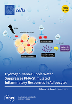
Cover Story (view full-size image): Reactive oxygen species (ROS)-induced oxidative stress in adipose tissue is associated with inflammation and the development of metabolic disorders. In this study, for the first time, we discovered that PMA (an inducer of endogenous ROS) could markedly increase lipid accumulation, the release of pro-inflammatory cytokine IL-6 and intracellular ROS generation in OP9 adipocytes. This finding suggests that ROS can initiate adipogenesis, hypertrophy of adipocytes and inflammatory responses in adipose tissue. Hydrogen nano-bubble water (HW), which contains more than one hundred million nano-sized hydrogen bubbles per mL (the nano-bubbles have excellent stability), could significantly repress PMA-induced ROS generation, lipid accumulation and IL-6 secretion in OP9 adipocytes. HW can be applied as a potential healthcare candidate for preventing metabolic disorders. View this paper
  • Issues are regarded as officially published after their release is announced to the table of contents alert mailing list.
  • You may sign up for e-mail alerts to receive table of contents of newly released issues.
  • PDF is the official format for papers published in both, html and pdf forms. To view the papers in pdf format, click on the "PDF Full-text" link, and use the free Adobe Reader to open them.
Order results
Result details
Section
Select all
Export citation of selected articles as:
21 pages, 5656 KiB  
Article
Deletion of the Actin-Associated Tropomyosin Tpm3 Leads to Reduced Cell Complexity in Cultured Hippocampal Neurons—New Insights into the Role of the C-Terminal Region of Tpm3.1
by Tamara Tomanić, Claire Martin, Holly Stefen, Esmeralda Parić, Peter Gunning and Thomas Fath
Cells 2021, 10(3), 715; https://doi.org/10.3390/cells10030715 - 23 Mar 2021
Cited by 8 | Viewed by 3172
Abstract
Tropomyosins (Tpms) have been described as master regulators of actin, with Tpm3 products shown to be involved in early developmental processes, and the Tpm3 isoform Tpm3.1 controlling changes in the size of neuronal growth cones and neurite growth. Here, we used primary mouse [...] Read more.
Tropomyosins (Tpms) have been described as master regulators of actin, with Tpm3 products shown to be involved in early developmental processes, and the Tpm3 isoform Tpm3.1 controlling changes in the size of neuronal growth cones and neurite growth. Here, we used primary mouse hippocampal neurons of C57/Bl6 wild type and Bl6Tpm3flox transgenic mice to carry out morphometric analyses in response to the absence of Tpm3 products, as well as to investigate the effect of C-terminal truncation on the ability of Tpm3.1 to modulate neuronal morphogenesis. We found that the knock-out of Tpm3 leads to decreased neurite length and complexity, and that the deletion of two amino acid residues at the C-terminus of Tpm3.1 leads to more detrimental changes in neurite morphology than the deletion of six amino acid residues. We also found that Tpm3.1 that lacks the 6 C-terminal amino acid residues does not associate with stress fibres, does not segregate to the tips of neurites, and does not impact the amount of the filamentous actin pool at the axonal growth cones, as opposed to Tpm3.1, which lacks the two C-terminal amino acid residues. Our study provides further insight into the role of both Tpm3 products and the C-terminus of Tpm3.1, and it forms the basis for future studies that aim to identify the molecular mechanisms underlying Tpm3.1 targeting to different subcellular compartments. Full article
(This article belongs to the Special Issue Molecular Mechanisms of Neuronal Actin Cytoskeleton Dynamics)
Show Figures

Figure 1

22 pages, 2109 KiB  
Review
HPV and Other Microbiota; Who’s Good and Who’s Bad: Effects of the Microbial Environment on the Development of Cervical Cancer—A Non-Systematic Review
by Matthias Läsche, Horst Urban, Julia Gallwas and Carsten Gründker
Cells 2021, 10(3), 714; https://doi.org/10.3390/cells10030714 - 23 Mar 2021
Cited by 14 | Viewed by 5665
Abstract
Cervical cancer is responsible for around 5% of all human cancers worldwide. It develops almost exclusively from an unsolved, persistent infection of the squamocolumnar transformation zone between the endo- and ecto-cervix with various high-risk (HR) human papillomaviruses (HPVs). The decisive turning point on [...] Read more.
Cervical cancer is responsible for around 5% of all human cancers worldwide. It develops almost exclusively from an unsolved, persistent infection of the squamocolumnar transformation zone between the endo- and ecto-cervix with various high-risk (HR) human papillomaviruses (HPVs). The decisive turning point on the way to persistent HPV infection and malignant transformation is an immune system weakened by pathobionts and oxidative stress and an injury to the cervical mucosa, often caused by sexual activities. Through these injury and healing processes, HPV viruses, hijacking activated keratinocytes, move into the basal layers of the cervical epithelium and then continue their development towards the distal prickle cell layer (Stratum spinosum). The microbial microenvironment of the cervical tissue determines the tissue homeostasis and the integrity of the protective mucous layer through the maintenance of a healthy immune and metabolic signalling. Pathological microorganisms and the resulting dysbiosis disturb this signalling. Thus, pathological inflammatory reactions occur, which manifest the HPV infection. About 90% of all women contract an HPV infection in the course of their lives. In about 10% of cases, the virus persists and cervical intra-epithelial neoplasia (CIN) develops. Approximately 1% of women with a high-risk HPV infection incur a cervical carcinoma after 10 to 20 years. In this non-systematic review article, we summarise how the sexually and microbial mediated pathogenesis of the cervix proceeds through aberrant immune and metabolism signalling via CIN to cervical carcinoma. We show how both the virus and the cancer benefit from the same changes in the immune and metabolic environment. Full article
(This article belongs to the Special Issue Metabolic Interactions in Tumor Microenvironment (TME))
Show Figures

Figure 1

30 pages, 915 KiB  
Review
Review: Tissue Engineering of Small-Diameter Vascular Grafts and Their In Vivo Evaluation in Large Animals and Humans
by Shu Fang, Ditte Gry Ellman and Ditte Caroline Andersen
Cells 2021, 10(3), 713; https://doi.org/10.3390/cells10030713 - 23 Mar 2021
Cited by 43 | Viewed by 5898
Abstract
To date, a wide range of materials, from synthetic to natural or a mixture of these, has been explored, modified, and examined as small-diameter tissue-engineered vascular grafts (SD-TEVGs) for tissue regeneration either in vitro or in vivo. However, very limited success has been [...] Read more.
To date, a wide range of materials, from synthetic to natural or a mixture of these, has been explored, modified, and examined as small-diameter tissue-engineered vascular grafts (SD-TEVGs) for tissue regeneration either in vitro or in vivo. However, very limited success has been achieved due to mechanical failure, thrombogenicity or intimal hyperplasia, and improvements of the SD-TEVG design are thus required. Here, in vivo studies investigating novel and relative long (10 times of the inner diameter) SD-TEVGs in large animal models and humans are identified and discussed, with emphasis on graft outcome based on model- and graft-related conditions. Only a few types of synthetic polymer-based SD-TEVGs have been evaluated in large-animal models and reflect limited success. However, some polymers, such as polycaprolactone (PCL), show favorable biocompatibility and potential to be further modified and improved in the form of hybrid grafts. Natural polymer- and cell-secreted extracellular matrix (ECM)-based SD-TEVGs tested in large animals still fail due to a weak strength or thrombogenicity. Similarly, native ECM-based SD-TEVGs and in-vitro-developed hybrid SD-TEVGs that contain xenogeneic molecules or matrix seem related to a harmful graft outcome. In contrast, allogeneic native ECM-based SD-TEVGs, in-vitro-developed hybrid SD-TEVGs with allogeneic banked human cells or isolated autologous stem cells, and in-body tissue architecture (IBTA)-based SD-TEVGs seem to be promising for the future, since they are suitable in dimension, mechanical strength, biocompatibility, and availability. Full article
Show Figures

Figure 1

18 pages, 739 KiB  
Review
Contemporary Mouse Models in Glioma Research
by William H. Hicks, Cylaina E. Bird, Jeffrey I. Traylor, Diana D. Shi, Tarek Y. El Ahmadieh, Timothy E. Richardson, Samuel K. McBrayer and Kalil G. Abdullah
Cells 2021, 10(3), 712; https://doi.org/10.3390/cells10030712 - 23 Mar 2021
Cited by 28 | Viewed by 12818
Abstract
Despite advances in understanding of the molecular pathogenesis of glioma, outcomes remain dismal. Developing successful treatments for glioma requires faithful in vivo disease modeling and rigorous preclinical testing. Murine models, including xenograft, syngeneic, and genetically engineered models, are used to study glioma-genesis, identify [...] Read more.
Despite advances in understanding of the molecular pathogenesis of glioma, outcomes remain dismal. Developing successful treatments for glioma requires faithful in vivo disease modeling and rigorous preclinical testing. Murine models, including xenograft, syngeneic, and genetically engineered models, are used to study glioma-genesis, identify methods of tumor progression, and test novel treatment strategies. Since the discovery of highly recurrent isocitrate dehydrogenase (IDH) mutations in lower-grade gliomas, there is increasing emphasis on effective modeling of IDH mutant brain tumors. Improvements in preclinical models that capture the phenotypic and molecular heterogeneity of gliomas are critical for the development of effective new therapies. Herein, we explore the current status, advancements, and challenges with contemporary murine glioma models. Full article
(This article belongs to the Special Issue Molecular Basis of Brain Tumors)
Show Figures

Figure 1

17 pages, 2946 KiB  
Article
Cell Surface Profiling of Retinal Müller Glial Cells Reveals Association to Immune Pathways after LPS Stimulation
by Lea Lorenz, Sieglinde Hirmer, Adrian Schmalen, Stefanie M. Hauck and Cornelia A. Deeg
Cells 2021, 10(3), 711; https://doi.org/10.3390/cells10030711 - 23 Mar 2021
Cited by 15 | Viewed by 3095
Abstract
Retinal Müller glial cells (RMG) are involved in virtually every retinal disease; however, the role of these glial cells in neuroinflammation is still poorly understood. Since cell surface proteins play a decisive role in immune system signaling pathways, this study aimed at characterizing [...] Read more.
Retinal Müller glial cells (RMG) are involved in virtually every retinal disease; however, the role of these glial cells in neuroinflammation is still poorly understood. Since cell surface proteins play a decisive role in immune system signaling pathways, this study aimed at characterizing the changes of the cell surface proteome of RMG after incubation with prototype immune system stimulant lipopolysaccharide (LPS). While mass spectrometric analysis of the human Müller glia cell line MIO-M1 revealed 507 cell surface proteins in total, with 18 proteins significantly more abundant after stimulation (ratio ≥ 2), the surfaceome of primary RMG comprised 1425 proteins, among them 79 proteins with significantly higher abundance in the stimulated state. Pathway analysis revealed notable association with immune system pathways such as “antigen presentation”, “immunoregulatory interactions between a lymphoid and a non-lymphoid cell” and “cell migration”. We could demonstrate a higher abundance of proteins that are usually ascribed to antigen-presenting cells (APCs) and function to interact with T-cells, suggesting that activated RMG might act as atypical APCs in the course of ocular neuroinflammation. Our data provide a detailed description of the unstimulated and stimulated RMG surfaceome and offer fundamental insights regarding the capacity of RMG to actively participate in neuroinflammation in the retina. Full article
Show Figures

Figure 1

19 pages, 788 KiB  
Review
The Inherited and Familial Component of Early-Onset Colorectal Cancer
by Maria Daca Alvarez, Isabel Quintana, Mariona Terradas, Pilar Mur, Francesc Balaguer and Laura Valle
Cells 2021, 10(3), 710; https://doi.org/10.3390/cells10030710 - 23 Mar 2021
Cited by 49 | Viewed by 6328
Abstract
Early-onset colorectal cancer (EOCRC), defined as that diagnosed before the age of 50, accounts for 10–12% of all new colorectal cancer (CRC) diagnoses. Epidemiological data indicate that EOCRC incidence is increasing, despite the observed heterogeneity among countries. Although the cause for such increase [...] Read more.
Early-onset colorectal cancer (EOCRC), defined as that diagnosed before the age of 50, accounts for 10–12% of all new colorectal cancer (CRC) diagnoses. Epidemiological data indicate that EOCRC incidence is increasing, despite the observed heterogeneity among countries. Although the cause for such increase remains obscure, ≈13% (range: 9–26%) of EOCRC patients carry pathogenic germline variants in known cancer predisposition genes, including 2.5% of patients with germline pathogenic variants in hereditary cancer genes traditionally not associated with CRC predisposition. Approximately 28% of EOCRC patients have family history of the disease. This article recapitulates current evidence on the inherited syndromes that predispose to EOCRC and its familial component. The evidence gathered support that all patients diagnosed with an EOCRC should be referred to a specialized genetic counseling service and offered somatic and germline pancancer multigene panel testing. The identification of a germline pathogenic variant in a known hereditary cancer gene has relevant implications for the clinical management of the patient and his/her relatives, and it may guide surgical and therapeutic decisions. The relative high prevalence of hereditary cancer syndromes and familial component among EOCRC patients supports further research that helps understand the genetic background, either monogenic or polygenic, behind this increasingly common disease. Full article
Show Figures

Graphical abstract

17 pages, 3909 KiB  
Review
Axonal Modulation of Striatal Dopamine Release by Local γ-Aminobutyric Acid (GABA) Signalling
by Bradley M. Roberts, Emanuel F. Lopes and Stephanie J. Cragg
Cells 2021, 10(3), 709; https://doi.org/10.3390/cells10030709 - 23 Mar 2021
Cited by 19 | Viewed by 6612
Abstract
Striatal dopamine (DA) release is critical for motivated actions and reinforcement learning, and is locally influenced at the level of DA axons by other striatal neurotransmitters. Here, we review a wealth of historical and more recently refined evidence indicating that DA output is [...] Read more.
Striatal dopamine (DA) release is critical for motivated actions and reinforcement learning, and is locally influenced at the level of DA axons by other striatal neurotransmitters. Here, we review a wealth of historical and more recently refined evidence indicating that DA output is inhibited by striatal γ-aminobutyric acid (GABA) acting via GABAA and GABAB receptors. We review evidence supporting the localisation of GABAA and GABAB receptors to DA axons, as well as the identity of the striatal sources of GABA that likely contribute to GABAergic modulation of DA release. We discuss emerging data outlining the mechanisms through which GABAA and GABAB receptors inhibit the amplitude as well as modulate the short-term plasticity of DA release. Furthermore, we highlight recent data showing that DA release is governed by plasma membrane GABA uptake transporters on striatal astrocytes, which determine ambient striatal GABA tone and, by extension, the tonic inhibition of DA release. Finally, we discuss how the regulation of striatal GABA-DA interactions represents an axis for dysfunction in psychomotor disorders associated with dysregulated DA signalling, including Parkinson’s disease, and could be a novel therapeutic target for drugs to modify striatal DA output. Full article
(This article belongs to the Special Issue Dopamine Signaling: From Synapses to Behavior)
Show Figures

Graphical abstract

21 pages, 1229 KiB  
Review
Aging and Interferons: Impacts on Inflammation and Viral Disease Outcomes
by Emily Feng, Elizabeth Balint, Sophie M. Poznanski, Ali A. Ashkar and Mark Loeb
Cells 2021, 10(3), 708; https://doi.org/10.3390/cells10030708 - 23 Mar 2021
Cited by 37 | Viewed by 7952
Abstract
As highlighted by the COVID-19 global pandemic, elderly individuals comprise the majority of cases of severe viral infection outcomes and death. A combined inability to control viral replication and exacerbated inflammatory immune activation in elderly patients causes irreparable immune-mediated tissue pathology in response [...] Read more.
As highlighted by the COVID-19 global pandemic, elderly individuals comprise the majority of cases of severe viral infection outcomes and death. A combined inability to control viral replication and exacerbated inflammatory immune activation in elderly patients causes irreparable immune-mediated tissue pathology in response to infection. Key to these responses are type I, II, and III interferons (IFNs), which are involved in inducing an antiviral response, as well as controlling and suppressing inflammation and immunopathology. IFNs support monocyte/macrophage-stimulated immune responses that clear infection and promote their immunosuppressive functions that prevent excess inflammation and immune-mediated pathology. The timing and magnitude of IFN responses to infection are critical towards their immunoregulatory functions and ability to prevent immunopathology. Aging is associated with multiple defects in the ability of macrophages and dendritic cells to produce IFNs in response to viral infection, leading to a dysregulation of inflammatory immune responses. Understanding the implications of aging on IFN-regulated inflammation will give critical insights on how to treat and prevent severe infection in vulnerable individuals. In this review, we describe the causes of impaired IFN production in aging, and the evidence to suggest that these impairments impact the regulation of the innate and adaptive immune response to infection, thereby causing disease pathology. Full article
(This article belongs to the Special Issue Macrophages during Inflammation)
Show Figures

Figure 1

18 pages, 2083 KiB  
Review
Immunologically Inert Nanostructures as Selective Therapeutic Tools in Inflammatory Diseases
by Laura Talamini, Eiji Matsuura, Luisa De Cola and Sylviane Muller
Cells 2021, 10(3), 707; https://doi.org/10.3390/cells10030707 - 23 Mar 2021
Cited by 6 | Viewed by 3694
Abstract
The current therapies based on immunosuppressant or new biologic drugs often show some limitations in term of efficacy and applicability, mainly because of their inadequate targeting and of unwanted adverse reactions they generate. To overcome these inherent problems, in the last decades, innovative [...] Read more.
The current therapies based on immunosuppressant or new biologic drugs often show some limitations in term of efficacy and applicability, mainly because of their inadequate targeting and of unwanted adverse reactions they generate. To overcome these inherent problems, in the last decades, innovative nanocarriers have been developed to encapsulate active molecules and offer novel promising strategies to efficiently modulate the immune system. This review provides an overview of how it is possible, exploiting the favorable features of nanocarriers, especially with regard to their immunogenicity, to improve the bioavailability of novel drugs that selectively target immune cells in the context of autoimmune disorders and inflammatory diseases. A focus is made on nanoparticles that selectively target neutrophils in inflammatory pathologies. Full article
(This article belongs to the Special Issue NETs in Infectious and Inflammatory Diseases)
Show Figures

Figure 1

20 pages, 1598 KiB  
Review
The Role of Chloroplast Membrane Lipid Metabolism in Plant Environmental Responses
by Ron Cook, Josselin Lupette and Christoph Benning
Cells 2021, 10(3), 706; https://doi.org/10.3390/cells10030706 - 23 Mar 2021
Cited by 40 | Viewed by 7546
Abstract
Plants are nonmotile life forms that are constantly exposed to changing environmental conditions during the course of their life cycle. Fluctuations in environmental conditions can be drastic during both day–night and seasonal cycles, as well as in the long term as the climate [...] Read more.
Plants are nonmotile life forms that are constantly exposed to changing environmental conditions during the course of their life cycle. Fluctuations in environmental conditions can be drastic during both day–night and seasonal cycles, as well as in the long term as the climate changes. Plants are naturally adapted to face these environmental challenges, and it has become increasingly apparent that membranes and their lipid composition are an important component of this adaptive response. Plants can remodel their membranes to change the abundance of different lipid classes, and they can release fatty acids that give rise to signaling compounds in response to environmental cues. Chloroplasts harbor the photosynthetic apparatus of plants embedded into one of the most extensive membrane systems found in nature. In part one of this review, we focus on changes in chloroplast membrane lipid class composition in response to environmental changes, and in part two, we will detail chloroplast lipid-derived signals. Full article
Show Figures

Graphical abstract

15 pages, 3953 KiB  
Review
Energy Metabolism in IDH1 Wild-Type and IDH1-Mutated Glioblastoma Stem Cells: A Novel Target for Therapy?
by Cornelis J.F. van Noorden, Vashendriya V.V. Hira, Amber J. van Dijck, Metka Novak, Barbara Breznik and Remco J. Molenaar
Cells 2021, 10(3), 705; https://doi.org/10.3390/cells10030705 - 22 Mar 2021
Cited by 19 | Viewed by 5350
Abstract
Cancer is a redox disease. Low levels of reactive oxygen species (ROS) are beneficial for cells and have anti-cancer effects. ROS are produced in the mitochondria during ATP production by oxidative phosphorylation (OXPHOS). In the present review, we describe ATP production in primary [...] Read more.
Cancer is a redox disease. Low levels of reactive oxygen species (ROS) are beneficial for cells and have anti-cancer effects. ROS are produced in the mitochondria during ATP production by oxidative phosphorylation (OXPHOS). In the present review, we describe ATP production in primary brain tumors, glioblastoma, in relation to ROS production. Differentiated glioblastoma cells mainly use glycolysis for ATP production (aerobic glycolysis) without ROS production, whereas glioblastoma stem cells (GSCs) in hypoxic periarteriolar niches use OXPHOS for ATP and ROS production, which is modest because of the hypoxia and quiescence of GSCs. In a significant proportion of glioblastoma, isocitrate dehydrogenase 1 (IDH1) is mutated, causing metabolic rewiring, and all cancer cells use OXPHOS for ATP and ROS production. Systemic therapeutic inhibition of glycolysis is not an option as clinical trials have shown ineffectiveness or unwanted side effects. We argue that systemic therapeutic inhibition of OXPHOS is not an option either because the anti-cancer effects of ROS production in healthy cells is inhibited as well. Therefore, we advocate to remove GSCs out of their hypoxic niches by the inhibition of their binding to niches to enable their differentiation and thus increase their sensitivity to radiotherapy and/or chemotherapy. Full article
(This article belongs to the Special Issue Molecular and Cellular Mechanisms of Cancers: Glioblastoma)
Show Figures

Figure 1

18 pages, 1935 KiB  
Review
Molecular Genetic Features of Cerebral Cavernous Malformations (CCM) Patients: An Overall View from Genes to Endothelial Cells
by Giulia Riolo, Claudia Ricci and Stefania Battistini
Cells 2021, 10(3), 704; https://doi.org/10.3390/cells10030704 - 22 Mar 2021
Cited by 22 | Viewed by 5595
Abstract
Cerebral cavernous malformations (CCMs) are vascular lesions that affect predominantly microvasculature in the brain and spinal cord. CCM can occur either in sporadic or familial form, characterized by autosomal dominant inheritance and development of multiple lesions throughout the patient’s life. Three genes associated [...] Read more.
Cerebral cavernous malformations (CCMs) are vascular lesions that affect predominantly microvasculature in the brain and spinal cord. CCM can occur either in sporadic or familial form, characterized by autosomal dominant inheritance and development of multiple lesions throughout the patient’s life. Three genes associated with CCM are known: CCM1/KRIT1 (krev interaction trapped 1), CCM2/MGC4607 (encoding a protein named malcavernin), and CCM3/PDCD10 (programmed cell death 10). All the mutations identified in these genes cause a loss of function and compromise the protein functions needed for maintaining the vascular barrier integrity. Loss of function of CCM proteins causes molecular disorganization and dysfunction of endothelial adherens junctions. In this review, we provide an overall vision of the CCM pathology, starting with the genetic bases of the disease, describing the role of the proteins, until we reach the cellular level. Thus, we summarize the genetics of CCM, providing a description of CCM genes and mutation features, provided an updated knowledge of the CCM protein structure and function, and discuss the molecular mechanisms through which CCM proteins may act within endothelial cells, particularly in endothelial barrier maintenance/regulation and in cellular signaling. Full article
Show Figures

Figure 1

17 pages, 29587 KiB  
Article
Oxidative Stress Conditions Result in Trapping of PHF-Core Tau (297–391) Intermediates
by Mahmoud B. Maina, Youssra K. Al-Hilaly, Gunasekhar Burra, Janet E. Rickard, Charles R. Harrington, Claude M. Wischik and Louise C. Serpell
Cells 2021, 10(3), 703; https://doi.org/10.3390/cells10030703 - 22 Mar 2021
Cited by 9 | Viewed by 4464
Abstract
The self-assembly of tau into paired helical filaments (PHFs) in neurofibrillary tangles (NFTs) is a significant event in Alzheimer’s disease (AD) pathogenesis. Numerous post-translational modifications enhance or inhibit tau assembly into NFTs. Oxidative stress, which accompanies AD, induces multiple post-translational modifications in proteins, [...] Read more.
The self-assembly of tau into paired helical filaments (PHFs) in neurofibrillary tangles (NFTs) is a significant event in Alzheimer’s disease (AD) pathogenesis. Numerous post-translational modifications enhance or inhibit tau assembly into NFTs. Oxidative stress, which accompanies AD, induces multiple post-translational modifications in proteins, including the formation of dityrosine (DiY) cross-links. Previous studies have revealed that metal-catalysed oxidation (MCO) using Cu2+ and H2O2 leads to the formation of DiY cross-links in two misfolding proteins, Aβ and α-synuclein, associated with AD and Parkinson’s disease respectively. The effect of MCO on tau remains unknown. Here, we examined the effect of MCO and ultra-violet oxidation to study the influence of DiY cross-linking on the self-assembly of the PHF-core tau fragment. We report that DiY cross-linking facilitates tau assembly into tau oligomers that fail to bind thioflavin S, lack β-sheet structure and prevents their elongation into filaments. At a higher concentration, Cu2+ (without H2O2) also facilitates the formation of these tau oligomers. The DiY cross-linked tau oligomers do not cause cell death. Our findings suggest that DiY cross-linking of pre-assembled tau promotes the formation of soluble tau oligomers that show no acute impact on cell viability. Full article
(This article belongs to the Collection New Insights into the Molecular Mechanisms of Neurodegeneration)
Show Figures

Figure 1

16 pages, 1219 KiB  
Review
Fenamates as Potential Therapeutics for Neurodegenerative Disorders
by Jaunetta Hill and Nasser H. Zawia
Cells 2021, 10(3), 702; https://doi.org/10.3390/cells10030702 - 22 Mar 2021
Cited by 17 | Viewed by 4806
Abstract
Neurodegenerative disorders are desperately lacking treatment options. It is imperative that drug repurposing be considered in the fight against neurodegenerative diseases. Fenamates have been studied for efficacy in treating several neurodegenerative diseases. The purpose of this review is to comprehensively present the past [...] Read more.
Neurodegenerative disorders are desperately lacking treatment options. It is imperative that drug repurposing be considered in the fight against neurodegenerative diseases. Fenamates have been studied for efficacy in treating several neurodegenerative diseases. The purpose of this review is to comprehensively present the past and current research on fenamates in the context of neurodegenerative diseases with a special emphasis on tolfenamic acid and Alzheimer’s disease. Furthermore, this review discusses the major molecular pathways modulated by fenamates. Full article
(This article belongs to the Collection Advances in Neurodegenerative Disease)
Show Figures

Graphical abstract

29 pages, 2107 KiB  
Review
Characterization of Apis mellifera Gastrointestinal Microbiota and Lactic Acid Bacteria for Honeybee Protection—A Review
by Adriana Nowak, Daria Szczuka, Anna Górczyńska, Ilona Motyl and Dorota Kręgiel
Cells 2021, 10(3), 701; https://doi.org/10.3390/cells10030701 - 22 Mar 2021
Cited by 64 | Viewed by 12272
Abstract
Numerous honeybee (Apis mellifera) products, such as honey, propolis, and bee venom, are used in traditional medicine to prevent illness and promote healing. Therefore, this insect has a huge impact on humans’ way of life and the environment. While the population [...] Read more.
Numerous honeybee (Apis mellifera) products, such as honey, propolis, and bee venom, are used in traditional medicine to prevent illness and promote healing. Therefore, this insect has a huge impact on humans’ way of life and the environment. While the population of A. mellifera is large, there is concern that widespread commercialization of beekeeping, combined with environmental pollution and the action of bee pathogens, has caused significant problems for the health of honeybee populations. One of the strategies to preserve the welfare of honeybees is to better understand and protect their natural microbiota. This paper provides a unique overview of the latest research on the features and functioning of A. mellifera. Honeybee microbiome analysis focuses on both the function and numerous factors affecting it. In addition, we present the characteristics of lactic acid bacteria (LAB) as an important part of the gut community and their special beneficial activities for honeybee health. The idea of probiotics for honeybees as a promising tool to improve their health is widely discussed. Knowledge of the natural gut microbiota provides an opportunity to create a broad strategy for honeybee vitality, including the development of modern probiotic preparations to use instead of conventional antibiotics, environmentally friendly biocides, and biological control agents. Full article
Show Figures

Graphical abstract

14 pages, 2740 KiB  
Article
Direct Stimulatory Effects of the CB2 Ligand JTE 907 in Human and Mouse Islets
by Inmaculada Ruz-Maldonado, Patricio Atanes, Guo Cai Huang, Bo Liu and Shanta J Persaud
Cells 2021, 10(3), 700; https://doi.org/10.3390/cells10030700 - 22 Mar 2021
Cited by 2 | Viewed by 2913
Abstract
Aims: The endocannabinoid system is a complex cell-signaling network through which endogenous cannabinoid ligands regulate cell function by interaction with CB1 and CB2 cannabinoid receptors, and with the novel cannabinoid receptor GPR55. CB1, CB2, and GPR55 are [...] Read more.
Aims: The endocannabinoid system is a complex cell-signaling network through which endogenous cannabinoid ligands regulate cell function by interaction with CB1 and CB2 cannabinoid receptors, and with the novel cannabinoid receptor GPR55. CB1, CB2, and GPR55 are expressed by islet β-cells where they modulate insulin secretion. We have previously shown that administration of the putative CB2 antagonist/inverse agonist JTE 907 to human islets did not affect the insulinotropic actions of CB2 agonists and it unexpectedly stimulated insulin secretion on its own. In this study, we evaluated whether the lack of antagonism could be related to the ability of JTE 907 to act as a GPR55 agonist. Materials and Methods: We used islets isolated from human donors and from Gpr55+/+ and Gpr55−/− mice and quantified the effects of incubation with 10 μM JTE 907 on dynamic insulin secretion, apoptosis, and β-cell proliferation by radioimmunoassay, luminescence caspase 3/7 activity, and immunofluorescence, respectively. We also measured islet IP1 and cAMP accumulation using fluorescence assays, and monitored [Ca2+]i elevations by Fura-2 single cell microfluorometry. Results: JTE 907 significantly stimulated insulin secretion from islets isolated from human donors and islets from Gpr55+/+ and Gpr55−/− mice. These stimulatory effects were accompanied by significant elevations of IP1 and [Ca2+]i, but there were no changes in cAMP generation. JTE 907 also significantly reduced cytokine-induced apoptosis in human and mouse islets and promoted human β-cell proliferation. Conclusion: Our observations show for the first time that JTE 907 acts as a Gq-coupled agonist in islets to stimulate insulin secretion and maintain β-cell mass in a GPR55-independent fashion. Full article
(This article belongs to the Special Issue Signal Transduction in the Islets of Langerhans)
Show Figures

Figure 1

17 pages, 2707 KiB  
Article
Phytoremediation of CYN, MC-LR and ANTX-a from Water by the Submerged Macrophyte Lemna trisulca
by Małgorzata Kucała, Michał Saładyga and Ariel Kaminski
Cells 2021, 10(3), 699; https://doi.org/10.3390/cells10030699 - 21 Mar 2021
Cited by 8 | Viewed by 2776
Abstract
Cyanotoxins are harmful to aquatic and water-related organisms. In this study, Lemna trisulca was tested as a phytoremediation agent for three common cyanotoxins produced by bloom-forming cyanobacteria. Cocultivation of L. trisulca with Dolichospermum flos-aquae in BG11 medium caused a release of the intracellular [...] Read more.
Cyanotoxins are harmful to aquatic and water-related organisms. In this study, Lemna trisulca was tested as a phytoremediation agent for three common cyanotoxins produced by bloom-forming cyanobacteria. Cocultivation of L. trisulca with Dolichospermum flos-aquae in BG11 medium caused a release of the intracellular pool of anatoxin-a into the medium and the adsorption of 92% of the toxin by the plant—after 14 days, the total amount of toxin decreased 3.17 times. Cocultivation with Raphidopsis raciborskii caused a 2.77-time reduction in the concentration of cylindrospermopsin (CYN) in comparison to the control (62% of the total pool of CYN was associated with the plant). The greatest toxin limitation was noted for cocultivation with Microcystis aeruginosa. After two weeks, the microcystin-LR (MC-LR) concentration decreased more than 310 times. The macrophyte also influenced the growth and development of cyanobacteria cells. Overall, 14 days of cocultivation reduced the biomass of D. flos-aquae, M. aeruginosa, and R. raciborskii by 8, 12, and 3 times, and chlorophyll a concentration in comparison to the control decreased by 17.5, 4.3, and 32.6 times, respectively. Additionally, the macrophyte stabilized the electrical conductivity (EC) and pH values of the water and affected the even uptake of cations and anions from the medium. The obtained results indicate the biotechnological potential of L. trisulca for limiting the development of harmful cyanobacterial blooms and their toxicity. Full article
(This article belongs to the Section Plant, Algae and Fungi Cell Biology)
Show Figures

Figure 1

23 pages, 8213 KiB  
Article
Microglial Cell Morphology and Phagocytic Activity Are Critically Regulated by the Neurosteroid Allopregnanolone: A Possible Role in Neuroprotection
by Valérie Jolivel, Susana Brun, Fabien Binamé, Jérémie Benyounes, Omar Taleb, Dominique Bagnard, Jérôme De Sèze, Christine Patte-Mensah and Ayikoe-Guy Mensah-Nyagan
Cells 2021, 10(3), 698; https://doi.org/10.3390/cells10030698 - 21 Mar 2021
Cited by 28 | Viewed by 3695
Abstract
Microglial cells are key players in neural pathogenesis and microglial function regulation appears to be pivotal in controlling neuroinflammatory/neurological diseases. Here, we investigated the effects and mechanism of action of neurosteroid allopregnanolone (ALLO) on murine microglial BV-2 cells and primary microglia in order [...] Read more.
Microglial cells are key players in neural pathogenesis and microglial function regulation appears to be pivotal in controlling neuroinflammatory/neurological diseases. Here, we investigated the effects and mechanism of action of neurosteroid allopregnanolone (ALLO) on murine microglial BV-2 cells and primary microglia in order to determine ALLO-induced immunomodulatory potential and to provide new insights for the development of both natural and safe neuroprotective strategies targeting microglia. Indeed, ALLO-treatment is increasingly suggested as beneficial in various models of neurological disorders but the underlying mechanisms have not been elucidated. Therefore, the microglial cells were cultured with various serum concentrations to mimic the blood-brain-barrier rupture and to induce their activation. Proliferation, viability, RT-qPCR, phagocytosis, and morphology analyzes, as well as migration with time-lapse imaging and quantitative morphodynamic methods, were combined to investigate ALLO actions on microglia. BV-2 cells express subunits of GABA-A receptor that mediates ALLO activity. ALLO (10µM) induced microglial cell process extension and decreased migratory capacity. Interestingly, ALLO modulated the phagocytic activity of BV-2 cells and primary microglia. Our results, which show a direct effect of ALLO on microglial morphology and phagocytic function, suggest that the natural neurosteroid-based approach may contribute to developing effective strategies against neurological disorders that are evoked by microglia-related abnormalities. Full article
(This article belongs to the Section Intracellular and Plasma Membranes)
Show Figures

Figure 1

16 pages, 3654 KiB  
Article
Mast Cell Mediated Regulation of Small Intestinal Chloride Malabsorption in SAMP1/YitFc Mouse Model of Spontaneous Chronic Ileitis
by M Motiur Rahman, Sheuli Afroz, Subha Arthur and Uma Sundaram
Cells 2021, 10(3), 697; https://doi.org/10.3390/cells10030697 - 21 Mar 2021
Cited by 4 | Viewed by 3562
Abstract
In Inflammatory Bowel Disease (IBD), malabsorption of electrolytes (NaCl) results in diarrhea. Inhibition of coupled NaCl absorption, mediated by the dual operation of Na:H and Cl:HCO3 exchangers on the brush border membrane (BBM) of the intestinal villus cells has been reported in [...] Read more.
In Inflammatory Bowel Disease (IBD), malabsorption of electrolytes (NaCl) results in diarrhea. Inhibition of coupled NaCl absorption, mediated by the dual operation of Na:H and Cl:HCO3 exchangers on the brush border membrane (BBM) of the intestinal villus cells has been reported in IBD. In the SAMP1/YitFcs (SAMP1) mice model of spontaneous ileitis, representing Crohn’s disease, DRA (Downregulated in Adenoma) mediated Cl:HCO3 exchange was shown to be inhibited secondary to diminished affinity of the exchanger for Cl. However, NHE3 mediated Na:H exchange remained unaffected. Mast cells and their secreted mediators are known to be increased in the IBD mucosa and can affect intestinal electrolyte absorption. However, how mast cell mediators may regulate Cl:HCO3 exchange in SAMP1 mice is unknown. Therefore, the aim of this study was to determine the effect of mast cell mediators on the downregulation of DRA in SAMP1 mice. Mast cell numbers and their degranulation marker enzyme (β-hexosaminidase) levels were significantly increased in SAMP1 mice compared to control AKR mice. However, treatment of SAMP1 mice with a mast cell stabilizer, ketotifen, restored the β-hexosaminidase enzyme levels to normal in the intestine, demonstrating stabilization of mast cells by ketotifen. Moreover, downregulation of Cl:HCO3 exchange activity was restored in ketotifen treated SAMP1 mice. Kinetic studies showed that ketotifen restored the altered affinity of Cl:HCO3 exchange in SAMP1 mice villus cells thus reinstating its activity to normal. Further, RT-qPCR, Western blot and immunofluorescence studies showed that the expression levels of DRA mRNA and BBM protein, respectively remained unaltered in all experimental conditions, supporting the kinetic data. Thus, inhibition of Cl:HCO3 exchange resulting in chloride malabsorption leading to diarrhea in IBD is likely mediated by mast cell mediators. Full article
(This article belongs to the Collection Mast Cells in Health and Diseases)
Show Figures

Figure 1

21 pages, 5166 KiB  
Article
Rapid and Progressive Loss of Multiple Retinal Cell Types in Cathepsin D-Deficient Mice—An Animal Model of CLN10 Disease
by Mahmoud Bassal, Junling Liu, Wanda Jankowiak, Paul Saftig and Udo Bartsch
Cells 2021, 10(3), 696; https://doi.org/10.3390/cells10030696 - 21 Mar 2021
Cited by 11 | Viewed by 3833
Abstract
Vision loss is among the characteristic symptoms of neuronal ceroid lipofuscinosis (NCL), a fatal neurodegenerative lysosomal storage disorder. Here, we performed an in-depth analysis of retinal degeneration at the molecular and cellular levels in mice lacking the lysosomal aspartyl protease cathepsin D, an [...] Read more.
Vision loss is among the characteristic symptoms of neuronal ceroid lipofuscinosis (NCL), a fatal neurodegenerative lysosomal storage disorder. Here, we performed an in-depth analysis of retinal degeneration at the molecular and cellular levels in mice lacking the lysosomal aspartyl protease cathepsin D, an animal model of congenital CLN10 disease. We observed an early-onset accumulation of storage material as indicated by elevated levels of saposin D and subunit C of the mitochondrial ATP synthase. The accumulation of storage material was accompanied by reactive astrogliosis and microgliosis, elevated expression of the autophagy marker sequestosome 1/p62 and a dysregulated expression of several lysosomal proteins. The number of cone photoreceptor cells was reduced as early as at postnatal day 5. At the end stage of the disease, the outer nuclear layer was almost atrophied, and all cones were lost. A significant loss of rod and cone bipolar cells, amacrine cells and ganglion cells was found at advanced stages of the disease. Results demonstrate that cathepsin D deficiency results in an early-onset and rapidly progressing retinal dystrophy that involves all retinal cell types. Data of the present study will serve as a reference for studies aimed at developing treatments for retinal degeneration in CLN10 disease. Full article
(This article belongs to the Collection Animal Models of Retinal Degeneration)
Show Figures

Figure 1

11 pages, 976 KiB  
Article
Endometriosis Is Associated with Functional Polymorphism in the Promoter of Heme Oxygenase 1 (HMOX1) Gene
by Łukasz Milewski, Aneta Ścieżyńska, Joanna Ponińska, Marta Soszyńska, Ewa Barcz, Piotr I. Roszkowski, Paweł Kamiński, Paweł Włodarski, Rafał Płoski and Jacek Malejczyk
Cells 2021, 10(3), 695; https://doi.org/10.3390/cells10030695 - 21 Mar 2021
Cited by 12 | Viewed by 2782
Abstract
Endometriosis is a common gynecological disorder characterized by the ectopic growth of endometrial-like tissue outside the uterine cavity. Etiopathogenesis of endometriosis is poorly understood; it is plausible, however, that the disease may be associated with oxidative stress related to local heme and iron [...] Read more.
Endometriosis is a common gynecological disorder characterized by the ectopic growth of endometrial-like tissue outside the uterine cavity. Etiopathogenesis of endometriosis is poorly understood; it is plausible, however, that the disease may be associated with oxidative stress related to local heme and iron metabolism. Therefore, the aim of the study was to reveal a possible association of endometriosis with a stress-inducible heme oxygenase 1 (HMOX1). For this purpose, 228 patients with clinically confirmed endometriosis and 415 control parous women from general Polish population were examined for functional –413A>T (rs2071746) single-nucleotide polymorphism (SNP) and (GT)n dinucleotide repeat length polymorphism in the promoter of HMOX1 gene. In addition, –413A>T SNP was assessed by the specific TaqMan® SNP Genotyping Assay, and (GT)n polymorphism was determined by PCR product size analysis. We found that endometriosis is associated with an increased frequency of −413A(GT)31,32 haplotype (OR (95%CI) = 1.27 (1.01–1.60), p = 0.0381) and −413A(GT)31,32 homozygous genotype [OR (95%CI) = 1.51 (1.06–2.17), p = 0.0238]. These data suggest that endometriosis is associated with functional polymorphism of HMOX1 gene, and this gene may play a part in the pathogenesis of this disorder. Full article
(This article belongs to the Special Issue Molecular and Cellular Aspects of Endometriosis)
Show Figures

Figure 1

26 pages, 1085 KiB  
Review
Molecular Basis of Neuronal Autophagy in Ageing: Insights from Caenorhabditis elegans
by Georgios Konstantinidis and Nektarios Tavernarakis
Cells 2021, 10(3), 694; https://doi.org/10.3390/cells10030694 - 21 Mar 2021
Cited by 12 | Viewed by 6160
Abstract
Autophagy is an evolutionarily conserved degradation process maintaining cell homeostasis. Induction of autophagy is triggered as a response to a broad range of cellular stress conditions, such as nutrient deprivation, protein aggregation, organelle damage and pathogen invasion. Macroautophagy involves the sequestration of cytoplasmic [...] Read more.
Autophagy is an evolutionarily conserved degradation process maintaining cell homeostasis. Induction of autophagy is triggered as a response to a broad range of cellular stress conditions, such as nutrient deprivation, protein aggregation, organelle damage and pathogen invasion. Macroautophagy involves the sequestration of cytoplasmic contents in a double-membrane organelle referred to as the autophagosome with subsequent degradation of its contents upon delivery to lysosomes. Autophagy plays critical roles in development, maintenance and survival of distinct cell populations including neurons. Consequently, age-dependent decline in autophagy predisposes animals for age-related diseases including neurodegeneration and compromises healthspan and longevity. In this review, we summarize recent advances in our understanding of the role of neuronal autophagy in ageing, focusing on studies in the nematode Caenorhabditis elegans. Full article
(This article belongs to the Special Issue Autophagy Meets Aging)
Show Figures

Figure 1

18 pages, 2789 KiB  
Article
Voluntary Wheel Running Did Not Alter Gene Expression in 5xfad Mice, but in Wild-Type Animals Exclusively after One-Day of Physical Activity
by Anna Wierczeiko, Lena Gammel, Konstantin Radyushkin, Vu Thu Thuy Nguyen, Hristo Todorov, Susanne Gerber and Kristina Endres
Cells 2021, 10(3), 693; https://doi.org/10.3390/cells10030693 - 20 Mar 2021
Cited by 3 | Viewed by 3866
Abstract
Physical activity is considered a promising preventive intervention to reduce the risk of developing Alzheimer’s disease (AD). However, the positive effect of therapeutic administration of physical activity has not been proven conclusively yet, likely due to confounding factors such as varying activity regimens [...] Read more.
Physical activity is considered a promising preventive intervention to reduce the risk of developing Alzheimer’s disease (AD). However, the positive effect of therapeutic administration of physical activity has not been proven conclusively yet, likely due to confounding factors such as varying activity regimens and life or disease stages. To examine the impact of different routines of physical activity in the early disease stages, we subjected young 5xFAD and wild-type mice to 1-day (acute) and 30-day (chronic) voluntary wheel running and compared them with age-matched sedentary controls. We observed a significant increase in brain lactate levels in acutely trained 5xFAD mice relative to all other experimental groups. Subsequent brain RNA-seq analysis did not reveal major differences in transcriptomic regulation between training durations in 5xFAD mice. In contrast, acute training yielded substantial gene expression changes in wild-type animals relative to their chronically trained and sedentary counterparts. The comparison of 5xFAD and wild-type mice showed the highest transcriptional differences in the chronic and sedentary groups, whereas acute training was associated with much fewer differentially expressed genes. In conclusion, our results suggest that different training durations did not affect the global transcriptome of 3-month-old 5xFAD mice, whereas acute running seemed to induce a similar transcriptional stress state in wild-type animals as already known for 5xFAD mice. Full article
(This article belongs to the Collection Advances in Neurodegenerative Disease)
Show Figures

Figure 1

15 pages, 2532 KiB  
Article
CALINCA—A Novel Pipeline for the Identification of lncRNAs in Podocyte Disease
by Sweta Talyan, Samantha Filipów, Michael Ignarski, Magdalena Smieszek, He Chen, Lucas Kühne, Linus Butt, Heike Göbel, K. Johanna R. Hoyer-Allo, Felix C. Koehler, Janine Altmüller, Paul Brinkkötter, Bernhard Schermer, Thomas Benzing, Martin Kann, Roman-Ulrich Müller and Christoph Dieterich
Cells 2021, 10(3), 692; https://doi.org/10.3390/cells10030692 - 20 Mar 2021
Cited by 3 | Viewed by 4304
Abstract
Diseases of the renal filtration unit—the glomerulus—are the most common cause of chronic kidney disease. Podocytes are the pivotal cell type for the function of this filter and focal-segmental glomerulosclerosis (FSGS) is a classic example of a podocytopathy leading to proteinuria and glomerular [...] Read more.
Diseases of the renal filtration unit—the glomerulus—are the most common cause of chronic kidney disease. Podocytes are the pivotal cell type for the function of this filter and focal-segmental glomerulosclerosis (FSGS) is a classic example of a podocytopathy leading to proteinuria and glomerular scarring. Currently, no targeted treatment of FSGS is available. This lack of therapeutic strategies is explained by a limited understanding of the defects in podocyte cell biology leading to FSGS. To date, most studies in the field have focused on protein-coding genes and their gene products. However, more than 80% of all transcripts produced by mammalian cells are actually non-coding. Here, long non-coding RNAs (lncRNAs) are a relatively novel class of transcripts and have not been systematically studied in FSGS to date. The appropriate tools to facilitate lncRNA research for the renal scientific community are urgently required due to a row of challenges compared to classical analysis pipelines optimized for coding RNA expression analysis. Here, we present the bioinformatic pipeline CALINCA as a solution for this problem. CALINCA automatically analyzes datasets from murine FSGS models and quantifies both annotated and de novo assembled lncRNAs. In addition, the tool provides in-depth information on podocyte specificity of these lncRNAs, as well as evolutionary conservation and expression in human datasets making this pipeline a crucial basis to lncRNA studies in FSGS. Full article
(This article belongs to the Special Issue Long Noncoding RNAs in Disease)
Show Figures

Figure 1

13 pages, 1485 KiB  
Article
A Molecular Networking Strategy: High-Throughput Screening and Chemical Analysis of Brazilian Cerrado Plant Extracts against Cancer Cells
by Patrícia C. Cortelo, Daniel P. Demarque, Renata G. Dusi, Lorena C. Albernaz, Raimundo Braz-Filho, Ekaterina I. Goncharova, Heidi R. Bokesch, Kirk R. Gustafson, John A. Beutler and Laila S. Espindola
Cells 2021, 10(3), 691; https://doi.org/10.3390/cells10030691 - 20 Mar 2021
Cited by 17 | Viewed by 4064
Abstract
Plants have historically been a rich source of successful anticancer drugs and chemotherapeutic agents, with research indicating that this trend will continue. In this contribution, we performed high-throughput cytotoxicity screening of 702 extracts from 95 plant species, representing 40 families of the Brazilian [...] Read more.
Plants have historically been a rich source of successful anticancer drugs and chemotherapeutic agents, with research indicating that this trend will continue. In this contribution, we performed high-throughput cytotoxicity screening of 702 extracts from 95 plant species, representing 40 families of the Brazilian Cerrado biome. Activity was investigated against the following cancer cell lines: colon (Colo205 and Km12), renal (A498 and U031), liver (HEP3B and SKHEP), and osteosarcoma (MG63 and MG63.3). Dose-response tests were conducted with 44 of the most active extracts, with 22 demonstrating IC50 values ranging from <1.3 to 20 µg/mL. A molecular networking strategy was formulated using the Global Natural Product Social Molecular Networking (GNPS) platform to visualize, analyze, and annotate the compounds present in 17 extracts active against NCI-60 cell lines. Significant cytotoxic activity was found for Salacia crassifolia, Salacia elliptica, Simarouba versicolor, Diospyros hispida, Schinus terebinthifolia, Casearia sylvestris var. lingua, Magonia pubescens, and Rapanea guianensis. Molecular networking resulted in the annotation of 27 compounds. This strategy provided an initial overview of a complex and diverse natural product data set, yielded a large amount of chemical information, identified patterns and known compounds, and assisted in defining priorities for further studies. Full article
Show Figures

Figure 1

16 pages, 6034 KiB  
Article
P-Glycoprotein Inhibitor Tariquidar Plays an Important Regulatory Role in Pigmentation in Larval Zebrafish
by Natalia Kasica, Piotr Jakubowski and Jerzy Kaleczyc
Cells 2021, 10(3), 690; https://doi.org/10.3390/cells10030690 - 20 Mar 2021
Cited by 6 | Viewed by 2876
Abstract
Zebrafish has emerged as a powerful model in studies dealing with pigment development and pathobiology of pigment diseases. Due to its conserved pigment pattern with established genetic background, the zebrafish is used for screening of active compounds influencing melanophore, iridophore, and xanthophore development [...] Read more.
Zebrafish has emerged as a powerful model in studies dealing with pigment development and pathobiology of pigment diseases. Due to its conserved pigment pattern with established genetic background, the zebrafish is used for screening of active compounds influencing melanophore, iridophore, and xanthophore development and differentiation. In our study, zebrafish embryos and larvae were used to investigate the influence of third-generation noncompetitive P-glycoprotein inhibitor, tariquidar (TQR), on pigmentation, including phenotype effects and changes in gene expression of chosen chromatophore differentiation markers. Five-day exposure to increasing TQR concentrations (1 µM, 10 µM, and 50 µM) resulted in a dose-dependent augmentation of the area covered with melanophores but a reduction in the area covered by iridophores. The observations were performed in three distinct regions—the eye, dorsal head, and tail. Moreover, TQR enhanced melanophore renewal after depigmentation caused by 0.2 mM 1-phenyl-2-thiourea (PTU) treatment. qPCR analysis performed in 56-h post-fertilization (hpf) embryos demonstrated differential expression patterns of genes related to pigment development and differentiation. The most substantial findings include those indicating that TQR had no significant influence on leukocyte tyrosine kinase, GTP cyclohydrolase 2, tyrosinase-related protein 1, and forkhead box D3, however, markedly upregulated tyrosinase, dopachrome tautomerase and melanocyte inducing transcription factor, and downregulated purine nucleoside phosphorylase 4a. The present study suggests that TQR is an agent with multidirectional properties toward pigment cell formation and distribution in the zebrafish larvae and therefore points to the involvement of P-glycoprotein in this process. Full article
(This article belongs to the Special Issue Fishing for Health: Zebrafish Models of Human Disease)
Show Figures

Figure 1

19 pages, 1660 KiB  
Review
Analysis of Nucleosides and Nucleotides in Plants: An Update on Sample Preparation and LC–MS Techniques
by Henryk Straube, Claus-Peter Witte and Marco Herde
Cells 2021, 10(3), 689; https://doi.org/10.3390/cells10030689 - 20 Mar 2021
Cited by 16 | Viewed by 6244
Abstract
Nucleotides fulfill many essential functions in plants. Compared to non-plant systems, these hydrophilic metabolites have not been adequately investigated in plants, especially the less abundant nucleotide species such as deoxyribonucleotides and modified or damaged nucleotides. Until recently, this was mainly due to a [...] Read more.
Nucleotides fulfill many essential functions in plants. Compared to non-plant systems, these hydrophilic metabolites have not been adequately investigated in plants, especially the less abundant nucleotide species such as deoxyribonucleotides and modified or damaged nucleotides. Until recently, this was mainly due to a lack of adequate methods for in-depth analysis of nucleotides and nucleosides in plants. In this review, we focus on the current state-of-the-art of nucleotide analysis in plants with liquid chromatography coupled to mass spectrometry and describe recent major advances. Tissue disruption, quenching, liquid–liquid and solid-phase extraction, chromatographic strategies, and peculiarities of nucleotides and nucleosides in mass spectrometry are covered. We describe how the different steps of the analytical workflow influence each other, highlight the specific challenges of nucleotide analysis, and outline promising future developments. The metabolite matrix of plants is particularly complex. Therefore, it is likely that nucleotide analysis methods that work for plants can be applied to other organisms as well. Although this review focuses on plants, we also discuss advances in nucleotide analysis from non-plant systems to provide an overview of the analytical techniques available for this challenging class of metabolites. Full article
(This article belongs to the Special Issue Metabolomics in Plant Research)
Show Figures

Graphical abstract

13 pages, 2747 KiB  
Article
miRNAs Potentially Involved in Post Lung Transplant-Obliterative Bronchiolitis: The Role of miR-21-5p
by Sara Bozzini, Laura Pandolfi, Elena Rossi, Simona Inghilleri, Michele Zorzetto, Giuseppina Ferrario, Stefano Di Carlo, Gianfranco Politano, Annalisa De Silvestri, Vanessa Frangipane, Michele Porzio, Romain Kessler, Fiorella Calabrese, Federica Meloni and Patrizia Morbini
Cells 2021, 10(3), 688; https://doi.org/10.3390/cells10030688 - 20 Mar 2021
Cited by 4 | Viewed by 2522
Abstract
Epigenetic changes, including miRNAs deregulation, have been suggested to play a significant role in development of obliterative bronchiolitis (OB) in transplanted lungs. Many studies have tried to identify ideal candidate miRNAs and the downstream pathways implicated in the bronchiolar fibro-obliterative process. Several candidate [...] Read more.
Epigenetic changes, including miRNAs deregulation, have been suggested to play a significant role in development of obliterative bronchiolitis (OB) in transplanted lungs. Many studies have tried to identify ideal candidate miRNAs and the downstream pathways implicated in the bronchiolar fibro-obliterative process. Several candidate miRNAs, previously indicated as possibly being associated with OB, were analyzed by combining the quantitative real time-polymerase chain reaction (qRT-PCR) and in situ hybridization (ISH) of lung tissues of OB affected patients. Disease and OB-lesion-specific expression of miR-21-5p was confirmed and by computational analysis we were able to identify the network of genes most probably associated miR-21-5p in the context of OB fibrogenesis. Among all potentially associated genes, STAT3 had a very high probability score. Immunohistochemistry showed that STAT3/miR-21-5p were co-over expressed in OB lesions, thus, suggesting miR-21-5p could regulate STAT3 expression. However, miR-21-5p inhibition in cultures of bronchiolitis obliterans syndrome (BOS) derived myofibroblasts did not significantly affect STAT3 mRNA and protein expression levels. This study demonstrates the specificity of miR-21-5p over-expression in OB lesions and contributes to existing knowledge on the miR-21-5p downstream pathway. Activation of STAT3 is associated with miR-21-5p upregulation, however, STAT-3 network activation is most likely complex and miR-21-5p is not the sole regulator of STAT3. Full article
(This article belongs to the Special Issue Immuno-Pathology in Organ and Cell Transplantation)
Show Figures

Figure 1

27 pages, 4633 KiB  
Review
Cell–Matrix Interactions in the Eye: From Cornea to Choroid
by Andrew E. Pouw, Mark A. Greiner, Razek G. Coussa, Chunhua Jiao, Ian C. Han, Jessica M. Skeie, John H. Fingert, Robert F. Mullins and Elliott H. Sohn
Cells 2021, 10(3), 687; https://doi.org/10.3390/cells10030687 - 20 Mar 2021
Cited by 52 | Viewed by 9716
Abstract
The extracellular matrix (ECM) plays a crucial role in all parts of the eye, from maintaining clarity and hydration of the cornea and vitreous to regulating angiogenesis, intraocular pressure maintenance, and vascular signaling. This review focuses on the interactions of the ECM for [...] Read more.
The extracellular matrix (ECM) plays a crucial role in all parts of the eye, from maintaining clarity and hydration of the cornea and vitreous to regulating angiogenesis, intraocular pressure maintenance, and vascular signaling. This review focuses on the interactions of the ECM for homeostasis of normal physiologic functions of the cornea, vitreous, retina, retinal pigment epithelium, Bruch’s membrane, and choroid as well as trabecular meshwork, optic nerve, conjunctiva and tenon’s layer as it relates to glaucoma. A variety of pathways and key factors related to ECM in the eye are discussed, including but not limited to those related to transforming growth factor-β, vascular endothelial growth factor, basic-fibroblastic growth factor, connective tissue growth factor, matrix metalloproteinases (including MMP-2 and MMP-9, and MMP-14), collagen IV, fibronectin, elastin, canonical signaling, integrins, and endothelial morphogenesis consistent of cellular activation-tubulogenesis and cellular differentiation-stabilization. Alterations contributing to disease states such as wound healing, diabetes-related complications, Fuchs endothelial corneal dystrophy, angiogenesis, fibrosis, age-related macular degeneration, retinal detachment, and posteriorly inserted vitreous base are also reviewed. Full article
(This article belongs to the Special Issue Vascular Signalling)
Show Figures

Graphical abstract

17 pages, 1183 KiB  
Review
Microglial Pruning: Relevance for Synaptic Dysfunction in Multiple Sclerosis and Related Experimental Models
by Maria Concetta Geloso and Nadia D’Ambrosi
Cells 2021, 10(3), 686; https://doi.org/10.3390/cells10030686 - 20 Mar 2021
Cited by 37 | Viewed by 6924
Abstract
Microglia, besides being able to react rapidly to a wide range of environmental changes, are also involved in shaping neuronal wiring. Indeed, they actively participate in the modulation of neuronal function by regulating the elimination (or “pruning”) of weaker synapses in both physiologic [...] Read more.
Microglia, besides being able to react rapidly to a wide range of environmental changes, are also involved in shaping neuronal wiring. Indeed, they actively participate in the modulation of neuronal function by regulating the elimination (or “pruning”) of weaker synapses in both physiologic and pathologic processes. Mounting evidence supports their crucial role in early synaptic loss, which is emerging as a hallmark of several neurodegenerative diseases, including multiple sclerosis (MS) and its preclinical models. MS is an inflammatory, immune-mediated pathology of the white matter in which demyelinating lesions may cause secondary neuronal death. Nevertheless, primitive grey matter (GM) damage is emerging as an important contributor to patients’ long-term disability, since it has been associated with early and progressive cognitive decline (CD), which seriously worsens the quality of life of MS patients. Widespread synapse loss even in the absence of demyelination, axon degeneration and neuronal death has been demonstrated in different GM structures, thus raising the possibility that synaptic dysfunction could be an early and possibly independent event in the neurodegenerative process associated with MS. This review provides an overview of microglial-dependent synapse elimination in the neuroinflammatory process that underlies MS and its experimental models. Full article
Show Figures

Figure 1

Previous Issue
Next Issue
Back to TopTop