Next Issue
Volume 11, August-2
Previous Issue
Volume 11, July-2


Cells, Volume 11, Issue 15 (August-1 2022) – 204 articles

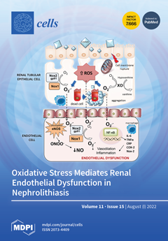
Cover Story (view full-size image): Urolithiasis is a risk factor for the development of chronic kidney disease that has been clinically correlated with cardiovascular disorders. Here, we first demonstrate that hyperoxaluria induces endothelial dysfunction mediated by oxidative stress in renal preglomerular arteries. Xanthine oxidase, pathway, Nox1 and in particular Nox2 contribute to the enhanced reactive oxygen species levels in the renal cortex of hyperoxaluric rats, which generates endothelial dysfunction in the nearby arteries. Inhibition of these enzymes restores impaired endothelial vasodilatation. An inflammatory response is also generated in the vascular endothelium through the activation of the redox-sensitive NF-κB pathway. The current data suggest that oxidative stress-mediated endothelial dysfunction induced by hyperoxaluria might contribute to the vascular renal damage associated with nephrolithiasis. View this paper
  • Issues are regarded as officially published after their release is announced to the table of contents alert mailing list.
  • You may sign up for e-mail alerts to receive table of contents of newly released issues.
  • PDF is the official format for papers published in both, html and pdf forms. To view the papers in pdf format, click on the "PDF Full-text" link, and use the free Adobe Reader to open them.
Order results
Result details
Section
Select all
Export citation of selected articles as:
29 pages, 3478 KiB  
Article
The Protective Action of Metformin against Pro-Inflammatory Cytokine-Induced Human Islet Cell Damage and the Mechanisms Involved
by Laura Giusti, Marta Tesi, Federica Ciregia, Lorella Marselli, Lorenzo Zallocco, Mara Suleiman, Carmela De Luca, Silvia Del Guerra, Mariachiara Zuccarini, Marco Trerotola, Decio L. Eizirik, Miriam Cnop, Maria R. Mazzoni, Piero Marchetti, Antonio Lucacchini and Maurizio Ronci
Cells 2022, 11(15), 2465; https://doi.org/10.3390/cells11152465 - 8 Aug 2022
Cited by 13 | Viewed by 3524
Abstract
Metformin, a drug widely used in type 2 diabetes (T2D), has been shown to protect human β-cells exposed to gluco- and/or lipotoxic conditions and those in islets from T2D donors. We assessed whether metformin could relieve the human β-cell stress induced by pro-inflammatory [...] Read more.
Metformin, a drug widely used in type 2 diabetes (T2D), has been shown to protect human β-cells exposed to gluco- and/or lipotoxic conditions and those in islets from T2D donors. We assessed whether metformin could relieve the human β-cell stress induced by pro-inflammatory cytokines (which mediate β-cells damage in type 1 diabetes, T1D) and investigated the underlying mechanisms using shotgun proteomics. Human islets were exposed to 50 U/mL interleukin-1β plus 1000 U/mL interferon-γ for 48 h, with or without 2.4 µg/mL metformin. Glucose-stimulated insulin secretion (GSIS) and caspase 3/7 activity were studied, and a shotgun label free proteomics analysis was performed. Metformin prevented the reduction of GSIS and the activation of caspase 3/7 induced by cytokines. Proteomics analysis identified more than 3000 proteins in human islets. Cytokines alone altered the expression of 244 proteins (145 up- and 99 down-regulated), while, in the presence of metformin, cytokine-exposure modified the expression of 231 proteins (128 up- and 103 downregulated). Among the proteins inversely regulated in the two conditions, we found proteins involved in vesicle motility, defense against oxidative stress (including peroxiredoxins), metabolism, protein synthesis, glycolysis and its regulation, and cytoskeletal proteins. Metformin inhibited pathways linked to inflammation, immune reactions, mammalian target of rapamycin (mTOR) signaling, and cell senescence. Some of the changes were confirmed by Western blot. Therefore, metformin prevented part of the deleterious actions of pro-inflammatory cytokines in human β-cells, which was accompanied by islet proteome modifications. This suggests that metformin, besides use in T2D, might be considered for β-cell protection in other types of diabetes, possibly including early T1D. Full article
Show Figures

Figure 1

12 pages, 1698 KiB  
Review
Efp/TRIM25 and Its Related Protein, TRIM47, in Hormone-Dependent Cancers
by Kotaro Azuma and Satoshi Inoue
Cells 2022, 11(15), 2464; https://doi.org/10.3390/cells11152464 - 8 Aug 2022
Cited by 2 | Viewed by 3327
Abstract
Increasing attention has been paid to the biological roles of tripartite motif-containing (TRIM) family proteins, which typically function as E3 ubiquitin ligases. Estrogen-responsive finger protein (Efp), a member of the TRIM family proteins, also known as TRIM25, was originally identified as a protein [...] Read more.
Increasing attention has been paid to the biological roles of tripartite motif-containing (TRIM) family proteins, which typically function as E3 ubiquitin ligases. Estrogen-responsive finger protein (Efp), a member of the TRIM family proteins, also known as TRIM25, was originally identified as a protein induced by estrogen and plays critical roles in promoting endocrine-related cancers, including breast cancer, endometrial cancer, and prostate cancer. The pathophysiological importance of Efp made us interested in the roles of other TRIM family proteins that share a similar structure with Efp. Based on a phylogenetic analysis of the C-terminal region of TRIM family proteins, we focused on TRIM47 as a protein belonging to the same branch as Efp. TRIM47 is a poor prognostic factor in both breast cancer and prostate cancer. Atypical lysine-27-like poly-ubiquitination was involved in the underlying mechanism causing endocrine resistance in breast cancer. We also discuss the functions of Efp and TRIM47 in other types of cancers and innate immunity by introducing substrates the are modified by poly-ubiquitination. Full article
(This article belongs to the Special Issue Mechanism of Nuclear Hormone Receptors in Cancer)
Show Figures

Figure 1

9 pages, 994 KiB  
Editorial
LL-37, a Multi-Faceted Amphipathic Peptide Involved in NETosis
by Marko Radic and Sylviane Muller
Cells 2022, 11(15), 2463; https://doi.org/10.3390/cells11152463 - 8 Aug 2022
Cited by 15 | Viewed by 3207
Abstract
Innate immunity responds to infections and inflammatory stimuli through a carefully choreographed set of interactions between cells, stimuli and their specific receptors. Of particular importance are endogenous peptides, which assume roles as defensins or alarmins, growth factors or wound repair inducers. LL-37, a [...] Read more.
Innate immunity responds to infections and inflammatory stimuli through a carefully choreographed set of interactions between cells, stimuli and their specific receptors. Of particular importance are endogenous peptides, which assume roles as defensins or alarmins, growth factors or wound repair inducers. LL-37, a proteolytic fragment of cathelicidin, fulfills the roles of a defensin by inserting into the membranes of bacterial pathogens, functions as alarmin in stimulating chemotaxis of innate immune cells, and alters the structure and efficacy of various cytokines. Here, we draw attention to the direct effect of LL-37 on neutrophils and the release of extracellular traps (NETs), as NETs have been established as mediators of immune defense against pathogens but also as important contributors to chronic disease and tissue pathogenesis. We propose a specific structural basis for LL-37 function, in part by highlighting the structural flexibility of LL-37 and its ability to adapt to distinct microenvironments and interacting counterparts. Full article
(This article belongs to the Special Issue NETs in Infectious and Inflammatory Diseases)
Show Figures

Figure 1

10 pages, 691 KiB  
Article
Assessment of a Serum Microrna Risk Score for Colorectal Cancer among Participants of Screening Colonoscopy at Various Stages of Colorectal Carcinogenesis
by Janhavi R. Raut, Megha Bhardwaj, Tobias Niedermaier, Kaya Miah, Petra Schrotz-King and Hermann Brenner
Cells 2022, 11(15), 2462; https://doi.org/10.3390/cells11152462 - 8 Aug 2022
Cited by 2 | Viewed by 2034
Abstract
We recently derived and validated a serum-based microRNA risk score (miR-score) which predicted colorectal cancer (CRC) occurrence with very high accuracy within 14 years of follow-up in a large population-based cohort. Here, we aimed to assess and compare the distribution of the miR-score [...] Read more.
We recently derived and validated a serum-based microRNA risk score (miR-score) which predicted colorectal cancer (CRC) occurrence with very high accuracy within 14 years of follow-up in a large population-based cohort. Here, we aimed to assess and compare the distribution of the miR-score among participants of screening colonoscopy at various stages of colorectal carcinogenesis. MicroRNAs (miRNAs) were profiled by quantitative-real-time-polymerase-chain-reaction in the serum samples of screening colonoscopy participants with CRC (n = 52), advanced colorectal adenoma (AA, n = 100), non-advanced colorectal adenoma (NAA, n = 88), and participants free of colorectal neoplasms (n = 173). The mean values of the miR-score were compared between groups by the Mann–Whitney U test. The associations of the miR-score with risk for colorectal neoplasms were evaluated using logistic regression analyses. MicroRNA risk scores were significantly higher among participants with AA than among those with NAA (p = 0.027) and those with CRC (p = 0.014), whereas no statistically significant difference was seen between those with NAA and those with no colorectal neoplasms (p = 0.127). When comparing adjacent groups, miR-scores were inversely associated with CRC versus AA and positively associated with AA versus NAA [odds ratio (OR), 0.37 (95% confidence interval (CI), 0.16–0.86) and OR, 2.22 (95% CI, 1.06–4.64) for the top versus bottom tertiles, respectively]. Our results are consistent with the hypothesis that a high miR-score may be indicative of an increased CRC risk by an increased tendency of progression from non-advanced to advanced colorectal neoplasms, along with a change of the miR-patterns after CRC manifestation. Full article
Show Figures

Figure 1

19 pages, 2110 KiB  
Article
Pharmacological Activation of Potassium Channel Kv11.1 with NS1643 Attenuates Triple Negative Breast Cancer Cell Migration by Promoting the Dephosphorylation of Caveolin-1
by Ying Jiang, Vitalyi Senyuk, Ke Ma, Hui Chen, Xiang Qin, Shun Li, Yiyao Liu, Saverio Gentile and Richard D. Minshall
Cells 2022, 11(15), 2461; https://doi.org/10.3390/cells11152461 - 8 Aug 2022
Cited by 6 | Viewed by 2818
Abstract
The prevention of metastasis is a central goal of cancer therapy. Caveolin-1 (Cav-1) is a structural membrane and scaffolding protein shown to be a key regulator of late-stage breast cancer metastasis. However, therapeutic strategies targeting Cav-1 are still lacking. Here, we demonstrate that [...] Read more.
The prevention of metastasis is a central goal of cancer therapy. Caveolin-1 (Cav-1) is a structural membrane and scaffolding protein shown to be a key regulator of late-stage breast cancer metastasis. However, therapeutic strategies targeting Cav-1 are still lacking. Here, we demonstrate that the pharmacological activation of potassium channel Kv11.1, which is uniquely expressed in MDA-MB-231 triple negative breast cancer cells (TNBCs) but not in normal MCF-10A cells, induces the dephosphorylation of Cav-1 Tyr-14 by promoting the Ca2+-dependent stimulation of protein tyrosine phosphatase 1B (PTP1B). Consequently, the dephosphorylation of Cav-1 resulted in its disassociation from β-catenin, which enabled the accumulation of β-catenin at cell borders, where it facilitated the formation of cell–cell adhesion complexes via interactions with R-cadherin and desmosomal proteins. Kv11.1 activation-dependent Cav-1 dephosphorylation induced with NS1643 also reduced cell migration and invasion, consistent with its ability to regulate focal adhesion dynamics. Thus, this study sheds light on a novel pharmacological mechanism of promoting Cav-1 dephosphorylation, which may prove to be effective at reducing metastasis and promoting contact inhibition. Full article
(This article belongs to the Collection Feature Papers in Cell Motility and Adhesion)
Show Figures

Figure 1

14 pages, 5952 KiB  
Article
Static Magnetic Fields Regulate T-Type Calcium Ion Channels and Mediate Mesenchymal Stem Cells Proliferation
by Haokaifeng Wu, Chuang Li, Muqaddas Masood, Zhen Zhang, Esther González-Almela, Alvaro Castells-Garcia, Gaoyang Zou, Xiaoduo Xu, Luqin Wang, Guoqing Zhao, Shengyong Yu, Ping Zhu, Bo Wang, Dajiang Qin and Jing Liu
Cells 2022, 11(15), 2460; https://doi.org/10.3390/cells11152460 - 8 Aug 2022
Cited by 23 | Viewed by 4024
Abstract
The static magnetic fields (SMFs) impact on biological systems, induce a variety of biological responses, and have been applied to the clinical treatment of diseases. However, the underlying mechanisms remain largely unclear. In this report, by using human mesenchymal stem cells (MSCs) as [...] Read more.
The static magnetic fields (SMFs) impact on biological systems, induce a variety of biological responses, and have been applied to the clinical treatment of diseases. However, the underlying mechanisms remain largely unclear. In this report, by using human mesenchymal stem cells (MSCs) as a model, we investigated the biological effect of SMFs at a molecular and cellular level. We showed that SMF exposure promotes MSC proliferation and activates the expression of transcriptional factors such as FOS (Fos Proto-Oncogene, AP-1 Transcription Factor Subunit) and EGR1 (Early Growth Response 1). In addition, the expression of signal-transduction proteins p-ERK1/2 and p-JNK oscillate periodically with SMF exposure time. Furthermore, we found that the inhibition of the T-type calcium ion channels negates the biological effects of SMFs on MSCs. Together, we revealed that the SMFs regulate T-type calcium ion channels and mediate MSC proliferation via the MAPK signaling pathways. Full article
(This article belongs to the Collection Magnetic Fields and Cells)
Show Figures

Figure 1

12 pages, 1271 KiB  
Review
Role of Spectrin in Endocytosis
by Donghai Li
Cells 2022, 11(15), 2459; https://doi.org/10.3390/cells11152459 - 8 Aug 2022
Cited by 2 | Viewed by 2532
Abstract
Cytoskeletal spectrin is found in (non)erythroid cells. Eukaryotic endocytosis takes place for internalizing cargos from extracellular milieu. The role of spectrin in endocytosis still remains poorly understood. Here, I summarize current knowledge of spectrin function, spectrin-based cytoskeleton and endocytosis of erythrocytes, and highlight [...] Read more.
Cytoskeletal spectrin is found in (non)erythroid cells. Eukaryotic endocytosis takes place for internalizing cargos from extracellular milieu. The role of spectrin in endocytosis still remains poorly understood. Here, I summarize current knowledge of spectrin function, spectrin-based cytoskeleton and endocytosis of erythrocytes, and highlight how spectrin contributes to endocytosis and working models in different types of cells. From an evolutionary viewpoint, I discuss spectrin and endocytosis in a range of organisms, particularly in plants and yeast where spectrin is absent. Together, the role of spectrin in endocytosis is related to its post-translational modification, movement/rearrangement, elimination (by proteases) and meshwork fencing. Full article
(This article belongs to the Section Intracellular and Plasma Membranes)
Show Figures

Figure 1

15 pages, 1440 KiB  
Review
Endogenous Retroviruses and Placental Evolution, Development, and Diversity
by Kazuhiko Imakawa, Kazuya Kusama, Tomoko Kaneko-Ishino, So Nakagawa, Koichi Kitao, Takayuki Miyazawa and Fumitoshi Ishino
Cells 2022, 11(15), 2458; https://doi.org/10.3390/cells11152458 - 8 Aug 2022
Cited by 14 | Viewed by 7803
Abstract
The main roles of placentas include physical protection, nutrient and oxygen import, export of gasses and fetal waste products, and endocrinological regulation. In addition to physical protection of the fetus, the placentas must provide immune protection throughout gestation. These basic functions are well-conserved; [...] Read more.
The main roles of placentas include physical protection, nutrient and oxygen import, export of gasses and fetal waste products, and endocrinological regulation. In addition to physical protection of the fetus, the placentas must provide immune protection throughout gestation. These basic functions are well-conserved; however, placentas are undoubtedly recent evolving organs with structural and cellular diversities. These differences have been explained for the last two decades through co-opting genes and gene control elements derived from transposable elements, including endogenous retroviruses (ERVs). However, the differences in placental structures have not been explained or characterized. This manuscript addresses the sorting of ERVs and their integration into the mammalian genomes and provides new ways to explain why placental structures have diverged. Full article
(This article belongs to the Special Issue Placental Development in Health and Disease)
Show Figures

Figure 1

21 pages, 6012 KiB  
Article
Genome-Wide Association Analysis Reveals Trait-Linked Markers for Grain Nutrient and Agronomic Traits in Diverse Set of Chickpea Germplasm
by Rajasekhar Srungarapu, Mahesh Damodhar Mahendrakar, Lal Ahamed Mohammad, Uttam Chand, Venkata Ramana Jagarlamudi, Kiran Prakash Kondamudi, Himabindu Kudapa and Srinivasan Samineni
Cells 2022, 11(15), 2457; https://doi.org/10.3390/cells11152457 - 8 Aug 2022
Cited by 11 | Viewed by 4326
Abstract
Chickpea is an inexpensive source of protein, minerals, and vitamins to the poor people living in arid and semi-arid regions of Southern Asia and Sub-Saharan Africa. New chickpea cultivars with enhanced levels of protein, Fe and Zn content are a medium-term strategy for [...] Read more.
Chickpea is an inexpensive source of protein, minerals, and vitamins to the poor people living in arid and semi-arid regions of Southern Asia and Sub-Saharan Africa. New chickpea cultivars with enhanced levels of protein, Fe and Zn content are a medium-term strategy for supplying essential nutrients for human health and reducing malnutrition. In the current study, a chickpea reference set of 280 accessions, including landraces, breeding lines, and advanced cultivars, was evaluated for grain protein, Fe, Zn content and agronomic traits over two seasons. Using a mid-density 5k SNP array, 4603 highly informative SNPs distributed across the chickpea genome were used for GWAS analysis. Population structure analysis revealed three subpopulations (K = 3). Linkage disequilibrium (LD) was extensive, and LD decay was relatively low. A total of 20 and 46 marker-trait associations (MTAs) were identified for grain nutrient and agronomic traits, respectively, using FarmCPU and BLINK models. Of which seven SNPs for grain protein, twelve for Fe, and one for Zn content were distributed on chromosomes 1, 4, 6, and 7. The marker S4_4477846 on chr4 was found to be co-associated with grain protein over seasons. The markers S1_11613376 and S1_2772537 co-associated with grain Fe content under NSII and pooled seasons and S7_9379786 marker under NSI and pooled seasons. The markers S4_31996956 co-associated with grain Fe and days to maturity. SNP annotation of associated markers were found to be related to gene functions of metal ion binding, transporters, protein kinases, transcription factors, and many more functions involved in plant metabolism along with Fe and protein homeostasis. The identified significant MTAs has potential use in marker-assisted selection for developing nutrient-rich chickpea cultivars after validation in the breeding populations. Full article
(This article belongs to the Special Issue Omics in Plant Genetics and Breeding)
Show Figures

Graphical abstract

20 pages, 107238 KiB  
Article
HSSG: Identification of Cancer Subtypes Based on Heterogeneity Score of A Single Gene
by Shanchen Pang, Wenhao Wu, Yuanyuan Zhang, Shudong Wang, Muyuan Niu, Kuijie Zhang and Wenjing Yin
Cells 2022, 11(15), 2456; https://doi.org/10.3390/cells11152456 - 8 Aug 2022
Cited by 1 | Viewed by 2032
Abstract
Cancer is a highly heterogeneous disease, which leads to the fact that even the same cancer can be further classified into different subtypes according to its pathology. With the multi-omics data widely used in cancer subtypes identification, effective feature selection is essential for [...] Read more.
Cancer is a highly heterogeneous disease, which leads to the fact that even the same cancer can be further classified into different subtypes according to its pathology. With the multi-omics data widely used in cancer subtypes identification, effective feature selection is essential for accurately identifying cancer subtypes. However, the feature selection in the existing cancer subtypes identification methods has the problem that the most helpful features cannot be selected from a biomolecular perspective, and the relationship between the selected features cannot be reflected. To solve this problem, we propose a method for feature selection to identify cancer subtypes based on the heterogeneity score of a single gene: HSSG. In the proposed method, the sample-similarity network of a single gene is constructed, and pseudo-F statistics calculates the heterogeneity score for cancer subtypes identification of each gene. Finally, we construct gene-gene networks using genes with higher heterogeneity scores and mine essential genes from the networks. From the seven TCGA data sets for three experiments, including cancer subtypes identification in single-omics data, the performance in feature selection of multi-omics data, and the effectiveness and stability of the selected features, HSSG achieves good performance in all. This indicates that HSSG can effectively select features for subtypes identification. Full article
(This article belongs to the Collection Computational Imaging for Biophotonics and Biomedicine)
Show Figures

Figure 1

17 pages, 4523 KiB  
Article
Estimating Biomass and Vitality of Microalgae for Monitoring Cultures: A Roadmap for Reliable Measurements
by Michael Schagerl, Rainer Siedler, Eliška Konopáčová and Sameh Samir Ali
Cells 2022, 11(15), 2455; https://doi.org/10.3390/cells11152455 - 8 Aug 2022
Cited by 24 | Viewed by 4026
Abstract
Estimating algal biomass is a prerequisite for monitoring growth of microalgae. Especially for large-scale production sites, the measurements must be robust, reliable, fast and easy to obtain. We compare the relevant parameters, discuss potential hurdles and provide recommendations to tackle these issues. The [...] Read more.
Estimating algal biomass is a prerequisite for monitoring growth of microalgae. Especially for large-scale production sites, the measurements must be robust, reliable, fast and easy to obtain. We compare the relevant parameters, discuss potential hurdles and provide recommendations to tackle these issues. The focus is on optical density and in vivo autofluorescence of chlorophyll, which have proven to be ideal candidates for monitoring purposes. Beyond biomass, cell vitality is also crucial for maintaining cultures. While maximizing biomass yield is often the primary consideration, some applications require adverse growth conditions for the synthesis of high-quality compounds. The non-invasive technique of pulse-amplified modulated (PAM) fluorescence measurements provides an ideal tool and is increasingly being employed due to ever more affordable devices. We compared three devices and studied the robustness of the dark fluorescence yield of photosystem II (Fv/Fm) at various cell densities. Although the so-called inner filter effects influence the fluorescence signal, the resulting Fv/Fm remain stable and robust over a wide range of cell densities due to mutual effects. Full article
(This article belongs to the Special Issue Growth and Division in Algae)
Show Figures

Figure 1

14 pages, 2327 KiB  
Article
Faster Serotonin Clearance in CA3 Region of Hippocampus and Antidepressant-like Effect of Decynium-22 in Juvenile Mice Are Putatively Linked to Increased Plasma Membrane Monoamine Transporter Function: Implications for Efficacy of Antidepressants in Juveniles
by Melodi A. Bowman, Jorge A. Gomez, Nathan C. Mitchell, Anne M. Wells, Melissa Vitela, Kyra M. Clarke, Rebecca E. Horton, Wouter Koek and Lynette C. Daws
Cells 2022, 11(15), 2454; https://doi.org/10.3390/cells11152454 - 8 Aug 2022
Cited by 2 | Viewed by 2122
Abstract
Selective serotonin reuptake inhibitors (SSRIs) are less efficacious in treating depression in children than in adults. SSRIs block serotonin uptake via the high-affinity, low-capacity serotonin transporter. However, the low-affinity, high-capacity organic cation transporter 3 (OCT3) and plasma membrane monoamine transporter (PMAT) are emerging [...] Read more.
Selective serotonin reuptake inhibitors (SSRIs) are less efficacious in treating depression in children than in adults. SSRIs block serotonin uptake via the high-affinity, low-capacity serotonin transporter. However, the low-affinity, high-capacity organic cation transporter 3 (OCT3) and plasma membrane monoamine transporter (PMAT) are emerging as important players in serotonin uptake. We hypothesized that OCT3 and/or PMAT are functionally upregulated in juveniles, thereby buffering SSRIs’ ability to enhance serotonergic neurotransmission. Unlike in adult mice, we found the OCT/PMAT blocker, decynium-22, to have standalone antidepressant-like effects in juveniles. Using in vivo high-speed chronoamperometry, we found that juveniles clear serotonin from the CA3 region of the hippocampus ~2-fold faster than adult mice. Cell density did not differ between ages, suggesting that faster serotonin clearance in juveniles is unrelated to faster diffusion through the extracellular matrix. Western blot and immunohistochemistry showed that juvenile mice have modestly greater expression of PMAT than adults, whereas OCT3 expression in the CA3 region of the hippocampus was similar between ages. Together, these data suggest that faster serotonin clearance and antidepressant-like effects of decynium-22 in juvenile mice may be due to functionally upregulated PMAT. Faster serotonin clearance via PMAT in juveniles may contribute to reduced therapeutic efficacy of SSRIs in children relative to adults. Full article
(This article belongs to the Special Issue Neurotransmitter Transporters in Health and Disease)
Show Figures

Figure 1

17 pages, 2785 KiB  
Article
Phaseolus vulgaris Erythroagglutinin (PHA-E)-Positive Ceruloplasmin Acts as a Potential Biomarker in Pancreatic Cancer Diagnosis
by Shanshan Sha, Yating Wang, Menglu Liu, Gang Liu, Ning Fan, Zhi Li and Weijie Dong
Cells 2022, 11(15), 2453; https://doi.org/10.3390/cells11152453 - 8 Aug 2022
Cited by 4 | Viewed by 2589
Abstract
Pancreatic cancer (PC) remains one of the top 10 causes of cancer-related death in recent years. Approximately 80% of PC patients are diagnosed at the middle or advanced stage and miss the opportunity for surgery. The demand for early diagnostic methods and reliable [...] Read more.
Pancreatic cancer (PC) remains one of the top 10 causes of cancer-related death in recent years. Approximately 80% of PC patients are diagnosed at the middle or advanced stage and miss the opportunity for surgery. The demand for early diagnostic methods and reliable biomarkers is increasing, although a number of tumor markers such as CA19-9 and CEA have already been utilized in clinics. In this study, we analyzed the alteration of N-glycan of serum glycoproteins by mass spectrometry and lectin blotting. The results showed that bisecting GlcNAc structures of glycoproteins are significantly increased in PC patients’ sera. With Phaseolus vulgaris Erythroagglutinin (PHA-E) lectin that specifically recognizes bisecting GlcNAc N-glycans, the serum glycoproteins bearing bisecting GlcNAc in PC patients’ sera were pulled down and identified by nano-LC-MS/MS. Among them, ceruloplasmin (Cp) was screened out with a satisfied sensitivity and specificity in identifying PC from acute pancreatitis patients (AUC: 0.757) and normal healthy persons (AUC: 0.972), suggesting a close association between Cp and PC development and diagnosis. To prove that, the Cp expression in tumor tissues of PC patients was examined. The results showed that Cp was significantly upregulated in PC tissues compared to that in adjacent normal tissues. All these results suggested that PHA-E-positive Cp could be a potential PC-specific glycoprotein marker to distinguish PC patients from acute pancreatitis patients and normal persons. Full article
Show Figures

Figure 1

18 pages, 5563 KiB  
Article
Endothelial Glycocalyx Morphology in Different Flow Regions of the Aqueous Outflow Pathway of Normal and Laser-Induced Glaucoma Monkey Eyes
by Shayna Sosnowik, David L. Swain, Neil Liu, Shan Fan, Carol B. Toris and Haiyan Gong
Cells 2022, 11(15), 2452; https://doi.org/10.3390/cells11152452 - 7 Aug 2022
Cited by 4 | Viewed by 2767
Abstract
Glycocalyx morphology was examined in the trabecular outflow pathway of monkey eyes with and without experimental glaucoma. Laser burns were administered along ~270 degrees of the trabecular meshwork (TM) of one eye (n = 6) or both eyes (n = 2) [...] Read more.
Glycocalyx morphology was examined in the trabecular outflow pathway of monkey eyes with and without experimental glaucoma. Laser burns were administered along ~270 degrees of the trabecular meshwork (TM) of one eye (n = 6) or both eyes (n = 2) of each monkey until intraocular pressure remained elevated. Portions of the TM were not laser-treated. Unlasered eyes (n = 6) served as controls. Enucleated eyes were perfused at 15 mmHg to measure the outflow facility, perfused with fluorescein to evaluate the outflow pattern, perfusion-fixed for glycocalyx labeling, and processed for electron microscopy. Coverage and thickness of the glycocalyx were measured in the TM, Schlemm’s canal (SC), collector channels (CCs), intrascleral veins (ISVs), and episcleral veins (ESVs) in non-lasered regions and high- and low-flow regions of controls. Compared to controls, laser-treated eyes had decreased outflow facility (p = 0.02). Glycocalyx thickness increased from the TM to ESVs in non-lasered regions and controls (p < 0.05). Glycocalyx coverage was generally greater distally in non-lasered regions (p < 0.05). In lasered regions, TM, SC, and CCs were partly to completely obliterated, and ISVs and ESVs displayed minimal glycocalyx. Whether the glycocalyx is decreased in the trabecular outflow pathway of human glaucomatous eyes warrants investigation. Full article
Show Figures

Figure 1

15 pages, 2244 KiB  
Article
Three Binding Conformations of BIO124 in the Pocket of the PICK1 PDZ Domain
by Amy O. Stevens, Samuel Luo and Yi He
Cells 2022, 11(15), 2451; https://doi.org/10.3390/cells11152451 - 7 Aug 2022
Cited by 4 | Viewed by 3058
Abstract
The PDZ family has drawn attention as possible drug targets because of the domains’ wide ranges of function and highly conserved binding pockets. The PICK1 PDZ domain has been proposed as a possible drug target because the interactions between the PICK1 PDZ domain [...] Read more.
The PDZ family has drawn attention as possible drug targets because of the domains’ wide ranges of function and highly conserved binding pockets. The PICK1 PDZ domain has been proposed as a possible drug target because the interactions between the PICK1 PDZ domain and the GluA2 subunit of the AMPA receptor have been shown to progress neurodegenerative diseases. BIO124 has been identified as a sub µM inhibitor of the PICK1–GluA2 interaction. Here, we use all-atom molecular dynamics simulations to reveal the atomic-level interaction pattern between the PICK1 PDZ domain and BIO124. Our simulations reveal three unique binding conformations of BIO124 in the PICK1 PDZ binding pocket, referred to here as state 0, state 1, and state 2. Each conformation is defined by a unique hydrogen bonding network and a unique pattern of hydrophobic interactions between BIO124 and the PICK1 PDZ domain. Interestingly, each conformation of BIO124 results in different dynamic changes to the PICK1 PDZ domain. Unlike states 1 and 2, state 0 induces dynamic coupling between BIO124 and the αA helix. Notably, this dynamic coupling with the αA helix is similar to what has been observed in other PDZ–ligand complexes. Our analysis indicates that the interactions formed between BIO124 and I35 may be the key to inducing dynamic coupling with the αA helix. Lastly, we suspect that the conformational shifts observed in our simulations may affect the stability and thus the overall effectiveness of BIO124. We propose that a physically larger inhibitor may be necessary to ensure sufficient interactions that permit stable binding between a drug and the PICK1 PDZ domain. Full article
Show Figures

Figure 1

12 pages, 2223 KiB  
Article
Spectral Library-Based Single-Cell Proteomics Resolves Cellular Heterogeneity
by Lakmini Senavirathna, Cheng Ma, Ru Chen and Sheng Pan
Cells 2022, 11(15), 2450; https://doi.org/10.3390/cells11152450 - 7 Aug 2022
Cited by 9 | Viewed by 3403
Abstract
Dissecting the proteome of cell types and states at single-cell resolution, while being highly challenging, has significant implications in basic science and biomedicine. Mass spectrometry (MS)-based single-cell proteomics represents an emerging technology for system-wide, unbiased profiling of proteins in single cells. However, significant [...] Read more.
Dissecting the proteome of cell types and states at single-cell resolution, while being highly challenging, has significant implications in basic science and biomedicine. Mass spectrometry (MS)-based single-cell proteomics represents an emerging technology for system-wide, unbiased profiling of proteins in single cells. However, significant challenges remain in analyzing an extremely small amount of proteins collected from a single cell, as a proteome-wide amplification of proteins is not currently feasible. Here, we report an integrated spectral library-based single-cell proteomics (SLB-SCP) platform that is ultrasensitive and well suited for a large-scale analysis. To overcome the low MS/MS signal intensity intrinsically associated with a single-cell analysis, this approach takes an alternative approach by extracting a breadth of information that specifically defines the physicochemical characteristics of a peptide from MS1 spectra, including monoisotopic mass, isotopic distribution, and retention time (hydrophobicity), and uses a spectral library for proteomic identification. This conceptually unique MS platform, coupled with the DIRECT sample preparation method, enabled identification of more than 2000 proteins in a single cell to distinguish different proteome landscapes associated with cellular types and heterogeneity. We characterized individual normal and cancerous pancreatic ductal cells (HPDE and PANC-1, respectively) and demonstrated the substantial difference in the proteomes between HPDE and PANC-1 at the single-cell level. A significant upregulation of multiple protein networks in cancer hallmarks was identified in the PANC-1 cells, functionally discriminating the PANC-1 cells from the HPDE cells. This integrated platform can be built on high-resolution MS and widely accepted proteomic software, making it possible for community-wide applications. Full article
(This article belongs to the Collection Deciphering the Proteome in Cell Biology and Diseases)
Show Figures

Figure 1

20 pages, 1229 KiB  
Review
Interplay of Developmental Hippo–Notch Signaling Pathways with the DNA Damage Response in Prostate Cancer
by Ioanna Mourkioti, Andriani Angelopoulou, Konstantinos Belogiannis, Nefeli Lagopati, Spyridon Potamianos, Efthymios Kyrodimos, Vassilis Gorgoulis and Angelos Papaspyropoulos
Cells 2022, 11(15), 2449; https://doi.org/10.3390/cells11152449 - 7 Aug 2022
Cited by 10 | Viewed by 3081
Abstract
Prostate cancer belongs in the class of hormone-dependent cancers, representing a major cause of cancer incidence in men worldwide. Since upon disease onset almost all prostate cancers are androgen-dependent and require active androgen receptor (AR) signaling for their survival, the primary treatment approach [...] Read more.
Prostate cancer belongs in the class of hormone-dependent cancers, representing a major cause of cancer incidence in men worldwide. Since upon disease onset almost all prostate cancers are androgen-dependent and require active androgen receptor (AR) signaling for their survival, the primary treatment approach has for decades relied on inhibition of the AR pathway via androgen deprivation therapy (ADT). However, following this line of treatment, cancer cell pools often become resistant to therapy, contributing to disease progression towards the significantly more aggressive castration-resistant prostate cancer (CRPC) form, characterized by poor prognosis. It is, therefore, of critical importance to elucidate the molecular mechanisms and signaling pathways underlying the progression of early-stage prostate cancer towards CRPC. In this review, we aim to shed light on the role of major signaling pathways including the DNA damage response (DDR) and the developmental Hippo and Notch pathways in prostate tumorigenesis. We recapitulate key evidence demonstrating the crosstalk of those pathways as well as with pivotal prostate cancer-related ‘hubs’ such as AR signaling, and evaluate the clinical impact of those interactions. Moreover, we attempt to identify molecules of the complex DDR–Hippo–Notch interplay comprising potentially novel therapeutic targets in the battle against prostate tumorigenesis. Full article
(This article belongs to the Special Issue Multitasking Proteins and Their Involvement in Pathogenesis)
Show Figures

Figure 1

22 pages, 874 KiB  
Review
Regulatory Roles of Noncoding RNAs in the Progression of Gastrointestinal Cancers and Health Disparities
by Aditi Kulkarni, Sharan Gayathrinathan, Soumya Nair, Anamika Basu, Taslim A. Al-Hilal and Sourav Roy
Cells 2022, 11(15), 2448; https://doi.org/10.3390/cells11152448 - 7 Aug 2022
Cited by 3 | Viewed by 3056
Abstract
Annually, more than a million individuals are diagnosed with gastrointestinal (GI) cancers worldwide. With the advancements in radio- and chemotherapy and surgery, the survival rates for GI cancer patients have improved in recent years. However, the prognosis for advanced-stage GI cancers remains poor. [...] Read more.
Annually, more than a million individuals are diagnosed with gastrointestinal (GI) cancers worldwide. With the advancements in radio- and chemotherapy and surgery, the survival rates for GI cancer patients have improved in recent years. However, the prognosis for advanced-stage GI cancers remains poor. Site-specific GI cancers share a few common risk factors; however, they are largely distinct in their etiologies and descriptive epidemiologic profiles. A large number of mutations or copy number changes associated with carcinogenesis are commonly found in noncoding DNA regions, which transcribe several noncoding RNAs (ncRNAs) that are implicated to regulate cancer initiation, metastasis, and drug resistance. In this review, we summarize the regulatory functions of ncRNAs in GI cancer development, progression, chemoresistance, and health disparities. We also highlight the potential roles of ncRNAs as therapeutic targets and biomarkers, mainly focusing on their ethnicity-/race-specific prognostic value, and discuss the prospects of genome-wide association studies (GWAS) to investigate the contribution of ncRNAs in GI tumorigenesis. Full article
(This article belongs to the Special Issue Regulatory Roles of Non-coding RNAs in Cancer)
Show Figures

Figure 1

19 pages, 3260 KiB  
Article
Investigation of Radiotracer Metabolic Stability In Vitro with CYP-Overexpressing Hepatoma Cell Lines
by Sandy Lemm, Susanne Köhler, Robert Wodtke, Friedrich Jung, Jan-Heiner Küpper, Jens Pietzsch and Markus Laube
Cells 2022, 11(15), 2447; https://doi.org/10.3390/cells11152447 - 7 Aug 2022
Cited by 3 | Viewed by 2300
Abstract
The characterization of novel radiotracers toward their metabolic stability is an essential part of their development. While in vitro methods such as liver microsome assays or ex vivo blood or tissue samples provide information on overall stability, little or no information is obtained [...] Read more.
The characterization of novel radiotracers toward their metabolic stability is an essential part of their development. While in vitro methods such as liver microsome assays or ex vivo blood or tissue samples provide information on overall stability, little or no information is obtained on cytochrome P450 (CYP) enzyme and isoform-specific contribution to the metabolic fate of individual radiotracers. Herein, we investigated recently established CYP-overexpressing hepatoblastoma cell lines (HepG2) for their suitability to study the metabolic stability of radiotracers in general and to gain insight into CYP isoform specificity. Wildtype HepG2 and CYP1A2-, CYP2C19-, and CYP3A4-overexpressing HepG2 cells were incubated with radiotracers, and metabolic turnover was analyzed. The optimized protocol, covering cell seeding in 96-well plates and analysis of supernatant by radio thin-layer-chromatography for higher throughput, was transferred to the evaluation of three 18F-labeled celecoxib-derived cyclooxygenase-2 inhibitors (coxibs). These investigations revealed time-dependent degradation of the intact radiotracers, as well as CYP isoform- and substrate-specific differences in their metabolic profiles. HepG2 CYP2C19 proved to be the cell line showing the highest metabolic turnover for each radiotracer studied here. Comparison with human and murine liver microsome assays showed good agreement with the human metabolite profile obtained by the HepG2 cell lines. Therefore, CYP-overexpressing HepG2 cells provide a good complement for assessing the metabolic stability of radiotracers and allow the analysis of the CYP isoform-specific contribution to the overall radiotracer metabolism. Full article
(This article belongs to the Collection Advances in Cell Culture and Tissue Engineering)
Show Figures

Figure 1

31 pages, 17296 KiB  
Article
Phylogeny and Fatty Acid Profiles of New Pinnularia (Bacillariophyta) Species from Soils of Vietnam
by Elena Kezlya, Yevhen Maltsev, Sergei Genkal, Zinaida Krivova and Maxim Kulikovskiy
Cells 2022, 11(15), 2446; https://doi.org/10.3390/cells11152446 - 7 Aug 2022
Cited by 8 | Viewed by 2875
Abstract
We studied the morphology, ultrastructure, and phylogeny of eight soil diatom strains assigned to the Pinnularia genus. Six of these strains, identified by us as new species, are described for the first time. We provide a comprehensive comparison with related species and include [...] Read more.
We studied the morphology, ultrastructure, and phylogeny of eight soil diatom strains assigned to the Pinnularia genus. Six of these strains, identified by us as new species, are described for the first time. We provide a comprehensive comparison with related species and include ecological data. Molecular phylogeny reconstruction using 18S rDNA and rbcL affiliates the new strains with different subclades within Pinnularia, including ‘borealis’, ‘grunowii’ and ‘stomatophora’. We also studied the fatty acid profiles in connection with the emerging biotechnological value of diatoms as a source of lipids. Stearic (36.0–64.4%), palmitic (20.1–30.4%), and palmitoleic (up to 20.8%) acids were the dominant fatty acids in the algae cultured on Waris-H + Si medium. High yields of saturated and monounsaturated fatty acids position the novel Pinnularia strains as a promising feedstock for biofuel production. Full article
(This article belongs to the Special Issue Growth and Division in Algae)
Show Figures

Figure 1

15 pages, 2690 KiB  
Review
Duplication and Segregation of Centrosomes during Cell Division
by Claude Prigent and Rustem Uzbekov
Cells 2022, 11(15), 2445; https://doi.org/10.3390/cells11152445 - 7 Aug 2022
Cited by 12 | Viewed by 6629
Abstract
During its division the cell must ensure the equal distribution of its genetic material in the two newly created cells, but it must also distribute organelles such as the Golgi apparatus, the mitochondria and the centrosome. DNA, the carrier of heredity, located in [...] Read more.
During its division the cell must ensure the equal distribution of its genetic material in the two newly created cells, but it must also distribute organelles such as the Golgi apparatus, the mitochondria and the centrosome. DNA, the carrier of heredity, located in the nucleus of the cell, has made it possible to define the main principles that regulate the progression of the cell cycle. The cell cycle, which includes interphase and mitosis, is essentially a nuclear cycle, or a DNA cycle, since the interphase stages names (G1, S, G2) phases are based on processes that occur exclusively with DNA. However, centrosome duplication and segregation are two equally important events for the two new cells that must inherit a single centrosome. The centrosome, long considered the center of the cell, is made up of two small cylinders, the centrioles, made up of microtubules modified to acquire a very high stability. It is the main nucleation center of microtubules in the cell. Apart from a few exceptions, each cell in G1 phase has only one centrosome, consisting in of two centrioles and pericentriolar materials (PCM), which must be duplicated before the cell divides so that the two new cells formed inherit a single centrosome. The centriole is also the origin of the primary cilia, motile cilia and flagella of some cells. Full article
(This article belongs to the Special Issue The Regulation of the Cell Cycle)
Show Figures

Figure 1

17 pages, 1231 KiB  
Review
Could Experimental Inflammation Provide Better Understanding of Migraines?
by Philip Victor Reducha, Lars Edvinsson and Kristian Agmund Haanes
Cells 2022, 11(15), 2444; https://doi.org/10.3390/cells11152444 - 6 Aug 2022
Cited by 14 | Viewed by 3524
Abstract
Migraines constitute a common neurological and headache disorder affecting around 15% of the world’s population. In addition to other mechanisms, neurogenic neuroinflammation has been proposed to play a part in migraine chronification, which includes peripheral and central sensitization. There is therefore considerable evidence [...] Read more.
Migraines constitute a common neurological and headache disorder affecting around 15% of the world’s population. In addition to other mechanisms, neurogenic neuroinflammation has been proposed to play a part in migraine chronification, which includes peripheral and central sensitization. There is therefore considerable evidence suggesting that inflammation in the intracranial meninges could be a key element in addition to calcitonin gene-related peptide (CGRP), leading to sensitization of trigeminal meningeal nociceptors in migraines. There are several studies that have utilized this approach, with a strong focus on using inflammatory animal models. Data from these studies show that the inflammatory process involves sensitization of trigeminovascular afferent nerve terminals. Further, by applying a wide range of different pharmacological interventions, insight has been gained on the pathways involved. Importantly, we discuss how animal models should be used with care and that it is important to evaluate outcomes in the light of migraine pathology. Full article
(This article belongs to the Special Issue Migraine Neuroscience: From Experimental Models to Target Therapy)
Show Figures

Figure 1

14 pages, 1445 KiB  
Article
lncRNA2919 Suppresses Rabbit Dermal Papilla Cell Proliferation via trans-Regulatory Actions
by Bohao Zhao, Jiali Li, Ming Liu, Shuaishuai Hu, Naisu Yang, Shuang Liang, Xiyu Zhang, Yingying Dai, Zhiyuan Bao, Yang Chen and Xinsheng Wu
Cells 2022, 11(15), 2443; https://doi.org/10.3390/cells11152443 - 6 Aug 2022
Cited by 5 | Viewed by 2141
Abstract
Hair follicles (HFs) are complex organs that grow cyclically during mammals’ growth and development. Long non-coding RNAs (lncRNAs) cannot be translated into proteins and play crucial roles in many biological processes. In our previous study, candidate lncRNAs associated with HF cyclic regeneration were [...] Read more.
Hair follicles (HFs) are complex organs that grow cyclically during mammals’ growth and development. Long non-coding RNAs (lncRNAs) cannot be translated into proteins and play crucial roles in many biological processes. In our previous study, candidate lncRNAs associated with HF cyclic regeneration were screened, and we identified that the novel lncRNA, lncRNA2919, was significantly expressed during catagen. Here, we identified that lncRNA2919 has no coding potentiality and is highly expressed in the cell nucleus, and downregulates HF growth and development-related genes, inhibits cell proliferation, and promotes cell apoptosis in rabbit dermal papilla cells. lncRNA2919 recruits STAT1 to form a compound. As a key transcription factor, STAT1 regulates the transcriptional expression of KRTAP11-1. Our study revealed that lncRNA2919 is involved in HF cyclic regeneration through the trans-regulatory lncRNA2919–STAT1–KRTAP11-1 axis. This study elucidates the mechanism through which lncRNA2919 regulates HF growth and development and the role of lncRNA2919 as a new therapeutic target in animal wool production and human hair-related disease treatment. Full article
Show Figures

Figure 1

21 pages, 3719 KiB  
Review
Immunoregulation via Cell Density and Quorum Sensing-like Mechanisms: An Underexplored Emerging Field with Potential Translational Implications
by Adrian A. Naoun, Itay Raphael and Thomas G. Forsthuber
Cells 2022, 11(15), 2442; https://doi.org/10.3390/cells11152442 - 6 Aug 2022
Cited by 2 | Viewed by 3506
Abstract
Quorum sensing (QS) was historically described as a mechanism by which bacteria detect and optimize their population density via gene regulation based on dynamic environmental cues. Recently, it was proposed that QS or similar mechanisms may have broader applications across different species and [...] Read more.
Quorum sensing (QS) was historically described as a mechanism by which bacteria detect and optimize their population density via gene regulation based on dynamic environmental cues. Recently, it was proposed that QS or similar mechanisms may have broader applications across different species and cell types. Indeed, emerging evidence shows that the mammalian immune system can also elicit coordinated responses on a population level to regulate cell density and function, thus suggesting that QS-like mechanisms may also be a beneficial trait of the immune system. In this review, we explore and discuss potential QS-like mechanisms deployed by the immune system to coordinate cellular-level responses, such as T cell responses mediated via the common gamma chain (γc) receptor cytokines and the aryl hydrocarbon receptors (AhRs). We present evidence regarding a novel role of QS as a multifunctional mechanism coordinating CD4+ and CD8+ T cell behavior during steady state and in response to infection, inflammatory diseases, and cancer. Successful clinical therapies such as adoptive cell transfer for cancer treatment may be re-evaluated to harness the effects of the QS mechanism(s) and enhance treatment responsiveness. Moreover, we discuss how signaling threshold perturbations through QS-like mediators may result in disturbances of the complex crosstalk between immune cell populations, undesired T cell responses, and induction of autoimmune pathology. Finally, we discuss the potential therapeutic role of modulating immune-system-related QS as a promising avenue to treat human diseases. Full article
(This article belongs to the Collection Advances in Immune Monitoring)
Show Figures

Figure 1

21 pages, 1473 KiB  
Article
The Gluopsins: Opsins without the Retinal Binding Lysine
by Martin Gühmann, Megan L. Porter and Michael J. Bok
Cells 2022, 11(15), 2441; https://doi.org/10.3390/cells11152441 - 6 Aug 2022
Cited by 9 | Viewed by 4114
Abstract
Opsins allow us to see. They are G-protein-coupled receptors and bind as ligand retinal, which is bound covalently to a lysine in the seventh transmembrane domain. This makes opsins light-sensitive. The lysine is so conserved that it is used to define a sequence [...] Read more.
Opsins allow us to see. They are G-protein-coupled receptors and bind as ligand retinal, which is bound covalently to a lysine in the seventh transmembrane domain. This makes opsins light-sensitive. The lysine is so conserved that it is used to define a sequence as an opsin and thus phylogenetic opsin reconstructions discard any sequence without it. However, recently, opsins were found that function not only as photoreceptors but also as chemoreceptors. For chemoreception, the lysine is not needed. Therefore, we wondered: Do opsins exists that have lost this lysine during evolution? To find such opsins, we built an automatic pipeline for reconstructing a large-scale opsin phylogeny. The pipeline compiles and aligns sequences from public sources, reconstructs the phylogeny, prunes rogue sequences, and visualizes the resulting tree. Our final opsin phylogeny is the largest to date with 4956 opsins. Among them is a clade of 33 opsins that have the lysine replaced by glutamic acid. Thus, we call them gluopsins. The gluopsins are mainly dragonfly and butterfly opsins, closely related to the RGR-opsins and the retinochromes. Like those, they have a derived NPxxY motif. However, what their particular function is, remains to be seen. Full article
(This article belongs to the Special Issue Eye Development and Evolution: Cellular and Molecular Events)
Show Figures

Figure 1

13 pages, 3772 KiB  
Communication
The Exocyst Is Required for CD36 Fatty Acid Translocase Trafficking and Free Fatty Acid Uptake in Skeletal Muscle Cells
by Nicole K. Nakamura, Darcy S. Tokunaga, Herena Y. Ha and Noemi Polgar
Cells 2022, 11(15), 2440; https://doi.org/10.3390/cells11152440 - 6 Aug 2022
Cited by 4 | Viewed by 2323
Abstract
In obesity, chronic membrane-localization of CD36 free fatty acid (FFA) translocase, but not other FFA transporters, enhances FFA uptake and intracellular lipid accumulation. This ectopic lipid accumulation promotes insulin resistance by inhibiting insulin-induced GLUT4 glucose transporter trafficking and glucose uptake. GLUT4 and CD36 [...] Read more.
In obesity, chronic membrane-localization of CD36 free fatty acid (FFA) translocase, but not other FFA transporters, enhances FFA uptake and intracellular lipid accumulation. This ectopic lipid accumulation promotes insulin resistance by inhibiting insulin-induced GLUT4 glucose transporter trafficking and glucose uptake. GLUT4 and CD36 cell surface delivery is triggered by insulin- and contraction-induced signaling, which share conserved downstream effectors. While we have gathered detailed knowledge on GLUT4 trafficking, the mechanisms regulating CD36 membrane delivery and subsequent FFA uptake in skeletal muscle are not fully understood. The exocyst trafficking complex facilitates the docking of membrane-bound vesicles, a process underlying the controlled surface delivery of fuel transporters. The exocyst regulates insulin-induced glucose uptake via GLUT4 membrane trafficking in adipocytes and skeletal muscle cells and plays a role in lipid uptake in adipocytes. Based on the high degree of conservation of the GLUT4 and CD36 trafficking mechanisms in adipose and skeletal muscle tissue, we hypothesized that the exocyst also contributes to lipid uptake in skeletal muscle and acts through the targeted plasma membrane delivery of CD36 in response to insulin and contraction. Here, we show that the exocyst complex is necessary for insulin- and contraction-induced CD36 membrane trafficking and FFA uptake in muscle cells. Full article
Show Figures

Figure 1

46 pages, 5516 KiB  
Review
Innovative Treatment Strategies to Accelerate Wound Healing: Trajectory and Recent Advancements
by Praveen Kolimi, Sagar Narala, Dinesh Nyavanandi, Ahmed Adel Ali Youssef and Narendar Dudhipala
Cells 2022, 11(15), 2439; https://doi.org/10.3390/cells11152439 - 6 Aug 2022
Cited by 136 | Viewed by 20406
Abstract
Wound healing is highly specialized dynamic multiple phase process for the repair of damaged/injured tissues through an intricate mechanism. Any failure in the normal wound healing process results in abnormal scar formation, and chronic state which is more susceptible to infections. Chronic wounds [...] Read more.
Wound healing is highly specialized dynamic multiple phase process for the repair of damaged/injured tissues through an intricate mechanism. Any failure in the normal wound healing process results in abnormal scar formation, and chronic state which is more susceptible to infections. Chronic wounds affect patients’ quality of life along with increased morbidity and mortality and are huge financial burden to healthcare systems worldwide, and thus requires specialized biomedical intensive treatment for its management. The clinical assessment and management of chronic wounds remains challenging despite the development of various therapeutic regimens owing to its painstakingly long-term treatment requirement and complex wound healing mechanism. Various conventional approaches such as cell therapy, gene therapy, growth factor delivery, wound dressings, and skin grafts etc., are being utilized for promoting wound healing in different types of wounds. However, all these abovementioned therapies are not satisfactory for all wound types, therefore, there is an urgent demand for the development of competitive therapies. Therefore, there is a pertinent requirement to develop newer and innovative treatment modalities for multipart therapeutic regimens for chronic wounds. Recent developments in advanced wound care technology includes nanotherapeutics, stem cells therapy, bioengineered skin grafts, and 3D bioprinting-based strategies for improving therapeutic outcomes with a focus on skin regeneration with minimal side effects. The main objective of this review is to provide an updated overview of progress in therapeutic options in chronic wounds healing and management over the years using next generation innovative approaches. Herein, we have discussed the skin function and anatomy, wounds and wound healing processes, followed by conventional treatment modalities for wound healing and skin regeneration. Furthermore, various emerging and innovative strategies for promoting quality wound healing such as nanotherapeutics, stem cells therapy, 3D bioprinted skin, extracellular matrix-based approaches, platelet-rich plasma-based approaches, and cold plasma treatment therapy have been discussed with their benefits and shortcomings. Finally, challenges of these innovative strategies are reviewed with a note on future prospects. Full article
(This article belongs to the Special Issue Cellular and Molecular Basis of Wound Healing)
Show Figures

Graphical abstract

15 pages, 3787 KiB  
Article
HHcy Induces Pyroptosis and Atherosclerosis via the Lipid Raft-Mediated NOX-ROS-NLRP3 Inflammasome Pathway in apoE−/− Mice
by Sijun Liu, Jun Tao, Fengqi Duan, Huangjing Li and Hongmei Tan
Cells 2022, 11(15), 2438; https://doi.org/10.3390/cells11152438 - 6 Aug 2022
Cited by 21 | Viewed by 3225
Abstract
Lipid rafts play important roles in signal transduction, particularly in responses to inflammatory processes. The current study aimed to identify whether lipid raft-mediated inflammation contributes to hyperhomocysteinemia (HHcy)-accelerated atherosclerosis (AS), and to investigate the underlying mechanisms. THP-1-derived macrophages were used for in vitro [...] Read more.
Lipid rafts play important roles in signal transduction, particularly in responses to inflammatory processes. The current study aimed to identify whether lipid raft-mediated inflammation contributes to hyperhomocysteinemia (HHcy)-accelerated atherosclerosis (AS), and to investigate the underlying mechanisms. THP-1-derived macrophages were used for in vitro experiments. ApoE−/− mice were fed a high-fat diet for 12 weeks to establish an AS model, and a high-fat plus high-methionine diet was used to induce HHcy. We found that homocysteine (Hcy) increased the expression of p22phox and p67phox and promoted their recruitment into lipid rafts (indicating the assembly of the NOX complex), thereby increasing ROS generation and NOX activity, NLRP3 inflammasome activation, and pyroptosis. Mechanistically, Hcy activated the NOX-ROS-NLRP3 inflammasome pathway and induced pyroptosis by increasing the expression of acid sphingomyelinase (ASM) to promote the formation of lipid raft clustering. Importantly, lipid raft-mediated pyroptosis was confirmed in HHcy mice, and HHcy-promoted macrophage recruitment in atherosclerotic lesions and HHcy-aggravated AS were blocked by the lipid raft disruptor methyl-β-cyclodextrin. The study findings indicate that Hcy promotes lipid raft clustering via the upregulation of ASM, which mediates the assembly of the NOX complex, causing an increase in ROS generation, NLRP3 inflammasome activation, and pyroptosis, and contributes to HHcy-induced AS. Full article
(This article belongs to the Collection Cellular and Molecular Mechanisms of Atherosclerosis)
Show Figures

Figure 1

4 pages, 213 KiB  
Editorial
Editorial to Summarize the Papers Published in the Special Issue “10th Anniversary of Cells—Advances in Cell Cycle”
by Zhixiang Wang
Cells 2022, 11(15), 2437; https://doi.org/10.3390/cells11152437 - 5 Aug 2022
Viewed by 1331
Abstract
To celebrate its 10th anniversary, the prestigious journal Cells launched a series of Special Issues in 2021 [...] Full article
(This article belongs to the Special Issue 10th Anniversary of Cells—Advances in Cell Cycle)
14 pages, 643 KiB  
Review
Role of Intestinal Dysbiosis and Nutrition in Rheumatoid Arthritis
by Malavikalakshmi Attur, Jose U Scher, Steven B. Abramson and Mukundan Attur
Cells 2022, 11(15), 2436; https://doi.org/10.3390/cells11152436 - 5 Aug 2022
Cited by 23 | Viewed by 4720
Abstract
Rheumatoid arthritis is a chronic systemic immune-mediated disease caused by genetic and environmental factors. It is often characterized by the generation of autoantibodies that lead to synovial inflammation and eventual multi-joint destruction. A growing number of studies have shown significant differences in the [...] Read more.
Rheumatoid arthritis is a chronic systemic immune-mediated disease caused by genetic and environmental factors. It is often characterized by the generation of autoantibodies that lead to synovial inflammation and eventual multi-joint destruction. A growing number of studies have shown significant differences in the gut microbiota composition of rheumatoid arthritis (RA) patients compared to healthy controls. Environmental factors, and changes in diet and nutrition are thought to play a role in developing this dysbiosis. This review aims to summarize the current knowledge of intestinal dysbiosis, the role of nutritional factors, and its implications in the pathogenesis of rheumatoid arthritis and autoimmunity. The future direction focuses on developing microbiome manipulation therapeutics for RA disease management. Full article
Show Figures

Figure 1

Previous Issue
Next Issue
Back to TopTop