Next Issue
Volume 11, November-2
Previous Issue
Volume 11, October-2


Cells, Volume 11, Issue 21 (November-1 2022) – 194 articles

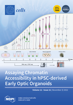
Cover Story (view full-size image): To investigate cellular and molecular changes in hPSC-derived developing early retinal organoids, we generated SIX6-GFP and VSX2-tdTomato (or VSX2-h2b-mRuby3) dual fluorescent reporters. These 3D organoids expressed GFP at day 15 and tdTomato (or mRuby3) at day 25, respectively. This enabled us to explore transcriptional- and chromatin-related changes using RNA-seq and ATAC-seq, from pluripotency through to early retina specification. Correlating gene transcription with chromatin accessibility in early eye field development showed that retinal cells underwent a clear change in the chromatin landscape, as well as gene expression profiles. Pathway analysis revealed a stepwise loss of pluripotency, while optic vesicle and retina pathways became progressively more prevalent. Considering both datasets in parallel provided an informative glimpse into the molecular nature of eye development. View this paper
  • Issues are regarded as officially published after their release is announced to the table of contents alert mailing list.
  • You may sign up for e-mail alerts to receive table of contents of newly released issues.
  • PDF is the official format for papers published in both, html and pdf forms. To view the papers in pdf format, click on the "PDF Full-text" link, and use the free Adobe Reader to open them.
Order results
Result details
Section
Select all
Export citation of selected articles as:
22 pages, 1696 KiB  
Review
NMR-Based Chromatography Readouts: Indispensable Tools to “Translate” Analytical Features into Molecular Structures
by Christoph Seger and Sonja Sturm
Cells 2022, 11(21), 3526; https://doi.org/10.3390/cells11213526 - 7 Nov 2022
Cited by 3 | Viewed by 2969
Abstract
Gaining structural information is a must to allow the unequivocal structural characterization of analytes from natural sources. In liquid state, NMR spectroscopy is almost the only possible alternative to HPLC-MS and hyphenating the effluent of an analyte separation device to the probe head [...] Read more.
Gaining structural information is a must to allow the unequivocal structural characterization of analytes from natural sources. In liquid state, NMR spectroscopy is almost the only possible alternative to HPLC-MS and hyphenating the effluent of an analyte separation device to the probe head of an NMR spectrometer has therefore been pursued for more than three decades. The purpose of this review article was to demonstrate that, while it is possible to use mass spectrometry and similar methods to differentiate, group, and often assign the differentiating variables to entities that can be recognized as single molecules, the structural characterization of these putative biomarkers usually requires the use of NMR spectroscopy. Full article
Show Figures

Figure 1

13 pages, 1279 KiB  
Review
The Versatile Role of miR-21 in Renal Homeostasis and Diseases
by Romain Larrue, Sandy Fellah, Cynthia Van der Hauwaert, Marie-Flore Hennino, Michaël Perrais, Arnaud Lionet, François Glowacki, Nicolas Pottier and Christelle Cauffiez
Cells 2022, 11(21), 3525; https://doi.org/10.3390/cells11213525 - 7 Nov 2022
Cited by 8 | Viewed by 2841
Abstract
MicroRNAs (miRNAs) are small, non-coding RNA species that control gene expression and confer robustness to biological processes. Over the last two decades, their important roles during kidney development, homeostasis and the treatment of diseases have been established, in particular during the onset and [...] Read more.
MicroRNAs (miRNAs) are small, non-coding RNA species that control gene expression and confer robustness to biological processes. Over the last two decades, their important roles during kidney development, homeostasis and the treatment of diseases have been established, in particular during the onset and progression of various forms of acute and chronic renal disorders. In recent years, miR-21, one of the best-characterized miRNAs to date, has received much attention in renal physiology in particular given its high degree of conservation and expression in kidneys, as well as its potent pathogenic role in various debilitating renal diseases. This review summarizes the current knowledge on miR-21’s involvement in both renal homeostasis and diseases, in particular its double-edged-sword role in acute versus chronic kidney injuries. Finally, we also discuss the potential of miR-21 as a biomarker and therapeutic target in renal diseases. Full article
(This article belongs to the Topic MicroRNA: Mechanisms of Action, Physio-Pathological Implications, and Disease Biomarkers)
(This article belongs to the Section Cell Microenvironment)
Show Figures

Graphical abstract

18 pages, 5445 KiB  
Article
The Protective Effects of Human Embryonic Stem Cell-Derived Mesenchymal Stem Cells in Noise-Induced Hearing Loss of Rats
by So Young Kim, Jeoung Eun Lee, Sung Hun Kang, So Min Lee, Jiwon Jeon and Dong Ryul Lee
Cells 2022, 11(21), 3524; https://doi.org/10.3390/cells11213524 - 7 Nov 2022
Cited by 1 | Viewed by 2332
Abstract
A few prior animal studies have suggested the transplantation or protective effects of mesenchymal stem cells (MSCs) in noise-induced hearing loss. This study intended to evaluate the fates of administered MSCs in the inner ears and the otoprotective effects of MSCs in the [...] Read more.
A few prior animal studies have suggested the transplantation or protective effects of mesenchymal stem cells (MSCs) in noise-induced hearing loss. This study intended to evaluate the fates of administered MSCs in the inner ears and the otoprotective effects of MSCs in the noise-induced hearing loss of rats. Human embryonic stem cell-derived MSCs (ES-MSCs) were systematically administered via the tail vein in adult rats. Eight-week-old Sprague-Dawley rats were randomly allocated to the control (n = 8), ES-MSC (n = 4), noise (n = 8), and ES-MSC+noise (n = 10) groups. In ES-MSC and ES-MSC+noise rats, 5 × 10⁵ ES-MSCs were injected via the tail vein. In noise and ES-MSC+noise rats, broadband noise with 115 dB SPL was exposed for 3 h daily for 5 days. The hearing levels were measured using auditory brainstem response (ABR) at 4, 8, 16, and 32 kHz. Cochlear histology was examined using H&E staining and cochlear whole mount immunofluorescence. The presence of human DNA was examined using Sry PCR, and the presence of human cytoplasmic protein was examined using STEM121 immunofluorescence staining. The protein expression levels of heat shock protein 70 (HSP70), apoptosis-inducing factor (AIF), poly (ADP-ribose) (PAR), PAR polymerase (PARP), caspase 3, and cleaved caspase 3 were estimated. The ES-MSC rats did not show changes in ABR thresholds following the administration of ES-MSCs. The ES-MSC+ noise rats demonstrated lower ABR thresholds at 4, 8, and 16 kHz than the noise rats. Cochlear spiral ganglial cells and outer hair cells were more preserved in the ES-MSC+ noise rats than in the noise rats. The Sry PCR bands were highly detected in lung tissue and less in cochlear tissue of ES-MSC+noise rats. Only a few STEM121-positivities were observed in the spiral ganglial cell area of ES-MSC and ES-MSC+noise rats. The protein levels of AIF, PAR, PARP, caspase 3, and cleaved caspase 3 were lower in the ES-MSC+noise rats than in the noise rats. The systemic injection of ES-MSCs preserved hearing levels and attenuated parthanatos and apoptosis in rats with noise-induced hearing loss. In addition, a tiny number of transplanted ES-MSCs were observed in the spiral ganglial areas. Full article
(This article belongs to the Special Issue Stem Cells and Hearing Loss)
Show Figures

Figure 1

27 pages, 3017 KiB  
Article
Branched-Chain Amino Acids Are Linked with Alzheimer’s Disease-Related Pathology and Cognitive Deficits
by Md Abu Bakkar Siddik, Caitlyn A. Mullins, Alyssa Kramer, Harsh Shah, Ritchel B. Gannaban, Masoud Zabet-Moghaddam, Ryan M. Huebinger, Vijay K. Hegde, Sheba M. J. MohanKumar, Puliyur S. MohanKumar and Andrew C. Shin
Cells 2022, 11(21), 3523; https://doi.org/10.3390/cells11213523 - 7 Nov 2022
Cited by 19 | Viewed by 5283
Abstract
Alzheimer’s disease (AD) is an irreversible neurodegenerative disorder with a complex pathophysiology. Type 2 diabetes (T2D) is a strong risk factor for AD that shares similar abnormal features including metabolic dysregulation and brain pathology such as amyloid and/or Tau deposits. Emerging evidence suggests [...] Read more.
Alzheimer’s disease (AD) is an irreversible neurodegenerative disorder with a complex pathophysiology. Type 2 diabetes (T2D) is a strong risk factor for AD that shares similar abnormal features including metabolic dysregulation and brain pathology such as amyloid and/or Tau deposits. Emerging evidence suggests that circulating branched-chain amino acids (BCAAs) are associated with T2D. While excess BCAAs are shown to be harmful to neurons, its connection to AD is poorly understood. Here we show that individuals with AD have elevated circulating BCAAs and their metabolites compared to healthy individuals, and that a BCAA metabolite is correlated with the severity of dementia. APPSwe mouse model of AD also displayed higher plasma BCAAs compared to controls. In pursuit of understanding a potential causality, BCAA supplementation to HT-22 neurons was found to reduce genes critical for neuronal health while increasing phosphorylated Tau. Moreover, restricting BCAAs from diet delayed cognitive decline and lowered AD-related pathology in the cortex and hippocampus in APP/PS1 mice. BCAA restriction for two months was sufficient to correct glycemic control and increased/restored dopamine that were severely reduced in APP/PS1 controls. Treating 5xFAD mice that show early brain pathology with a BCAA-lowering compound recapitulated the beneficial effects of BCAA restriction on brain pathology and neurotransmitters including norepinephrine and serotonin. Collectively, this study reveals a positive association between circulating BCAAs and AD. Our findings suggest that BCAAs impair neuronal functions whereas BCAA-lowering alleviates AD-related pathology and cognitive decline, thus establishing a potential causal link between BCAAs and AD progression. Full article
(This article belongs to the Section Cells of the Nervous System)
Show Figures

Graphical abstract

18 pages, 3156 KiB  
Article
Premature Macrophage Activation by Stored Red Blood Cell Transfusion Halts Liver Regeneration Post-Partial Hepatectomy in Rats
by Nathalie Abudi, Omri Duev, Tal Asraf, Simcha Blank, Idit Matot and Rinat Abramovitch
Cells 2022, 11(21), 3522; https://doi.org/10.3390/cells11213522 - 7 Nov 2022
Cited by 1 | Viewed by 2368
Abstract
Liver resection is a common treatment for various conditions and often requires blood transfusions to compensate for operative blood loss. As partial hepatectomy (PHx) is frequently performed in patients with a pre-damaged liver, avoiding further injury is of paramount clinical importance. Our aim [...] Read more.
Liver resection is a common treatment for various conditions and often requires blood transfusions to compensate for operative blood loss. As partial hepatectomy (PHx) is frequently performed in patients with a pre-damaged liver, avoiding further injury is of paramount clinical importance. Our aim was to study the impact of red blood cell (RBC) resuscitation on liver regeneration. We assessed the impact of RBC storage time on liver regeneration following 50% PHx in rats and explored possible contributing molecular mechanisms using immunohistochemistry, RNA-Seq, and macrophage depletion. The liver was successfully regenerated after PHx when rats were transfused with fresh RBCs (F-RBCs). However, in rats resuscitated with stored RBCs (S-RBCs), the regeneration process was disrupted, as detected by delayed hepatocyte proliferation and lack of hypertrophy. The delayed regeneration was associated with elevated numbers of hemorrhage-activated liver macrophages (Mhem) secreting HO-1. Depletion of macrophages prior to PHx and transfusion improved the regeneration process. Gene expression profiling revealed alterations in numerous genes belonging to critical pathways, including cell cycle and DNA replication, and genes associated with immune cell activation, such as chemokine signaling and platelet activation and adhesion. Our results implicate activated macrophages in delayed liver regeneration following S-RBC transfusion via HO-1 and PAI-1 overexpression. Full article
(This article belongs to the Special Issue Liver Injury and Regeneration: From Basic to Translational Research)
Show Figures

Figure 1

22 pages, 2250 KiB  
Review
Neutrophil Extracellular Traps in Asthma: Friends or Foes?
by Remo Poto, Mohamed Shamji, Gianni Marone, Stephen R. Durham, Guy W. Scadding and Gilda Varricchi
Cells 2022, 11(21), 3521; https://doi.org/10.3390/cells11213521 - 7 Nov 2022
Cited by 13 | Viewed by 4750
Abstract
Asthma is a chronic inflammatory disease characterized by variable airflow limitation and airway hyperresponsiveness. A plethora of immune and structural cells are involved in asthma pathogenesis. The roles of neutrophils and their mediators in different asthma phenotypes are largely unknown. Neutrophil extracellular traps [...] Read more.
Asthma is a chronic inflammatory disease characterized by variable airflow limitation and airway hyperresponsiveness. A plethora of immune and structural cells are involved in asthma pathogenesis. The roles of neutrophils and their mediators in different asthma phenotypes are largely unknown. Neutrophil extracellular traps (NETs) are net-like structures composed of DNA scaffolds, histones and granular proteins released by activated neutrophils. NETs were originally described as a process to entrap and kill a variety of microorganisms. NET formation can be achieved through a cell-death process, termed NETosis, or in association with the release of DNA from viable neutrophils. NETs can also promote the resolution of inflammation by degrading cytokines and chemokines. NETs have been implicated in the pathogenesis of various non-infectious conditions, including autoimmunity, cancer and even allergic disorders. Putative surrogate NET biomarkers (e.g., double-strand DNA (dsDNA), myeloperoxidase-DNA (MPO-DNA), and citrullinated histone H3 (CitH3)) have been found in different sites/fluids of patients with asthma. Targeting NETs has been proposed as a therapeutic strategy in several diseases. However, different NETs and NET components may have alternate, even opposite, consequences on inflammation. Here we review recent findings emphasizing the pathogenic and therapeutic potential of NETs in asthma. Full article
(This article belongs to the Special Issue Metabolic Inflammation and Cellular Immunity)
Show Figures

Figure 1

15 pages, 2474 KiB  
Article
Arginine Reduces Glycation in γ2 Subunit of AMPK and Pathologies in Alzheimer’s Disease Model Mice
by Rui Zhu, Ying Lei, Fangxiao Shi, Qing Tian and Xinwen Zhou
Cells 2022, 11(21), 3520; https://doi.org/10.3390/cells11213520 - 7 Nov 2022
Cited by 8 | Viewed by 2011
Abstract
The metabolism disorders are a common convergence of Alzheimer’s disease (AD) and type 2 diabetes mellitus (T2DM). The characteristics of AD are senile plaques and neurofibrillary tangles (NFTs) composed by deposits of amyloid−β (Aβ) and phosphorylated tau, respectively. Advanced glycation end−products (AGEs) are [...] Read more.
The metabolism disorders are a common convergence of Alzheimer’s disease (AD) and type 2 diabetes mellitus (T2DM). The characteristics of AD are senile plaques and neurofibrillary tangles (NFTs) composed by deposits of amyloid−β (Aβ) and phosphorylated tau, respectively. Advanced glycation end−products (AGEs) are a stable modification of proteins by non−enzymatic reactions, which could result in the protein dysfunction. AGEs are associated with some disease developments, such as diabetes mellitus and AD, but the effects of the glycated γ2 subunit of AMPK on its activity and the roles in AD onset are unknown. Methods: We studied the effect of glycated γ2 subunit of AMPK on its activity in N2a cells. In 3 × Tg mice, we administrated L−arginine once every two days for 45 days and evaluated the glycation level of γ2 subunit and function of AMPK and alternation of pathologies. Results: The glycation level of γ2 subunit was significantly elevated in 3 × Tg mice as compared with control mice, meanwhile, the level of pT172−AMPK was obviously lower in 3 × Tg mice than that in control mice. Moreover, we found that arginine protects the γ2 subunit of AMPK from glycation, preserves AMPK function, and improves pathologies and cognitive deficits in 3 × Tg mice. Conclusions: Arginine treatment decreases glycated γ2 subunit of AMPK and increases p−AMPK levels in 3 × Tg mice, suggesting that reduced glycation of the γ2 subunit could ameliorate AMPK function and become a new target for AD therapy in the future. Full article
Show Figures

Graphical abstract

16 pages, 3878 KiB  
Article
Dynamic Balance between PTH1R-Dependent Signal Cascades Determines Its Pro- or Anti-Osteogenic Effects on MSC
by Konstantin Kulebyakin, Pyotr Tyurin-Kuzmin, Leila Sozaeva, Nikita Voloshin, Mikhail Nikolaev, Vadim Chechekhin, Maxim Vigovskiy, Veronika Sysoeva, Elizaveta Korchagina, Daria Naida and Maria Vorontsova
Cells 2022, 11(21), 3519; https://doi.org/10.3390/cells11213519 - 7 Nov 2022
Cited by 4 | Viewed by 2528
Abstract
Parathyroid hormone (PTH) is one of the key regulators of calcium and phosphate metabolism in the body, controlling bone metabolism and ion excretion by the kidneys. At present, attempts to use PTH as a therapeutic agent have been associated with side-effects, the nature [...] Read more.
Parathyroid hormone (PTH) is one of the key regulators of calcium and phosphate metabolism in the body, controlling bone metabolism and ion excretion by the kidneys. At present, attempts to use PTH as a therapeutic agent have been associated with side-effects, the nature of which is not always clear and predictable. In addition, it is known that in vivo impairment of PTH post-receptor signaling is associated with atypical differentiation behavior not only of bone cells, but also of connective tissues, including adipose tissue. In this work, we studied the functional responses of multipotent mesenchymal stromal cells (MSCs) to the action of PTH at the level of single cells. We used MSCs isolated from the periosteum and subcutaneous adipose tissue to compare characteristics of cell responses to PTH. We found that the hormone can activate three key responses via its receptor located on the surface of MSCs: single transients of calcium, calcium oscillations, and hormone-activated smooth increase in intracellular calcium. These types of calcium responses led to principally different cellular responses of MSCs. The cAMP-dependent smooth increase of intracellular calcium was associated with pro-osteogenic action of PTH, whereas phospholipase C dependent calcium oscillations led to a decrease in osteogenic differentiation intensity. Different variants of calcium responses are in dynamic equilibrium. Suppression of one type of response leads to increased activation of another type and, accordingly, to a change in the effect of PTH on cell differentiation. Full article
(This article belongs to the Special Issue Advances in Calcium Signaling)
Show Figures

Figure 1

14 pages, 3532 KiB  
Article
Bta-miR-199a-3p Inhibits LPS-Induced Inflammation in Bovine Mammary Epithelial Cells via the PI3K/AKT/NF-κB Signaling Pathway
by Yuhang Li, Qianqian Ren, Xingping Wang, Zhuoma Luoreng and Dawei Wei
Cells 2022, 11(21), 3518; https://doi.org/10.3390/cells11213518 - 7 Nov 2022
Cited by 9 | Viewed by 2363
Abstract
Mastitis is characterized by inflammatory damage to mammary gland tissue, which could decline milk production and quality and significantly affect the economic benefits of ranching. MicroRNAs (miRNAs), such as miR-199a-3p, are novel therapeutic targets in inflammation, and their regulation is an effective strategy [...] Read more.
Mastitis is characterized by inflammatory damage to mammary gland tissue, which could decline milk production and quality and significantly affect the economic benefits of ranching. MicroRNAs (miRNAs), such as miR-199a-3p, are novel therapeutic targets in inflammation, and their regulation is an effective strategy for inflammation control. Despite its importance in humans and animals, the molecular mechanism of bovine miR-199a-3p (bta-miR-199a-3p) in dairy cow mastitis and bovine mammary epithelial cell (bMEC) inflammation is unclear. In our study, a bovine mammary epithelial cell line (MAC-T) induced by lipopolysaccharide (LPS) was used as an inflammatory cell model to investigate the molecular mechanism of bta-miR-199a-3p in the MAC-T inflammatory response. bta-miR-199a-3p was up-regulated in the LPS-induced MAC-T cells, while CD2-associated protein (CD2AP) was revealed as its target gene in a double luciferase reporter gene experiment. In addition, the overexpression of bta-miR-199a-3p negatively regulated the expression of CD2AP and the activation of the phosphatidylinositol 3-kinase (PI3K)/AKT/nuclear factor kappa-B (NF-κB) signaling pathway. These subsequently inhibited the secretion of related inflammatory factors (TNF-α, IL-1β, and IL-6) and the expression of apoptotic genes (CASP3 and CASP9), thereby alleviating the LPS-challenged inflammatory response in the MAC-T cells. Silencing of bta-miR-199a-3p, however, reversed the above effects. Thus, bta-miR-199a-3p inhibits LPS-induced inflammation in bMECs by directly targeting CD2AP and regulating the PI3K/AKT/NF-κB signaling pathway. This study reveals the potential regulatory mechanism of bta-miR-199a-3p in bMEC inflammatory immune response and may serve as a useful target for the treatment of mastitis. Full article
(This article belongs to the Collection Regulatory Functions of microRNAs)
Show Figures

Figure 1

16 pages, 3644 KiB  
Article
PI3K Isoform-Specific Regulation of Leader and Follower Cell Function for Collective Migration and Proliferation in Response to Injury
by Morgan D. Basta, A. Sue Menko and Janice L. Walker
Cells 2022, 11(21), 3515; https://doi.org/10.3390/cells11213515 - 7 Nov 2022
Cited by 2 | Viewed by 2086
Abstract
To ensure proper wound healing it is important to elucidate the signaling cues that coordinate leader and follower cell behavior to promote collective migration and proliferation for wound healing in response to injury. Using an ex vivo post-cataract surgery wound healing model we [...] Read more.
To ensure proper wound healing it is important to elucidate the signaling cues that coordinate leader and follower cell behavior to promote collective migration and proliferation for wound healing in response to injury. Using an ex vivo post-cataract surgery wound healing model we investigated the role of class I phosphatidylinositol-3-kinase (PI3K) isoforms in this process. Our findings revealed a specific role for p110α signaling independent of Akt for promoting the collective migration and proliferation of the epithelium for wound closure. In addition, we found an important role for p110α signaling in orchestrating proper polarized cytoskeletal organization within both leader and wounded epithelial follower cells to coordinate their function for wound healing. p110α was necessary to signal the formation and persistence of vimentin rich-lamellipodia extensions by leader cells and the reorganization of actomyosin into stress fibers along the basal domains of the wounded lens epithelial follower cells for movement. Together, our study reveals a critical role for p110α in the collective migration of an epithelium in response to wounding. Full article
(This article belongs to the Special Issue New Advances in Lens Biology and Pathology)
Show Figures

Figure 1

23 pages, 3735 KiB  
Article
Bioinformatics Analysis and Validation of the Role of Lnc-RAB11B-AS1 in the Development and Prognosis of Hepatocellular Carcinoma
by Dedong Wang, Xiangzhi Hu, Jinbin Chen, Boheng Liang, Lin Zhang, Pengzhe Qin and Di Wu
Cells 2022, 11(21), 3517; https://doi.org/10.3390/cells11213517 - 6 Nov 2022
Cited by 4 | Viewed by 2483
Abstract
Lnc-RAB11B-AS1 is reported to be dysregulated in several types of cancers and can function as both an oncogene and tumor suppressor gene. To evaluate the potential role of lnc-RAB11B-AS1 in hepatocellular carcinoma (HCC), we investigated and evaluated its expression in HCC based on [...] Read more.
Lnc-RAB11B-AS1 is reported to be dysregulated in several types of cancers and can function as both an oncogene and tumor suppressor gene. To evaluate the potential role of lnc-RAB11B-AS1 in hepatocellular carcinoma (HCC), we investigated and evaluated its expression in HCC based on the data mining of a series of public databases, including TCGA, GEO, ICGC, HPA, DAVID, cBioPortal, GeneMIANA, TIMER, and ENCORI. The data showed downregulation of lnc-RAB11B-AS1 in HCC and was accompanied by the synchronous downregulation of the targeted RAB11B mRNA and its protein. Low expression of lnc-RAB11B-AS1 was associated with shorter overall survival (OS) and disease-free survival (DFS) of HCC patients, PD1/PD-L1 was correlated with low expression of RAB11B. Furthermore, Gene Ontology (GO) functional annotation and Kyoto Encyclopedia of Genes and Genomes (KEGG) pathway enrichment analysis showed a correlation between immune cell change and non-alcoholic fatty liver disease. The above findings revealed that lnc-RAB11B-AS1 was down-regulated in HCC and closely associated with the clinical stage of the HCC patients, suggesting that lnc-RAB11B-AS1 could be a possible predictor for HCC and a potential new therapeutic target for the treatment of HCC. Full article
(This article belongs to the Section Cell Methods)
Show Figures

Figure 1

42 pages, 1992 KiB  
Review
Generation of Lens Progenitor Cells and Lentoid Bodies from Pluripotent Stem Cells: Novel Tools for Human Lens Development and Ocular Disease Etiology
by Aleš Cvekl and Michael John Camerino
Cells 2022, 11(21), 3516; https://doi.org/10.3390/cells11213516 - 6 Nov 2022
Cited by 8 | Viewed by 3778
Abstract
In vitro differentiation of human pluripotent stem cells (hPSCs) into specialized tissues and organs represents a powerful approach to gain insight into those cellular and molecular mechanisms regulating human development. Although normal embryonic eye development is a complex process, generation of ocular organoids [...] Read more.
In vitro differentiation of human pluripotent stem cells (hPSCs) into specialized tissues and organs represents a powerful approach to gain insight into those cellular and molecular mechanisms regulating human development. Although normal embryonic eye development is a complex process, generation of ocular organoids and specific ocular tissues from pluripotent stem cells has provided invaluable insights into the formation of lineage-committed progenitor cell populations, signal transduction pathways, and self-organization principles. This review provides a comprehensive summary of recent advances in generation of adenohypophyseal, olfactory, and lens placodes, lens progenitor cells and three-dimensional (3D) primitive lenses, “lentoid bodies”, and “micro-lenses”. These cells are produced alone or “community-grown” with other ocular tissues. Lentoid bodies/micro-lenses generated from human patients carrying mutations in crystallin genes demonstrate proof-of-principle that these cells are suitable for mechanistic studies of cataractogenesis. Taken together, current and emerging advanced in vitro differentiation methods pave the road to understand molecular mechanisms of cataract formation caused by the entire spectrum of mutations in DNA-binding regulatory genes, such as PAX6, SOX2, FOXE3, MAF, PITX3, and HSF4, individual crystallins, and other genes such as BFSP1, BFSP2, EPHA2, GJA3, GJA8, LIM2, MIP, and TDRD7 represented in human cataract patients. Full article
(This article belongs to the Special Issue New Advances in Lens Biology and Pathology)
Show Figures

Figure 1

23 pages, 2733 KiB  
Article
The Potential for Placental Activation of PPARγ to Improve the Angiogenic Profile in Preeclampsia
by Brooke Grimaldi, Hamid-Reza Kohan-Ghadr and Sascha Drewlo
Cells 2022, 11(21), 3514; https://doi.org/10.3390/cells11213514 - 6 Nov 2022
Cited by 6 | Viewed by 2297
Abstract
Preeclampsia (PE) is one of the most common causes of maternal-fetal morbidity and mortality world-wide. While the underlying causes of PE remain elusive, aberrant trophoblast differentiation and function are thought to cause an imbalance of secreted angiogenic proteins resulting in systemic endothelial dysfunction [...] Read more.
Preeclampsia (PE) is one of the most common causes of maternal-fetal morbidity and mortality world-wide. While the underlying causes of PE remain elusive, aberrant trophoblast differentiation and function are thought to cause an imbalance of secreted angiogenic proteins resulting in systemic endothelial dysfunction and organ damage in the mother. The placental dysfunction is also characterized by a reduction of the transcription factor, peroxisome proliferator activated receptor γ (PPARγ) which normally promotes trophoblast differentiation and healthy placental function. This study aimed to understand how placental activation of PPARγ effects the secretion of angiogenic proteins and subsequently endothelial function. To study this, healthy and PE placental tissues were cultured with or without the PPARγ agonist, Rosiglitazone, and a Luminex assay was performed to measure secreted proteins from the placenta. To assess the angiogenic effects of placental activation of PPARγ, human umbilical vein endothelial cells (HUVECs) were cultured with the placental conditioned media and the net angiogenic potential of these cells was measured by a tube formation assay. This is the first study to show PPARγ’s beneficial effect on the angiogenic profile in the human preeclamptic placenta through the reduction of anti-angiogenic angiopoietin-2 and soluble endoglin and the upregulation of pro-angiogenic placental growth factor, fibroblast growth factor-2, heparin-binding epidermal growth factor, and follistatin. The changes in the angiogenic profile were supported by the increased angiogenic potential observed in the HUVECs when cultured with conditioned media from rosiglitazone-treated preeclamptic placentas. The restoration of these disrupted pathways by activation of PPARγ in the preeclamptic placenta offers potential to improve placental and endothelial function in PE. Full article
(This article belongs to the Special Issue The Role of PPARs in Disease II)
Show Figures

Figure 1

11 pages, 3532 KiB  
Article
The Atypical Antipsychotic Lurasidone Affects Brain but Not Liver Cytochrome P450 2D (CYP2D) Activity. A Comparison with Other Novel Neuroleptics and Significance for Drug Treatment of Schizophrenia
by Przemysław J. Danek and Władysława A. Daniel
Cells 2022, 11(21), 3513; https://doi.org/10.3390/cells11213513 - 6 Nov 2022
Cited by 3 | Viewed by 2681
Abstract
The aim of this work was to study the effect of prolonged lurasidone administration on the cytochrome 2D (CYP2D) expression and activity in the rat liver and selected brain structures involved in the therapeutic or side effects of this neuroleptic. Male Wistar rats [...] Read more.
The aim of this work was to study the effect of prolonged lurasidone administration on the cytochrome 2D (CYP2D) expression and activity in the rat liver and selected brain structures involved in the therapeutic or side effects of this neuroleptic. Male Wistar rats received lurasidone (1 mg/kg ip.) for two weeks. The activity of CYP2D was measured in brain and liver microsomes as the rate of bufuralol 1′-hydroxylation. The CYP2D protein level was determined in microsomes by Western blot analysis. The CYP2D gene expression was estimated in liver tissue by a qRT-PCR method. Lurasidone decreased the activity and protein level of CYP2D in the frontal cortex but increased them in the striatum, nucleus accumbens, brain stem, substantia nigra, and the remainder of the brain. The neuroleptic did not affect CYP2D in the hippocampus, hypothalamus, and cerebellum. In the liver, lurasidone did not affect the CYP2D activity and protein level, though it enhanced the mRNA of CYP2D1 without affecting that of CYP2D2, CYP2D3, CYP2D4, and CYP2D5. In conclusion, lurasidone regulates brain (but not liver) CYP2D activity/protein level in a region-dependent manner, which is similar to that of other atypical neuroleptics (iloperidone and asenapine) as concerns the frontal cortex (down-regulation) and nigrostriatal pathway (up-regulation) and may be of pharmacological significance. However, further molecular studies with selective receptor agonists are necessary to find out which individual monoaminergic receptors/signaling pathways are involved in the regulation of the rat CYP2D4 and human CYP2D6 enzyme in particular brain structures. Full article
Show Figures

Graphical abstract

16 pages, 3747 KiB  
Article
A Balance between Transmembrane-Mediated ER/Golgi Retention and Forward Trafficking Signals in Glycophorin-Anion Exchanger-1 Interaction
by Kate Hsu, Ting-Ying Lee, Jian-Yi Lin and Pin-Lung Chen
Cells 2022, 11(21), 3512; https://doi.org/10.3390/cells11213512 - 6 Nov 2022
Cited by 2 | Viewed by 2094
Abstract
Anion exchanger-1 (AE1) is the main erythroid Cl/HCO3 transporter that supports CO2 transport. Glycophorin A (GPA), a component of the AE1 complexes, facilitates AE1 expression and anion transport, but Glycophorin B (GPB) does not. Here, we dissected the [...] Read more.
Anion exchanger-1 (AE1) is the main erythroid Cl/HCO3 transporter that supports CO2 transport. Glycophorin A (GPA), a component of the AE1 complexes, facilitates AE1 expression and anion transport, but Glycophorin B (GPB) does not. Here, we dissected the structural components of GPA/GPB involved in glycophorin-AE1 trafficking by comparing them with three GPB variants—GPBhead (lacking the transmembrane domain [TMD]), GPBtail (mainly the TMD), and GP.Mur (glycophorin B-A-B hybrid). GPB-derived GP.Mur bears an O-glycopeptide that encompasses the R18 epitope, which is present in GPA but not GPB. By flow cytometry, AE1 expression in the control erythrocytes increased with the GPA-R18 expression; GYP.Mur+/+ erythrocytes bearing both GP.Mur and GPA expressed more R18 epitopes and more AE1 proteins. In contrast, heterologously expressed GPBtail and GPB were predominantly localized in the Golgi apparatus of HEK-293 cells, whereas GBhead was diffuse throughout the cytosol, suggesting that glycophorin transmembrane encoded an ER/Golgi retention signal. AE1 coexpression could reduce the ER/Golgi retention of GPB, but not of GPBtail or GPBhead. Thus, there are forward-trafficking and transmembrane-driven ER/Golgi retention signals encoded in the glycophorin sequences. How the balance between these opposite trafficking signals could affect glycophorin sorting into AE1 complexes and influence erythroid anion transport remains to be explored. Full article
(This article belongs to the Topic Advances in Red Blood Cells Research)
Show Figures

Figure 1

19 pages, 1191 KiB  
Review
Mitochondrial Cardiomyopathy: Molecular Epidemiology, Diagnosis, Models, and Therapeutic Management
by Jinjuan Yang, Shaoxiang Chen, Fuyu Duan, Xiuxiu Wang, Xiaoxian Zhang, Boonxuan Lian, Meng Kou, Zhixin Chiang, Ziyue Li and Qizhou Lian
Cells 2022, 11(21), 3511; https://doi.org/10.3390/cells11213511 - 6 Nov 2022
Cited by 10 | Viewed by 3693
Abstract
Mitochondrial cardiomyopathy (MCM) is characterized by abnormal heart-muscle structure and function, caused by mutations in the nuclear genome or mitochondrial DNA. The heterogeneity of gene mutations and various clinical presentations in patients with cardiomyopathy make its diagnosis, molecular mechanism, and therapeutics great challenges. [...] Read more.
Mitochondrial cardiomyopathy (MCM) is characterized by abnormal heart-muscle structure and function, caused by mutations in the nuclear genome or mitochondrial DNA. The heterogeneity of gene mutations and various clinical presentations in patients with cardiomyopathy make its diagnosis, molecular mechanism, and therapeutics great challenges. This review describes the molecular epidemiology of MCM and its clinical features, reviews the promising diagnostic tests applied for mitochondrial diseases and cardiomyopathies, and details the animal and cellular models used for modeling cardiomyopathy and to investigate disease pathogenesis in a controlled in vitro environment. It also discusses the emerging therapeutics tested in pre-clinical and clinical studies of cardiac regeneration. Full article
Show Figures

Figure 1

14 pages, 3035 KiB  
Article
Volatile Compounds in Actinomycete Communities: A New Tool for Biosynthetic Gene Cluster Activation, Cooperative Growth Promotion, and Drug Discovery
by Lorena Cuervo, Carmen Méndez, José A. Salas, Carlos Olano and Mónica G. Malmierca
Cells 2022, 11(21), 3510; https://doi.org/10.3390/cells11213510 - 5 Nov 2022
Cited by 10 | Viewed by 3179
Abstract
The increasing appearance of multiresistant pathogens, as well as emerging diseases, has highlighted the need for new strategies to discover natural compounds that can be used as therapeutic alternatives, especially in the genus Streptomyces, which is one of the largest producers of [...] Read more.
The increasing appearance of multiresistant pathogens, as well as emerging diseases, has highlighted the need for new strategies to discover natural compounds that can be used as therapeutic alternatives, especially in the genus Streptomyces, which is one of the largest producers of bioactive metabolites. In recent years, the study of volatile compounds (VOCs) has raised interest because of the variety of their biological properties in addition to their involvement in cell communication. In this work, we analyze the implications of VOCs as mediating molecules capable of inducing the activation of biosynthetic pathways of bioactive compounds in surrounding Actinomycetes. For this purpose, several strains of Streptomyces were co-cultured in chamber devices that allowed VOC exchange while avoiding physical contact. In several of those strains, secondary metabolism was activated by VOCs emitted by companion strains, resulting in increased antibiotic production and synthesis of new VOCs. This study shows a novel strategy to exploit the metabolic potential of Actinomycetes as well as emphasizes the importance of studying the interactions between different microorganisms sharing the same ecological niche. Full article
Show Figures

Figure 1

37 pages, 1443 KiB  
Review
Integration of O-GlcNAc into Stress Response Pathways
by Kamau M. M. Fahie, Kyriakos N. Papanicolaou and Natasha E. Zachara
Cells 2022, 11(21), 3509; https://doi.org/10.3390/cells11213509 - 5 Nov 2022
Cited by 27 | Viewed by 5027
Abstract
The modification of nuclear, mitochondrial, and cytosolic proteins by O-linked βN-acetylglucosamine (O-GlcNAc) has emerged as a dynamic and essential post-translational modification of mammalian proteins. O-GlcNAc is cycled on and off over 5000 proteins in response to diverse stimuli impacting protein function and, in [...] Read more.
The modification of nuclear, mitochondrial, and cytosolic proteins by O-linked βN-acetylglucosamine (O-GlcNAc) has emerged as a dynamic and essential post-translational modification of mammalian proteins. O-GlcNAc is cycled on and off over 5000 proteins in response to diverse stimuli impacting protein function and, in turn, epigenetics and transcription, translation and proteostasis, metabolism, cell structure, and signal transduction. Environmental and physiological injury lead to complex changes in O-GlcNAcylation that impact cell and tissue survival in models of heat shock, osmotic stress, oxidative stress, and hypoxia/reoxygenation injury, as well as ischemic reperfusion injury. Numerous mechanisms that appear to underpin O-GlcNAc-mediated survival include changes in chaperone levels, impacts on the unfolded protein response and integrated stress response, improvements in mitochondrial function, and reduced protein aggregation. Here, we discuss the points at which O-GlcNAc is integrated into the cellular stress response, focusing on the roles it plays in the cardiovascular system and in neurodegeneration. Full article
(This article belongs to the Special Issue Molecular Mechanism of Stress, Stress Response, and Adaptation)
Show Figures

Graphical abstract

12 pages, 2168 KiB  
Review
Targeting Ferroptosis Holds Potential for Intervertebral Disc Degeneration Therapy
by Jiaxing Chen, Xinyu Yang, Yi Feng, Qiaochu Li, Jingjin Ma, Linbang Wang and Zhengxue Quan
Cells 2022, 11(21), 3508; https://doi.org/10.3390/cells11213508 - 5 Nov 2022
Cited by 17 | Viewed by 3383
Abstract
Intervertebral disc degeneration (IVDD) is a common pathological condition responsible for lower back pain, which can significantly increase economic and social burdens. Although considerable efforts have been made to identify potential mechanisms of disc degeneration, the treatment of IVDD is not satisfactory. Ferroptosis, [...] Read more.
Intervertebral disc degeneration (IVDD) is a common pathological condition responsible for lower back pain, which can significantly increase economic and social burdens. Although considerable efforts have been made to identify potential mechanisms of disc degeneration, the treatment of IVDD is not satisfactory. Ferroptosis, a recently reported form of regulated cell death (RCD), is characterized by iron-dependent lipid peroxidation and has been demonstrated to be responsible for a variety of degenerative diseases. Accumulating evidence suggests that ferroptosis is implicated in IVDD by decreasing viability and increasing extracellular matrix degradation of nucleus pulposus cells, annulus fibrosus cells, or endplate chondrocytes. In this review, we summarize the literature regarding ferroptosis of intervertebral disc cells and discuss its molecular pathways and biomarkers for treating IVDD. Importantly, ferroptosis is verified as a promising therapeutic target for IVDD. Full article
Show Figures

Figure 1

19 pages, 1623 KiB  
Review
Current Status and Prospects of Targeted Therapy for Osteosarcoma
by Zunguo Hu, Shuang Wen, Zijun Huo, Qing Wang, Jiantao Zhao, Zihao Wang, Yanchun Chen, Lingyun Zhang, Fenghua Zhou, Zhangyu Guo, Huancai Liu and Shuanhu Zhou
Cells 2022, 11(21), 3507; https://doi.org/10.3390/cells11213507 - 5 Nov 2022
Cited by 37 | Viewed by 3694
Abstract
Osteosarcoma (OS) is a highly malignant tumor occurring in bone tissue with a high propensity to metastasize, and its underlying mechanisms remain largely elusive. The OS prognosis is poor, and improving the survival of OS patients remains a challenge. Current treatment methods such [...] Read more.
Osteosarcoma (OS) is a highly malignant tumor occurring in bone tissue with a high propensity to metastasize, and its underlying mechanisms remain largely elusive. The OS prognosis is poor, and improving the survival of OS patients remains a challenge. Current treatment methods such as surgical approaches, chemotherapeutic drugs, and immunotherapeutic drugs remain ineffective. As research progresses, targeted therapy is gradually becoming irreplaceable. In this review, several treatment modalities for osteosarcoma, such as surgery, chemotherapy, and immunotherapy, are briefly described, followed by a discussion of targeted therapy, the important targets, and new technologies for osteosarcoma treatment. Full article
(This article belongs to the Special Issue Advances in Bone Metabolism)
Show Figures

Figure 1

12 pages, 4352 KiB  
Article
LncKCNQ1OT1 Promotes the Odontoblastic Differentiation of Dental Pulp Stem Cells via Regulating hsa-miR-153-3p/RUNX2 Axis
by Xiaohui Lu, Jiawen Zhang, Yuanzhou Lu, Jing Xing, Min Lian, Guijuan Feng, Dan Huang, Chenfei Wang, Nimei Shen and Xingmei Feng
Cells 2022, 11(21), 3506; https://doi.org/10.3390/cells11213506 - 5 Nov 2022
Cited by 2 | Viewed by 1869
Abstract
This study aimed to explore the role of LncKCNQ1OT1/hsa-miR-153-3p/RUNX2 in the odontoblastic differentiation of human dental pulp stem cells (DPSCs) and its possible mechanism. The expression of LncKCNQ1OT1, hsa-miR-153-3p, and RUNX2 in the odontoblastic differentiation was detected by qRT-PCR. Interaction between LncKCNQ1OT1 and [...] Read more.
This study aimed to explore the role of LncKCNQ1OT1/hsa-miR-153-3p/RUNX2 in the odontoblastic differentiation of human dental pulp stem cells (DPSCs) and its possible mechanism. The expression of LncKCNQ1OT1, hsa-miR-153-3p, and RUNX2 in the odontoblastic differentiation was detected by qRT-PCR. Interaction between LncKCNQ1OT1 and hsa-miR-153-3p and interaction between hsa-miR-153-3p and RUNX2 were detected by dual-luciferase assay. The cell viability of DPSCs was detected by CCK-8, and the effect of LncKCNQ1OT1 and hsa-miR-153-3p on the odontoblastic differentiation of DPSCs was observed by alizarin red staining, alkaline phosphatase (ALP) activity assay, and Western blot for RUNX2, DSPP, and DMP-1. The results showed, during odontoblastic differentiation of DPSCs, the expression of LncKCNQ1OT1 increased, hsa-miR-153-3p expression decreased, and RUNX2 expression increased. Dual-luciferase assay showed that LncKCNQ1OT1 sponges hsa-miR-153-3p and hsa-miR-153-3p targets on RUNX2. After LncKCNQ1OT1 and hsa-miR-153-3p expressions of DPSCs were changed, the cell viability was not notably changed, but the odontoblastic differentiation was notably changed, which was confirmed with Alizarin Red staining, ALP activity, and Western blot for RUNX2, DSPP, and DMP-1. The results indicate that LncKCNQ1OT1 promotes the odontoblastic differentiation of DPSCs via regulating hsa-miR-153-3p/RUNX2 axis, which may provide a therapeutic clue for odontogenesis. Full article
Show Figures

Figure 1

16 pages, 3303 KiB  
Article
MK-2206 Alleviates Renal Fibrosis by Suppressing the Akt/mTOR Signaling Pathway In Vivo and In Vitro
by Meiling Chen, Yihang Yu, Tao Mi, Qitong Guo, Bin Xiang, Xiaomao Tian, Liming Jin, Chunlan Long, Lianju Shen, Xing Liu, Jianbo Pan, Yuanyuan Zhang, Tao Xu, Deying Zhang and Guanghui Wei
Cells 2022, 11(21), 3505; https://doi.org/10.3390/cells11213505 - 5 Nov 2022
Cited by 10 | Viewed by 2756
Abstract
Renal fibrosis is a common pathological feature of various kidney diseases, leading to irreversible renal failure and end-stage renal disease. However, there are still no effective treatments to reverse renal fibrosis. This study aimed to explore the potential mechanism of a targeted drug [...] Read more.
Renal fibrosis is a common pathological feature of various kidney diseases, leading to irreversible renal failure and end-stage renal disease. However, there are still no effective treatments to reverse renal fibrosis. This study aimed to explore the potential mechanism of a targeted drug for fibrosis. Here, unilateral ureteral obstruction (UUO)-treated mice and a TGF-β1-treated human renal tubular epithelial cell line (HK-2 cells) were used as models of renal fibrosis. Based on the changes of mRNA in UUO kidneys detected by transcriptome sequencing, MK-2206, an Akt inhibitor, was predicted as a potential drug to alleviate renal fibrosis through bioinformatics. We dissolved UUO mice with MK-2206 by gastric gavage and cultured TGF-β-induced HK-2 cells with MK-2206. Histopathological examinations were performed after MK-2206 intervention, and the degree of renal fibrosis, as well as the expression of Akt/mTOR pathway-related proteins, were evaluated by immunohistochemical staining, immunofluorescence staining, and Western blot. The results showed that MK-2206 significantly improved the pathological structure of the kidney. Furthermore, MK-2206 intervention effectively inhibited UUO- and TGF-β1-induced epithelial-mesenchymal transition, fibroblast activation, and extracellular matrix deposition. Mechanistically, MK-2206 treatment attenuated the activation of the Akt/mTOR signaling pathway. Taken together, our study revealed for the first time that MK-2206 is a promising drug for the improvement of renal fibrosis. Full article
(This article belongs to the Special Issue Recent Advances in Development and Progression of Kidney Diseases)
Show Figures

Figure 1

15 pages, 2879 KiB  
Article
The HSF1-CPT1a Pathway Is Differentially Regulated in NAFLD Progression
by Wiebke Breternitz, Friedrich Sandkühler, Frauke Grohmann, Jochen Hampe, Mario Brosch, Alexander Herrmann, Clemens Schafmayer, Christian Meinhardt, Stefan Schreiber, Alexander Arlt and Claudia Geismann
Cells 2022, 11(21), 3504; https://doi.org/10.3390/cells11213504 - 4 Nov 2022
Cited by 3 | Viewed by 2615
Abstract
Obesity and obesity-associated diseases represent one of the key health challenges of our time. In this context, aberrant hepatic lipid accumulation is a central pathological aspect of non-alcoholic fatty liver disease (NAFLD). By comparing methylation signatures of liver biopsies before and after bariatric [...] Read more.
Obesity and obesity-associated diseases represent one of the key health challenges of our time. In this context, aberrant hepatic lipid accumulation is a central pathological aspect of non-alcoholic fatty liver disease (NAFLD). By comparing methylation signatures of liver biopsies before and after bariatric surgery, we recently demonstrated the strong enrichment of differentially methylated heat shock factor 1 (HSF1) binding sites (>400-fold) in the process of liver remodeling, indicating a crucial role of HSF1 in modulating central aspects of NAFLD pathogenesis. Using cellular models of NAFLD, we were able to show that HSF1 is activated during fat accumulation in hepatocytes, mimicking conditions in patients before bariatric surgery. This induction was abolished by starving the cells, mimicking the situation after bariatric surgery. Regarding this connection, carnitine palmitoyltransferase 1 isoform A (CTP1a), a central regulator of lipid beta-oxidation, was identified as a HSF1 target gene by promoter analysis and HSF1 knockdown experiments. Finally, pharmacological activation of HSF1 through celastrol reduced fat accumulation in the cells in a HSF1-dependent manner. In conclusion, we were able to confirm the relevance of HSF1 activity and described a functional HSF1-CPT1a pathway in NAFLD pathogenesis. Full article
(This article belongs to the Special Issue Metabolic Regulation: Cell Growth and Proliferation)
Show Figures

Figure 1

18 pages, 923 KiB  
Review
Advanced Glycation End Products and Inflammation in Type 1 Diabetes Development
by Chenping Du, Rani O. Whiddett, Irina Buckle, Chen Chen, Josephine M. Forbes and Amelia K. Fotheringham
Cells 2022, 11(21), 3503; https://doi.org/10.3390/cells11213503 - 4 Nov 2022
Cited by 23 | Viewed by 3887
Abstract
Type 1 diabetes (T1D) is an autoimmune disease in which the β-cells of the pancreas are attacked by the host’s immune system, ultimately resulting in hyperglycemia. It is a complex multifactorial disease postulated to result from a combination of genetic and environmental factors. [...] Read more.
Type 1 diabetes (T1D) is an autoimmune disease in which the β-cells of the pancreas are attacked by the host’s immune system, ultimately resulting in hyperglycemia. It is a complex multifactorial disease postulated to result from a combination of genetic and environmental factors. In parallel with increasing prevalence of T1D in genetically stable populations, highlighting an environmental component, consumption of advanced glycation end products (AGEs) commonly found in in Western diets has increased significantly over the past decades. AGEs can bind to cell surface receptors including the receptor for advanced glycation end products (RAGE). RAGE has proinflammatory roles including in host–pathogen defense, thereby influencing immune cell behavior and can activate and cause proliferation of immune cells such as islet infiltrating CD8+ and CD4+ T cells and suppress the activity of T regulatory cells, contributing to β-cell injury and hyperglycemia. Insights from studies of individuals at risk of T1D have demonstrated that progression to symptomatic onset and diagnosis can vary, ranging from months to years, providing a window of opportunity for prevention strategies. Interaction between AGEs and RAGE is believed to be a major environmental risk factor for T1D and targeting the AGE-RAGE axis may act as a potential therapeutic strategy for T1D prevention. Full article
(This article belongs to the Topic Inflammation: The Cause of All Diseases)
Show Figures

Figure 1

18 pages, 3043 KiB  
Article
Palmitate Inhibits Mouse Macrophage Efferocytosis by Activating an mTORC1-Regulated Rho Kinase 1 Pathway: Therapeutic Implications for the Treatment of Obesity
by László Sós, Éva Garabuczi, Tibor Sághy, Gábor Mocsár and Zsuzsa Szondy
Cells 2022, 11(21), 3502; https://doi.org/10.3390/cells11213502 - 4 Nov 2022
Cited by 6 | Viewed by 2794
Abstract
Every day, billions of our cells die and get cleared without inducing inflammation. When, clearance is improper, uncleared cells undergo secondary necrosis and trigger inflammation. In addition, proper efferocytosis would be required for inducing resolution of inflammation, thus clearance deficiencies in the long [...] Read more.
Every day, billions of our cells die and get cleared without inducing inflammation. When, clearance is improper, uncleared cells undergo secondary necrosis and trigger inflammation. In addition, proper efferocytosis would be required for inducing resolution of inflammation, thus clearance deficiencies in the long term lead to development of various chronic inflammatory diseases. Increasing evidence indicates that obesity, itself being a low-grade inflammatory disease, predisposes to a variety of other chronic inflammatory diseases. Previous studies indicated that this later might be partially related to an impaired efferocytosis induced by increased uptake of circulating saturated fatty acids by macrophages in obese people. Here, we show that palmitate inhibits efferocytosis by bone marrow-derived macrophages in a dose-dependent manner. Palmitate triggers autophagy but also activates an energy-sensing mTORC1/ROCK1 signaling pathway, which interferes with the autophagosome–lysosome fusion, resulting in accumulation of the cellular membranes in autophagosomes. We propose that lack of sufficient plasma membrane supply attenuates efferocytosis of palmitate-exposed macrophages. AMP-activated protein kinase activators lead to mTORC1 inhibition and, consequently, released the palmitate-induced efferocytosis block in macrophages. Thus, they might be useful in the treatment of obesity not only by affecting metabolism thought so far. ROCK1 inhibitors could also be considered. Full article
Show Figures

Figure 1

21 pages, 2211 KiB  
Article
Secreted Factors and Extracellular Vesicles Account for the Immunomodulatory and Tissue Regenerative Properties of Bone-Marrow-Derived Mesenchymal Stromal Cells for Osteoarthritis
by Enrico Ragni, Carlotta Perucca Orfei and Laura de Girolamo
Cells 2022, 11(21), 3501; https://doi.org/10.3390/cells11213501 - 4 Nov 2022
Cited by 5 | Viewed by 2157
Abstract
Bone-marrow-derived mesenchymal stromal cells (BMSCs) showed therapeutic potential in the treatment of musculoskeletal diseases, including osteoarthritis (OA). Their soluble mediators and extracellular vesicles (EVs), which make up the secretome, suppress immune response, attenuate inflammation and promote cartilage repair. EVs, as well as the [...] Read more.
Bone-marrow-derived mesenchymal stromal cells (BMSCs) showed therapeutic potential in the treatment of musculoskeletal diseases, including osteoarthritis (OA). Their soluble mediators and extracellular vesicles (EVs), which make up the secretome, suppress immune response, attenuate inflammation and promote cartilage repair. EVs, as well as the whole secretome, have been investigated as cell free approaches for OA although, to date, a disease-tailored molecular fingerprint is missing. In this study, soluble mediators and miRNAs were sifted in the BMSCs’ secretome and EVs, respectively, and analyzed in the frame of cell types and factors involved in OA. The majority of identified molecules repress the activation of immune cells and the production of OA-related inflammatory mediators, as well as promote cartilage protection by acting on both chondrocytes homeostasis and extracellular matrix-degrading enzymes. These data provide the molecular ground for the therapeutic potential of BMSCs for regenerative applications for OA and support the use of secretome or EVs as cell-free applications in joint diseases. Full article
Show Figures

Figure 1

22 pages, 80706 KiB  
Article
Only Acute but Not Chronic Thrombocytopenia Protects Mice against Left Ventricular Dysfunction after Acute Myocardial Infarction
by Friedrich Reusswig, Amin Polzin, Meike Klier, Matthias Achim Dille, Aysel Ayhan, Marcel Benkhoff, Celina Lersch, Anika Prinz, Simone Gorressen, Jens Walter Fischer, Malte Kelm and Margitta Elvers
Cells 2022, 11(21), 3500; https://doi.org/10.3390/cells11213500 - 4 Nov 2022
Cited by 9 | Viewed by 1980
Abstract
Background: Platelets are major players of thrombosis and inflammation after acute myocardial infarction (AMI). The impact of thrombocytopenia on platelet-induced cellular processes post AMI is not well defined. Methods: The left anterior descending artery was ligated in C57/Bl6 mice and in two thrombocytopenic [...] Read more.
Background: Platelets are major players of thrombosis and inflammation after acute myocardial infarction (AMI). The impact of thrombocytopenia on platelet-induced cellular processes post AMI is not well defined. Methods: The left anterior descending artery was ligated in C57/Bl6 mice and in two thrombocytopenic mouse models to induce AMI. Results: Platelets from STEMI patients and from C57/Bl6 mice displayed enhanced platelet activation after AMI. This allows platelets to migrate into the infarct but not into the remote zone of the left ventricle. Acute thrombocytopenia by antibody-induced platelet depletion resulted in reduced infarct size and improved cardiac function 24 h and 21 days post AMI. This was due to reduced platelet-mediated inflammation after 24 h and reduced scar formation after 21 days post AMI. The collagen composition and interstitial collagen content in the left ventricle were altered due to platelet interaction with cardiac fibroblasts. Acute inflammation was also significantly reduced in Mpl−/− mice with chronic thrombocytopenia, but cardiac remodeling was unaltered. Consequently, left ventricular function, infarct size and scar formation in Mpl−/− mice were comparable to controls. Conclusion: This study discovers a novel role for platelets in cardiac remodeling and reveals that acute but not chronic thrombocytopenia protects left ventricular function post AMI. Full article
(This article belongs to the Special Issue Impact of Platelet Defects on Pathophysiological Processes)
Show Figures

Figure 1

20 pages, 4921 KiB  
Article
Multiplexed Imaging Reveals the Spatial Relationship of the Extracellular Acidity-Targeting pHLIP with Necrosis, Hypoxia, and the Integrin-Targeting cRGD Peptide
by Zhao-Hui Jin, Atsushi B. Tsuji, Mélissa Degardin, Pascal Dumy, Didier Boturyn and Tatsuya Higashi
Cells 2022, 11(21), 3499; https://doi.org/10.3390/cells11213499 - 4 Nov 2022
Cited by 1 | Viewed by 2782
Abstract
pH (low) insertion peptides (pHLIPs) have been developed for cancer imaging and therapy targeting the acidic extracellular microenvironment. However, the characteristics of intratumoral distribution (ITD) of pHLIPs are not yet fully understood. This study aimed to reveal the details of the ITD of [...] Read more.
pH (low) insertion peptides (pHLIPs) have been developed for cancer imaging and therapy targeting the acidic extracellular microenvironment. However, the characteristics of intratumoral distribution (ITD) of pHLIPs are not yet fully understood. This study aimed to reveal the details of the ITD of pHLIPs and their spatial relationship with other tumor features of concern. The fluorescent dye-labeled pHLIPs were intravenously administered to subcutaneous xenograft mouse models of U87MG and IGR-OV1 expressing αVβ3 integrins (using large necrotic tumors). The αVβ3 integrin-targeting Cy5.5-RAFT-c(-RGDfK-)4 was used as a reference. In vivo and ex vivo fluorescence imaging, whole-tumor section imaging, fluorescence microscopy, and multiplexed fluorescence colocalization analysis were performed. The ITD of fluorescent dye-labeled pHLIPs was heterogeneous, having a high degree of colocalization with necrosis. A direct one-to-one comparison of highly magnified images revealed the cellular localization of pHLIP in pyknotic, karyorrhexis, and karyolytic necrotic cells. pHLIP and hypoxia were spatially contiguous but not overlapping cellularly. The hypoxic region was found between the ITDs of pHLIP and the cRGD peptide and the Ki-67 proliferative activity remained detectable in the pHLIP-accumulated regions. The results provide a better understanding of the characteristics of ITD of pHLIPs, leading to new insights into the theranostic applications of pHLIPs. Full article
Show Figures

Figure 1

21 pages, 3597 KiB  
Article
Src Family Kinases Facilitate the Crosstalk between CGRP and Cytokines in Sensitizing Trigeminal Ganglion via Transmitting CGRP Receptor/PKA Pathway
by Lingdi Nie, Kai Sun, Ziyang Gong, Haoyang Li, John P. Quinn and Minyan Wang
Cells 2022, 11(21), 3498; https://doi.org/10.3390/cells11213498 - 4 Nov 2022
Cited by 10 | Viewed by 3528
Abstract
The communication between calcitonin gene-related peptide (CGRP) and cytokines plays a prominent role in maintaining trigeminal ganglion (TG) and trigeminovascular sensitization. However, the underlying regulatory mechanism is elusive. In this study, we explored the hypothesis that Src family kinases (SFKs) activity facilitates the [...] Read more.
The communication between calcitonin gene-related peptide (CGRP) and cytokines plays a prominent role in maintaining trigeminal ganglion (TG) and trigeminovascular sensitization. However, the underlying regulatory mechanism is elusive. In this study, we explored the hypothesis that Src family kinases (SFKs) activity facilitates the crosstalk between CGRP and cytokines in sensitizing TG. Mouse TG tissue culture was performed to study CGRP release by enzyme-linked immunosorbent assay, cytokine release by multiplex assay, cytokine gene expression by quantitative polymerase chain reaction, and phosphorylated SFKs level by western blot. The results demonstrated that a SFKs activator, pYEEI (YGRKKRRQRRREPQY(PO3H2)EEIPIYL) alone, did not alter CGRP release or the inflammatory cytokine interleukin-1β (IL-1β) gene expression in the mouse TG. In contrast, a SFKs inhibitor, saracatinib, restored CGRP release, the inflammatory cytokines IL-1β, C-X-C motif ligand 1, C-C motif ligand 2 (CCL2) release, and IL-1β, CCL2 gene expression when the mouse TG was pre-sensitized with hydrogen peroxide and CGRP respectively. Consistently with this, the phosphorylated SFKs level was increased by both hydrogen peroxide and CGRP in the mouse TG, which was reduced by a CGRP receptor inhibitor BIBN4096 and a protein kinase A (PKA) inhibitor PKI (14–22) Amide. The present study demonstrates that SFKs activity plays a pivotal role in facilitating the crosstalk between CGRP and cytokines by transmitting CGRP receptor/PKA signaling to potentiate TG sensitization and ultimately trigeminovascular sensitization. Full article
(This article belongs to the Special Issue Migraine Neuroscience: From Experimental Models to Target Therapy)
Show Figures

Figure 1

16 pages, 3440 KiB  
Article
Role of lncRNA Has2os in Skeletal Muscle Differentiation and Regeneration
by Wanxin Chen, Weicai Chen, Peng Liu, Shiyu Qian, Shuang Tao, Mengchun Huang, Wanyi Xu, Cuiping Li, Xiaoyan Chen, Huizhu Lin, Zhenshu Qin, Jianxi Lu and Shujuan Xie
Cells 2022, 11(21), 3497; https://doi.org/10.3390/cells11213497 - 4 Nov 2022
Cited by 8 | Viewed by 2459
Abstract
Long non-coding RNAs (lncRNAs) regulate a series of physiological processes and play an important role in development, metabolism and disease. Our previous studies showed that lncRNAs involved in skeletal muscle differentiation. Here, we demonstrated that lncRNA Has2os is highly expressed in skeletal muscle [...] Read more.
Long non-coding RNAs (lncRNAs) regulate a series of physiological processes and play an important role in development, metabolism and disease. Our previous studies showed that lncRNAs involved in skeletal muscle differentiation. Here, we demonstrated that lncRNA Has2os is highly expressed in skeletal muscle and significantly elevated during skeletal cell differentiation. The knockdown of Has2os inhibited myocyte fusion and impeded the expression of the myogenic factors MyHC and Mef2C. Mechanically, Has2os regulates skeletal muscle differentiation by inhibiting the JNK/MAPK signaling pathway. Furthermore, we also revealed that Has2os is involved in the early stage of regeneration after muscle injury, and the JNK/MAPK signaling pathway is activated at both protein and mRNA levels during early repair. Our results demonstrate the new function of lncRNA Has2os, which plays crucial roles during skeletal muscle differentiation and muscle regeneration, providing a basis for the therapy of lncRNA-related muscle diseases. Full article
(This article belongs to the Section Tissues and Organs)
Show Figures

Figure 1

Previous Issue
Back to TopTop