Next Issue
Volume 12, August-1
Previous Issue
Volume 12, July-1


Cells, Volume 12, Issue 14 (July-2 2023) – 112 articles

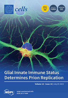
Cover Story (view full-size image): The prominent pathological change in prion-affected brains is neuroinflammation and aggregation of pathogenic prions. We investigated the impact of innate immune responses on prion replication in vitro. Glial cells from the cerebellum were susceptible to HY, but resistant to DY prion as determined by Western blot analysis, immunocytochemistry, and animal bioassay. However, glial cells from the cerebral cortex were refractory to both strains. Priming glial cells with lipopolysaccharide decreased prion replication, whereas pre-treatment with dexamethasone inhibiting innate immunity increased susceptibility to DY infection. Our results suggest that neuroinflammation resulting from prion infection might be an attempt to resolve prion propagation, implying a therapeutic potential of innate immune modulation in the early stages of prion disease. View this paper
  • Issues are regarded as officially published after their release is announced to the table of contents alert mailing list.
  • You may sign up for e-mail alerts to receive table of contents of newly released issues.
  • PDF is the official format for papers published in both, html and pdf forms. To view the papers in pdf format, click on the "PDF Full-text" link, and use the free Adobe Reader to open them.
Order results
Result details
Section
Select all
Export citation of selected articles as:
14 pages, 2860 KiB  
Article
Epigenetic Age Acceleration in Frontotemporal Lobar Degeneration: A Comprehensive Analysis in the Blood and Brain
by Megha Murthy, Patrizia Rizzu, Peter Heutink, Jonathan Mill, Tammaryn Lashley and Conceição Bettencourt
Cells 2023, 12(14), 1922; https://doi.org/10.3390/cells12141922 - 24 Jul 2023
Cited by 5 | Viewed by 1747
Abstract
Frontotemporal lobar degeneration (FTLD) includes a heterogeneous group of disorders pathologically characterized by the degeneration of the frontal and temporal lobes. In addition to major genetic contributors of FTLD such as mutations in MAPT, GRN, and C9orf72, recent work has [...] Read more.
Frontotemporal lobar degeneration (FTLD) includes a heterogeneous group of disorders pathologically characterized by the degeneration of the frontal and temporal lobes. In addition to major genetic contributors of FTLD such as mutations in MAPT, GRN, and C9orf72, recent work has identified several epigenetic modifications including significant differential DNA methylation in DLX1, and OTUD4 loci. As aging remains one of the major risk factors for FTLD, we investigated the presence of accelerated epigenetic aging in FTLD compared to controls. We calculated epigenetic age in both peripheral blood and brain tissues of multiple FTLD subtypes using several DNA methylation clocks, i.e., DNAmClockMulti, DNAmClockHannum, DNAmClockCortical, GrimAge, and PhenoAge, and determined age acceleration and its association with different cellular proportions and clinical traits. Significant epigenetic age acceleration was observed in the peripheral blood of both frontotemporal dementia (FTD) and progressive supranuclear palsy (PSP) patients compared to controls with DNAmClockHannum, even after accounting for confounding factors. A similar trend was observed with both DNAmClockMulti and DNAmClockCortical in post-mortem frontal cortex tissue of PSP patients and in FTLD cases harboring GRN mutations. Our findings support that increased epigenetic age acceleration in the peripheral blood could be an indicator for PSP and to a smaller extent, FTD. Full article
(This article belongs to the Special Issue Advances in the Understanding of Frontotemporal Dementia)
Show Figures

Figure 1

16 pages, 2182 KiB  
Article
Intercorrelation of Molecular Biomarkers and Clinical Phenotype Measures in Fragile X Syndrome
by Ramkumar Aishworiya, Mei-Hung Chi, Marwa Zafarullah, Guadalupe Mendoza, Matthew Dominic Ponzini, Kyoungmi Kim, Hazel Maridith Barlahan Biag, Angela John Thurman, Leonard Abbeduto, David Hessl, Jamie Leah Randol, Francois V. Bolduc, Sebastien Jacquemont, Sarah Lippé, Paul Hagerman, Randi Hagerman, Andrea Schneider and Flora Tassone
Cells 2023, 12(14), 1920; https://doi.org/10.3390/cells12141920 - 24 Jul 2023
Cited by 2 | Viewed by 1891
Abstract
This study contributes to a greater understanding of the utility of molecular biomarkers to identify clinical phenotypes of fragile X syndrome (FXS). Correlations of baseline clinical trial data (molecular measures—FMR1 mRNA, CYFIP1 mRNA, MMP9 and FMRP protein expression levels, nonverbal IQ, body [...] Read more.
This study contributes to a greater understanding of the utility of molecular biomarkers to identify clinical phenotypes of fragile X syndrome (FXS). Correlations of baseline clinical trial data (molecular measures—FMR1 mRNA, CYFIP1 mRNA, MMP9 and FMRP protein expression levels, nonverbal IQ, body mass index and weight, language level, NIH Toolbox, adaptive behavior rating, autism, and other mental health correlates) of 59 participants with FXS ages of 6–32 years are reported. FMR1 mRNA expression levels correlated positively with adaptive functioning levels, expressive language, and specific NIH Toolbox measures. The findings of a positive correlation of MMP-9 levels with obesity, CYFIP1 mRNA with mood and autistic symptoms, and FMR1 mRNA expression level with better cognitive, language, and adaptive functions indicate potential biomarkers for specific FXS phenotypes. These may be potential markers for future clinical trials for targeted treatments of FXS. Full article
Show Figures

Figure 1

19 pages, 5052 KiB  
Article
Evolving Acquired Vemurafenib Resistance in a BRAF V600E Mutant Melanoma PDTX Model to Reveal New Potential Targets
by József Tóvári, Diána Vári-Mező, Sára Eszter Surguta, Andrea Ladányi, Attila Kigyós and Mihály Cserepes
Cells 2023, 12(14), 1919; https://doi.org/10.3390/cells12141919 - 24 Jul 2023
Cited by 6 | Viewed by 2079
Abstract
Malignant melanoma is challenging to treat, and metastatic cases need chemotherapy strategies. Targeted inhibition of commonly mutant BRAF V600E by inhibitors is efficient but eventually leads to resistance and progression in the vast majority of cases. Numerous studies investigated the mechanisms of resistance [...] Read more.
Malignant melanoma is challenging to treat, and metastatic cases need chemotherapy strategies. Targeted inhibition of commonly mutant BRAF V600E by inhibitors is efficient but eventually leads to resistance and progression in the vast majority of cases. Numerous studies investigated the mechanisms of resistance in melanoma cell lines, and an increasing number of in vivo or clinical data are accumulating. In most cases, bypassing BRAF and resulting reactivation of the MAPK signaling, as well as alternative PI3K-AKT signaling activation are reported. However, several unique changes were also shown. We developed and used a patient-derived tumor xenograft (PDTX) model to screen resistance evolution in mice in vivo, maintaining tumor heterogeneity. Our results showed no substantial activation of the canonical pathways; however, RNAseq and qPCR data revealed several altered genes, such as GPR39, CD27, SLC15A3, IFI27, PDGFA, and ABCB1. Surprisingly, p53 activity, leading to apoptotic cell death, was unchanged. The found biomarkers can confer resistance in a subset of melanoma patients via immune modulation, microenvironment changes, or drug elimination. Our resistance model can be further used in testing specific inhibitors that could be used in future drug development, and combination therapy testing that can overcome inhibitor resistance in melanoma. Full article
Show Figures

Figure 1

6 pages, 262 KiB  
Editorial
Questions about Using the Induced Membrane Technique to Manage Cases of Congenital Tibial Pseudarthrosis
by Céline Klein, Florelle Gindraux, Alain-Charles Masquelet, Romuald Mentaverri and Richard Gouron
Cells 2023, 12(14), 1918; https://doi.org/10.3390/cells12141918 - 24 Jul 2023
Viewed by 1205
Abstract
The induced membrane technique is an innovative approach for repairing critical bone defects and has been applied recently in patients with congenital pseudarthrosis of the tibia (CPT). CPT is frequently associated with neurofibromatosis type 1 (NF1). Here, we briefly describe the clinical results [...] Read more.
The induced membrane technique is an innovative approach for repairing critical bone defects and has been applied recently in patients with congenital pseudarthrosis of the tibia (CPT). CPT is frequently associated with neurofibromatosis type 1 (NF1). Here, we briefly describe the clinical results of the induced membrane technique in NF1-deficient patients with CPT and in an animal model of CPT. Furthermore, we discuss the hypotheses used to explain inconsistent outcomes for the induced membrane technique in CPT–especially when associated with NF1. Full article
(This article belongs to the Special Issue Mineralized Tissues Repair and Regeneration 2.0)
23 pages, 4743 KiB  
Article
MicroRNA-Based Discovery of Biomarkers, Therapeutic Targets, and Repositioning Drugs for Breast Cancer
by Qing Ye, Rebecca A. Raese, Dajie Luo, Juan Feng, Wenjun Xin, Chunlin Dong, Yong Qian and Nancy Lan Guo
Cells 2023, 12(14), 1917; https://doi.org/10.3390/cells12141917 - 23 Jul 2023
Cited by 6 | Viewed by 2738
Abstract
Breast cancer treatment can be improved with biomarkers for early detection and individualized therapy. A set of 86 microRNAs (miRNAs) were identified to separate breast cancer tumors from normal breast tissues (n = 52) with an overall accuracy of 90.4%. Six miRNAs [...] Read more.
Breast cancer treatment can be improved with biomarkers for early detection and individualized therapy. A set of 86 microRNAs (miRNAs) were identified to separate breast cancer tumors from normal breast tissues (n = 52) with an overall accuracy of 90.4%. Six miRNAs had concordant expression in both tumors and breast cancer patient blood samples compared with the normal control samples. Twelve miRNAs showed concordant expression in tumors vs. normal breast tissues and patient survival (n = 1093), with seven as potential tumor suppressors and five as potential oncomiRs. From experimentally validated target genes of these 86 miRNAs, pan-sensitive and pan-resistant genes with concordant mRNA and protein expression associated with in-vitro drug response to 19 NCCN-recommended breast cancer drugs were selected. Combined with in-vitro proliferation assays using CRISPR-Cas9/RNAi and patient survival analysis, MEK inhibitors PD19830 and BRD-K12244279, pilocarpine, and tremorine were discovered as potential new drug options for treating breast cancer. Multi-omics biomarkers of response to the discovered drugs were identified using human breast cancer cell lines. This study presented an artificial intelligence pipeline of miRNA-based discovery of biomarkers, therapeutic targets, and repositioning drugs that can be applied to many cancer types. Full article
Show Figures

Graphical abstract

27 pages, 996 KiB  
Review
RNA-Based Liquid Biopsy in Head and Neck Cancer
by Jacek Kabzinski, Aleksandra Kucharska-Lusina and Ireneusz Majsterek
Cells 2023, 12(14), 1916; https://doi.org/10.3390/cells12141916 - 23 Jul 2023
Cited by 8 | Viewed by 2482
Abstract
Head and neck cancer (HNC) is a prevalent and diverse group of malignancies with substantial morbidity and mortality rates. Early detection and monitoring of HNC are crucial for improving patient outcomes. Liquid biopsy, a non-invasive diagnostic approach, has emerged as a promising tool [...] Read more.
Head and neck cancer (HNC) is a prevalent and diverse group of malignancies with substantial morbidity and mortality rates. Early detection and monitoring of HNC are crucial for improving patient outcomes. Liquid biopsy, a non-invasive diagnostic approach, has emerged as a promising tool for cancer detection and monitoring. In this article, we review the application of RNA-based liquid biopsy in HNC. Various types of RNA, including messenger RNA (mRNA), microRNA (miRNA), long non-coding RNA (lncRNA), small nuclear RNA (snRNA), small nucleolar RNA (snoRNA), circular RNA (circRNA) and PIWI-interacting RNA (piRNA), are explored as potential biomarkers in HNC liquid-based diagnostics. The roles of RNAs in HNC diagnosis, metastasis, tumor resistance to radio and chemotherapy, and overall prognosis are discussed. RNA-based liquid biopsy holds great promise for the early detection, prognosis, and personalized treatment of HNC. Further research and validation are necessary to translate these findings into clinical practice and improve patient outcomes. Full article
Show Figures

Figure 1

13 pages, 3645 KiB  
Article
MG53 Mitigates Nitrogen Mustard-Induced Skin Injury
by Haichang Li, Zhongguang Li, Xiuchun Li, Chuanxi Cai, Serena Li Zhao, Robert E. Merritt, Xinyu Zhou, Tao Tan, Valerie Bergdall and Jianjie Ma
Cells 2023, 12(14), 1915; https://doi.org/10.3390/cells12141915 - 23 Jul 2023
Cited by 3 | Viewed by 2028
Abstract
Sulfur mustard (SM) and nitrogen mustard (NM) are vesicant agents that cause skin injury and blistering through complicated cellular events, involving DNA damage, free radical formation, and lipid peroxidation. The development of therapeutic approaches targeting the multi-cellular process of tissue injury repair can [...] Read more.
Sulfur mustard (SM) and nitrogen mustard (NM) are vesicant agents that cause skin injury and blistering through complicated cellular events, involving DNA damage, free radical formation, and lipid peroxidation. The development of therapeutic approaches targeting the multi-cellular process of tissue injury repair can potentially provide effective countermeasures to combat vesicant-induced dermal lesions. MG53 is a vital component of cell membrane repair. Previous studies have demonstrated that topical application of recombinant human MG53 (rhMG53) protein has the potential to promote wound healing. In this study, we further investigate the role of MG53 in NM-induced skin injury. Compared with wild-type mice, mg53−/− mice are more susceptible to NM-induced dermal injuries, whereas mice with sustained elevation of MG53 in circulation are resistant to dermal exposure of NM. Exposure of keratinocytes and human follicle stem cells to NM causes elevation of oxidative stress and intracellular aggregation of MG53, thus compromising MG53′s intrinsic cell membrane repair function. Topical rhMG53 application mitigates NM-induced dermal injury in mice. Histologic examination reveals the therapeutic benefits of rhMG53 are associated with the preservation of epidermal integrity and hair follicle structure in mice with dermal NM exposure. Overall, these findings identify MG53 as a potential therapeutic agent to mitigate vesicant-induced skin injuries. Full article
(This article belongs to the Special Issue Toward Understanding Wound Repair Mechanism)
Show Figures

Figure 1

19 pages, 4207 KiB  
Article
Application-Oriented Bulk Cryopreservation of Human iPSCs in Cryo Bags Followed by Direct Inoculation in Scalable Suspension Bioreactors for Expansion and Neural Differentiation
by Ina Meiser, Monica Alstrup, Elham Khalesi, Bianca Stephan, Anna M. Speicher, Julia Majer, Chee Keong Kwok, Julia C. Neubauer, Mattias Hansson and Heiko Zimmermann
Cells 2023, 12(14), 1914; https://doi.org/10.3390/cells12141914 - 22 Jul 2023
Cited by 2 | Viewed by 1914
Abstract
Stem cell-based therapies are promising tools for regenerative medicine and require bulk numbers of high-quality cells. Currently, cells are produced on demand and have a limited shelf-life as conventional cryopreservation is primarily designed for stock keeping. We present a study on bulk cryopreservation [...] Read more.
Stem cell-based therapies are promising tools for regenerative medicine and require bulk numbers of high-quality cells. Currently, cells are produced on demand and have a limited shelf-life as conventional cryopreservation is primarily designed for stock keeping. We present a study on bulk cryopreservation of the human iPSC lines UKKi011-A and BIONi010-C-41. By increasing cell concentration and volume, compared to conventional cryopreservation routines in cryo vials, one billion cells were frozen in 50 mL cryo bags. Upon thawing, the cells were immediately seeded in scalable suspension-based bioreactors for expansion to assess the stemness maintenance and for neural differentiation to assess their differentiation potential on the gene and protein levels. Both the conventional and bulk cryo approach show comparative results regarding viability and aggregation upon thawing and bioreactor inoculation. Reduced performance compared to the non-frozen control was compensated within 3 days regarding biomass yield. Stemness was maintained upon thawing in expansion. In neural differentiation, a delay of the neural marker expression on day 4 was compensated at day 9. We conclude that cryopreservation in cryo bags, using high cell concentrations and volumes, does not alter the cells’ fate and is a suitable technology to avoid pre-cultivation and enable time- and cost-efficient therapeutic approaches with bulk cell numbers. Full article
(This article belongs to the Special Issue iPS Cells (iPSCs) for Modelling and Treatment of Human Diseases 2022)
Show Figures

Graphical abstract

19 pages, 4277 KiB  
Article
Innate Immunity and Sex: Distinct Inflammatory Profiles Associated with Murine Pain in Acute Synovitis
by Natália Valdrighi, Arjen B. Blom, Juliana P. Vago, Henk M. van Beuningen, Elly L. Vitters, Monique M. Helsen, Birgitte Walgreen, Onno J. Arntz, Marije I. Koenders, Peter M. van der Kraan, Esmeralda N. Blaney Davidson and Fons A. J. van de Loo
Cells 2023, 12(14), 1913; https://doi.org/10.3390/cells12141913 - 22 Jul 2023
Viewed by 1566
Abstract
Joint pain severity in arthritic diseases differs between sexes and is often more pronounced in women. This disparity is thought to stem from biological mechanisms, particularly innate immunity, yet the understanding of sex-specific differences in arthritic pain remains incomplete. This study aims to [...] Read more.
Joint pain severity in arthritic diseases differs between sexes and is often more pronounced in women. This disparity is thought to stem from biological mechanisms, particularly innate immunity, yet the understanding of sex-specific differences in arthritic pain remains incomplete. This study aims to investigate these disparities using an innate immunity-driven inflammation model induced by intra-articular injections of Streptococcus Cell Wall fragments to mimic both acute and pre-sensitized joint conditions. Nociceptive behavior was evaluated via gait analysis and static weight-bearing, and inflammation was evaluated via joint histology and the synovial gene expression involved in immune response. Although acute inflammation and pain severity were comparable between sexes, distinct associations between synovial inflammatory gene expression and static nociceptive behavior emerged. These associations delineated sex-specific relationships with pain, highlighting differential gene interactions (Il6 versus Cybb on day 1 and Cyba/Gas6 versus Nos2 on day 8) between sexes. In conclusion, our study found that, despite similar pain severity between sexes, the association of inflammatory synovial genes revealed sex-specific differences in the molecular inflammatory mechanisms underlying pain. These findings suggest a path towards more personalized treatment strategies for pain management in arthritis and other inflammatory joint diseases. Full article
(This article belongs to the Special Issue The Signaling and Cellular Mechanisms of Pain)
Show Figures

Graphical abstract

12 pages, 3934 KiB  
Perspective
Myeloid-Derived Suppressor Cells (MDSCs) in Ovarian Cancer—Looking Back and Forward
by Karolina Okła
Cells 2023, 12(14), 1912; https://doi.org/10.3390/cells12141912 - 22 Jul 2023
Cited by 4 | Viewed by 1955
Abstract
Myeloid-derived suppressor cells (MDSCs) play a significant role in the immune system and have been extensively studied in cancer. MDSCs are a heterogeneous population of myeloid cells that accumulate in the tumor microenvironment. Consequently, the high abundance of these cells often leads to [...] Read more.
Myeloid-derived suppressor cells (MDSCs) play a significant role in the immune system and have been extensively studied in cancer. MDSCs are a heterogeneous population of myeloid cells that accumulate in the tumor microenvironment. Consequently, the high abundance of these cells often leads to immunosuppression, tumor growth, treatment failure, and poor prognosis. Ovarian cancer ranks fifth in cancer deaths among women, accounting for more deaths than any other cancer of the female genital tract. Currently, there is a lack of effective clinical strategies for the treatment of ovarian cancer. Although several studies underline the negative role of human MDSCs in ovarian cancer, this topic is still understudied. The works on MDSCs are summarized here, along with an explanation of why focusing on these cells would be a promising approach for treating ovarian cancer patients. Full article
(This article belongs to the Section Cellular Immunology)
Show Figures

Figure 1

16 pages, 960 KiB  
Review
Liquid Biopsy in Neurological Diseases
by Sunny Malhotra, Mari Carmen Martín Miras, Agustín Pappolla, Xavier Montalban and Manuel Comabella
Cells 2023, 12(14), 1911; https://doi.org/10.3390/cells12141911 - 22 Jul 2023
Cited by 9 | Viewed by 4635
Abstract
The most recent and non-invasive approach for studying early-stage biomarkers is liquid biopsy. This implies the extraction and analysis of non-solid biological tissues (serum, plasma, saliva, urine, and cerebrospinal fluid) without undergoing invasive procedures to determine disease prognosis. Liquid biopsy can be used [...] Read more.
The most recent and non-invasive approach for studying early-stage biomarkers is liquid biopsy. This implies the extraction and analysis of non-solid biological tissues (serum, plasma, saliva, urine, and cerebrospinal fluid) without undergoing invasive procedures to determine disease prognosis. Liquid biopsy can be used for the screening of several components, such as extracellular vesicles, microRNAs, cell-free DNA, cell-free mitochondrial and nuclear DNA, circulating tumour cells, circulating tumour DNA, transfer RNA, and circular DNA or RNA derived from body fluids. Its application includes early disease diagnosis, the surveillance of disease activity, and treatment response monitoring, with growing evidence for validating this methodology in cancer, liver disease, and central nervous system (CNS) disorders. This review will provide an overview of mentioned liquid biopsy components, which could serve as valuable biomarkers for the evaluation of complex neurological conditions, including Alzheimer’s disease, Parkinson’s disease, amyotrophic lateral sclerosis, multiple sclerosis, epilepsy, stroke, traumatic brain injury, CNS tumours, and neuroinfectious diseases. Furthermore, this review highlights the future directions and potential limitations associated with liquid biopsy. Full article
(This article belongs to the Special Issue Liquid Biopsy Components in Neurological Diseases)
Show Figures

Figure 1

15 pages, 1198 KiB  
Review
The Emerging Role of Innate Lymphoid Cells (ILCs) and Alarmins in Celiac Disease: An Update on Pathophysiological Insights, Potential Use as Disease Biomarkers, and Therapeutic Implications
by Angela Rizzi, Mario Di Gioacchino, Luca Gammeri, Riccardo Inchingolo, Raffaella Chini, Francesca Santilli, Eleonora Nucera and Sebastiano Gangemi
Cells 2023, 12(14), 1910; https://doi.org/10.3390/cells12141910 - 21 Jul 2023
Cited by 3 | Viewed by 1963
Abstract
Celiac disease (CD) is an intestinal disease that develops in genetically predisposed individuals and is triggered by the ingestion of gluten. CD was considered a Th1-disease. Today, the role of Th17, IL-21, and IL-17A lymphocytes is well known. Inflammation is regulated by the [...] Read more.
Celiac disease (CD) is an intestinal disease that develops in genetically predisposed individuals and is triggered by the ingestion of gluten. CD was considered a Th1-disease. Today, the role of Th17, IL-21, and IL-17A lymphocytes is well known. Inflammation is regulated by the activity of gluten-specific CD4+ T lymphocytes that produce pro-inflammatory cytokines, including IFN-γ, TNF-α, and IL-21, perpetuating the Th1 response. These cytokines determine an inflammatory state of the small intestine, with consequent epithelial infiltration of lymphocytes and an alteration of the architecture of the duodenal mucosa. B cells produce antibodies against tissue transglutaminase and against deamidated gliadin. Although the role of the adaptive immune response is currently known, the evidence about the role of innate immunity cells is still poorly understood. Epithelial damage determines the release of damage-associated molecular patterns (DAMPs), also known as alarmins. Together with the intestinal epithelial cells and the type 1 innate lymphoid cells (ILC1s), alarmins like TSLP, IL-33, and HMGB1 could have a fundamental role in the genesis and maintenance of inflammation. Our study aims to evaluate the evidence in the literature about the role of ILCs and alarmins in celiac disease, evaluating the possible future diagnostic and therapeutic implications. Full article
(This article belongs to the Section Cellular Immunology)
Show Figures

Figure 1

17 pages, 2268 KiB  
Article
Early Detection of Pre-Cancerous and Cancerous Cells Using Raman Spectroscopy-Based Machine Learning
by Uraib Sharaha, Daniel Hania, Itshak Lapidot, Ahmad Salman and Mahmoud Huleihel
Cells 2023, 12(14), 1909; https://doi.org/10.3390/cells12141909 - 21 Jul 2023
Cited by 4 | Viewed by 1907
Abstract
Cancer is the most common and fatal disease around the globe, with an estimated 19 million newly diagnosed patients and approximately 10 million deaths annually. Patients with cancer struggle daily due to difficult treatments, pain, and financial and social difficulties. Detecting the disease [...] Read more.
Cancer is the most common and fatal disease around the globe, with an estimated 19 million newly diagnosed patients and approximately 10 million deaths annually. Patients with cancer struggle daily due to difficult treatments, pain, and financial and social difficulties. Detecting the disease in its early stages is critical in increasing the likelihood of recovery and reducing the financial burden on the patient and society. Currently used methods for the diagnosis of cancer are time-consuming, producing discomfort and anxiety for patients and significant medical waste. The main goal of this study is to evaluate the potential of Raman spectroscopy-based machine learning for the identification and characterization of precancerous and cancerous cells. As a representative model, normal mouse primary fibroblast cells (NFC) as healthy cells; a mouse fibroblast cell line (NIH/3T3), as precancerous cells; and fully malignant mouse fibroblasts (MBM-T) as cancerous cells were used. Raman spectra were measured from three different sites of each of the 457 investigated cells and analyzed by principal component analysis (PCA) and linear discriminant analysis (LDA). Our results showed that it was possible to distinguish between the normal and abnormal (precancerous and cancerous) cells with a success rate of 93.1%; this value was 93.7% when distinguishing between normal and precancerous cells and 80.2% between precancerous and cancerous cells. Moreover, there was no influence of the measurement site on the differentiation between the different examined biological systems. Full article
(This article belongs to the Special Issue Research Advances in Cell Methods)
Show Figures

Figure 1

2 pages, 1733 KiB  
Correction
Correction: Cheng et al. Glutathione S-Transferases S1, Z1 and A1 Serve as Prognostic Factors in Glioblastoma and Promote Drug Resistance through Antioxidant Pathways. Cells 2022, 11, 3232
by Bo Cheng, Yu Wang, Abiola Abdulrahman Ayanlaja, Jing Zhu, Piniel Alphayo Kambey, Ziqiang Qiu, Caiyi Zhang and Wei Hu
Cells 2023, 12(14), 1908; https://doi.org/10.3390/cells12141908 - 21 Jul 2023
Cited by 1 | Viewed by 785
Abstract
The authors wish to make the following change to their paper [...] Full article
11 pages, 1383 KiB  
Article
Staphylococcus aureus Sensitivity to Membrane Disrupting Antibacterials Is Increased under Microgravity
by Hyochan Jang, Seong Yeol Choi and Robert J. Mitchell
Cells 2023, 12(14), 1907; https://doi.org/10.3390/cells12141907 - 21 Jul 2023
Cited by 3 | Viewed by 2149
Abstract
In a survey of the International Space Station (ISS), the most common pathogenic bacterium identified in samples from the air, water and surfaces was Staphylococcus aureus. While growth under microgravity is known to cause physiological changes in microbial pathogens, including shifts in [...] Read more.
In a survey of the International Space Station (ISS), the most common pathogenic bacterium identified in samples from the air, water and surfaces was Staphylococcus aureus. While growth under microgravity is known to cause physiological changes in microbial pathogens, including shifts in antibacterial sensitivity, its impact on S. aureus is not well understood. Using high-aspect ratio vessels (HARVs) to generate simulated microgravity (SMG) conditions in the lab, we found S. aureus lipid profiles are altered significantly, with a higher presence of branch-chained fatty acids (BCFAs) (14.8% to 35.4%) with a concomitant reduction (41.3% to 31.4%) in straight-chain fatty acids (SCFAs) under SMG. This shift significantly increased the sensitivity of this pathogen to daptomycin, a membrane-acting antibiotic, leading to 12.1-fold better killing under SMG. Comparative assays with two additional compounds, i.e., SDS and violacein, confirmed S. aureus is more susceptible to membrane-disrupting agents, with 0.04% SDS and 0.6 mg/L violacein resulting in 22.9- and 12.8-fold better killing in SMG than normal gravity, respectively. As humankind seeks to establish permanent colonies in space, these results demonstrate the increased potency of membrane-active antibacterials to control the presence and spread of S. aureus, and potentially other pathogens. Full article
(This article belongs to the Special Issue New Insights into Microgravity and Space Biology)
Show Figures

Figure 1

21 pages, 5688 KiB  
Article
Autophagy Inhibition with Chloroquine Increased Pro-Apoptotic Potential of New Aziridine-Hydrazide Hydrazone Derivatives against Glioblastoma Cells
by Monika Witusik-Perkowska, Pola Głowacka, Adam M. Pieczonka, Ewa Świderska, Agnieszka Pudlarz, Michał Rachwalski, Julia Szymańska, Magdalena Zakrzewska, Dariusz J. Jaskólski and Janusz Szemraj
Cells 2023, 12(14), 1906; https://doi.org/10.3390/cells12141906 - 21 Jul 2023
Cited by 5 | Viewed by 2040
Abstract
Tumor therapy escape due to undesired side effects induced by treatment, such as prosurvival autophagy or cellular senescence, is one of the key mechanisms of resistance that eventually leads to tumor dormancy and recurrence. Glioblastoma is the most frequent and practically incurable neoplasm [...] Read more.
Tumor therapy escape due to undesired side effects induced by treatment, such as prosurvival autophagy or cellular senescence, is one of the key mechanisms of resistance that eventually leads to tumor dormancy and recurrence. Glioblastoma is the most frequent and practically incurable neoplasm of the central nervous system; thus, new treatment modalities have been investigated to find a solution more effective than the currently applied standards based on temozolomide. The present study examined the newly synthesized compounds of aziridine–hydrazide hydrazone derivatives to determine their antineoplastic potential against glioblastoma cells in vitro. Although the output of our investigation clearly demonstrates their proapoptotic activity, the cytotoxic effect appeared to be blocked by treatment-induced autophagy, the phenomenon also detected in the case of temozolomide action. The addition of an autophagy inhibitor, chloroquine, resulted in a significant increase in apoptosis triggered by the tested compounds, as well as temozolomide. The new aziridine–hydrazide hydrazone derivatives, which present cytotoxic potential against glioblastoma cells comparable to or even higher than that of temozolomide, show promising results and, thus, should be further investigated as antineoplastic agents. Moreover, our findings suggest that the combination of an apoptosis inducer with an autophagy inhibitor could optimize chemotherapeutic efficiency, and the addition of an autophagy inhibitor should be considered as an optional adjunctive therapy minimizing the risk of tumor escape from treatment. Full article
(This article belongs to the Special Issue Cell Death Mechanisms and Therapeutic Opportunities in Glioblastoma)
Show Figures

Figure 1

26 pages, 2210 KiB  
Review
Management of Rheumatoid Arthritis: Possibilities and Challenges of Mesenchymal Stromal/Stem Cell-Based Therapies
by Yusuke Shimizu, Edward Hosea Ntege, Chinatsu Azuma, Fuminari Uehara, Takashi Toma, Kotaro Higa, Hiroki Yabiku, Naoki Matsuura, Yoshikazu Inoue and Hiroshi Sunami
Cells 2023, 12(14), 1905; https://doi.org/10.3390/cells12141905 - 21 Jul 2023
Cited by 3 | Viewed by 3053
Abstract
Rheumatoid arthritis (RA) is a highly prevalent, chronic, and progressive autoimmune disorder primarily affecting joints and muscles. The associated inflammation, pain, and motor restriction negatively impact patient quality of life (QOL) and can even contribute to premature mortality. Further, conventional treatments such as [...] Read more.
Rheumatoid arthritis (RA) is a highly prevalent, chronic, and progressive autoimmune disorder primarily affecting joints and muscles. The associated inflammation, pain, and motor restriction negatively impact patient quality of life (QOL) and can even contribute to premature mortality. Further, conventional treatments such as antiinflammatory drugs are only symptomatic. Substantial progress has been made on elucidating the etiopathology of overt RA, in particular the contributions of innate and adaptive immune system dysfunction to chronic inflammation. Although the precise mechanisms underlying onset and progression remain elusive, the discovery of new drug targets, early diagnosis, and new targeted treatments have greatly improved the prognosis and QOL of patients with RA. However, a sizable proportion of patients develop severe adverse effects, exhibit poor responses, or cannot tolerate long-term use of these drugs, necessitating more effective and safer therapeutic alternatives. Mounting preclinical and clinical evidence suggests that the transplantation of multipotent adult stem cells such as mesenchymal stromal/stem cells is a safe and effective treatment strategy for controlling chronic inflammation and promoting tissue regeneration in patients with intractable diseases, including RA. This review describes the current status of MSC-based therapies for RA as well as the opportunities and challenges to broader clinical application. Full article
Show Figures

Figure 1

23 pages, 1454 KiB  
Review
Unravelling the Role of PARP1 in Homeostasis and Tumorigenesis: Implications for Anti-Cancer Therapies and Overcoming Resistance
by Taylor Lovsund, Fatemeh Mashayekhi, Amira Fitieh, James Stafford and Ismail Hassan Ismail
Cells 2023, 12(14), 1904; https://doi.org/10.3390/cells12141904 - 21 Jul 2023
Viewed by 1994
Abstract
Detailing the connection between homeostatic functions of enzymatic families and eventual progression into tumorigenesis is crucial to our understanding of anti-cancer therapies. One key enzyme group involved in this process is the Poly (ADP-ribose) polymerase (PARP) family, responsible for an expansive number of [...] Read more.
Detailing the connection between homeostatic functions of enzymatic families and eventual progression into tumorigenesis is crucial to our understanding of anti-cancer therapies. One key enzyme group involved in this process is the Poly (ADP-ribose) polymerase (PARP) family, responsible for an expansive number of cellular functions, featuring members well established as regulators of DNA repair, genomic stability and beyond. Several PARP inhibitors (PARPi) have been approved for clinical use in a range of cancers, with many more still in trials. Unfortunately, the occurrence of resistance to PARPi therapy is growing in prevalence and requires the introduction of novel counter-resistance mechanisms to maintain efficacy. In this review, we summarize the updated understanding of the vast homeostatic functions the PARP family mediates and pin the importance of PARPi therapies as anti-cancer agents while discussing resistance mechanisms and current up-and-coming counter-strategies for countering such resistance. Full article
Show Figures

Figure 1

17 pages, 1699 KiB  
Review
Targeting Soluble Guanylyl Cyclase during Ischemia and Reperfusion
by Eric H. Mace, Melissa J. Kimlinger, Frederic T. Billings IV and Marcos G. Lopez
Cells 2023, 12(14), 1903; https://doi.org/10.3390/cells12141903 - 21 Jul 2023
Cited by 3 | Viewed by 2891
Abstract
Ischemia and reperfusion (IR) damage organs and contribute to many disease states. Few effective treatments exist that attenuate IR injury. The augmentation of nitric oxide (NO) signaling remains a promising therapeutic target for IR injury. NO binds to soluble guanylyl cyclase (sGC) to [...] Read more.
Ischemia and reperfusion (IR) damage organs and contribute to many disease states. Few effective treatments exist that attenuate IR injury. The augmentation of nitric oxide (NO) signaling remains a promising therapeutic target for IR injury. NO binds to soluble guanylyl cyclase (sGC) to regulate vasodilation, maintain endothelial barrier integrity, and modulate inflammation through the production of cyclic-GMP in vascular smooth muscle. Pharmacologic sGC stimulators and activators have recently been developed. In preclinical studies, sGC stimulators, which augment the reduced form of sGC, and activators, which activate the oxidized non-NO binding form of sGC, increase vasodilation and decrease cardiac, cerebral, renal, pulmonary, and hepatic injury following IR. These effects may be a result of the improved regulation of perfusion and decreased oxidative injury during IR. sGC stimulators are now used clinically to treat some chronic conditions such as heart failure and pulmonary hypertension. Clinical trials of sGC activators have been terminated secondary to adverse side effects including hypotension. Additional clinical studies to investigate the effects of sGC stimulation and activation during acute conditions, such as IR, are warranted. Full article
Show Figures

Figure 1

14 pages, 4794 KiB  
Article
FITC-Labeled RGD Peptides as Novel Contrast Agents for Functional Fluorescent Angiographic Detection of Retinal and Choroidal Neovascularization
by Seung Woo Choi, Hye Kyoung Hong, Jehwi Jeon, Ji Young Choi, Minah Kim, Pilhan Kim, Byung Chul Lee and Se Joon Woo
Cells 2023, 12(14), 1902; https://doi.org/10.3390/cells12141902 - 21 Jul 2023
Cited by 2 | Viewed by 2274
Abstract
The development of choroidal neovascularization (CNV) is a crucial factor in the pathophysiology and prognosis of exudative age-related macular degeneration (AMD). Therefore, the detection of CNV is essential for establishing an appropriate diagnosis and treatment plan. Current ophthalmic imaging techniques, such as fundus [...] Read more.
The development of choroidal neovascularization (CNV) is a crucial factor in the pathophysiology and prognosis of exudative age-related macular degeneration (AMD). Therefore, the detection of CNV is essential for establishing an appropriate diagnosis and treatment plan. Current ophthalmic imaging techniques, such as fundus fluorescent angiography and optical coherence tomography, have limitations in accurately visualizing CNV lesions and expressing CNV activity, owing to issues such as excessive dye leakage with pooling and the inability to provide functional information. Here, using the arginine−glycine−aspartic acid (RGD) peptide’s affinity for integrin αvβ3, which is expressed in the neovascular endothelial cells in ocular tissues, we propose the use of fluorescein isothiocyanate (FITC)-labeled RGD peptide as a novel dye for effective molecular imaging of CNV. FITC-labeled RGD peptides (FITC-RGD2), prepared by bioconjugation of one FITC molecule with two RGD peptides, demonstrated better visualization and precise localization of CNV lesions than conventional fluorescein dyes in laser-induced CNV rodent models, as assessed using various imaging techniques, including a commercially available clinical fundus camera (Optos). These results suggest that FITC-RGD2 can serve as an effective novel dye for the diagnosis of neovascular retinal diseases, including AMD, by enabling early detection and treatment of disease occurrence and recurrence after treatment. Full article
(This article belongs to the Special Issue Vascular Growth Factors in Health and Diseases)
Show Figures

Figure 1

17 pages, 3271 KiB  
Article
Cell Surface Vibrations Distinguish Malignant from Benign Cells
by Ishay Wohl, Julia Sajman and Eilon Sherman
Cells 2023, 12(14), 1901; https://doi.org/10.3390/cells12141901 - 21 Jul 2023
Viewed by 1593
Abstract
The mechanical properties of living cells, including their shape, rigidity, and internal dynamics play a crucial role in their physiology and pathology. Still, the relations between the physiological cell state and its rigidity and surface vibrations remain poorly understood. Here, we have employed [...] Read more.
The mechanical properties of living cells, including their shape, rigidity, and internal dynamics play a crucial role in their physiology and pathology. Still, the relations between the physiological cell state and its rigidity and surface vibrations remain poorly understood. Here, we have employed AFM measurements on T cells and found a negative relation between cell surface stiffness and its vibrations. Blocking T-type Ca++-channels using Mibefradil reduced cortical actin tension in these cells and enhanced their membrane vibrations and dissipation of intracellular mechanical work to the cell surroundings. We also found increased vibrations of cell membranes in five different malignant cells lines derived from T cell leukemia, lung, prostate, bladder, and melanoma cancers, as compared to their corresponding benign cells. This was demonstrated by utilizing TIRF microscopy in single cells and dynamic laser speckles measurements in an in vitro model of multiple cells in a tissue. Our results show that cell membrane vibrations and dissipation of mechanical work are higher in malignant cells relative to benign cells. Accordingly, these properties may be used to detect and monitor cellular and tissue malignancies. Full article
(This article belongs to the Special Issue Actin-Myosin Cytoskeleton Regulation and Function Series 2)
Show Figures

Graphical abstract

24 pages, 3224 KiB  
Article
The Genetic Structures and Molecular Mechanisms Underlying Ear Traits in Maize (Zea mays L.)
by Zhenying Dong, Yanbo Wang, Jianxi Bao, Ya’nan Li, Zechao Yin, Yan Long and Xiangyuan Wan
Cells 2023, 12(14), 1900; https://doi.org/10.3390/cells12141900 - 21 Jul 2023
Cited by 5 | Viewed by 3156
Abstract
Maize (Zea mays L.) is one of the world’s staple food crops. In order to feed the growing world population, improving maize yield is a top priority for breeding programs. Ear traits are important determinants of maize yield, and are mostly quantitatively [...] Read more.
Maize (Zea mays L.) is one of the world’s staple food crops. In order to feed the growing world population, improving maize yield is a top priority for breeding programs. Ear traits are important determinants of maize yield, and are mostly quantitatively inherited. To date, many studies relating to the genetic and molecular dissection of ear traits have been performed; therefore, we explored the genetic loci of the ear traits that were previously discovered in the genome-wide association study (GWAS) and quantitative trait locus (QTL) mapping studies, and refined 153 QTL and 85 quantitative trait nucleotide (QTN) clusters. Next, we shortlisted 19 common intervals (CIs) that can be detected simultaneously by both QTL mapping and GWAS, and 40 CIs that have pleiotropic effects on ear traits. Further, we predicted the best possible candidate genes from 71 QTL and 25 QTN clusters that could be valuable for maize yield improvement. Full article
Show Figures

Figure 1

24 pages, 4584 KiB  
Article
Tissue Inhibitor of Metalloproteinases-1 Interacts with CD74 to Promote AKT Signaling, Monocyte Recruitment Responses, and Vascular Smooth Muscle Cell Proliferation
by Simon Ebert, Lan Zang, Noor Ismail, Michael Otabil, Adrian Fröhlich, Virginia Egea, Susann Ács, Mikkel Hoeberg, Marie-Luise Berres, Christian Weber, José M. A. Moreira, Christian Ries, Jürgen Bernhagen and Omar El Bounkari
Cells 2023, 12(14), 1899; https://doi.org/10.3390/cells12141899 - 20 Jul 2023
Cited by 6 | Viewed by 2400
Abstract
Tissue inhibitor of metalloproteinases-1 (TIMP-1), an important regulator of matrix metalloproteinases (MMPs), has recently been shown to interact with CD74, a receptor for macrophage migration inhibitory factor (MIF). However, the biological effects mediated by TIMP-1 through CD74 remain largely unexplored. Using sequence alignment [...] Read more.
Tissue inhibitor of metalloproteinases-1 (TIMP-1), an important regulator of matrix metalloproteinases (MMPs), has recently been shown to interact with CD74, a receptor for macrophage migration inhibitory factor (MIF). However, the biological effects mediated by TIMP-1 through CD74 remain largely unexplored. Using sequence alignment and in silico protein–protein docking analysis, we demonstrated that TIMP-1 shares residues with both MIF and MIF-2, crucial for CD74 binding, but not for CXCR4. Subcellular colocalization, immunoprecipitation, and internalization experiments supported these findings, demonstrating that TIMP-1 interacts with surface-expressed CD74, resulting in its internalization in a dose-dependent manner, as well as with a soluble CD74 ectodomain fragment (sCD74). This prompted us to study the effects of the TIMP-1–CD74 axis on monocytes and vascular smooth muscle cells (VSCMs) to assess its impact on vascular inflammation. A phospho-kinase array revealed the activation of serine/threonine kinases by TIMP-1 in THP-1 pre-monocytes, in particular AKT. Similarly, TIMP-1 dose-dependently triggered the phosphorylation of AKT and ERK1/2 in primary human monocytes. Importantly, Transwell migration, 3D-based Chemotaxis, and flow adhesion assays demonstrated that TIMP-1 engagement of CD74 strongly promotes the recruitment response of primary human monocytes, while live cell imaging studies revealed a profound activating effect on VSMC proliferation. Finally, re-analysis of scRNA-seq data highlighted the expression patterns of TIMP-1 and CD74 in human atherosclerotic lesions, thus, together with our experimental data, indicating a role for the TIMP-1–CD74 axis in vascular inflammation and atherosclerosis. Full article
Show Figures

Figure 1

16 pages, 4648 KiB  
Article
A Postmortem MRI Study of Cerebrovascular Disease and Iron Content at End-Stage of Fragile X-Associated Tremor/Ataxia Syndrome
by Jun Yi Wang, Gerard J. Sonico, Maria Jimena Salcedo-Arellano, Randi J. Hagerman and Veronica Martinez-Cerdeno
Cells 2023, 12(14), 1898; https://doi.org/10.3390/cells12141898 - 20 Jul 2023
Cited by 1 | Viewed by 1388
Abstract
Brain changes at the end-stage of fragile X-associated tremor/ataxia syndrome (FXTAS) are largely unknown due to mobility impairment. We conducted a postmortem MRI study of FXTAS to quantify cerebrovascular disease, brain atrophy and iron content, and examined their relationships using principal component analysis [...] Read more.
Brain changes at the end-stage of fragile X-associated tremor/ataxia syndrome (FXTAS) are largely unknown due to mobility impairment. We conducted a postmortem MRI study of FXTAS to quantify cerebrovascular disease, brain atrophy and iron content, and examined their relationships using principal component analysis (PCA). Intracranial hemorrhage (ICH) was observed in 4/17 FXTAS cases, among which one was confirmed by histologic staining. Compared with seven control brains, FXTAS cases showed higher ratings of T2-hyperintensities (indicating cerebral small vessel disease) in the cerebellum, globus pallidus and frontoparietal white matter, and significant atrophy in the cerebellar white matter, red nucleus and dentate nucleus. PCA of FXTAS cases revealed negative associations of T2-hyperintensity ratings with anatomic volumes and iron content in the white matter, hippocampus and amygdala, that were independent from a highly correlated number of regions with ICH and iron content in subcortical nuclei. Post-hoc analysis confirmed PCA findings and further revealed increased iron content in the white matter, hippocampus and amygdala in FXTAS cases compared to controls, after adjusting for T2-hyperintensity ratings. These findings indicate that both ischemic and hemorrhagic brain damage may occur in FXTAS, with the former being marked by demyelination/iron depletion and atrophy, and the latter by ICH and iron accumulation in basal ganglia. Full article
Show Figures

Figure 1

35 pages, 5074 KiB  
Review
The Role of Mitochondrial Dynamics and Mitotic Fission in Regulating the Cell Cycle in Cancer and Pulmonary Arterial Hypertension: Implications for Dynamin-Related Protein 1 and Mitofusin2 in Hyperproliferative Diseases
by Pierce Colpman, Asish Dasgupta and Stephen L. Archer
Cells 2023, 12(14), 1897; https://doi.org/10.3390/cells12141897 - 20 Jul 2023
Cited by 7 | Viewed by 4861
Abstract
Mitochondria, which generate ATP through aerobic respiration, also have important noncanonical functions. Mitochondria are dynamic organelles, that engage in fission (division), fusion (joining) and translocation. They also regulate intracellular calcium homeostasis, serve as oxygen-sensors, regulate inflammation, participate in cellular and organellar quality control [...] Read more.
Mitochondria, which generate ATP through aerobic respiration, also have important noncanonical functions. Mitochondria are dynamic organelles, that engage in fission (division), fusion (joining) and translocation. They also regulate intracellular calcium homeostasis, serve as oxygen-sensors, regulate inflammation, participate in cellular and organellar quality control and regulate the cell cycle. Mitochondrial fission is mediated by the large GTPase, dynamin-related protein 1 (Drp1) which, when activated, translocates to the outer mitochondrial membrane (OMM) where it interacts with binding proteins (Fis1, MFF, MiD49 and MiD51). At a site demarcated by the endoplasmic reticulum, fission proteins create a macromolecular ring that divides the organelle. The functional consequence of fission is contextual. Physiological fission in healthy, nonproliferating cells mediates organellar quality control, eliminating dysfunctional portions of the mitochondria via mitophagy. Pathological fission in somatic cells generates reactive oxygen species and triggers cell death. In dividing cells, Drp1-mediated mitotic fission is critical to cell cycle progression, ensuring that daughter cells receive equitable distribution of mitochondria. Mitochondrial fusion is regulated by the large GTPases mitofusin-1 (Mfn1) and mitofusin-2 (Mfn2), which fuse the OMM, and optic atrophy 1 (OPA-1), which fuses the inner mitochondrial membrane. Mitochondrial fusion mediates complementation, an important mitochondrial quality control mechanism. Fusion also favors oxidative metabolism, intracellular calcium homeostasis and inhibits cell proliferation. Mitochondrial lipids, cardiolipin and phosphatidic acid, also regulate fission and fusion, respectively. Here we review the role of mitochondrial dynamics in health and disease and discuss emerging concepts in the field, such as the role of central versus peripheral fission and the potential role of dynamin 2 (DNM2) as a fission mediator. In hyperproliferative diseases, such as pulmonary arterial hypertension and cancer, Drp1 and its binding partners are upregulated and activated, positing mitochondrial fission as an emerging therapeutic target. Full article
(This article belongs to the Special Issue Molecular Pathogenesis of PH: Group 1 and Beyond)
Show Figures

Figure 1

23 pages, 2472 KiB  
Article
A Shiga Toxin B-Subunit-Based Lectibody Boosts T Cell Cytotoxicity towards Gb3-Positive Cancer Cells
by Jana Tomisch, Vincent Busse, Francesca Rosato, Olga N. Makshakova, Pavel Salavei, Anna-Sophia Kittel, Emilie Gillon, Levin Lataster, Anne Imberty, Ana Valeria Meléndez and Winfried Römer
Cells 2023, 12(14), 1896; https://doi.org/10.3390/cells12141896 - 20 Jul 2023
Cited by 3 | Viewed by 2399
Abstract
Aberrant glycosylation plays a crucial role in tumour progression and invasiveness. Tumour-associated carbohydrate antigens (TACAs) represent a valuable set of targets for immunotherapeutic approaches. The poor immunogenicity of glycan structures, however, requires a more effective and well-directed way of targeting TACAs on the [...] Read more.
Aberrant glycosylation plays a crucial role in tumour progression and invasiveness. Tumour-associated carbohydrate antigens (TACAs) represent a valuable set of targets for immunotherapeutic approaches. The poor immunogenicity of glycan structures, however, requires a more effective and well-directed way of targeting TACAs on the surface of cancer cells than antibodies. The glycosphingolipid globotriaosylceramide (Gb3) is a well-established TACA present in a multitude of cancer types. Its overexpression has been linked to metastasis, invasiveness, and multidrug resistance. In the present study, we propose to use a dimeric fragment of the Shiga toxin B-subunit (StxB) to selectively target Gb3-positive cancer cells in a StxB-scFv UCHT1 lectibody. The lectibody, comprised of a lectin and the UCHT1 antibody fragment, was produced in E. coli and purified via Ni-NTA affinity chromatography. Specificity of the lectibody towards Gb3-positive cancer cell lines and specificity towards the CD3 receptor on T cells, was assessed using flow cytometry. We evaluated the efficacy of the lectibody in redirecting T cell cytotoxicity towards Gb3-overexpressing cancer cells in luciferase-based cytotoxicity in vitro assays. The StxB-scFv UCHT1 lectibody has proven specific for Gb3 and could induce the killing of up to 80% of Gb3-overexpressing cancer cells in haemorrhagic and solid tumours. The lectibody developed in this study, therefore, highlights the potential that lectibodies and lectins in general have for usage in immunotherapeutic approaches to boost the efficacy of established cancer treatments. Full article
(This article belongs to the Section Intracellular and Plasma Membranes)
Show Figures

Figure 1

31 pages, 3860 KiB  
Review
Revisiting Two Decades of Research Focused on Targeting APE1 for Cancer Therapy: The Pros and Cons
by Matilde Clarissa Malfatti, Alessia Bellina, Giulia Antoniali and Gianluca Tell
Cells 2023, 12(14), 1895; https://doi.org/10.3390/cells12141895 - 20 Jul 2023
Cited by 13 | Viewed by 2335
Abstract
APE1 is an essential endodeoxyribonuclease of the base excision repair pathway that maintains genome stability. It was identified as a pivotal factor favoring tumor progression and chemoresistance through the control of gene expression by a redox-based mechanism. APE1 is overexpressed and serum-secreted in [...] Read more.
APE1 is an essential endodeoxyribonuclease of the base excision repair pathway that maintains genome stability. It was identified as a pivotal factor favoring tumor progression and chemoresistance through the control of gene expression by a redox-based mechanism. APE1 is overexpressed and serum-secreted in different cancers, representing a prognostic and predictive factor and a promising non-invasive biomarker. Strategies directly targeting APE1 functions led to the identification of inhibitors showing potential therapeutic value, some of which are currently in clinical trials. Interestingly, evidence indicates novel roles of APE1 in RNA metabolism that are still not fully understood, including its activity in processing damaged RNA in chemoresistant phenotypes, regulating onco-miRNA maturation, and oxidized RNA decay. Recent data point out a control role for APE1 in the expression and sorting of onco-miRNAs within secreted extracellular vesicles. This review is focused on giving a portrait of the pros and cons of the last two decades of research aiming at the identification of inhibitors of the redox or DNA-repair functions of APE1 for the definition of novel targeted therapies for cancer. We will discuss the new perspectives in cancer therapy emerging from the unexpected finding of the APE1 role in miRNA processing for personalized therapy. Full article
Show Figures

Figure 1

27 pages, 3351 KiB  
Review
KV Channel-Interacting Proteins in the Neurological and Cardiovascular Systems: An Updated Review
by Le-Yi Wu, Yu-Juan Song, Cheng-Lin Zhang and Jie Liu
Cells 2023, 12(14), 1894; https://doi.org/10.3390/cells12141894 - 20 Jul 2023
Cited by 3 | Viewed by 2303
Abstract
KV channel-interacting proteins (KChIP1-4) belong to a family of Ca2+-binding EF-hand proteins that are able to bind to the N-terminus of the KV4 channel α-subunits. KChIPs are predominantly expressed in the brain and heart, where they contribute to [...] Read more.
KV channel-interacting proteins (KChIP1-4) belong to a family of Ca2+-binding EF-hand proteins that are able to bind to the N-terminus of the KV4 channel α-subunits. KChIPs are predominantly expressed in the brain and heart, where they contribute to the maintenance of the excitability of neurons and cardiomyocytes by modulating the fast inactivating-KV4 currents. As the auxiliary subunit, KChIPs are critically involved in regulating the surface protein expression and gating properties of KV4 channels. Mechanistically, KChIP1, KChIP2, and KChIP3 promote the translocation of KV4 channels to the cell membrane, accelerate voltage-dependent activation, and slow the recovery rate of inactivation, which increases KV4 currents. By contrast, KChIP4 suppresses KV4 trafficking and eliminates the fast inactivation of KV4 currents. In the heart, IKs, ICa,L, and INa can also be regulated by KChIPs. ICa,L and INa are positively regulated by KChIP2, whereas IKs is negatively regulated by KChIP2. Interestingly, KChIP3 is also known as downstream regulatory element antagonist modulator (DREAM) because it can bind directly to the downstream regulatory element (DRE) on the promoters of target genes that are implicated in the regulation of pain, memory, endocrine, immune, and inflammatory reactions. In addition, all the KChIPs can act as transcription factors to repress the expression of genes involved in circadian regulation. Altered expression of KChIPs has been implicated in the pathogenesis of several neurological and cardiovascular diseases. For example, KChIP2 is decreased in failing hearts, while loss of KChIP2 leads to increased susceptibility to arrhythmias. KChIP3 is increased in Alzheimer’s disease and amyotrophic lateral sclerosis, but decreased in epilepsy and Huntington’s disease. In the present review, we summarize the progress of recent studies regarding the structural properties, physiological functions, and pathological roles of KChIPs in both health and disease. We also summarize the small-molecule compounds that regulate the function of KChIPs. This review will provide an overview and update of the regulatory mechanism of the KChIP family and the progress of targeted drug research as a reference for researchers in related fields. Full article
(This article belongs to the Special Issue Research Advances Related to Cardiovascular System)
Show Figures

Figure 1

26 pages, 14396 KiB  
Article
Differentiation Induction of Mesenchymal Stem Cells by a Au Delivery Platform
by Meng-Yin Yang, Cheng-Di Chiu, Yi-Chun Ke, Yi-Chin Yang, Kai-Bo Chang, Chien-Min Chen, Hsu-Tung Lee, Chien-Lun Tang, Bai-Shuan Liu and Huey-Shan Hung
Cells 2023, 12(14), 1893; https://doi.org/10.3390/cells12141893 - 19 Jul 2023
Viewed by 1993
Abstract
Au decorated with type I collagen (Col) was used as a core material to cross-link with stromal cell-derived factor 1α (SDF1α) in order to investigate biological performance. The Au-based nanoparticles were subjected to physicochemical determination using scanning electron microscopy (SEM), dynamic light scattering [...] Read more.
Au decorated with type I collagen (Col) was used as a core material to cross-link with stromal cell-derived factor 1α (SDF1α) in order to investigate biological performance. The Au-based nanoparticles were subjected to physicochemical determination using scanning electron microscopy (SEM), dynamic light scattering (DLS) and ultraviolet–visible (UV-Vis) and Fourier-transform infrared spectroscopy (FTIR). Mesenchymal stem cells (MSCs) were used to evaluate the biocompatibility of this nanoparticle using the MTT assay and measuring reactive oxygen species (ROS) production. Also, the biological effects of the SDF-1α-conjugated nanoparticles (Au-Col-SDF1α) were assessed and the mechanisms were explored. Furthermore, we investigated the cell differentiation-inducing potential of these conjugated nanoparticles on MSCs toward endothelial cells, neurons, osteoblasts and adipocytes. We then ultimately explored the process of cell entry and transportation of the nanoparticles. Using a mouse animal model and retro-orbital sinus injection, we traced in vivo biodistribution to determine the biosafety of the Au-Col-SDF1α nanoparticles. In summary, our results indicate that Au-Col is a promising drug delivery system; it can be used to carry SDF1α to improve MSC therapeutic efficiency. Full article
(This article belongs to the Topic Stem Cell Differentiation and Applications)
Show Figures

Graphical abstract

13 pages, 1911 KiB  
Article
Expression of Interferon Regulatory Factor 8 (IRF8) and Its Association with Infections in Dialysis Patients
by Justa Friebus-Kardash, Fei Kuang, Tobias Peitz, Thamer A. Hamdan, Ute Eisenberger, Kristina Boss, Andreas Kribben, Karl Sebastian Lang and Michael Jahn
Cells 2023, 12(14), 1892; https://doi.org/10.3390/cells12141892 - 19 Jul 2023
Cited by 1 | Viewed by 1215
Abstract
Patients on dialysis have dysfunctions of innate and adaptive immune system responses. The transcriptional factor IRF8 (interferon regulatory factor 8) is primarily expressed in plasmacytoid cells (pDCs) and myeloid dendritic cells (mDCs), playing a crucial role in the maturation of dendritic cells, monocytes, [...] Read more.
Patients on dialysis have dysfunctions of innate and adaptive immune system responses. The transcriptional factor IRF8 (interferon regulatory factor 8) is primarily expressed in plasmacytoid cells (pDCs) and myeloid dendritic cells (mDCs), playing a crucial role in the maturation of dendritic cells, monocytes, and macrophages, and contributing to protection against bacterial infections. The current study analyzed the expression patterns of IRF8 and assessed its association with the risk of infections in 79 dialysis patients compared to 44 healthy controls. Different subsets of leukocytes and the intracellular expression of IRF8 were measured using flow cytometry. Compared to the healthy controls, the dialysis patients showed significantly reduced numbers of pDCs and significantly increased numbers of natural killer cells and classical and intermediate monocytes. The dialysis patients exhibited decreased numbers of IRF8-positive dendritic cells (pDC p < 0.001, mDC1 p < 0.001, mDC2 p = 0.005) and increased numbers of IRF8-positive monocytes (p < 0.001). IRF8 expression in pDC, mDC, and classical monocytes was lower in the dialysis patients than in the controls. Dialysis patients who required hospitalization due to infections within one year of follow-up displayed significantly reduced IRF8 expression levels in pDCs compared to patients without such infections (p = 0.04). Our results suggest that reduced IRF8 expression in pDCs is a potential risk factor predisposing dialysis patients to serious infections. Full article
(This article belongs to the Special Issue Role of Innate Immunity in Chronic Kidney Diseases)
Show Figures

Figure 1

Previous Issue
Next Issue
Back to TopTop