Next Issue
Volume 6, December
Previous Issue
Volume 6, June


Cells, Volume 6, Issue 3 (September 2017) – 15 articles

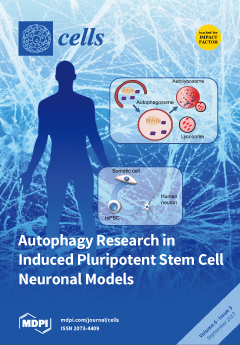
Cover Story (view full-size image): Autophagy cytoplasmic quality control protects neurons against operational stress and cell death. Human neurodegenerative diseases are often associated with declining autophagic activity, highlighting the need for better human neuronal disease models. Human neurons can be generated from somatic cells following pluripotency reprogramming (hiPSCs) and neural induction. Here, we discuss the potential of hiPSC-derived neurons for autophagy research in human neurodegenerative disease models in vitro. View this paper
  • Issues are regarded as officially published after their release is announced to the table of contents alert mailing list.
  • You may sign up for e-mail alerts to receive table of contents of newly released issues.
  • PDF is the official format for papers published in both, html and pdf forms. To view the papers in pdf format, click on the "PDF Full-text" link, and use the free Adobe Reader to open them.
Order results
Result details
Section
Select all
Export citation of selected articles as:
3061 KiB  
Review
Assessing Autophagy in Sciatic Nerves of a Rat Model that Develops Inflammatory Autoimmune Peripheral Neuropathies
by Susana Brun, Nicolas Schall, Hélène Jeltsch-David, Jérôme de Sèze and Sylviane Muller
Cells 2017, 6(3), 30; https://doi.org/10.3390/cells6030030 - 18 Sep 2017
Cited by 9 | Viewed by 11924
Abstract
The rat sciatic nerve has attracted widespread attention as an excellent model system for studying autophagy alterations in peripheral neuropathies. In our laboratory, we have developed an original rat model, which we used currently in routine novel drug screening and to evaluate treatment [...] Read more.
The rat sciatic nerve has attracted widespread attention as an excellent model system for studying autophagy alterations in peripheral neuropathies. In our laboratory, we have developed an original rat model, which we used currently in routine novel drug screening and to evaluate treatment strategies for chronic inflammatory demyelinating polyneuropathy (CIDP) and other closely related diseases. Lewis rats injected with the S-palmitoylated P0(180-199) peptide develop a chronic, sometimes relapsing-remitting type of disease. Our model fulfills electrophysiological criteria of demyelination with axonal degeneration, confirmed by immunohistopathology and several typical features of CIDP. We have set up a series of techniques that led us to examine the failures of autophagy pathways in the sciatic nerve of these model rats and to follow the possible improvement of these defects after treatment. Based on these newly introduced methods, a novel area of investigation is now open and will allow us to more thoroughly examine important features of certain autophagy pathways occurring in sciatic nerves. Full article
(This article belongs to the Special Issue Assays to Monitor Autophagy in Model Systems)
Show Figures

Figure 1

2136 KiB  
Article
Delayed Activation Kinetics of Th2- and Th17 Cells Compared to Th1 Cells
by Andrea Duechting, Anna Przybyla, Stefanie Kuerten and Paul V. Lehmann
Cells 2017, 6(3), 29; https://doi.org/10.3390/cells6030029 - 12 Sep 2017
Cited by 21 | Viewed by 6753
Abstract
During immune responses, different classes of T cells arise: Th1, Th2, and Th17. Mobilizing the right class plays a critical role in successful host defense and therefore defining the ratios of Th1/Th2/Th17 cells within the antigen-specific T cell repertoire is critical for immune [...] Read more.
During immune responses, different classes of T cells arise: Th1, Th2, and Th17. Mobilizing the right class plays a critical role in successful host defense and therefore defining the ratios of Th1/Th2/Th17 cells within the antigen-specific T cell repertoire is critical for immune monitoring purposes. Antigen-specific Th1, Th2, and Th17 cells can be detected by challenging peripheral blood mononuclear cells (PBMC) with antigen, and establishing the numbers of T cells producing the respective lead cytokine, IFN-γ and IL-2 for Th1 cells, IL-4 and IL-5 for Th2, and IL-17 for Th-17 cells, respectively. Traditionally, these cytokines are measured within 6 h in flow cytometry. We show here that 6 h of stimulation is sufficient to detect peptide-induced production of IFN-γ, but 24 h are required to reveal the full frequency of protein antigen-specific Th1 cells. Also the detection of IL-2 producing Th1 cells requires 24 h stimulation cultures. Measurements of IL-4 producing Th2 cells requires 48-h cultures and 96 h are required for frequency measurements of IL-5 and IL-17 secreting T cells. Therefore, accounting for the differential secretion kinetics of these cytokines is critical for the accurate determination of the frequencies and ratios of antigen-specific Th1, Th2, and Th17 cells. Full article
(This article belongs to the Special Issue Recent Advances in ELISPOT Research)
Show Figures

Figure 1

2420 KiB  
Review
Lamin B Receptor: Interplay between Structure, Function and Localization
by Eleni Nikolakaki, Ilias Mylonis and Thomas Giannakouros
Cells 2017, 6(3), 28; https://doi.org/10.3390/cells6030028 - 31 Aug 2017
Cited by 41 | Viewed by 16412
Abstract
Lamin B receptor (LBR) is an integral protein of the inner nuclear membrane, containing a hydrophilic N-terminal end protruding into the nucleoplasm, eight hydrophobic segments that span the membrane and a short, nucleoplasmic C-terminal tail. Two seemingly unrelated functions have been [...] Read more.
Lamin B receptor (LBR) is an integral protein of the inner nuclear membrane, containing a hydrophilic N-terminal end protruding into the nucleoplasm, eight hydrophobic segments that span the membrane and a short, nucleoplasmic C-terminal tail. Two seemingly unrelated functions have been attributed to LBR. Its N-terminal domain tethers heterochromatin to the nuclear periphery, thus contributing to the shape of interphase nuclear architecture, while its transmembrane domains exhibit sterol reductase activity. Mutations within the transmembrane segments result in defects in cholesterol synthesis and are associated with diseases such as the Pelger–Huët anomaly and Greenberg skeletal dysplasia, whereas no such harmful mutations related to the anchoring properties of LBR have been reported so far. Recent evidence suggests a dynamic regulation of LBR expression levels, structural organization, localization and function, in response to various signals. The molecular mechanisms underlying this dynamic behavior have not yet been fully unraveled. Here, we provide an overview of the current knowledge of the interplay between the structure, function and localization of LBR, and hint at the interconnection of the two distinct functions of LBR. Full article
(This article belongs to the Collection Lamins and Laminopathies)
Show Figures

Figure 1

2598 KiB  
Review
Approaches for Studying Autophagy in Caenorhabditis elegans
by Yanfang Chen, Vincent Scarcelli and Renaud Legouis
Cells 2017, 6(3), 27; https://doi.org/10.3390/cells6030027 - 30 Aug 2017
Cited by 34 | Viewed by 8930
Abstract
Macroautophagy (hereafter referred to as autophagy) is an intracellular degradative process, well conserved among eukaryotes. By engulfing cytoplasmic constituents into the autophagosome for degradation, this process is involved in the maintenance of cellular homeostasis. Autophagy induction triggers the formation of a cup-shaped double [...] Read more.
Macroautophagy (hereafter referred to as autophagy) is an intracellular degradative process, well conserved among eukaryotes. By engulfing cytoplasmic constituents into the autophagosome for degradation, this process is involved in the maintenance of cellular homeostasis. Autophagy induction triggers the formation of a cup-shaped double membrane structure, the phagophore, which progressively elongates and encloses materials to be removed. This double membrane vesicle, which is called an autophagosome, fuses with lysosome and forms the autolysosome. The inner membrane of the autophagosome, along with engulfed compounds, are degraded by lysosomal enzymes, which enables the recycling of carbohydrates, amino acids, nucleotides, and lipids. In response to various factors, autophagy can be induced for non-selective degradation of bulk cytoplasm. Autophagy is also able to selectively target cargoes and organelles such as mitochondria or peroxisome, functioning as a quality control system. The modification of autophagy flux is involved in developmental processes such as resistance to stress conditions, aging, cell death, and multiple pathologies. So, the use of animal models is essential for understanding these processes in the context of different cell types throughout the entire lifespan. For almost 15 years, the nematode Caenorhabditis elegans has emerged as a powerful model to analyze autophagy in physiological or pathological contexts. This review presents a rapid overview of physiological processes involving autophagy in Caenorhabditis elegans, the different assays used to monitor autophagy, their drawbacks, and specific tools for the analyses of selective autophagy. Full article
(This article belongs to the Special Issue Assays to Monitor Autophagy in Model Systems)
Show Figures

Figure 1

226 KiB  
Review
The Potential of Targeting Brain Pathology with Ascl1/Mash1
by Bor Luen Tang
Cells 2017, 6(3), 26; https://doi.org/10.3390/cells6030026 - 23 Aug 2017
Cited by 16 | Viewed by 5081
Abstract
The proneural factor Achaete-scute complex-like 1 (Ascl1/Mash1) acts as a pioneering transcription factor that initializes neuronal reprogramming. It drives neural progenitors and non-neuronal cells to exit the cell cycle, and promotes neuronal differentiation by activating neuronal target genes, even those that are normally [...] Read more.
The proneural factor Achaete-scute complex-like 1 (Ascl1/Mash1) acts as a pioneering transcription factor that initializes neuronal reprogramming. It drives neural progenitors and non-neuronal cells to exit the cell cycle, and promotes neuronal differentiation by activating neuronal target genes, even those that are normally repressed. Importantly, force-expression of Ascl1 was shown to drive proliferative reactive astroglia formed during stroke and glioblastoma stem cells towards neuronal differentiation, and this could potentially diminish CNS damage resulting from their proliferation. As a pro-neural factor, Ascl1 also has the general effect of enhancing neurite growth by damaged or surviving neurons. Here, a hypothesis that brain pathologies associated with traumatic/ischemic injury and malignancy could be targeted with pro-neural factors that drives neuronal differentiation is formulated and explored. Although a good number of caveats exist, exogenous over-expression of Ascl1, alone or in combination with other factors, may be worth further consideration as a therapeutic approach in brain injury and cancer. Full article
499 KiB  
Review
Assessment of Autophagy in Neurons and Brain Tissue
by Irene Benito-Cuesta, Héctor Diez, Lara Ordoñez and Francisco Wandosell
Cells 2017, 6(3), 25; https://doi.org/10.3390/cells6030025 - 23 Aug 2017
Cited by 43 | Viewed by 9378
Abstract
Autophagy is a complex process that controls the transport of cytoplasmic components into lysosomes for degradation. This highly conserved proteolytic system involves dynamic and complex processes, using similar molecular elements and machinery from yeast to humans. Moreover, autophagic dysfunction may contribute to a [...] Read more.
Autophagy is a complex process that controls the transport of cytoplasmic components into lysosomes for degradation. This highly conserved proteolytic system involves dynamic and complex processes, using similar molecular elements and machinery from yeast to humans. Moreover, autophagic dysfunction may contribute to a broad spectrum of mammalian diseases. Indeed, in adult tissues, where the capacity for regeneration or cell division is low or absent (e.g., in the mammalian brain), the accumulation of proteins/peptides that would otherwise be recycled or destroyed may have pathological implications. Indeed, such changes are hallmarks of pathologies, like Alzheimer’s, Prion or Parkinson’s disease, known as proteinopathies. However, it is still unclear whether such dysfunction is a cause or an effect in these conditions. One advantage when analysing autophagy in the mammalian brain is that almost all the markers described in different cell lineages and systems appear to be present in the brain, and even in neurons. By contrast, the mixture of cell types present in the brain and the differentiation stage of such neurons, when compared with neurons in culture, make translating basic research to the clinic less straightforward. Thus, the purpose of this review is to describe and discuss the methods available to monitor autophagy in neurons and in the mammalian brain, a process that is not yet fully understood, focusing primarily on mammalian macroautophagy. We will describe some general features of neuronal autophagy that point to our focus on neuropathologies in which macroautophagy may be altered. Indeed, we centre this review around the hypothesis that enhanced autophagy may be able to provide therapeutic benefits in some brain pathologies, like Alzheimer’s disease, considering this pathology as one of the most prevalent proteinopathies. Full article
(This article belongs to the Special Issue Assays to Monitor Autophagy in Model Systems)
Show Figures

Figure 1

2757 KiB  
Review
Induced Pluripotent Stem Cell Neuronal Models for the Study of Autophagy Pathways in Human Neurodegenerative Disease
by Natalia Jiménez-Moreno, Petros Stathakos, Maeve A. Caldwell and Jon D. Lane
Cells 2017, 6(3), 24; https://doi.org/10.3390/cells6030024 - 11 Aug 2017
Cited by 18 | Viewed by 11800
Abstract
Human induced pluripotent stem cells (hiPSCs) are invaluable tools for research into the causes of diverse human diseases, and have enormous potential in the emerging field of regenerative medicine. Our ability to reprogramme patient cells to become hiPSCs, and to subsequently direct their [...] Read more.
Human induced pluripotent stem cells (hiPSCs) are invaluable tools for research into the causes of diverse human diseases, and have enormous potential in the emerging field of regenerative medicine. Our ability to reprogramme patient cells to become hiPSCs, and to subsequently direct their differentiation towards those classes of neurons that are vulnerable to stress, is revealing how genetic mutations cause changes at the molecular level that drive the complex pathogeneses of human neurodegenerative diseases. Autophagy dysregulation is considered to be a major contributor in neural decline during the onset and progression of many human neurodegenerative diseases, meaning that a better understanding of the control of non-selective and selective autophagy pathways (including mitophagy) in disease-affected classes of neurons is needed. To achieve this, it is essential that the methodologies commonly used to study autophagy regulation under basal and stressed conditions in standard cell-line models are accurately applied when using hiPSC-derived neuronal cultures. Here, we discuss the roles and control of autophagy in human stem cells, and how autophagy contributes to neural differentiation in vitro. We also describe how autophagy-monitoring tools can be applied to hiPSC-derived neurons for the study of human neurodegenerative disease in vitro. Full article
(This article belongs to the Special Issue Assays to Monitor Autophagy in Model Systems)
Show Figures

Figure 1

3896 KiB  
Review
Assays to Monitor Autophagy in Saccharomyces cerevisiae
by Raffaela Torggler, Daniel Papinski and Claudine Kraft
Cells 2017, 6(3), 23; https://doi.org/10.3390/cells6030023 - 13 Jul 2017
Cited by 45 | Viewed by 13801
Abstract
Autophagy is an intracellular process responsible for the degradation and recycling of cytoplasmic components. It selectively removes harmful cellular material and enables the cell to survive starvation by mobilizing nutrients via the bulk degradation of cytoplasmic components. While research over the last decades [...] Read more.
Autophagy is an intracellular process responsible for the degradation and recycling of cytoplasmic components. It selectively removes harmful cellular material and enables the cell to survive starvation by mobilizing nutrients via the bulk degradation of cytoplasmic components. While research over the last decades has led to the discovery of the key factors involved in autophagy, the pathway is not yet completely understood. The first studies of autophagy on a molecular level were conducted in the yeast Saccharomyces cerevisiae. Building up on these studies, many homologs have been found in higher eukaryotes. Yeast remains a highly relevant model organism for studying autophagy, with a wide range of established methods to elucidate the molecular details of the autophagy pathway. In this review, we provide an overview of methods to study both selective and bulk autophagy, including intermediate steps in the yeast Saccharomyces cerevisiae. We compare different assays, discuss their advantages and limitations and list potential applications. Full article
(This article belongs to the Special Issue Assays to Monitor Autophagy in Model Systems)
Show Figures

Figure 1

2013 KiB  
Review
Exploring Autophagy in Drosophila
by Péter Lőrincz, Caroline Mauvezin and Gábor Juhász
Cells 2017, 6(3), 22; https://doi.org/10.3390/cells6030022 - 12 Jul 2017
Cited by 56 | Viewed by 11963
Abstract
Autophagy is a catabolic process in eukaryotic cells promoting bulk or selective degradation of cellular components within lysosomes. In recent decades, several model systems were utilized to dissect the molecular machinery of autophagy and to identify the impact of this cellular “self-eating” process [...] Read more.
Autophagy is a catabolic process in eukaryotic cells promoting bulk or selective degradation of cellular components within lysosomes. In recent decades, several model systems were utilized to dissect the molecular machinery of autophagy and to identify the impact of this cellular “self-eating” process on various physiological and pathological processes. Here we briefly discuss the advantages and limitations of using the fruit fly Drosophila melanogaster, a popular model in cell and developmental biology, to apprehend the main pathway of autophagy in a complete animal. Full article
(This article belongs to the Special Issue Assays to Monitor Autophagy in Model Systems)
Show Figures

Figure 1

3664 KiB  
Review
Studying Autophagy in Zebrafish
by Benan John Mathai, Annemarie H. Meijer and Anne Simonsen
Cells 2017, 6(3), 21; https://doi.org/10.3390/cells6030021 - 9 Jul 2017
Cited by 54 | Viewed by 12031
Abstract
Autophagy is an evolutionarily conserved catabolic process which allows lysosomal degradation of complex cytoplasmic components into basic biomolecules that are recycled for further cellular use. Autophagy is critical for cellular homeostasis and for degradation of misfolded proteins and damaged organelles as well as [...] Read more.
Autophagy is an evolutionarily conserved catabolic process which allows lysosomal degradation of complex cytoplasmic components into basic biomolecules that are recycled for further cellular use. Autophagy is critical for cellular homeostasis and for degradation of misfolded proteins and damaged organelles as well as intracellular pathogens. The role of autophagy in protection against age-related diseases and a plethora of other diseases is now coming to light; assisted by several divergent eukaryotic model systems ranging from yeast to mice. We here give an overview of different methods used to analyse autophagy in zebrafish—a relatively new model for studying autophagy—and briefly discuss what has been done so far and possible future directions. Full article
(This article belongs to the Special Issue Assays to Monitor Autophagy in Model Systems)
Show Figures

Figure 1

243 KiB  
Review
Assays to Monitor Autophagy Progression in Cell Cultures
by Idil Orhon and Fulvio Reggiori
Cells 2017, 6(3), 20; https://doi.org/10.3390/cells6030020 - 7 Jul 2017
Cited by 45 | Viewed by 8232
Abstract
The vast number of implications of autophagy in multiple areas of life sciences and medicine has attracted the interest of numerous scientists that aim to unveil the role of this process in specific physiological and pathological contexts. Cell cultures are one of the [...] Read more.
The vast number of implications of autophagy in multiple areas of life sciences and medicine has attracted the interest of numerous scientists that aim to unveil the role of this process in specific physiological and pathological contexts. Cell cultures are one of the most frequently used experimental setup for the investigation of autophagy. As a result, it is essential to assess this highly regulated molecular pathway with efficient and reliable methods. Each method has its own advantages and disadvantages. Here, we present a review summarizing the most established assays used to monitor autophagy induction and progression in cell cultures, in order to guide researchers in the selection of the most optimal solution for their experimental setup and design. Full article
(This article belongs to the Special Issue Assays to Monitor Autophagy in Model Systems)
4132 KiB  
Review
Vascular Mechanobiology: Towards Control of In Situ Regeneration
by Eline E. Van Haaften, Carlijn V. C. Bouten and Nicholas A. Kurniawan
Cells 2017, 6(3), 19; https://doi.org/10.3390/cells6030019 - 3 Jul 2017
Cited by 42 | Viewed by 9012
Abstract
The paradigm of regenerative medicine has recently shifted from in vitro to in situ tissue engineering: implanting a cell-free, biodegradable, off-the-shelf available scaffold and inducing the development of functional tissue by utilizing the regenerative potential of the body itself. This approach offers a [...] Read more.
The paradigm of regenerative medicine has recently shifted from in vitro to in situ tissue engineering: implanting a cell-free, biodegradable, off-the-shelf available scaffold and inducing the development of functional tissue by utilizing the regenerative potential of the body itself. This approach offers a prospect of not only alleviating the clinical demand for autologous vessels but also circumventing the current challenges with synthetic grafts. In order to move towards a hypothesis-driven engineering approach, we review three crucial aspects that need to be taken into account when regenerating vessels: (1) the structure-function relation for attaining mechanical homeostasis of vascular tissues, (2) the environmental cues governing cell function, and (3) the available experimental platforms to test instructive scaffolds for in situ tissue engineering. The understanding of cellular responses to environmental cues leads to the development of computational models to predict tissue formation and maturation, which are validated using experimental platforms recapitulating the (patho)physiological micro-environment. With the current advances, a progressive shift is anticipated towards a rational and effective approach of building instructive scaffolds for in situ vascular tissue regeneration. Full article
(This article belongs to the Special Issue Tissue Regeneration and Fibrosis)
Show Figures

Figure 1

3212 KiB  
Review
Methods to Monitor and Quantify Autophagy in the Social Amoeba Dictyostelium discoideum
by Eunice Domínguez-Martín, Elena Cardenal-Muñoz, Jason S. King, Thierry Soldati, Roberto Coria and Ricardo Escalante
Cells 2017, 6(3), 18; https://doi.org/10.3390/cells6030018 - 3 Jul 2017
Cited by 22 | Viewed by 10765
Abstract
Autophagy is a eukaryotic catabolic pathway that degrades and recycles cellular components to maintain homeostasis. It can target protein aggregates, superfluous biomolecular complexes, dysfunctional and damaged organelles, as well as pathogenic intracellular microbes. Autophagy is a dynamic process in which the different stages [...] Read more.
Autophagy is a eukaryotic catabolic pathway that degrades and recycles cellular components to maintain homeostasis. It can target protein aggregates, superfluous biomolecular complexes, dysfunctional and damaged organelles, as well as pathogenic intracellular microbes. Autophagy is a dynamic process in which the different stages from initiation to final degradation of cargo are finely regulated. Therefore, the study of this process requires the use of a palette of techniques, which are continuously evolving and whose interpretation is not trivial. Here, we present the social amoeba Dictyostelium discoideum as a relevant model to study autophagy. Several methods have been developed based on the tracking and observation of autophagosomes by microscopy, analysis of changes in expression of autophagy genes and proteins, and examination of the autophagic flux with various techniques. In this review, we discuss the pros and cons of the currently available techniques to assess autophagy in this organism. Full article
(This article belongs to the Special Issue Assays to Monitor Autophagy in Model Systems)
Show Figures

Figure 1

4852 KiB  
Review
Standard Immunohistochemical Assays to Assess Autophagy in Mammalian Tissue
by Wim Martinet, Lynn Roth and Guido R. Y. De Meyer
Cells 2017, 6(3), 17; https://doi.org/10.3390/cells6030017 - 30 Jun 2017
Cited by 28 | Viewed by 7922
Abstract
Autophagy is a highly conserved lysosomal degradation pathway with major impact on diverse human pathologies. Despite the development of different methodologies to detect autophagy both in vitro and in vivo, monitoring autophagy in tissue via immunohistochemical techniques is hampered due to the lack [...] Read more.
Autophagy is a highly conserved lysosomal degradation pathway with major impact on diverse human pathologies. Despite the development of different methodologies to detect autophagy both in vitro and in vivo, monitoring autophagy in tissue via immunohistochemical techniques is hampered due to the lack of biomarkers. Immunohistochemical detection of a punctate pattern of ATG8/MAP1LC3 proteins is currently the most frequently used approach to detect autophagy in situ, but it depends on a highly sensitive detection method and is prone to misinterpretation. Moreover, reliable MAP1LC3 immunohistochemical staining requires correct tissue processing and high-quality, isoform-specific antibodies. Immunohistochemical analysis of other autophagy-related protein targets such as SQSTM1, ubiquitin, ATG5 or lysosomal proteins is not recommended as marker for autophagic activity in tissue for multiple reasons including aspecific labeling of cellular structures and a lack of differential protein expression during autophagy initiation. To better understand the role of autophagy in human disease, novel biomarkers for visualization of the autophagic process with standard histology techniques are urgently needed. Full article
(This article belongs to the Special Issue Assays to Monitor Autophagy in Model Systems)
Show Figures

Figure 1

2748 KiB  
Review
Assessing Autophagy in Mouse Models and Patients with Systemic Autoimmune Diseases
by Fengjuan Wang, Baihui Li, Nicolas Schall, Maud Wilhelm and Sylviane Muller
Cells 2017, 6(3), 16; https://doi.org/10.3390/cells6030016 - 28 Jun 2017
Cited by 32 | Viewed by 8421
Abstract
Autophagy is a tightly regulated mechanism that allows cells to renew themselves through the lysosomal degradation of proteins, which are misfolded or produced in excess, and of damaged organelles. In the context of immunity, recent research has specially attempted to clarify its roles [...] Read more.
Autophagy is a tightly regulated mechanism that allows cells to renew themselves through the lysosomal degradation of proteins, which are misfolded or produced in excess, and of damaged organelles. In the context of immunity, recent research has specially attempted to clarify its roles in infection, inflammation and autoimmunity. Autophagy has emerged as a spotlight in several molecular pathways and trafficking events that participate to innate and adaptive immunity. Deregulation of autophagy has been associated to several autoimmune diseases, in particular to systemic lupus erythematosus. Nowadays, however, experimental data on the implication of autophagy in animal models of autoimmunity or patients remain limited. In our investigations, we use Murphy Roths Large (MRL)/lymphoproliferation (lpr) lupus-prone mice as a mouse model for lupus and secondary Sjögren’s syndrome, and, herein, we describe methods applied routinely to analyze different autophagic pathways in different lymphoid organs and tissues (spleen, lymph nodes, salivary glands). We also depict some techniques used to analyze autophagy in lupus patient’s blood samples. These methods can be adapted to the analysis of autophagy in other mouse models of autoinflammatory diseases. The understanding of autophagy implication in autoimmune diseases could prove to be very useful for developing novel immunomodulatory strategies. Our attention should be focused on the fact that autophagy processes are interconnected and that distinct pathways can be independently hyper-activated or downregulated in distinct organs and tissues of the same individual. Full article
(This article belongs to the Special Issue Assays to Monitor Autophagy in Model Systems)
Show Figures

Figure 1

Previous Issue
Next Issue
Back to TopTop