Next Issue
Volume 7, November
Previous Issue
Volume 7, September


Cells, Volume 7, Issue 10 (October 2018) – 39 articles

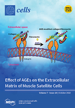
Cover Story (view full-size image): The skeletal muscle is the core tissue of insulin-mediated glucose uptake within the body. The extracellular matrix (ECM) provides integrity and biochemical signals and plays a crucial role in myogenesis; it also undergoes remodelling upon injury, which is related to insulin resistance (IR). Here, we reviewed the relationship between the ECM, IR, and skeletal muscle and discussed briefly the clinical and non-clinical aspect of ECM remodelling and IR. View the paper here.
  • Issues are regarded as officially published after their release is announced to the table of contents alert mailing list.
  • You may sign up for e-mail alerts to receive table of contents of newly released issues.
  • PDF is the official format for papers published in both, html and pdf forms. To view the papers in pdf format, click on the "PDF Full-text" link, and use the free Adobe Reader to open them.
Order results
Result details
Section
Select all
Export citation of selected articles as:
19 pages, 1148 KiB  
Review
Is there a Chance to Promote Arteriogenesis by DPP4 Inhibitors Even in Type 2 Diabetes? A Critical Review
by Srinivasan Vedantham, Anna-Kristina Kluever and Elisabeth Deindl
Cells 2018, 7(10), 181; https://doi.org/10.3390/cells7100181 - 22 Oct 2018
Cited by 10 | Viewed by 5835
Abstract
Cardiovascular diseases (CVD) are still the prevailing cause of death not only in industrialized countries, but even worldwide. Type 2 diabetes mellitus (type 2 DM) and hyperlipidemia, a metabolic disorder that is often associated with diabetes, are major risk factors for developing CVD. [...] Read more.
Cardiovascular diseases (CVD) are still the prevailing cause of death not only in industrialized countries, but even worldwide. Type 2 diabetes mellitus (type 2 DM) and hyperlipidemia, a metabolic disorder that is often associated with diabetes, are major risk factors for developing CVD. Recently, clinical trials proved the safety of gliptins in treating patients with type 2 DM. Gliptins are dipeptidyl-peptidase 4 (DPP4/CD26) inhibitors, which stabilize glucagon-like peptide-1 (GLP-1), thereby increasing the bioavailability of insulin. Moreover, blocking DPP4 results in increased levels of stromal cell derived factor 1 (SDF-1). SDF-1 has been shown in pre-clinical animal studies to improve heart function and survival after myocardial infarction, and to promote arteriogenesis, the growth of natural bypasses, compensating for the function of an occluded artery. Clinical trials, however, failed to demonstrate a superiority of gliptins compared to placebo treated type 2 DM patients in terms of cardiovascular (CV) outcomes. This review highlights the function of DPP4 inhibitors in type 2 DM, and in treating cardiovascular diseases, with special emphasis on arteriogenesis. It critically addresses the potency of currently available gliptins and gives rise to hope by pointing out the most relevant questions that need to be resolved. Full article
Show Figures

Figure 1

12 pages, 1925 KiB  
Article
The Relationship between Turgor Pressure Change and Cell Hydraulics of Midrib Parenchyma Cells in the Leaves of Zea mays
by Yangmin X. Kim, Burkhard Stumpf, Jwakyung Sung and Sang Joon Lee
Cells 2018, 7(10), 180; https://doi.org/10.3390/cells7100180 - 22 Oct 2018
Cited by 8 | Viewed by 5615
Abstract
Leaf dehydration decreases water potential and cell turgor pressure. Therefore, changes in cell turgor pressure may regulate water transport across plant cell membranes. Using a cell pressure probe, the hydraulic properties of parenchyma cells in the midrib of maize (Zea mays L.) [...] Read more.
Leaf dehydration decreases water potential and cell turgor pressure. Therefore, changes in cell turgor pressure may regulate water transport across plant cell membranes. Using a cell pressure probe, the hydraulic properties of parenchyma cells in the midrib of maize (Zea mays L.) leaves were measured (half time T 1 / 2 of water exchange in cells as a measure of hydraulic conductivity Lp). Using intact plants with root systems encased in a pressure chamber, the root systems were pressurized and the turgor pressure in leaf cells increased by increments up to 0.3 MPa. However, the increase in the cell turgor did not increase but stabilized T 1 / 2 values. Increased water potential in leaf cells seemed to have stabilizing effects on the T 1 / 2 probably due to enhanced water availability. When the cell turgor decreased by 0.1 MPa to 0.3 MPa with releasing the pressure in the pressure chamber, T 1 / 2 was temporarily increased to a large degree, a factor of up to 13 within 30 min. Full article
(This article belongs to the Special Issue Aquaporins)
Show Figures

Figure 1

19 pages, 1438 KiB  
Review
The Expanding Role of Vesicles Containing Aquaporins
by M Carmen Martinez-Ballesta, Paula Garcia-Ibañez, Lucía Yepes-Molina, Juan José Rios and Micaela Carvajal
Cells 2018, 7(10), 179; https://doi.org/10.3390/cells7100179 - 22 Oct 2018
Cited by 14 | Viewed by 5267
Abstract
In animals and plants, membrane vesicles containing proteins have been defined as key for biological systems involving different processes such as trafficking or intercellular communication. Docking and fusion of vesicles to the plasma membrane occur in living cells in response to different stimuli, [...] Read more.
In animals and plants, membrane vesicles containing proteins have been defined as key for biological systems involving different processes such as trafficking or intercellular communication. Docking and fusion of vesicles to the plasma membrane occur in living cells in response to different stimuli, such as environmental changes or hormones, and therefore play an important role in cell homeostasis as vehicles for certain proteins or other substances. Because aquaporins enhance the water permeability of membranes, their role as proteins immersed in vesicles formed of natural membranes is a recent topic of study. They regulate numerous physiological processes and could hence serve new biotechnological purposes. Thus, in this review, we have explored the physiological implications of the trafficking of aquaporins, the mechanisms that control their transit, and the proteins that coregulate the migration. In addition, the importance of exosomes containing aquaporins in the cell-to-cell communication processes in animals and plants have been analyzed, together with their potential uses in biomedicine or biotechnology. The properties of aquaporins make them suitable for use as biomarkers of different aquaporin-related diseases when they are included in exosomes. Finally, the fact that these proteins could be immersed in biomimetic membranes opens future perspectives for new biotechnological applications. Full article
(This article belongs to the Special Issue Aquaporins)
Show Figures

Figure 1

17 pages, 6729 KiB  
Article
Mechanisms of Age-Dependent Loss of Dietary Restriction Protective Effects in Acute Kidney Injury
by Nadezda V. Andrianova, Stanislovas S. Jankauskas, Ljubava D. Zorova, Irina B. Pevzner, Vasily A. Popkov, Denis N. Silachev, Egor Y. Plotnikov and Dmitry B. Zorov
Cells 2018, 7(10), 178; https://doi.org/10.3390/cells7100178 - 22 Oct 2018
Cited by 20 | Viewed by 5488
Abstract
Dietary restriction (DR) is one of the most efficient approaches ameliorating the severity of different pathological conditions including aging. We investigated the protective potential of short-term DR in the model of acute kidney injury (AKI) in young and old rats. In kidney tissue, [...] Read more.
Dietary restriction (DR) is one of the most efficient approaches ameliorating the severity of different pathological conditions including aging. We investigated the protective potential of short-term DR in the model of acute kidney injury (AKI) in young and old rats. In kidney tissue, the levels of autophagy and mitophagy were examined, and proliferative properties of renal cells obtained from rats of different age were compared. DR afforded a significant nephroprotection to ischemic kidneys of young rats. However, in old rats, DR did not provide such beneficial effect. On the assessment of the autophagy marker, the LC3 II/LC3 I ratio, and after staining the tissue with LysoTracker Green, we concluded that in old rats activity of the autophagic-lysosomal system decreased. Mitophagy, as assessed by the levels of PINK-1, was also deteriorated in old animals. Renal cells from old rats showed impaired proliferative capacity, a worse rate of recovery after ischemic injury, increased levels of oxidative stress, accumulation of lipofuscin granules and lower mitochondria membrane potential. The results suggest that the loss of DR benefits in old animals could be due to deterioration in the autophagy/mitophagy flux. Full article
(This article belongs to the Special Issue Autophagy in Tissue Injury and Homeostasis)
Show Figures

Figure 1

14 pages, 3107 KiB  
Brief Report
Targeting DNA Binding for NF-κB as an Anticancer Approach in Hepatocellular Carcinoma
by Po Yee Chung, Pik Ling Lam, Yuanyuan Zhou, Jessica Gasparello, Alessia Finotti, Adriana Chilin, Giovanni Marzaro, Roberto Gambari, Zhaoxiang Bian, Wai Ming Kwok, Wai Yeung Wong, Xi Wang, Alfred King-yin Lam, Albert Sun-chi Chan, Xingshu Li, Jessica Yuen Wuen Ma, Chung Hin Chui, Kim Hung Lam and Johnny Cheuk On Tang
Cells 2018, 7(10), 177; https://doi.org/10.3390/cells7100177 - 22 Oct 2018
Cited by 12 | Viewed by 4094
Abstract
Quinoline core has been shown to possess a promising role in the development of anticancer agents. However, the correlation between its broad spectrum of bioactivity and the underlying mechanism of actions is poorly understood. The present study, with the use of bioinformatics approaches, [...] Read more.
Quinoline core has been shown to possess a promising role in the development of anticancer agents. However, the correlation between its broad spectrum of bioactivity and the underlying mechanism of actions is poorly understood. The present study, with the use of bioinformatics approaches, reported a series of designed molecules which integrated quinoline core and sulfonyl moiety, with the objective of evaluating the substituent and linker effects on anticancer activities and associated mechanistic targets. We identified potent compounds (1h, 2h, 5 and 8) exhibiting significant anticancer effects towards liver cancer cells (Hep3B) with the 3-(4,5-dimethylthiazol-2-yl)-5-(3-carboxymethoxyphenyl)-2-(4-sulfophenyl)-2H-tetrazolium (MTS) relative values of cytotoxicity below 0.40, a value in the range of doxorubicin positive control with the value of 0.12. Bulky substituents and the presence of bromine atom, as well as the presence of sulfonamide linkage, are likely the favorable structural components for molecules exerting a strong anticancer effect. To the best of our knowledge, our findings obtained from chemical synthesis, in vitro cytotoxicity, bioinformatics-based molecular docking analysis (similarity ensemble approach, SEA),and electrophoretic mobility shift assay provided the first evidence in correlation to the anticancer activities of the selected compound 5 with the modulation on the binding of transcription factor NF-κB to its target DNA. Accordingly, compound 5 represented a lead structure for the development of quinoline-based NF-κB inhibitors and this work added novel information on the understanding of the mechanism of action for bioactive sulfonyl-containing quinoline compounds against hepatocellular carcinoma. Full article
(This article belongs to the Section Cell Signaling)
Show Figures

Graphical abstract

30 pages, 2136 KiB  
Review
Inhibitory-κB Kinase (IKK) α and Nuclear Factor-κB (NFκB)-Inducing Kinase (NIK) as Anti-Cancer Drug Targets
by Andrew Paul, Joanne Edwards, Christopher Pepper and Simon Mackay
Cells 2018, 7(10), 176; https://doi.org/10.3390/cells7100176 - 20 Oct 2018
Cited by 63 | Viewed by 14212
Abstract
The cellular kinases inhibitory-κB kinase (IKK) α and Nuclear Factor-κB (NF-κB)-inducing kinase (NIK) are well recognised as key central regulators and drivers of the non-canonical NF-κB cascade and as such dictate the initiation and development of defined transcriptional responses associated with the liberation [...] Read more.
The cellular kinases inhibitory-κB kinase (IKK) α and Nuclear Factor-κB (NF-κB)-inducing kinase (NIK) are well recognised as key central regulators and drivers of the non-canonical NF-κB cascade and as such dictate the initiation and development of defined transcriptional responses associated with the liberation of p52-RelB and p52-p52 NF-κB dimer complexes. Whilst these kinases and downstream NF-κB complexes transduce pro-inflammatory and growth stimulating signals that contribute to major cellular processes, they also play a key role in the pathogenesis of a number of inflammatory-based conditions and diverse cancer types, which for the latter may be a result of background mutational status. IKKα and NIK, therefore, represent attractive targets for pharmacological intervention. Here, specifically in the cancer setting, we reflect on the potential pathophysiological role(s) of each of these kinases, their associated downstream signalling outcomes and the stimulatory and mutational mechanisms leading to their increased activation. We also consider the downstream coordination of transcriptional events and phenotypic outcomes illustrative of key cancer ‘Hallmarks’ that are now increasingly perceived to be due to the coordinated recruitment of both NF-κB-dependent as well as NF-κB–independent signalling. Furthermore, as these kinases regulate the transition from hormone-dependent to hormone-independent growth in defined tumour subsets, potential tumour reactivation and major cytokine and chemokine species that may have significant bearing upon tumour-stromal communication and tumour microenvironment it reiterates their potential to be drug targets. Therefore, with the emergence of small molecule kinase inhibitors targeting each of these kinases, we consider medicinal chemistry efforts to date and those evolving that may contribute to the development of viable pharmacological intervention strategies to target a variety of tumour types. Full article
(This article belongs to the Special Issue NF-κB in Cancer)
Show Figures

Figure 1

10 pages, 1762 KiB  
Article
Iron Depletion Affects Genes Encoding Mitochondrial Electron Transport Chain and Genes of Non­Oxidative Metabolism, Pyruvate Kinase and Lactate Dehydrogenase, in Primary Human Cardiac Myocytes Cultured upon Mechanical Stretch
by Magdalena Dziegala, Kamil A. Kobak, Monika Kasztura, Jacek Bania, Krystian Josiak, Waldemar Banasiak, Piotr Ponikowski and Ewa A. Jankowska
Cells 2018, 7(10), 175; https://doi.org/10.3390/cells7100175 - 20 Oct 2018
Cited by 17 | Viewed by 4219
Abstract
(1) Background: Oxidative energy metabolism is presumed to rely on the optimal iron supply. Primary human cardiac myocytes (HCM) exposed to different iron availability conditions during mechanical stretch are anticipated to demonstrate expression changes of genes involved in aerobic and anaerobic metabolic pathways. [...] Read more.
(1) Background: Oxidative energy metabolism is presumed to rely on the optimal iron supply. Primary human cardiac myocytes (HCM) exposed to different iron availability conditions during mechanical stretch are anticipated to demonstrate expression changes of genes involved in aerobic and anaerobic metabolic pathways. (2) Methods: HCM were cultured for 48 h either in static conditions and upon mechanical stretch at the optimal versus reduced versus increased iron concentrations. We analyzed the expression of pyruvate kinase (PKM2), lactate dehydrogenase A (LDHA), and mitochondrial complexes I–V at the mRNA and protein levels. The concentration of l-lactate was assessed by means of lactate oxidase method-based kit. (3) Results: Reduced iron concentrations during mechanical work caused a decreased expression of complexes I–V (all p < 0.05). The expression of PKM2 and LDHA, as well as the medium concentration of l-lactate, was increased in these conditions (both p < 0.05). HCM exposed to the increased iron concentration during mechanical effort demonstrated a decreased expression of mitochondrial complexes (all p < 0.01); however, a decrement was smaller than in case of iron chelation (p < 0.05). The iron-enriched medium caused a decrease in expression of LDHA and did not influence the concentration of l-lactate. (4) Conclusions: During mechanical effort, the reduced iron availability enhances anaerobic glycolysis and extracellular lactate production, whilst decreasing mitochondrial aerobic pathway in HCM. Iron enrichment during mechanical effort may be protective in the context of intracellular protein machinery of non-oxidative metabolism with no effect on the extracellular lactate concentration. Full article
(This article belongs to the Section Intracellular and Plasma Membranes)
Show Figures

Graphical abstract

22 pages, 1704 KiB  
Review
Aquaporins and Their Regulation after Spinal Cord Injury
by Andrea M. Halsey, Alex C. Conner, Roslyn M. Bill, Ann Logan and Zubair Ahmed
Cells 2018, 7(10), 174; https://doi.org/10.3390/cells7100174 - 18 Oct 2018
Cited by 22 | Viewed by 6797
Abstract
After injury to the spinal cord, edema contributes to the underlying detrimental pathophysiological outcomes that lead to worsening of function. Several related membrane proteins called aquaporins (AQPs) regulate water movement in fluid transporting tissues including the spinal cord. Within the cord, AQP1, 4 [...] Read more.
After injury to the spinal cord, edema contributes to the underlying detrimental pathophysiological outcomes that lead to worsening of function. Several related membrane proteins called aquaporins (AQPs) regulate water movement in fluid transporting tissues including the spinal cord. Within the cord, AQP1, 4 and 9 contribute to spinal cord injury (SCI)-induced edema. AQP1, 4 and 9 are expressed in a variety of cells including astrocytes, neurons, ependymal cells, and endothelial cells. This review discusses some of the recent findings of the involvement of AQP in SCI and highlights the need for further study of these proteins to develop effective therapies to counteract the negative effects of SCI-induced edema. Full article
(This article belongs to the Special Issue Aquaporins)
Show Figures

Figure 1

17 pages, 1919 KiB  
Article
Subcutaneous Maturation of Neural Stem Cell-Loaded Hydrogels Forms Region-Specific Neuroepithelium
by Mahmoud Farrag and Nic D. Leipzig
Cells 2018, 7(10), 173; https://doi.org/10.3390/cells7100173 - 17 Oct 2018
Cited by 13 | Viewed by 4572
Abstract
A combinatorial approach integrating stem cells and capable of exploiting available cues is likely needed to regenerate lost neural tissues and ultimately restore neurologic functions. This study investigates the effects of the subcutaneous maturation of adult-derived neural stem cell (aNSCs) seeded into biomaterial [...] Read more.
A combinatorial approach integrating stem cells and capable of exploiting available cues is likely needed to regenerate lost neural tissues and ultimately restore neurologic functions. This study investigates the effects of the subcutaneous maturation of adult-derived neural stem cell (aNSCs) seeded into biomaterial constructs on aNSC differentiation and ultimate regional neuronal identity as a first step toward a future spinal cord injury treatment. To achieve this, we encapsulated rat aNSCs in chitosan-based hydrogels functionalized with immobilized azide-tagged interferon-γ inside a chitosan conduit. Then, we implanted these constructs in the subcutaneous tissues in the backs of rats in the cervical, thoracic, and lumbar regions for 4, 6, and 8 weeks. After harvesting the scaffolds, we analyzed cell differentiation qualitatively using immunohistochemical analysis and quantitatively using RT-qPCR. Results revealed that the hydrogels supported aNSC survival and differentiation up to 4 weeks in the subcutaneous environment as marked by the expression of several neurogenesis markers. Most interesting, the aNSCs expressed region-specific Hox genes corresponding to their region of implantation. This study lays the groundwork for further translational work to recapitulate the potentially undiscovered patterning cues in the subcutaneous tissue and provide support for the conceptual premise that our bioengineering approach can form caudalized region-specific neuroepithelium. Full article
(This article belongs to the Special Issue Advances in Stem Cells and Regenerative Medicine)
Show Figures

Figure 1

10 pages, 2200 KiB  
Article
Insertion Defects of Mitochondrially Encoded Proteins Burden the Mitochondrial Quality Control System
by Braulio Vargas Möller-Hergt, Andreas Carlström, Tamara Suhm and Martin Ott
Cells 2018, 7(10), 172; https://doi.org/10.3390/cells7100172 - 17 Oct 2018
Cited by 4 | Viewed by 3488
Abstract
The mitochondrial proteome contains proteins from two different genetic systems. Proteins are either synthesized in the cytosol and imported into the different compartments of the organelle or directly produced in the mitochondrial matrix. To ensure proteostasis, proteins are monitored by the mitochondrial quality [...] Read more.
The mitochondrial proteome contains proteins from two different genetic systems. Proteins are either synthesized in the cytosol and imported into the different compartments of the organelle or directly produced in the mitochondrial matrix. To ensure proteostasis, proteins are monitored by the mitochondrial quality control system, which will degrade non-native polypeptides. Defective mitochondrial membrane proteins are degraded by membrane-bound AAA-proteases. These proteases are regulated by factors promoting protein turnover or preventing their degradation. Here we determined genetic interactions between the mitoribosome receptors Mrx15 and Mba1 with the quality control system. We show that simultaneous absence of Mrx15 and the regulators of the i-AAA protease Mgr1 and Mgr3 provokes respiratory deficiency. Surprisingly, mutants lacking Mrx15 were more tolerant against proteotoxic stress. Furthermore, yeast cells became hypersensitive against proteotoxic stress upon deletion of MBA1. Contrary to Mrx15, Mba1 cooperates with the regulators of the m-AAA and i-AAA proteases. Taken together, these results suggest that membrane protein insertion and mitochondrial AAA-proteases are functionally coupled, possibly reflecting an early quality control step during mitochondrial protein synthesis. Full article
(This article belongs to the Special Issue Cellular Stress Response in Health and Disease)
Show Figures

Graphical abstract

21 pages, 3928 KiB  
Article
Interplay between Endoplasmic Reticular Stress and Survivin in Colonic Epithelial Cells
by Rohit Gundamaraju, Ravichandra Vemuri, Wai Chin Chong, Stephen Myers, Shaghayegh Norouzi, Madhur D. Shastri and Rajaraman Eri
Cells 2018, 7(10), 171; https://doi.org/10.3390/cells7100171 - 15 Oct 2018
Cited by 17 | Viewed by 5699
Abstract
Sustained endoplasmic reticular stress (ERS) is implicated in aggressive metastasis of cancer cells and increased tumor cell proliferation. Cancer cells activate the unfolded protein response (UPR), which aids in cellular survival and adaptation to harsh conditions. Inhibition of apoptosis, in contrast, is a [...] Read more.
Sustained endoplasmic reticular stress (ERS) is implicated in aggressive metastasis of cancer cells and increased tumor cell proliferation. Cancer cells activate the unfolded protein response (UPR), which aids in cellular survival and adaptation to harsh conditions. Inhibition of apoptosis, in contrast, is a mechanism adopted by cancer cells with the help of the inhibitor of an apoptosis (IAP) class of proteins such as Survivin to evade cell death and gain a proliferative advantage. In this study, we aimed to reveal the interrelation between ERS and Survivin. We initially verified the expression of Survivin in Winnie (a mouse model of chronic ERS) colon tissues by using immunohistochemistry (IHC) and immunofluorescence (IF) in comparison with wild type Blk6 mice. Additionally, we isolated the goblet cells and determined the expression of Survivin by IF and protein validation. Tunicamycin was utilized at a concentration of 10 µg/mL to induce ERS in the LS174T cell line and the gene expression of the ERS markers was measured. This was followed by determination of inflammatory cytokines. Inhibition of ERS was carried out by 4Phenyl Butyric acid (4PBA) at a concentration of 10 mM to assess whether there was a reciprocation effect. The downstream cell death assays including caspase 3/7, Annexin V, and poly(ADP-ribose) polymerase (PARP) cleavage were evaluated in the presence of ERS and absence of ERS, which was followed by a proliferative assay (EdU click) with and without ERS. Correspondingly, we inhibited Survivin by YM155 at a concentration of 100 nM and observed the succeeding ERS markers and inflammatory markers. We also verified the caspase 3/7 assay. Our results demonstrate that ERS inhibition not only significantly reduced the UPR genes (Grp78, ATF6, PERK and XBP1) along with Survivin but also downregulated the inflammatory markers such as IL8, IL4, and IL6, which suggests a positive correlation between ERS and the inhibition of apoptosis. Furthermore, we provided evidence that ERS inhibition promoted apoptosis in LS174T cells and shortened the proliferation rate. Moreover, Survivin inhibition by YM155 led to a comparable effect as that of ERS inhibition, which includes attenuation of ERS genes and inflammatory markers as well as the promotion of programmed cell death via the caspase 3/7 pathway. Together, our results propose the interrelation between ERS and inhibition of apoptosis assigning a molecular and therapeutic target for cancer treatment. Full article
(This article belongs to the Special Issue Cellular Stress Response in Health and Disease)
Show Figures

Graphical abstract

12 pages, 2825 KiB  
Article
Samp1 Mislocalization in Emery-Dreifuss Muscular Dystrophy
by Elisabetta Mattioli, Marta Columbaro, Mohammed Hakim Jafferali, Elisa Schena, Einar Hallberg and Giovanna Lattanzi
Cells 2018, 7(10), 170; https://doi.org/10.3390/cells7100170 - 15 Oct 2018
Cited by 8 | Viewed by 4386
Abstract
LMNA linked-Emery-Dreifuss muscular dystrophy (EDMD2) is a rare disease characterized by muscle weakness, muscle wasting, and cardiomyopathy with conduction defects. The mutated protein lamin A/C binds several nuclear envelope components including the Linker of Nucleoskeleton and Cytoskeleton (LINC) complex and the inner nuclear [...] Read more.
LMNA linked-Emery-Dreifuss muscular dystrophy (EDMD2) is a rare disease characterized by muscle weakness, muscle wasting, and cardiomyopathy with conduction defects. The mutated protein lamin A/C binds several nuclear envelope components including the Linker of Nucleoskeleton and Cytoskeleton (LINC) complex and the inner nuclear membrane protein Samp1 (Spindle Associated Membrane Protein 1). Considering that Samp1 is upregulated during muscle cell differentiation and it is involved in nuclear movement, we hypothesized that it could be part of the protein platform formed by LINC proteins and prelamin A at the myotube nuclear envelope and, as previously demonstrated for those proteins, could be affected in EDMD2. Our results show that Samp1 is uniformly distributed at the nuclear periphery of normal human myotubes and committed myoblasts, but its anchorage at the nuclear poles is related to the presence of farnesylated prelamin A and it is disrupted by the loss of prelamin A farnesylation. Moreover, Samp1 is absent from the nuclear poles in EDMD2 myotubes, which shows that LMNA mutations associated with muscular dystrophy, due to reduced prelamin A levels in muscle cell nuclei, impair Samp1 anchorage. Conversely, SUN1 pathogenetic mutations do not alter Samp1 localization in myotubes, which suggests that Samp1 lies upstream of SUN1 in nuclear envelope protein complexes. The hypothesis that Samp1 is part of the protein platform that regulates microtubule nucleation from the myotube nuclear envelope in concert with pericentrin and LINC components warrants future investigation. As a whole, our data identify Samp1 as a new contributor to EDMD2 pathogenesis and our data are relevant to the understanding of nuclear clustering occurring in laminopathic muscle. Full article
(This article belongs to the Collection Lamins and Laminopathies)
Show Figures

Figure 1

19 pages, 8046 KiB  
Article
The Effect of Ethanol on Telomere Dynamics and Regulation in Human Cells
by Tomer Harpaz, Heba Abumock, Einat Beery, Yonatan Edel, Meir Lahav, Uri Rozovski and Orit Uziel
Cells 2018, 7(10), 169; https://doi.org/10.3390/cells7100169 - 15 Oct 2018
Cited by 24 | Viewed by 4680
Abstract
Telomeres (TLs) protect chromosome ends from chromosomal fusion and degradation, thus conferring genomic stability, and play crucial roles in cellular aging and disease. Recent studies have found a correlation between environmental, physiological and even mental stresses on TL dynamics in humans. However, the [...] Read more.
Telomeres (TLs) protect chromosome ends from chromosomal fusion and degradation, thus conferring genomic stability, and play crucial roles in cellular aging and disease. Recent studies have found a correlation between environmental, physiological and even mental stresses on TL dynamics in humans. However, the causal relationship between stress and TL length and the molecular mechanisms underlying that relationship are far from being understood. This study describes the effect of moderate concentrations of ethanol, equivalent to social drinking, on human TL dynamics and partially elucidates the mechanism mediating this effect. The exposure of Immortalized human foreskin fibroblast, primary human foreskin fibroblast and human hepatocellular carcinoma cells to 25 mM ethanol for one week moderately shortened telomeres in all cells. Similar TL shortening was obtained following cells’ exposure to 25 µM acetaldehyde (AcH) and to a much lower extent after exposure to 4-methylpyrazolean, an inhibitor of alcoholdehydrogenase, suggesting that AcH plays a key role in ethanol-dependent telomere shortening. Telomerase activity was not involved in this effect. TRF2 and several TRF2 binding proteins increased their binding to TLs after ethanol treatment, implying their involvement in this effect. The methylation status of several sub-telomeric regions increased in response to EtOH exposure. Gene expression profiling showed distinct patterns in cells treated with EtOH and in cells recovered from EtOH. In addition to cellular ageing, the described telomere shortening may contribute to the carcinogenic potential of acute alcohol consumption; both are associated with the shortening of TLs and provide new insights regarding the moderate consumption of alcohol referred to as “social drinking.” Full article
(This article belongs to the Special Issue The Role of Telomere Biology in Aging and Human Disease)
Show Figures

Figure 1

14 pages, 3821 KiB  
Review
Real-Time Determination of the Cell-Cycle Position of Individual Cells within Live Tumors Using FUCCI Cell-Cycle Imaging
by Shuya Yano and Robert M. Hoffman
Cells 2018, 7(10), 168; https://doi.org/10.3390/cells7100168 - 14 Oct 2018
Cited by 22 | Viewed by 7030
Abstract
Most cytotoxic agents have limited efficacy for solid cancers. Cell-cycle phase analysis at the single-cell level in solid tumors has shown that the majority of cancer cells in tumors is not cycling and is therefore resistant to cytotoxic chemotherapy. Intravital cell-cycle imaging within [...] Read more.
Most cytotoxic agents have limited efficacy for solid cancers. Cell-cycle phase analysis at the single-cell level in solid tumors has shown that the majority of cancer cells in tumors is not cycling and is therefore resistant to cytotoxic chemotherapy. Intravital cell-cycle imaging within tumors demonstrated the cell-cycle position and distribution of cancer cells within a tumor, and cell-cycle dynamics during chemotherapy. Understanding cell-cycle dynamics within tumors should provide important insights into novel treatment strategies. Full article
(This article belongs to the Special Issue Innovative Methods to Monitor Single Live Cells)
Show Figures

Graphical abstract

26 pages, 1447 KiB  
Review
Extracellular Vesicles and Matrix Remodeling Enzymes: The Emerging Roles in Extracellular Matrix Remodeling, Progression of Diseases and Tissue Repair
by Muhammad Nawaz, Neelam Shah, Bruna Riedo Zanetti, Marco Maugeri, Renata Nacasaki Silvestre, Farah Fatima, Luciano Neder and Hadi Valadi
Cells 2018, 7(10), 167; https://doi.org/10.3390/cells7100167 - 13 Oct 2018
Cited by 137 | Viewed by 13961
Abstract
Extracellular vesicles (EVs) are membrane enclosed micro- and nano-sized vesicles that are secreted from almost every species, ranging from prokaryotes to eukaryotes, and from almost every cell type studied so far. EVs contain repertoire of bioactive molecules such as proteins (including enzymes and [...] Read more.
Extracellular vesicles (EVs) are membrane enclosed micro- and nano-sized vesicles that are secreted from almost every species, ranging from prokaryotes to eukaryotes, and from almost every cell type studied so far. EVs contain repertoire of bioactive molecules such as proteins (including enzymes and transcriptional factors), lipids, carbohydrates and nucleic acids including DNA, coding and non-coding RNAs. The secreted EVs are taken up by neighboring cells where they release their content in recipient cells, or can sail through body fluids to reach distant organs. Since EVs transport bioactive cargo between cells, they have emerged as novel mediators of extra- and intercellular activities in local microenvironment and inter-organ communications distantly. Herein, we review the activities of EV-associated matrix-remodeling enzymes such as matrix metalloproteinases, heparanases, hyaluronidases, aggrecanases, and their regulators such as extracellular matrix metalloproteinase inducers and tissue inhibitors of metalloproteinases as novel means of matrix remodeling in physiological and pathological conditions. We discuss how such EVs act as novel mediators of extracellular matrix degradation to prepare a permissive environment for various pathological conditions such as cancer, cardiovascular diseases, arthritis and metabolic diseases. Additionally, the roles of EV-mediated matrix remodeling in tissue repair and their potential applications as organ therapies have been reviewed. Collectively, this knowledge could benefit the development of new approaches for tissue engineering. Full article
(This article belongs to the Section Intracellular and Plasma Membranes)
Show Figures

Figure 1

19 pages, 1307 KiB  
Review
The Dual Role of TAM Receptors in Autoimmune Diseases and Cancer: An Overview
by Martha Wium, Juliano D. Paccez and Luiz F. Zerbini
Cells 2018, 7(10), 166; https://doi.org/10.3390/cells7100166 - 12 Oct 2018
Cited by 48 | Viewed by 8484
Abstract
Receptor tyrosine kinases (RTKs) regulate cellular processes by converting signals from the extracellular environment to the cytoplasm and nucleus. Tyro3, Axl, and Mer (TAM) receptors form an RTK family that plays an intricate role in tissue maintenance, phagocytosis, and inflammation as well as [...] Read more.
Receptor tyrosine kinases (RTKs) regulate cellular processes by converting signals from the extracellular environment to the cytoplasm and nucleus. Tyro3, Axl, and Mer (TAM) receptors form an RTK family that plays an intricate role in tissue maintenance, phagocytosis, and inflammation as well as cell proliferation, survival, migration, and development. Defects in TAM signaling are associated with numerous autoimmune diseases and different types of cancers. Here, we review the structure of TAM receptors, their ligands, and their biological functions. We discuss the role of TAM receptors and soluble circulating TAM receptors in the autoimmune diseases systemic lupus erythematosus (SLE) and multiple sclerosis (MS). Lastly, we discuss the effect of TAM receptor deregulation in cancer and explore the therapeutic potential of TAM receptors in the treatment of diseases. Full article
(This article belongs to the Special Issue Receptor Tyrosine Kinases in Health and Disease)
Show Figures

Graphical abstract

10 pages, 248 KiB  
Review
Viral Persistence and Chronicity in Hepatitis C Virus Infection: Role of T-Cell Apoptosis, Senescence and Exhaustion
by Muttiah Barathan, Rosmawati Mohamed, Yean K. Yong, Meganathan Kannan, Jamuna Vadivelu, Alireza Saeidi, Marie Larsson and Esaki Muthu Shankar
Cells 2018, 7(10), 165; https://doi.org/10.3390/cells7100165 - 12 Oct 2018
Cited by 23 | Viewed by 5297
Abstract
Hepatitis C virus (HCV) represents a challenging global health threat to ~200 million infected individuals. Clinical data suggest that only ~10–15% of acutely HCV-infected individuals will achieve spontaneous viral clearance despite exuberant virus-specific immune responses, which is largely attributed to difficulties in recognizing [...] Read more.
Hepatitis C virus (HCV) represents a challenging global health threat to ~200 million infected individuals. Clinical data suggest that only ~10–15% of acutely HCV-infected individuals will achieve spontaneous viral clearance despite exuberant virus-specific immune responses, which is largely attributed to difficulties in recognizing the pathognomonic symptoms during the initial stages of exposure to the virus. Given the paucity of a suitable small animal model, it is also equally challenging to study the early phases of viral establishment. Further, the host factors contributing to HCV chronicity in a vast majority of acutely HCV-infected individuals largely remain unexplored. The last few years have witnessed a surge in studies showing that HCV adopts myriad mechanisms to disconcert virus-specific immune responses in the host to establish persistence, which includes, but is not limited to viral escape mutations, viral growth at privileged sites, and antagonism. Here we discuss a few hitherto poorly explained mechanisms employed by HCV that are believed to lead to chronicity in infected individuals. A better understanding of these mechanisms would aid the design of improved therapeutic targets against viral establishment in susceptible individuals. Full article
(This article belongs to the Special Issue Hepatitis C Virus and Host Interactions)
18 pages, 1621 KiB  
Article
Influence of Molecular Design on the Targeting Properties of ABD-Fused Mono- and Bi-Valent Anti-HER3 Affibody Therapeutic Constructs
by Mohamed Altai, Charles Dahlsson Leitao, Sara S. Rinne, Anzhelika Vorobyeva, Christina Atterby, Stefan Ståhl, Vladimir Tolmachev, John Löfblom and Anna Orlova
Cells 2018, 7(10), 164; https://doi.org/10.3390/cells7100164 - 11 Oct 2018
Cited by 23 | Viewed by 5045
Abstract
Overexpression of human epidermal growth factor receptor type 3 (HER3) is associated with tumour cell resistance to HER-targeted therapies. Monoclonal antibodies (mAbs) targeting HER3 are currently being investigated for treatment of various types of cancers. Cumulative evidence suggests that affibody molecules may be [...] Read more.
Overexpression of human epidermal growth factor receptor type 3 (HER3) is associated with tumour cell resistance to HER-targeted therapies. Monoclonal antibodies (mAbs) targeting HER3 are currently being investigated for treatment of various types of cancers. Cumulative evidence suggests that affibody molecules may be appropriate alternatives to mAbs. We previously reported a fusion construct (3A3) containing two HER3-targeting affibody molecules flanking an engineered albumin-binding domain (ABD035) included for the extension of half-life in circulation. The 3A3 fusion protein (19.7 kDa) was shown to delay tumour growth in mice bearing HER3-expressing xenografts and was equipotent to the mAb seribantumab. Here, we have designed and explored a series of novel formats of anti-HER3 affibody molecules fused to the ABD in different orientations. All constructs inhibited heregulin-induced phosphorylation in HER3-expressing BxPC-3 and DU-145 cell lines. Biodistribution studies demonstrated extended the half-life of all ABD-fused constructs, although at different levels. The capacity of our ABD-fused proteins to accumulate in HER3-expressing tumours was demonstrated in nude mice bearing BxPC-3 xenografts. Formats where the ABD was located on the C-terminus of affibody binding domains (3A, 33A, and 3A3) provided the best tumour targeting properties in vivo. Further development of these promising candidates for treatment of HER3-overexpressing tumours is therefore justified. Full article
(This article belongs to the Special Issue Epidermal Growth Factor Receptor Signaling)
Show Figures

Figure 1

15 pages, 1421 KiB  
Review
AAA Proteases: Guardians of Mitochondrial Function and Homeostasis
by Magdalena Opalińska and Hanna Jańska
Cells 2018, 7(10), 163; https://doi.org/10.3390/cells7100163 - 11 Oct 2018
Cited by 35 | Viewed by 8125
Abstract
Mitochondria are dynamic, semi-autonomous organelles that execute numerous life-sustaining tasks in eukaryotic cells. Functioning of mitochondria depends on the adequate action of versatile proteinaceous machineries. Fine-tuning of mitochondrial activity in response to cellular needs involves continuous remodeling of organellar proteome. This process not [...] Read more.
Mitochondria are dynamic, semi-autonomous organelles that execute numerous life-sustaining tasks in eukaryotic cells. Functioning of mitochondria depends on the adequate action of versatile proteinaceous machineries. Fine-tuning of mitochondrial activity in response to cellular needs involves continuous remodeling of organellar proteome. This process not only includes modulation of various biogenetic pathways, but also the removal of superfluous proteins by adenosine triphosphate (ATP)-driven proteolytic machineries. Accordingly, all mitochondrial sub-compartments are under persistent surveillance of ATP-dependent proteases. Particularly important are highly conserved two inner mitochondrial membrane-bound metalloproteases known as m-AAA and i-AAA (ATPases associated with diverse cellular activities), whose mis-functioning may lead to impaired organellar function and consequently to development of severe diseases. Herein, we discuss the current knowledge of yeast, mammalian, and plant AAA proteases and their implications in mitochondrial function and homeostasis maintenance. Full article
(This article belongs to the Special Issue Mitochondrial Biology in Health and Disease)
Show Figures

Figure 1

21 pages, 1619 KiB  
Review
Chronic Infections: A Possible Scenario for Autophagy and Senescence Cross-Talk
by Milton O. Aguilera, Laura R. Delgui, Patricia S. Romano and María I. Colombo
Cells 2018, 7(10), 162; https://doi.org/10.3390/cells7100162 - 10 Oct 2018
Cited by 13 | Viewed by 5233
Abstract
Multiple tissues and systems in the organism undergo modifications during aging due to an accumulation of damaged proteins, lipids, and genetic material. To counteract this process, the cells are equipped with specific mechanisms, such as autophagy and senescence. Particularly, the immune system undergoes [...] Read more.
Multiple tissues and systems in the organism undergo modifications during aging due to an accumulation of damaged proteins, lipids, and genetic material. To counteract this process, the cells are equipped with specific mechanisms, such as autophagy and senescence. Particularly, the immune system undergoes a process called immunosenescence, giving rise to a chronic inflammatory status of the organism, with a decreased ability to counteract antigens. The obvious result of this process is a reduced defence capacity. Currently, there is evidence that some pathogens are able to accelerate the immunosenescence process for their own benefit. Although to date numerous reports show the autophagy–senescence relationship, or the connection between pathogens with autophagy or senescence, the link between the three actors remains unexplored. In this review, we have summarized current knowledge about important issues related to aging, senescence, and autophagy. Full article
(This article belongs to the Special Issue Autophagy in Age-Related Human Diseases)
Show Figures

Graphical abstract

19 pages, 498 KiB  
Review
Pathogenic Role of Immune Cells in Rheumatoid Arthritis: Implications in Clinical Treatment and Biomarker Development
by Hooi-Yeen Yap, Sabrina Zi-Yi Tee, Magdelyn Mei-Theng Wong, Sook-Khuan Chow, Suat-Cheng Peh and Sin-Yeang Teow
Cells 2018, 7(10), 161; https://doi.org/10.3390/cells7100161 - 9 Oct 2018
Cited by 301 | Viewed by 15749
Abstract
Rheumatoid arthritis (RA) is a chronic, autoimmune, systemic, inflammatory disorder that affects synovial joints, both small and large joints, in a symmetric pattern. This disorder usually does not directly cause death but significantly reduces the quality of life and life expectancy of patients [...] Read more.
Rheumatoid arthritis (RA) is a chronic, autoimmune, systemic, inflammatory disorder that affects synovial joints, both small and large joints, in a symmetric pattern. This disorder usually does not directly cause death but significantly reduces the quality of life and life expectancy of patients if left untreated. There is no cure for RA but, patients are usually on long-term disease modifying anti-rheumatic drugs (DMARDs) to suppress the joint inflammation, to minimize joint damage, to preserve joint function, and to keep the disease in remission. RA is strongly associated with various immune cells and each of the cell type contributes differently to the disease pathogenesis. Several types of immunomodulatory molecules mainly cytokines secreted from immune cells mediate pathogenesis of RA, hence complicating the disease treatment and management. There are various treatments for RA depending on the severity of the disease and more importantly, the patient’s response towards the given drugs. Early diagnosis of RA and treatment with (DMARDs) are known to significantly improve the treatment outcome of patients. Sensitive biomarkers are crucial in early detection of disease as well as to monitor the disease activity and progress. This review aims to discuss the pathogenic role of various immune cells and immunological molecules in RA. This review also highlights the importance of understanding the immune cells in treating RA and in exploring novel biomarkers. Full article
Show Figures

Figure 1

17 pages, 6375 KiB  
Article
A Novel t(8;14)(q24;q11) Rearranged Human Cell Line as a Model for Mechanistic and Drug Discovery Studies of NOTCH1-Independent Human T-Cell Leukemia
by Valeria Tosello, Gloria Milani, Annalisa Martines, Nadia Macri, Wouder Van Loocke, Filip Matthijssens, Barbara Buldini, Sonia Minuzzo, Deborah Bongiovanni, Richard Fabian Schumacher, Alberto Amadori, Pieter Van Vlierberghe and Erich Piovan
Cells 2018, 7(10), 160; https://doi.org/10.3390/cells7100160 - 9 Oct 2018
Cited by 10 | Viewed by 4564
Abstract
MYC-translocated T-lineage acute lymphoblastic leukemia (T-ALL) is a rare subgroup of T-ALL associated with CDKN2A/B deletions, PTEN inactivation, and absence of NOTCH1 or FBXW7 mutations. This subtype of T-ALL has been associated with induction failure and aggressive disease. Identification of drug targets [...] Read more.
MYC-translocated T-lineage acute lymphoblastic leukemia (T-ALL) is a rare subgroup of T-ALL associated with CDKN2A/B deletions, PTEN inactivation, and absence of NOTCH1 or FBXW7 mutations. This subtype of T-ALL has been associated with induction failure and aggressive disease. Identification of drug targets and mechanistic insights for this disease are still limited. Here, we established a human NOTCH1-independent MYC-translocated T-ALL cell line that maintains the genetic and phenotypic characteristics of the parental leukemic clone at diagnosis. The University of Padua T-cell acute lymphoblastic leukemia 13 (UP-ALL13) cell line has all the main features of the above described MYC-translocated T-ALL. Interestingly, UP-ALL13 was found to harbor a heterozygous R882H DNMT3A mutation typically found in myeloid leukemia. Chromatin immunoprecipitation coupled with high-throughput sequencing for histone H3 lysine 27 (H3K27) acetylation revealed numerous putative super-enhancers near key transcription factors, including MYC, MYB, and LEF1. Marked cytotoxicity was found following bromodomain-containing protein 4 (BRD4) inhibition with AZD5153, suggesting a strict dependency of this particular subtype of T-ALL on the activity of super-enhancers. Altogether, this cell line may be a useful model system for dissecting the signaling pathways implicated in NOTCH1-independent T-ALL and for the screening of targeted anti-leukemia agents specific for this T-ALL subgroup. Full article
Show Figures

Graphical abstract

18 pages, 2701 KiB  
Article
Pathophysiological Alterations of Redox Signaling and Endocannabinoid System in Granulocytes and Plasma of Psoriatic Patients
by Ewa Ambrożewicz, Piotr Wójcik, Adam Wroński, Wojciech Łuczaj, Anna Jastrząb, Neven Žarković and Elżbieta Skrzydlewska
Cells 2018, 7(10), 159; https://doi.org/10.3390/cells7100159 - 6 Oct 2018
Cited by 62 | Viewed by 5360
Abstract
Inflammatory granulocytes are characterized by an oxidative burst, which may promote oxidative stress and lipid modification both in affected tissues and on a systemic level. On the other hand, redox signaling involving lipid peroxidation products acting as second messengers of free radicals play [...] Read more.
Inflammatory granulocytes are characterized by an oxidative burst, which may promote oxidative stress and lipid modification both in affected tissues and on a systemic level. On the other hand, redox signaling involving lipid peroxidation products acting as second messengers of free radicals play important yet not fully understood roles in the pathophysiology of inflammation and various stress-associated disorders. Therefore, the aim of this study was to evaluate the onset of oxidative stress and alterations of enzyme-dependent lipid metabolism resulting from redox imbalance in granulocytes and plasma obtained from patients with psoriasis vulgaris or psoriatic arthritis in comparison to the healthy subjects. The results obtained revealed enhanced activity of pro-oxidant enzymes nicotinamide adenine dinucleotide phosphate (NADPH) and xanthine oxidases in granulocytes with a decrease of enzymatic and non-enzymatic antioxidants in the plasma of psoriatic patients. The nuclear factor erythroid 2–related factor 2 (Nrf2) and its regulators were increased in both forms of psoriasis while heme oxygenase 1 levels were increased only in psoriasis vulgaris. The redox imbalance was associated with decreased levels of phospholipids and of free polyunsaturated fatty acids but with enhanced activity of enzymes involved in lipid metabolism (phospholipase A2, acetylhydrolase PAF, cyclooxygenases 1 and 2) and increased lipid peroxidation products 4-hydroxynonenal, isoprostanes, and neuroprostanes. Increased endocannabinoids and G protein-coupled receptor 55 were observed in both forms of the disease while expression of the cannabinoid type 1 receptor (CB1) was increased only in patients with psoriatic arthritis, which is opposite to the cannabinoid type 2 receptor. This receptor was increased only in psoriasis vulgaris. Changes in protein expression promoted the apoptosis of granulocytes by increased caspases mainly in psoriasis vulgaris. This study indicates that inhibition of the Nrf2 pathway in psoriatic arthritis promotes a redox imbalance. In addition, increased expression of CB1 receptors leads to increased oxidative stress, lipid modifications, and inflammation, which, in turn, may promote the progression of psoriasis into the advanced, arthritic form of the disease. Full article
(This article belongs to the Special Issue 2018 Select Papers by Cells’ Editorial Board Members)
Show Figures

Figure 1

12 pages, 3358 KiB  
Article
Tumor–Stroma Cross-Talk in Human Pancreatic Ductal Adenocarcinoma: A Focus on the Effect of the Extracellular Matrix on Tumor Cell Phenotype and Invasive Potential
by Patrizia Procacci, Claudia Moscheni, Patrizia Sartori, Michele Sommariva and Nicoletta Gagliano
Cells 2018, 7(10), 158; https://doi.org/10.3390/cells7100158 - 5 Oct 2018
Cited by 44 | Viewed by 6077
Abstract
The extracellular matrix (ECM) in the tumor microenvironment modulates the cancer cell phenotype, especially in pancreatic ductal adenocarcinoma (PDAC), a tumor characterized by an intense desmoplastic reaction. Because the epithelial-to-mesenchymal transition (EMT), a process that provides cancer cells with a metastatic phenotype, plays [...] Read more.
The extracellular matrix (ECM) in the tumor microenvironment modulates the cancer cell phenotype, especially in pancreatic ductal adenocarcinoma (PDAC), a tumor characterized by an intense desmoplastic reaction. Because the epithelial-to-mesenchymal transition (EMT), a process that provides cancer cells with a metastatic phenotype, plays an important role in PDAC progression, the authors aimed to explore in vitro the interactions between human PDAC cells and ECM components of the PDAC microenvironment, focusing on the expression of EMT markers and matrix metalloproteinases (MMPs) that are able to digest the basement membrane during tumor invasion. EMT markers and the invasive potential of HPAF-II, HPAC, and PL45 cells grown on different ECM substrates (fibronectin, laminin, and collagen) were analyzed. While N-cadherin, αSMA, and type I collagen were not significantly affected by ECM components, the E-cadherin/β-catenin complex was highly expressed in all the experimental conditions, and E-cadherin was upregulated by collagen in PL45 cells. Cell migration was unaffected by fibronectin and delayed by laminin. In contrast, collagen significantly stimulated cell migration and the secretion of MMPs. This study’s results showed that ECM components impacted cell migration and invasive potential differently. Collagen exerted a more evident effect, providing new insights into the understanding of the intricate interplay between ECM molecules and cancer cells, in order to find novel therapeutic targets for PDAC treatment. Full article
(This article belongs to the Special Issue Extracellular Matrix Remodeling)
Show Figures

Figure 1

13 pages, 1254 KiB  
Review
Crosstalk between NF-κB and Nucleoli in the Regulation of Cellular Homeostasis
by Jingyu Chen and Lesley A. Stark
Cells 2018, 7(10), 157; https://doi.org/10.3390/cells7100157 - 5 Oct 2018
Cited by 30 | Viewed by 6123
Abstract
Nucleoli are emerging as key sensors of cellular stress and regulators of the downstream consequences on proliferation, metabolism, senescence, and apoptosis. NF-κB signalling is activated in response to a similar plethora of stresses, which leads to modulation of cell growth and death programs. [...] Read more.
Nucleoli are emerging as key sensors of cellular stress and regulators of the downstream consequences on proliferation, metabolism, senescence, and apoptosis. NF-κB signalling is activated in response to a similar plethora of stresses, which leads to modulation of cell growth and death programs. While nucleolar and NF-κB pathways are distinct, it is increasingly apparent that they converge at multiple levels. Exposure of cells to certain insults causes a specific type of nucleolar stress that is characterised by degradation of the PolI complex component, TIF-IA, and increased nucleolar size. Recent studies have shown that this atypical nucleolar stress lies upstream of cytosolic IκB degradation and NF-κB nuclear translocation. Under these stress conditions, the RelA component of NF-κB accumulates within functionally altered nucleoli to trigger a nucleophosmin dependent, apoptotic pathway. In this review, we will discuss these points of crosstalk and their relevance to anti-tumour mechanism of aspirin and small molecule CDK4 inhibitors. We will also briefly the discuss how crosstalk between nucleoli and NF-κB signalling may be more broadly relevant to the regulation of cellular homeostasis and how it may be exploited for therapeutic purpose. Full article
(This article belongs to the Special Issue NF-κB in Cancer)
Show Figures

Graphical abstract

16 pages, 2233 KiB  
Review
The Role of Hydrogen Peroxide in Redox-Dependent Signaling: Homeostatic and Pathological Responses in Mammalian Cells
by Noemi Di Marzo, Elisa Chisci and Roberto Giovannoni
Cells 2018, 7(10), 156; https://doi.org/10.3390/cells7100156 - 4 Oct 2018
Cited by 208 | Viewed by 20541
Abstract
Hydrogen peroxide (H2O2) is an important metabolite involved in most of the redox metabolism reactions and processes of the cells. H2O2 is recognized as one of the main molecules in the sensing, modulation and signaling of [...] Read more.
Hydrogen peroxide (H2O2) is an important metabolite involved in most of the redox metabolism reactions and processes of the cells. H2O2 is recognized as one of the main molecules in the sensing, modulation and signaling of redox metabolism, and it is acting as a second messenger together with hydrogen sulfide (H2S) and nitric oxide (NO). These second messengers activate in turn a cascade of downstream proteins via specific oxidations leading to a metabolic response of the cell. This metabolic response can determine proliferation, survival or death of the cell depending on which downstream pathways (homeostatic, pathological, or protective) have been activated. The cells have several sources of H2O2 and cellular systems strictly control its concentration in different subcellular compartments. This review summarizes research on the role played by H2O2 in signaling pathways of eukaryotic cells and how this signaling leads to homeostatic or pathological responses. Full article
Show Figures

Figure 1

11 pages, 634 KiB  
Review
Allogeneic CAR-T Cells: More than Ease of Access?
by Charlotte Graham, Agnieszka Jozwik, Andrea Pepper and Reuben Benjamin
Cells 2018, 7(10), 155; https://doi.org/10.3390/cells7100155 - 1 Oct 2018
Cited by 128 | Viewed by 12124
Abstract
Patient derived anti-CD19 chimeric antigen receptor-T (CAR-T) cells are a powerful tool in achieving a complete remission in a range of B-cell malignancies, most notably B-acute lymphoblastic leukaemia (B-ALL) and diffuse large B-cell lymphoma (DLBCL). However, there are limitations, including inability to manufacture [...] Read more.
Patient derived anti-CD19 chimeric antigen receptor-T (CAR-T) cells are a powerful tool in achieving a complete remission in a range of B-cell malignancies, most notably B-acute lymphoblastic leukaemia (B-ALL) and diffuse large B-cell lymphoma (DLBCL). However, there are limitations, including inability to manufacture CAR-T cells from the patient’s own T cells, disease progression and death prior to return of engineered cells. T cell dysfunction is known to occur in cancer patients, and several groups have recently described differences in CAR-T cells generated from chronic lymphocytic leukaemia (CLL) patients compared with those from a healthy donor. This is thought to contribute to the low response rate in this disease group. Healthy donor, gene-edited CAR-T cells which do not require human leucocyte antigen (HLA) matching have the potential to provide an ‘off the shelf’ product, overcoming the manufacturing difficulties of producing CAR-T cells for each individual patient. They may also provide a more functional, potent product for malignancies such as CLL, where T cell dysfunction is common and frequently cannot be fully reversed during the manufacturing process. Here we review the potential benefits and obstacles for healthy donor, allogeneic CAR-T cells. Full article
(This article belongs to the Special Issue Emerging Cellular Therapies: T Cells and Beyond)
Show Figures

Figure 1

26 pages, 3419 KiB  
Review
The Interplay among PINK1/PARKIN/Dj-1 Network during Mitochondrial Quality Control in Cancer Biology: Protein Interaction Analysis
by Celia Salazar, Paula Ruiz-Hincapie and Lina María Ruiz
Cells 2018, 7(10), 154; https://doi.org/10.3390/cells7100154 - 29 Sep 2018
Cited by 42 | Viewed by 10499
Abstract
PARKIN (E3 ubiquitin ligase PARK2), PINK1 (PTEN induced kinase 1) and DJ-1 (PARK7) are proteins involved in autosomal recessive parkinsonism, and carcinogenic processes. In damaged mitochondria, PINK1’s importing into the inner mitochondrial membrane is prevented, PARKIN presents a partial mitochondrial [...] Read more.
PARKIN (E3 ubiquitin ligase PARK2), PINK1 (PTEN induced kinase 1) and DJ-1 (PARK7) are proteins involved in autosomal recessive parkinsonism, and carcinogenic processes. In damaged mitochondria, PINK1’s importing into the inner mitochondrial membrane is prevented, PARKIN presents a partial mitochondrial localization at the outer mitochondrial membrane and DJ-1 relocates to mitochondria when oxidative stress increases. Depletion of these proteins result in abnormal mitochondrial morphology. PINK1, PARKIN, and DJ-1 participate in mitochondrial remodeling and actively regulate mitochondrial quality control. In this review, we highlight that PARKIN, PINK1, and DJ-1 should be regarded as having an important role in Cancer Biology. The STRING database and Gene Ontology (GO) enrichment analysis were performed to consolidate knowledge of well-known protein interactions for PINK1, PARKIN, and DJ-1 and envisage new ones. The enrichment analysis of KEGG pathways showed that the PINK1/PARKIN/DJ-1 network resulted in Parkinson disease as the main feature, while the protein DJ-1 showed enrichment in prostate cancer and p53 signaling pathway. Some predicted transcription factors regulating PINK1, PARK2 (PARKIN) and PARK7 (DJ-1) gene expression are related to cell cycle control. We can therefore suggest that the interplay among PINK1/PARKIN/DJ-1 network during mitochondrial quality control in cancer biology may occur at the transcriptional level. Further analysis, like a systems biology approach, will be helpful in the understanding of PINK1/PARKIN/DJ-1 network. Full article
(This article belongs to the Special Issue Mitochondrial Biology in Health and Disease)
Show Figures

Graphical abstract

12 pages, 1185 KiB  
Article
Estrogen Modulates Glycerol Permeability in Sertoli Cells through Downregulation of Aquaporin-9
by Raquel L. Bernardino, David F. Carrageta, Ana M. Silva, Giuseppe Calamita, Marco G. Alves, Graça Soveral and Pedro F. Oliveira
Cells 2018, 7(10), 153; https://doi.org/10.3390/cells7100153 - 28 Sep 2018
Cited by 23 | Viewed by 5684
Abstract
High 17β-Estradiol (E2) levels are known to cause alterations of spermatogenesis and environments throughout the male reproductive tract. Sertoli cells (SCs) ensure an adequate environment inside the seminiferous tubule. Glycerol stands as essential for the maintenance of blood–testis barrier created by SCs, however, [...] Read more.
High 17β-Estradiol (E2) levels are known to cause alterations of spermatogenesis and environments throughout the male reproductive tract. Sertoli cells (SCs) ensure an adequate environment inside the seminiferous tubule. Glycerol stands as essential for the maintenance of blood–testis barrier created by SCs, however, the role of E2 in this process is not known. Herein, we hypothesized that the effect of E2 on glycerol permeability in mouse SCs (mSCs) could be mediated by aquaglyceroporins. The expression of aquaglyceroporins was assessed by RT-PCR and qRT-PCR. Glycerol permeability was evaluated by stopped-flow light scattering. We were able to identify the expression of AQP3 and AQP9 in mSCs where AQP9 is more abundant than AQP3. Our results show that high E2 levels decrease AQP9 mRNA abundance with no influence on AQP3 in mSCs. Interestingly, high E2 levels decreased mSCs’ permeability to glycerol, while downregulating AQP9 expression, thus suggesting a novel mechanism by which E2 modulates fluid secretion in the testis. In conclusion, E2 is an important regulator of mSCs physiology and secretion through changes in AQP9 expression and function. Thus, alterations in glycerol permeability induced by E2 may be the cause for male infertility in cases associated with the presence of high E2 levels. Full article
(This article belongs to the Special Issue Aquaporins)
Show Figures

Figure 1

12 pages, 930 KiB  
Review
Emerging Picture of Deuterosome-Dependent Centriole Amplification in MCCs
by Umama Shahid and Priyanka Singh
Cells 2018, 7(10), 152; https://doi.org/10.3390/cells7100152 - 27 Sep 2018
Cited by 18 | Viewed by 6805
Abstract
Multiciliated cells (MCCs) have several hair-like structures called cilia, which are required to propel substances on their surface. A cilium is organized from a basal body which resembles a hollow microtubule structure called a centriole. In terminally differentiated MCCs, hundreds of new basal [...] Read more.
Multiciliated cells (MCCs) have several hair-like structures called cilia, which are required to propel substances on their surface. A cilium is organized from a basal body which resembles a hollow microtubule structure called a centriole. In terminally differentiated MCCs, hundreds of new basal bodies/centrioles are formed via two parallel pathways: the centriole- and deuterosome-dependent pathways. The deuterosome-dependent pathway is also referred to as “de novo” because unlike the centriole-dependent pathway which requires pre-existing centrioles, in the de novo pathway multiple new centrioles are organized around non-microtubule structures called deuterosomes. In the last five years, some deuterosome-specific markers have been identified and concurrent advancements in the super-resolution techniques have significantly contributed to gaining insights about the major stages of centriole amplification during ciliogenesis. Altogether, a new picture is emerging which also challenges the previous notion that deuterosome pathway is de novo. This review is primarily focused on studies that have contributed towards the better understanding of deuterosome-dependent centriole amplification and presents a developing model about the major stages identified during this process. Full article
(This article belongs to the Collection Cilia and Flagella: Structure, Function and Beyond)
Show Figures

Graphical abstract

Previous Issue
Next Issue
Back to TopTop