Next Issue
Volume 7, December
Previous Issue
Volume 7, October


Cells, Volume 7, Issue 11 (November 2018) – 41 articles

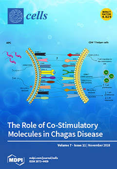
Cover Story (view full-size image): The heterogeneity of clinical manifestations in chronic Chagas disease is one of the features of this disease that still need to be unraveled by science. It is still unknown what causes some patients infected with Trypanossoma cruzi (T. cruzi) to develop cardiac and/or severe digestive clinical outcomes, while others patients do not present symptoms related to these pathologies. It is well known that interactions between immune cells such as monocytes and lymphocytes drive immune responses. Further, the co-stimulatory molecules expressed by monocytes induce T lymphocytes activation, thereby triggering cellular immune responses. However, little is known about the expression and function of these molecules in Chagas disease. Therefore, understanding the role of co-stimulatory molecules can elucidate important aspects of T. cruzi infection and of the development of cardiac changes inherent to the disease [...] Read more.
  • Issues are regarded as officially published after their release is announced to the table of contents alert mailing list.
  • You may sign up for e-mail alerts to receive table of contents of newly released issues.
  • PDF is the official format for papers published in both, html and pdf forms. To view the papers in pdf format, click on the "PDF Full-text" link, and use the free Adobe Reader to open them.
Order results
Result details
Section
Select all
Export citation of selected articles as:
12 pages, 1840 KiB  
Article
Lack of LTβR Increases Susceptibility of IPEC-J2 Cells to Porcine Epidemic Diarrhea Virus
by Tawfeek Altawaty, Lulu Liu, Hongyong Zhang, Cong Tao, Shaohua Hou, Kui Li and Yanfang Wang
Cells 2018, 7(11), 222; https://doi.org/10.3390/cells7110222 - 21 Nov 2018
Cited by 15 | Viewed by 4088
Abstract
The essential requirement of the lymphotoxin beta receptor (LTβR) in the development and maintenance of peripheral lymphoid organs is well recognized. Evidence shows that LTβR is involved in various cellular processes; however, whether it plays a role in maintaining the cellular function of [...] Read more.
The essential requirement of the lymphotoxin beta receptor (LTβR) in the development and maintenance of peripheral lymphoid organs is well recognized. Evidence shows that LTβR is involved in various cellular processes; however, whether it plays a role in maintaining the cellular function of intestinal porcine enterocytes (IPEC-J2), specifically during porcine epidemic diarrhea virus (PEDV) infection, remains unknown. In this study, we generated LTβR null IPEC-J2 cells using CRISPR/Cas9 to examine the importance of LTβR in cell proliferation, apoptosis, and the response to PEDV infection. Our results showed that the lack of LTβR leads to significantly decreased cell proliferation, potentially due to S phase arrest in LTβR−/− IPEC-J2 cells. Label-free digital holographic microscopy was used to record the three-dimensional morphology of both cell types for up to 72 hours and revealed significantly increased numbers of LTβR−/− cells undergoing apoptosis. Furthermore, we found that PEDV-infected LTβR−/− null IPEC-J2 cells exhibited significant suppression of nuclear factor kappa-light-chain-enhancer of activated B cells (NFκB) target genes (interleukin (IL)-6 and IL-8) and mucosal barrier integrity-related genes (vascular cell adhesion molecule 1 (VCAM1) and IL-22), which may explain why LTβR−/− cells are more susceptible to PEDV infection. Collectively, our data not only demonstrate the key role of LTβR in intestinal porcine enterocytes, but also provide data for the improved understanding of the cellular response to PEDV infection. Full article
Show Figures

Figure 1

18 pages, 1743 KiB  
Review
Controlling the Gatekeeper: Therapeutic Targeting of Nuclear Transport
by Friederike K. Kosyna and Reinhard Depping
Cells 2018, 7(11), 221; https://doi.org/10.3390/cells7110221 - 21 Nov 2018
Cited by 57 | Viewed by 10759
Abstract
Nuclear transport receptors of the karyopherin superfamily of proteins transport macromolecules from one compartment to the other and are critical for both cell physiology and pathophysiology. The nuclear transport machinery is tightly regulated and essential to a number of key cellular processes since [...] Read more.
Nuclear transport receptors of the karyopherin superfamily of proteins transport macromolecules from one compartment to the other and are critical for both cell physiology and pathophysiology. The nuclear transport machinery is tightly regulated and essential to a number of key cellular processes since the spatiotemporally expression of many proteins and the nuclear transporters themselves is crucial for cellular activities. Dysregulation of the nuclear transport machinery results in localization shifts of specific cargo proteins and associates with the pathogenesis of disease states such as cancer, inflammation, viral illness and neurodegenerative diseases. Therefore, inhibition of the nuclear transport system has future potential for therapeutic intervention and could contribute to the elucidation of disease mechanisms. In this review, we recapitulate clue findings in the pathophysiological significance of nuclear transport processes and describe the development of nuclear transport inhibitors. Finally, clinical implications and results of the first clinical trials are discussed for the most promising nuclear transport inhibitors. Full article
(This article belongs to the Special Issue Nuclear Transport in Ageing and Diseases)
Show Figures

Figure 1

16 pages, 2288 KiB  
Article
New Insights into the Runt Domain of RUNX2 in Melanoma Cell Proliferation and Migration
by Michela Deiana, Luca Dalle Carbonare, Michela Serena, Samuele Cheri, Francesca Parolini, Alberto Gandini, Giulia Marchetto, Giulio Innamorati, Marcello Manfredi, Emilio Marengo, Jessica Brandi, Daniela Cecconi, Antonio Mori, Maria Mihaela Mina, Franco Antoniazzi, Monica Mottes, Natascia Tiso, Giovanni Malerba, Donato Zipeto and Maria Teresa Valenti
Cells 2018, 7(11), 220; https://doi.org/10.3390/cells7110220 - 20 Nov 2018
Cited by 21 | Viewed by 6247
Abstract
The mortality rate for malignant melanoma (MM) is very high, since it is highly invasive and resistant to chemotherapeutic treatments. The modulation of some transcription factors affects cellular processes in MM. In particular, a higher expression of the osteogenic master gene RUNX2 has [...] Read more.
The mortality rate for malignant melanoma (MM) is very high, since it is highly invasive and resistant to chemotherapeutic treatments. The modulation of some transcription factors affects cellular processes in MM. In particular, a higher expression of the osteogenic master gene RUNX2 has been reported in melanoma cells, compared to normal melanocytes. By analyzing public databases for recurrent RUNX2 genetic and epigenetic modifications in melanoma, we found that the most common RUNX2 genetic alteration that exists in transcription upregulation is, followed by genomic amplification, nucleotide substitution and multiple changes. Additionally, altered RUNX2 is involved in unchecked pathways promoting tumor progression, Epithelial Mesenchymal Transition (EMT), and metastasis. In order to investigate further the role of RUNX2 in melanoma development and to identify a therapeutic target, we applied the CRISPR/Cas9 technique to explore the role of the RUNT domain of RUNX2 in a melanoma cell line. RUNT-deleted cells showed reduced proliferation, increased apoptosis, and reduced EMT features, suggesting the involvement of the RUNT domain in different pathways. In addition, del-RUNT cells showed a downregulation of genes involved in migration ability. In an in vivo zebrafish model, we observed that wild-type melanoma cells migrated in 81% of transplanted fishes, while del-RUNT cells migrated in 58%. All these findings strongly suggest the involvement of the RUNT domain in melanoma metastasis and cell migration and indicate RUNX2 as a prospective target in MM therapy. Full article
(This article belongs to the Section Cell Motility and Adhesion)
Show Figures

Graphical abstract

19 pages, 645 KiB  
Review
MicroRNAs as Biomarkers in Amyotrophic Lateral Sclerosis
by Claudia Ricci, Carlotta Marzocchi and Stefania Battistini
Cells 2018, 7(11), 219; https://doi.org/10.3390/cells7110219 - 20 Nov 2018
Cited by 79 | Viewed by 7646
Abstract
Amyotrophic lateral sclerosis (ALS) is an incurable and fatal disorder characterized by the progressive loss of motor neurons in the cerebral cortex, brain stem, and spinal cord. Sporadic ALS form accounts for the majority of patients, but in 1–13.5% of cases the disease [...] Read more.
Amyotrophic lateral sclerosis (ALS) is an incurable and fatal disorder characterized by the progressive loss of motor neurons in the cerebral cortex, brain stem, and spinal cord. Sporadic ALS form accounts for the majority of patients, but in 1–13.5% of cases the disease is inherited. The diagnosis of ALS is mainly based on clinical assessment and electrophysiological examinations with a history of symptom progression and is then made with a significant delay from symptom onset. Thus, the identification of biomarkers specific for ALS could be of a fundamental importance in the clinical practice. An ideal biomarker should display high specificity and sensitivity for discriminating ALS from control subjects and from ALS-mimics and other neurological diseases, and should then monitor disease progression within individual patients. microRNAs (miRNAs) are considered promising biomarkers for neurodegenerative diseases, since they are remarkably stable in human body fluids and can reflect physiological and pathological processes relevant for ALS. Here, we review the state of the art of miRNA biomarker identification for ALS in cerebrospinal fluid (CSF), blood and muscle tissue; we discuss advantages and disadvantages of different approaches, and underline the limits but also the great potential of this research for future practical applications. Full article
(This article belongs to the Special Issue Regulatory microRNA)
Show Figures

Figure 1

13 pages, 578 KiB  
Review
Emerging Roles of Diacylglycerol-Sensitive TRPC4/5 Channels
by Michael Mederos y Schnitzler, Thomas Gudermann and Ursula Storch
Cells 2018, 7(11), 218; https://doi.org/10.3390/cells7110218 - 20 Nov 2018
Cited by 23 | Viewed by 5355
Abstract
Transient receptor potential classical or canonical 4 (TRPC4) and TRPC5 channels are members of the classical or canonical transient receptor potential (TRPC) channel family of non-selective cation channels. TRPC4 and TRPC5 channels are widely accepted as receptor-operated cation channels that are activated in [...] Read more.
Transient receptor potential classical or canonical 4 (TRPC4) and TRPC5 channels are members of the classical or canonical transient receptor potential (TRPC) channel family of non-selective cation channels. TRPC4 and TRPC5 channels are widely accepted as receptor-operated cation channels that are activated in a phospholipase C-dependent manner, following the Gq/11 protein-coupled receptor activation. However, their precise activation mechanism has remained largely elusive for a long time, as the TRPC4 and TRPC5 channels were considered as being insensitive to the second messenger diacylglycerol (DAG) in contrast to the other TRPC channels. Recent findings indicate that the C-terminal interactions with the scaffolding proteins Na+/H+ exchanger regulatory factor 1 and 2 (NHERF1 and NHERF2) dynamically regulate the DAG sensitivity of the TRPC4 and TRPC5 channels. Interestingly, the C-terminal NHERF binding suppresses, while the dissociation of NHERF enables, the DAG sensitivity of the TRPC4 and TRPC5 channels. This leads to the assumption that all of the TRPC channels are DAG sensitive. The identification of the regulatory function of the NHERF proteins in the TRPC4/5-NHERF protein complex offers a new starting point to get deeper insights into the molecular basis of TRPC channel activation. Future studies will have to unravel the physiological and pathophysiological functions of this multi-protein channel complex. Full article
(This article belongs to the Special Issue TRP Channels in Health and Disease)
Show Figures

Figure 1

14 pages, 2740 KiB  
Article
Mechanisms of TQ-6, a Novel Ruthenium-Derivative Compound, against Lipopolysaccharide-Induced In Vitro Macrophage Activation and Liver Injury in Experimental Mice: The Crucial Role of p38 MAPK and NF-κB Signaling
by Chih-Hsuan Hsia, Marappan Velusamy, Thanasekaran Jayakumar, Yen-Jen Chen, Chih-Wei Hsia, Jie-Heng Tsai, Ruei-Dun Teng and Joen-Rong Sheu
Cells 2018, 7(11), 217; https://doi.org/10.3390/cells7110217 - 19 Nov 2018
Cited by 11 | Viewed by 4508
Abstract
Several studies have reported that metal complexes exhibit anti-inflammatory activities; however, the molecular mechanism is not well understood. In this study, we used a potent ruthenium (II)-derived compound, [Ru(η6-cymene)2-(1H-benzoimidazol-2-yl)-quinoline Cl]BF4 (TQ-6), to investigate the molecular mechanisms underlying the anti-inflammatory effects against lipopolysaccharide (LPS)-induced [...] Read more.
Several studies have reported that metal complexes exhibit anti-inflammatory activities; however, the molecular mechanism is not well understood. In this study, we used a potent ruthenium (II)-derived compound, [Ru(η6-cymene)2-(1H-benzoimidazol-2-yl)-quinoline Cl]BF4 (TQ-6), to investigate the molecular mechanisms underlying the anti-inflammatory effects against lipopolysaccharide (LPS)-induced macrophage activation and liver injury in mice. Treating LPS-stimulated RAW 264.7 cells with TQ-6 suppressed nitric oxide (NO) production and inducible nitric oxide synthase (iNOS) expression in a concentration-dependent manner. The LPS-induced expression of tumor necrosis factor alpha (TNF-α) and interleukin-1 beta (IL-1β) were reduced in TQ-6-treated cells. TQ-6 suppressed, LPS-stimulated p38 MAPK phosphorylation, IκBα degradation, and p65 nuclear translocation in cells. Consistent with the in vitro studies, TQ-6 also suppressed the expression of iNOS, TNF-α, and p65 in the mouse model with acute liver injury induced by LPS. The present study showed that TQ-6 could protect against LPS-induced in vitro inflammation in macrophage and in vivo liver injury in mice, and suggested that NF-κB could be a promising target for protecting against LPS-induced inflammation and liver injury by TQ-6. Therefore, TQ-6 can be a potential therapeutic agent for treating inflammatory diseases. Full article
(This article belongs to the Special Issue Protein Kinases: Signalling and Disease)
Show Figures

Figure 1

27 pages, 9553 KiB  
Article
Synthesis and Biological Evaluation of Novel Triple-Modified Colchicine Derivatives as Potent Tubulin-Targeting Anticancer Agents
by Urszula Majcher, Greta Klejborowska, Magdalena Kaik, Ewa Maj, Joanna Wietrzyk, Mahshad Moshari, Jordane Preto, Jack A. Tuszynski and Adam Huczyński
Cells 2018, 7(11), 216; https://doi.org/10.3390/cells7110216 - 19 Nov 2018
Cited by 24 | Viewed by 6844
Abstract
Specific modifications of colchicine followed by synthesis of its analogues have been tested in vitro with the objective of lowering colchicine toxicity. Our previous studies have clearly shown the anticancer potential of double-modified colchicine derivatives in C-7 and C-10 positions. Here, a series [...] Read more.
Specific modifications of colchicine followed by synthesis of its analogues have been tested in vitro with the objective of lowering colchicine toxicity. Our previous studies have clearly shown the anticancer potential of double-modified colchicine derivatives in C-7 and C-10 positions. Here, a series of novel triple-modified colchicine derivatives is reported. They have been obtained following a four-step strategy. In vitro cytotoxicity of these compounds has been evaluated against four human tumor cell lines (A549, MCF-7, LoVo, and LoVo/DX). Additionally, the mode of binding of the synthesized compounds was evaluated in silico using molecular docking to a 3D structure of β-tubulin based on crystallographic data from the Protein Data Bank and homology methodology. Binding free energy estimates, binding poses, and MlogP values of the compounds were obtained. All triple-modified colchicine derivatives were shown to be active at nanomolar concentrations against three of the investigated cancer cell lines (A549, MCF-7, LoVo). Four of them also showed higher potency against tumor cells over normal cells as confirmed by their high selectivity index values. A vast majority of the synthesized derivatives exhibited several times higher cytotoxicity than colchicine, doxorubicin, and cisplatin. Full article
(This article belongs to the Special Issue Tubulin: Structure, Functions and Roles in Disease)
Show Figures

Graphical abstract

11 pages, 2072 KiB  
Review
Revisiting Rab7 Functions in Mammalian Autophagy: Rab7 Knockout Studies
by Yoshihiko Kuchitsu and Mitsunori Fukuda
Cells 2018, 7(11), 215; https://doi.org/10.3390/cells7110215 - 19 Nov 2018
Cited by 68 | Viewed by 10332
Abstract
Rab7 (or Ypt7 in yeast) is one of the well-characterized members of the Rab family small GTPases, which serve as master regulators of membrane trafficking in eukaryotes. It localizes to late endosomes and lysosomes and has multiple functions in the autophagic pathway as [...] Read more.
Rab7 (or Ypt7 in yeast) is one of the well-characterized members of the Rab family small GTPases, which serve as master regulators of membrane trafficking in eukaryotes. It localizes to late endosomes and lysosomes and has multiple functions in the autophagic pathway as well as in the endocytic pathway. Because Rab7/Ypt7 has previously been shown to regulate the autophagosome-lysosome fusion step in yeast and fruit flies (i.e., autophagosome accumulation has been observed in both Ypt7-knockout [KO] yeast and Rab7-knockdown fruit flies), it is widely assumed that Rab7 regulates the autophagosome-lysosome fusion step in mammals. A recent analysis of Rab7-KO mammalian cultured cells, however, has revealed that Rab7 is essential for autolysosome maturation (i.e., autolysosome accumulation has been observed in Rab7-KO cells), but not for autophagosome-lysosome fusion, under nutrient-rich conditions. Thus, although Rab7/Ypt7 itself is essential for the proper progression of autophagy in eukaryotes, the function of Rab7/Ypt7 in autophagy in yeast/fruit flies and mammals must be different. In this review article, we describe novel roles of Rab7 in mammalian autophagy and discuss its functional diversification during evolution. Full article
Show Figures

Graphical abstract

19 pages, 3736 KiB  
Article
UBE2E1 Is Preferentially Expressed in the Cytoplasm of Slow-Twitch Fibers and Protects Skeletal Muscles from Exacerbated Atrophy upon Dexamethasone Treatment
by Cécile Polge, Julien Aniort, Andrea Armani, Agnès Claustre, Cécile Coudy-Gandilhon, Clara Tournebize, Christiane Deval, Lydie Combaret, Daniel Béchet, Marco Sandri, Didier Attaix and Daniel Taillandier
Cells 2018, 7(11), 214; https://doi.org/10.3390/cells7110214 - 16 Nov 2018
Cited by 9 | Viewed by 4441
Abstract
Skeletal muscle mass is reduced during many diseases or physiological situations (disuse, aging), which results in decreased strength and increased mortality. Muscle mass is mainly controlled by the ubiquitin-proteasome system (UPS), involving hundreds of ubiquitinating enzymes (E2s and E3s) that target their dedicated [...] Read more.
Skeletal muscle mass is reduced during many diseases or physiological situations (disuse, aging), which results in decreased strength and increased mortality. Muscle mass is mainly controlled by the ubiquitin-proteasome system (UPS), involving hundreds of ubiquitinating enzymes (E2s and E3s) that target their dedicated substrates for subsequent degradation. We recently demonstrated that MuRF1, an E3 ubiquitin ligase known to bind to sarcomeric proteins (telethonin, α-actin, myosins) during catabolic situations, interacts with 5 different E2 enzymes and that these E2-MuRF1 couples are able to target telethonin, a small sarcomeric protein, for degradation. Amongst the E2s interacting with MuRF1, E2E1 was peculiar as the presence of the substrate was necessary for optimal MuRF1-E2E1 interaction. In this work, we focused on the putative role of E2E1 during skeletal muscle atrophy. We found that E2E1 expression was restricted to type I and type IIA muscle fibers and was not detectable in type IIB fibers. This strongly suggests that E2E1 targets are fiber-specific and may be strongly linked to the contractile and metabolic properties of the skeletal muscle. However, E2E1 knockdown was not sufficient for preserving the protein content in C2C12 myotubes subjected to a catabolic state (dexamethasone treatment), suggesting that E2E1 is not involved in the development of muscle atrophy. By contrast, E2E1 knockdown aggravated the atrophying process in both catabolic C2C12 myotubes and the Tibialis anterior muscle of mice, suggesting that E2E1 has a protective effect on muscle mass. Full article
(This article belongs to the Special Issue Ubiquitination in Health and Disease)
Show Figures

Figure 1

18 pages, 1983 KiB  
Article
Proteomic Signatures of Clostridium difficile Stressed with Metronidazole, Vancomycin, or Fidaxomicin
by Sandra Maaß, Andreas Otto, Dirk Albrecht, Katharina Riedel, Anke Trautwein-Schult and Dörte Becher
Cells 2018, 7(11), 213; https://doi.org/10.3390/cells7110213 - 15 Nov 2018
Cited by 14 | Viewed by 4753
Abstract
The anaerobic pathogen Clostridium difficile is of growing significance for the health care system due to its increasing incidence and mortality. As C. difficile infection is both supported and treated by antibiotics, a deeper knowledge on how antimicrobial agents affect the physiology of [...] Read more.
The anaerobic pathogen Clostridium difficile is of growing significance for the health care system due to its increasing incidence and mortality. As C. difficile infection is both supported and treated by antibiotics, a deeper knowledge on how antimicrobial agents affect the physiology of this important pathogen may help to understand and prevent the development and spreading of antibiotic resistant strains. As the proteomic response of a cell to stress aims at counteracting the harmful effects of this stress, it can be expected that the pattern of a pathogen’s responses to antibiotic treatment will be dependent on the antibiotic mechanism of action. Hence, every antibiotic treatment is expected to result in a specific proteomic signature characterizing its mode of action. In the study presented here, the proteomic response of C. difficile 630∆erm to vancomycin, metronidazole, and fidaxomicin stress was investigated on the level of protein abundance and protein synthesis based on 2D PAGE. The quantification of 425 proteins of C. difficile allowed the deduction of proteomic signatures specific for each drug treatment. Indeed, these proteomic signatures indicate very specific cellular responses to each antibiotic with only little overlap of the responses. Whereas signature proteins for vancomycin stress fulfil various cellular functions, the proteomic signature of metronidazole stress is characterized by alterations of proteins involved in protein biosynthesis and protein degradation as well as in DNA replication, recombination, and repair. In contrast, proteins differentially expressed after fidaxomicin treatment can be assigned to amino acid biosynthesis, transcription, cell motility, and the cell envelope functions. Notably, the data provided by this study hint also at so far unknown antibiotic detoxification mechanisms. Full article
(This article belongs to the Special Issue Cellular Stress Response in Health and Disease)
Show Figures

Graphical abstract

16 pages, 1027 KiB  
Review
Mechanism of Resistance to Epidermal Growth Factor Receptor-Tyrosine Kinase Inhibitors and a Potential Treatment Strategy
by Tatsuya Nagano, Motoko Tachihara and Yoshihiro Nishimura
Cells 2018, 7(11), 212; https://doi.org/10.3390/cells7110212 - 15 Nov 2018
Cited by 206 | Viewed by 15035
Abstract
Treatment with epidermal growth factor receptor tyrosine kinase inhibitors (EGFR-TKIs) improves the overall survival of patients with EGFR-mutated non-small-cell lung cancer (NSCLC). First-generation EGFR-TKIs (e.g., gefitinib and erlotinib) or second-generation EGFR-TKIs (e.g., afatinib and dacomitinib) are effective for the treatment of EGFR [...] Read more.
Treatment with epidermal growth factor receptor tyrosine kinase inhibitors (EGFR-TKIs) improves the overall survival of patients with EGFR-mutated non-small-cell lung cancer (NSCLC). First-generation EGFR-TKIs (e.g., gefitinib and erlotinib) or second-generation EGFR-TKIs (e.g., afatinib and dacomitinib) are effective for the treatment of EGFR-mutated NSCLC, especially in patients with EGFR exon 19 deletions or an exon 21 L858R mutation. However, almost all cases experience disease recurrence after 1 to 2 years due to acquired resistance. The EGFR T790M mutation in exon 20 is the most frequent alteration associated with the development of acquired resistance. Osimertinib—a third-generation EGFR-TKI—targets the T790M mutation and has demonstrated high efficacy against EGFR-mutated lung cancer. However, the development of acquired resistance to third-generation EGFR-TKI, involving the cysteine residue at codon 797 mutation, has been observed. Other mechanisms of acquired resistance include the activation of alternative pathways or downstream targets and histological transformation (i.e., epithelial–mesenchymal transition or conversion to small-cell lung cancer). Furthermore, the development of primary resistance through overexpression of the hepatocyte growth factor and suppression of Bcl-2-like protein 11 expression may lead to problems. In this report, we review these mechanisms and discuss therapeutic strategies to overcome resistance to EGFR-TKIs. Full article
(This article belongs to the Special Issue Epidermal Growth Factor Receptor Signaling)
Show Figures

Graphical abstract

11 pages, 2321 KiB  
Article
The Intermolecular Interaction of Ephexin4 Leads to Autoinhibition by Impeding Binding of RhoG
by Kwanhyeong Kim, Juyeon Lee, Hyunji Moon, Sang-Ah Lee, Deokhwan Kim, Susumin Yang, Dae-Hee Lee, Gwangrog Lee and Daeho Park
Cells 2018, 7(11), 211; https://doi.org/10.3390/cells7110211 - 15 Nov 2018
Cited by 7 | Viewed by 3704
Abstract
Ephexin4 is a guanine nucleotide-exchange factor (GEF) for RhoG and is involved in various RhoG-related cellular processes such as phagocytosis of apoptotic cells and migration of cancer cells. Ephexin4 forms an oligomer via an intermolecular interaction, and its GEF activity is increased in [...] Read more.
Ephexin4 is a guanine nucleotide-exchange factor (GEF) for RhoG and is involved in various RhoG-related cellular processes such as phagocytosis of apoptotic cells and migration of cancer cells. Ephexin4 forms an oligomer via an intermolecular interaction, and its GEF activity is increased in the presence of Elmo, an Ephexin4-interacting protein. However, it is uncertain if and how Ephexin4 is autoinhibited. Here, using an Ephexin4 mutant that abrogated the intermolecular interaction, we report that this interaction impeded binding of RhoG to Ephexin4 and thus inhibited RhoG activation. Mutation of the glutamate residue at position 295, which is a highly conserved residue located in the region of Ephexin4 required for the intermolecular interaction, to alanine (Ephexin4E295A) disrupted the intermolecular interaction and increased binding of RhoG, resulting in augmented RhoG activation. In addition, phagocytosis of apoptotic cells and formation of membrane ruffles were increased more by expression of Ephexin4E295A than by expression of wild-type Ephexin4. Taken together, our data suggest that Ephexin4 is autoinhibited through its intermolecular interaction, which impedes binding of RhoG. Full article
Show Figures

Figure 1

13 pages, 10071 KiB  
Article
Systemic Hypertension Effects on the Ciliary Body and Iris. An Immunofluorescence Study with Aquaporin 1, Aquaporin 4, and Na+, K+ ATPase in Hypertensive Rats
by Ibrahim González-Marrero, Luis G. Hernández-Abad, Emilia M. Carmona-Calero, Leandro Castañeyra-Ruiz, José A. Abreu-Reyes and Agustín Castañeyra-Perdomo
Cells 2018, 7(11), 210; https://doi.org/10.3390/cells7110210 - 13 Nov 2018
Cited by 8 | Viewed by 5712
Abstract
Aquaporin 1 (AQP1) and aquaporin 4 (AQP4) have been identified in the eye as playing an essential role in the formation of the aqueous humor along with the Na+/K+ ATPase pump. Different authors have described the relationship between blood pressure, [...] Read more.
Aquaporin 1 (AQP1) and aquaporin 4 (AQP4) have been identified in the eye as playing an essential role in the formation of the aqueous humor along with the Na+/K+ ATPase pump. Different authors have described the relationship between blood pressure, aqueous humor production, and intraocular pressure with different conclusions, with some authors supporting a positive correlation between blood pressure and intraocular pressure while others disagree. The aim of this work was to study the effect of high blood pressure on the proteins involved in the production of aqueous humor in the ciliary body (CB) and iris. For this purpose, we used the eyes of spontaneously hypertensive rats (SHR) and their control Wistar-Kyoto rats (WKY). Immunofluorescence was performed in different eye structures to analyze the effects of hypertension in the expression of AQP1, AQP4, and the Na+/K+ ATPase α1 and α2 subunits. The results showed an increase in AQP1 and Na+/K+ ATPase α1 and a decrease in AQP4 and Na+/K+ ATPase α2 in the CB of SHR, while an increase in AQP4 and no significant differences in AQP1 were found in the iris. Therefore, systemic hypertension produced changes in the proteins implicated in the movement of water in the CB and iris that could influence the production rate of aqueous humor, which would be affected depending on the duration of systemic hypertension. Full article
(This article belongs to the Special Issue Aquaporins)
Show Figures

Figure 1

24 pages, 3856 KiB  
Review
Aquaporins: More Than Functional Monomers in a Tetrameric Arrangement
by Marcelo Ozu, Luciano Galizia, Cynthia Acuña and Gabriela Amodeo
Cells 2018, 7(11), 209; https://doi.org/10.3390/cells7110209 - 11 Nov 2018
Cited by 35 | Viewed by 7223
Abstract
Aquaporins (AQPs) function as tetrameric structures in which each monomer has its own permeable pathway. The combination of structural biology, molecular dynamics simulations, and experimental approaches has contributed to improve our knowledge of how protein conformational changes can challenge its transport capacity, rapidly [...] Read more.
Aquaporins (AQPs) function as tetrameric structures in which each monomer has its own permeable pathway. The combination of structural biology, molecular dynamics simulations, and experimental approaches has contributed to improve our knowledge of how protein conformational changes can challenge its transport capacity, rapidly altering the membrane permeability. This review is focused on evidence that highlights the functional relationship between the monomers and the tetramer. In this sense, we address AQP permeation capacity as well as regulatory mechanisms that affect the monomer, the tetramer, or tetramers combined in complex structures. We therefore explore: (i) water permeation and recent evidence on ion permeation, including the permeation pathway controversy—each monomer versus the central pore of the tetramer—and (ii) regulatory mechanisms that cannot be attributed to independent monomers. In particular, we discuss channel gating and AQPs that sense membrane tension. For the latter we propose a possible mechanism that includes the monomer (slight changes of pore shape, the number of possible H-bonds between water molecules and pore-lining residues) and the tetramer (interactions among monomers and a positive cooperative effect). Full article
(This article belongs to the Special Issue Aquaporins)
Show Figures

Figure 1

33 pages, 1673 KiB  
Review
Hedgehog Signaling in Cancer: A Prospective Therapeutic Target for Eradicating Cancer Stem Cells
by Ita Novita Sari, Lan Thi Hanh Phi, Nayoung Jun, Yoseph Toni Wijaya, Sanghyun Lee and Hyog Young Kwon
Cells 2018, 7(11), 208; https://doi.org/10.3390/cells7110208 - 10 Nov 2018
Cited by 145 | Viewed by 8486
Abstract
The Hedgehog (Hh) pathway is a signaling cascade that plays a crucial role in many fundamental processes, including embryonic development and tissue homeostasis. Moreover, emerging evidence has suggested that aberrant activation of Hh is associated with neoplastic transformations, malignant tumors, and drug resistance [...] Read more.
The Hedgehog (Hh) pathway is a signaling cascade that plays a crucial role in many fundamental processes, including embryonic development and tissue homeostasis. Moreover, emerging evidence has suggested that aberrant activation of Hh is associated with neoplastic transformations, malignant tumors, and drug resistance of a multitude of cancers. At the molecular level, it has been shown that Hh signaling drives the progression of cancers by regulating cancer cell proliferation, malignancy, metastasis, and the expansion of cancer stem cells (CSCs). Thus, a comprehensive understanding of Hh signaling during tumorigenesis and development of chemoresistance is necessary in order to identify potential therapeutic strategies to target various human cancers and their relapse. In this review, we discuss the molecular basis of the Hh signaling pathway and its abnormal activation in several types of human cancers. We also highlight the clinical development of Hh signaling inhibitors for cancer therapy as well as CSC-targeted therapy. Full article
(This article belongs to the Collection Hedgehog Signal Transduction in Physiology and Disease)
Show Figures

Figure 1

20 pages, 2798 KiB  
Article
Molecular Basis of Aquaporin-7 Permeability Regulation by pH
by Andreia F. Mósca, Andreia De Almeida, Darren Wragg, Ana P. Martins, Farzana Sabir, Stefano Leoni, Teresa F. Moura, Catarina Prista, Angela Casini and Graça Soveral
Cells 2018, 7(11), 207; https://doi.org/10.3390/cells7110207 - 10 Nov 2018
Cited by 30 | Viewed by 6190
Abstract
The aquaglyceroporin AQP7, a family member of aquaporin membrane channels, facilitates the permeation of water and glycerol through cell membranes and is crucial for body lipid and energy homeostasis. Regulation of glycerol permeability via AQP7 is considered a promising therapeutic strategy towards fat-related [...] Read more.
The aquaglyceroporin AQP7, a family member of aquaporin membrane channels, facilitates the permeation of water and glycerol through cell membranes and is crucial for body lipid and energy homeostasis. Regulation of glycerol permeability via AQP7 is considered a promising therapeutic strategy towards fat-related metabolic complications. Here, we used a yeast aqy-null strain for heterologous expression and functional analysis of human AQP7 and investigated its regulation by pH. Using a combination of in vitro and in silico approaches, we found that AQP7 changes from fully permeable to virtually closed at acidic pH, and that Tyr135 and His165 facing the extracellular environment are crucial residues for channel permeability. Moreover, instead of reducing the pore size, the protonation of key residues changes AQP7’s protein surface electrostatic charges, which, in turn, may decrease glycerol’s binding affinity to the pore, resulting in decreased permeability. In addition, since some pH-sensitive residues are located at the monomer-monomer interface, decreased permeability may result from cooperativity between AQP7’s monomers. Considering the importance of glycerol permeation via AQP7 in multiple pathophysiological conditions, this mechanism of hAQP7 pH-regulation may help the design of selective modulators targeting aquaglyceroporin-related disorders. Full article
(This article belongs to the Special Issue Aquaporins)
Show Figures

Graphical abstract

24 pages, 21752 KiB  
Article
Matrix Metalloproteinase-1 and Acid Phosphatase in the Degradation of the Lamina Propria of Eruptive Pathway of Rat Molars
by José Paulo De Pizzol Júnior, Estela Sasso-Cerri and Paulo Sérgio Cerri
Cells 2018, 7(11), 206; https://doi.org/10.3390/cells7110206 - 10 Nov 2018
Cited by 20 | Viewed by 5578
Abstract
The comprehension of dental pathogenesis and disorders derived from eruption failure requires a deep understanding of the molecular mechanisms underlying normal tooth eruption. As intense remodelling is needed during tooth eruption, we hypothesize that matrix metalloproteinase-1 (MMP-1) and acid phosphatase (ACP) play a [...] Read more.
The comprehension of dental pathogenesis and disorders derived from eruption failure requires a deep understanding of the molecular mechanisms underlying normal tooth eruption. As intense remodelling is needed during tooth eruption, we hypothesize that matrix metalloproteinase-1 (MMP-1) and acid phosphatase (ACP) play a role in the eruptive pathway degradation. We evaluated MMP-1-immunoexpression and the collagen content in the lamina propria at different eruptive phases. Immunohistochemistry and ultrastructural cytochemistry for detection of ACP were also performed. In the maxillary sections containing first molars of 9-, 11-, 13-, and 16-day-old rats, the birefringent collagen of eruptive pathway was quantified. MMP-1 and ACP-2 immunohistochemical reactions were performed and the number of MMP-1-immunolabelled cells was computed. Data were analyzed by one-way ANOVA and Tukey post-test (p ≤ 0.05). ACP cytochemistry was evaluated in specimens incubated in sodium β-glycerophosphate. In the eruptive pathway of 13- and 16-day-old rats, the number of MMP-1-immunolabelled cells increased concomitantly to reduction of collagen in the lamina propria. Enhanced ACP-2-immunolabelling was observed in the lamina propria of 13- and 16-day-old rats. Fibroblasts and macrophages showed lysosomes and vacuoles containing fragmented material reactive to ACP. MMP-1 degrades extracellular matrix, including collagen fibers, being responsible for the reduction in the collagen content during tooth eruption. The enhanced ACP activity at the mucosal penetration stage indicates that this enzyme plays a role in the degradation of remnant material, which is engulfed by macrophages and fibroblasts of the eruptive pathway. Therefore, enzymatic failure in the eruptive pathway may disturbs tooth eruption. Full article
(This article belongs to the Special Issue Extracellular Matrix Remodeling)
Show Figures

Graphical abstract

16 pages, 5830 KiB  
Article
The Heterochromatin Landscape in Migrating Cells and the Importance of H3K27me3 for Associated Transcriptome Alterations
by Tamar Segal, Mali Salmon-Divon and Gabi Gerlitz
Cells 2018, 7(11), 205; https://doi.org/10.3390/cells7110205 - 9 Nov 2018
Cited by 22 | Viewed by 5579
Abstract
H3K9me3, H3K27me3, and H4K20me1 are epigenetic markers associated with chromatin condensation and transcriptional repression. Previously, we found that migration of melanoma cells is associated with and dependent on global chromatin condensation that includes a global increase in these markers. Taken together with more [...] Read more.
H3K9me3, H3K27me3, and H4K20me1 are epigenetic markers associated with chromatin condensation and transcriptional repression. Previously, we found that migration of melanoma cells is associated with and dependent on global chromatin condensation that includes a global increase in these markers. Taken together with more recent reports by others suggests it is a general signature of migrating cells. Here, to learn about the function of these markers in migrating cells, we mapped them by ChIP-seq analysis. This analysis revealed that induction of migration leads to expansion of these markers along the genome and to an increased overlapping between them. Significantly, induction of migration led to a higher increase in H3K9me3 and H4K20me1 signals at repetitive elements than at protein-coding genes, while an opposite pattern was found for H3K27me3. Transcriptome analysis revealed 182 altered genes following induction of migration, of which 33% are dependent on H3K27me3 for these changes. H3K27me3 was also required to prevent changes in the expression of 501 other genes upon induction of migration. Taken together, our results suggest that heterochromatinization in migrating cells is global and not restricted to specific genomic loci and that H3K27me3 is a key component in executing a migration-specific transcriptional plan. Full article
(This article belongs to the Section Cell Nuclei: Function, Transport and Receptors)
Show Figures

Figure 1

13 pages, 1073 KiB  
Article
Calcium Pathways in Human Neutrophils—The Extended Effects of Thapsigargin and ML-9
by Daniela Ribeiro, Marisa Freitas, Sílvia Rocha, José L. F. C. Lima, Félix Carvalho and Eduarda Fernandes
Cells 2018, 7(11), 204; https://doi.org/10.3390/cells7110204 - 9 Nov 2018
Cited by 13 | Viewed by 5986
Abstract
In neutrophils, intracellular Ca2+ levels are regulated by several transporters and pathways, namely SERCA [sarco(endo)plasmic reticulum Ca2+-ATPase], SOCE (store-operated calcium entry), and ROCE (receptor-operated calcium entry). However, the exact mechanisms involved in the communication among these transporters are still unclear. [...] Read more.
In neutrophils, intracellular Ca2+ levels are regulated by several transporters and pathways, namely SERCA [sarco(endo)plasmic reticulum Ca2+-ATPase], SOCE (store-operated calcium entry), and ROCE (receptor-operated calcium entry). However, the exact mechanisms involved in the communication among these transporters are still unclear. In the present study, thapsigargin, an irreversible inhibitor of SERCA, and ML-9, a broadly used SOCE inhibitor, were applied in human neutrophils to better understand their effects on Ca2+ pathways in these important cells of the immune system. The thapsigargin and ML-9 effects in the intracellular free Ca2+ flux were evaluated in freshly isolated human neutrophils, using a microplate reader for monitoring fluorimetric kinetic readings. The obtained results corroborate the general thapsigargin-induced intracellular pattern of Ca2+ fluctuation, but it was also observed a much more extended effect in time and a clear sustained increase of Ca2+ levels due to its influx by SOCE. Moreover, it was obvious that ML-9 enhanced the thapsigargin-induced emptying of the internal stores. Indeed, ML-9 does not have this effect by itself, which indicates that, in neutrophils, thapsigargin does not act only on the influx by SOCE, but also by other Ca2+ pathways, that, in the future, should be further explored. Full article
Show Figures

Graphical abstract

19 pages, 2552 KiB  
Article
miR-338-3p Is Regulated by Estrogens through GPER in Breast Cancer Cells and Cancer-Associated Fibroblasts (CAFs)
by Adele Vivacqua, Anna Sebastiani, Anna Maria Miglietta, Damiano Cosimo Rigiracciolo, Francesca Cirillo, Giulia Raffaella Galli, Marianna Talia, Maria Francesca Santolla, Rosamaria Lappano, Francesca Giordano, Maria Luisa Panno and Marcello Maggiolini
Cells 2018, 7(11), 203; https://doi.org/10.3390/cells7110203 - 9 Nov 2018
Cited by 30 | Viewed by 4816
Abstract
Estrogens acting through the classic estrogen receptors (ERs) and the G protein estrogen receptor (GPER) regulate the expression of diverse miRNAs, small sequences of non-coding RNA involved in several pathophysiological conditions, including breast cancer. In order to provide novel insights on miRNAs regulation [...] Read more.
Estrogens acting through the classic estrogen receptors (ERs) and the G protein estrogen receptor (GPER) regulate the expression of diverse miRNAs, small sequences of non-coding RNA involved in several pathophysiological conditions, including breast cancer. In order to provide novel insights on miRNAs regulation by estrogens in breast tumor, we evaluated the expression of 754 miRNAs by TaqMan Array in ER-negative and GPER-positive SkBr3 breast cancer cells and cancer-associated fibroblasts (CAFs) upon 17β-estradiol (E2) treatment. Various miRNAs were regulated by E2 in a peculiar manner in SkBr3 cancer cells and CAFs, while miR-338-3p displayed a similar regulation in both cell types. By METABRIC database analysis we ascertained that miR-338-3p positively correlates with overall survival in breast cancer patients, according to previous studies showing that miR-338-3p may suppress the growth and invasion of different cancer cells. Well-fitting with these data, a miR-338-3p mimic sequence decreased and a miR-338-3p inhibitor sequence rescued the expression of genes and the proliferative effects induced by E2 through GPER in SkBr3 cancer cells and CAFs. Altogether, our results provide novel evidence on the molecular mechanisms by which E2 may regulate miR-338-3p toward breast cancer progression. Full article
(This article belongs to the Special Issue Regulatory microRNA)
Show Figures

Figure 1

9 pages, 2145 KiB  
Editorial
Comparative Biology of Centrosomal Structures in Eukaryotes
by Ralph Gräf
Cells 2018, 7(11), 202; https://doi.org/10.3390/cells7110202 - 8 Nov 2018
Cited by 7 | Viewed by 6188
Abstract
The centrosome is not only the largest and most sophisticated protein complex within a eukaryotic cell, in the light of evolution, it is also one of its most ancient organelles. This special issue of “Cells” features representatives of three main, structurally [...] Read more.
The centrosome is not only the largest and most sophisticated protein complex within a eukaryotic cell, in the light of evolution, it is also one of its most ancient organelles. This special issue of “Cells” features representatives of three main, structurally divergent centrosome types, i.e., centriole-containing centrosomes, yeast spindle pole bodies (SPBs), and amoebozoan nucleus-associated bodies (NABs). Here, I discuss their evolution and their key-functions in microtubule organization, mitosis, and cytokinesis. Furthermore, I provide a brief history of centrosome research and highlight recently emerged topics, such as the role of centrioles in ciliogenesis, the relationship of centrosomes and centriolar satellites, the integration of centrosomal structures into the nuclear envelope and the involvement of centrosomal components in non-centrosomal microtubule organization. Full article
(This article belongs to the Section Intracellular and Plasma Membranes)
Show Figures

Figure 1

15 pages, 1043 KiB  
Article
Exploring the Effect of Rotenone—A Known Inducer of Parkinson’s Disease—On Mitochondrial Dynamics in Dictyostelium discoideum
by Ethan Chernivec, Jacie Cooper and Kari Naylor
Cells 2018, 7(11), 201; https://doi.org/10.3390/cells7110201 - 8 Nov 2018
Cited by 25 | Viewed by 6562
Abstract
Current treatments for Parkinson’s disease (PD) only alleviate symptoms doing little to inhibit the onset and progression of the disease, thus we must research the mechanism of Parkinson’s. Rotenone is a known inducer of parkinsonian conditions in rats; we use rotenone to induce [...] Read more.
Current treatments for Parkinson’s disease (PD) only alleviate symptoms doing little to inhibit the onset and progression of the disease, thus we must research the mechanism of Parkinson’s. Rotenone is a known inducer of parkinsonian conditions in rats; we use rotenone to induce parkinsonian cellular conditions in Dictyostelium discoideum. In our model we primarily focus on mitochondrial dynamics. We found that rotenone disrupts the actin and microtubule cytoskeleton but mitochondrial morphology remains intact. Rotenone stimulates mitochondrial velocity while inhibiting mitochondrial fusion, increases reactive oxygen species (ROS) but has no effect on ATP levels. Antioxidants have been shown to decrease some PD symptoms thus we added ascorbic acid to our rotenone treated cells. Ascorbic acid administration suggests that rotenone effects may be specific to the disruption of the cytoskeleton rather than the increase in ROS. Our results imply that D. discoideum may be a valid cellular PD model and that the rotenone induced velocity increase and loss of fusion could prevent mitochondria from effectively providing energy and other mitochondrial products in high demand areas. The combination of these defects in mitochondrial dynamics and increased ROS could result in degeneration of neurons in PD. Full article
(This article belongs to the Special Issue Mitochondrial Biology in Health and Disease)
Show Figures

Graphical abstract

20 pages, 1147 KiB  
Review
The Role of Co-Stimulatory Molecules in Chagas Disease
by Bruna F. Pinto, Nayara I. Medeiros, Tereza C. M. Fontes-Cal, Isabela M. Naziazeno, Rodrigo Correa-Oliveira, Walderez O. Dutra and Juliana A. S. Gomes
Cells 2018, 7(11), 200; https://doi.org/10.3390/cells7110200 - 7 Nov 2018
Cited by 4 | Viewed by 6375
Abstract
Chagas disease, caused by Trypanosoma cruzi, is a potentially life-threatening tropical disease endemic to Latin American countries that affects approximately 8 million people. In the chronic phase of the disease, individuals are classified as belonging to the indeterminate clinical form or to [...] Read more.
Chagas disease, caused by Trypanosoma cruzi, is a potentially life-threatening tropical disease endemic to Latin American countries that affects approximately 8 million people. In the chronic phase of the disease, individuals are classified as belonging to the indeterminate clinical form or to the cardiac and/or digestive forms when clinical symptoms are apparent. The relationship between monocytes and lymphocytes may be an important point to help clarify the complexity that surrounds the clinical symptoms of the chronic phase of Chagas disease. The co-stimulatory signals are essential to determining the magnitude of T cell response to the antigen. The signals are known to determine the regulation of subsequent adaptive immune response. However, little is known about the expression and function of these molecules in Chagas disease. Therefore, this review aims to discuss the possible role of main pathways of co-stimulatory molecule-receptor interactions in this pathology that could be crucial to understand the disease dynamics. Full article
(This article belongs to the Section Cell Signaling)
Show Figures

Graphical abstract

20 pages, 3402 KiB  
Article
A Novel Circular RNA Generated by FGFR2 Gene Promotes Myoblast Proliferation and Differentiation by Sponging miR-133a-5p and miR-29b-1-5p
by Xiaolan Chen, Hongjia Ouyang, Zhijun Wang, Biao Chen and Qinghua Nie
Cells 2018, 7(11), 199; https://doi.org/10.3390/cells7110199 - 6 Nov 2018
Cited by 74 | Viewed by 5135
Abstract
It is well known that fibroblast growth factor receptor 2 (FGFR2) interacts with its ligand of fibroblast growth factor (FGF) therefore exerting biological functions on cell proliferation and differentiation. In this study, we first reported that the FGFR2 gene [...] Read more.
It is well known that fibroblast growth factor receptor 2 (FGFR2) interacts with its ligand of fibroblast growth factor (FGF) therefore exerting biological functions on cell proliferation and differentiation. In this study, we first reported that the FGFR2 gene could generate a circular RNA of circFGFR2, which regulates skeletal muscle development by sponging miRNA. In our previous study of circular RNA sequencing, we found that circFGFR2, generated by exon 3–6 of FGFR2 gene, differentially expressed during chicken embryo skeletal muscle development. The purpose of this study was to reveal the real mechanism of how circFGFR2 affects skeletal muscle development in chicken. In this study, cell proliferation was analyzed by both flow cytometry analysis of the cell cycle and 5-ethynyl-2′-deoxyuridine (EdU) assays. Cell differentiation was determined by analysis of the expression of the differentiation marker gene and Myosin heavy chain (MyHC) immunofluorescence. The results of flow cytometry analysis of the cell cycle and EdU assays showed that, overexpression of circFGFR2 accelerated the proliferation of myoblast and QM-7 cells, whereas knockdown of circFGFR2 with siRNA reduced the proliferation of both cells. Meanwhile, overexpression of circFGFR2 accelerated the expression of myogenic differentiation 1 (MYOD), myogenin (MYOG) and the formation of myotubes, and knockdown of circFGFR2 showed contrary effects in myoblasts. Results of luciferase reporter assay and biotin-coupled miRNA pull down assay further showed that circFGFR2 could directly target two binding sites of miR-133a-5p and one binding site of miR-29b-1-5p, and further inhibited the expression and activity of these two miRNAs. In addition, we demonstrated that both miR-133a-5p and miR-29b-1-5p inhibited myoblast proliferation and differentiation, while circFGFR2 could eliminate the inhibition effects of the two miRNAs as indicated by rescue experiments. Altogether, our data revealed that a novel circular RNA of circFGFR2 could promote skeletal muscle proliferation and differentiation by sponging miR-133a-5p and miR-29b-1-5p. Full article
(This article belongs to the Special Issue Regulatory microRNA)
Show Figures

Figure 1

14 pages, 972 KiB  
Review
The Roles of Endo-Lysosomes in Unconventional Protein Secretion
by Juhyung Lee and Yihong Ye
Cells 2018, 7(11), 198; https://doi.org/10.3390/cells7110198 - 3 Nov 2018
Cited by 24 | Viewed by 10143
Abstract
Protein secretion in general depends on signal sequence (also named leader sequence), a hydrophobic segment located at or close to the NH2-terminus of a secretory or membrane protein. This sequence guides the entry of nascent polypeptides into the lumen or membranes of the [...] Read more.
Protein secretion in general depends on signal sequence (also named leader sequence), a hydrophobic segment located at or close to the NH2-terminus of a secretory or membrane protein. This sequence guides the entry of nascent polypeptides into the lumen or membranes of the endoplasmic reticulum (ER) for folding, assembly, and export. However, evidence accumulated in recent years has suggested the existence of a collection of unconventional protein secretion (UPS) mechanisms that are independent of the canonical vesicular trafficking route between the ER and the plasma membrane (PM). These UPS mechanisms export soluble proteins bearing no signal sequence. The list of UPS cargos is rapidly expanding, along with the implicated biological functions, but molecular mechanisms accountable for the secretion of leaderless proteins are still poorly defined. This review summarizes our current understanding of UPS mechanisms with an emphasis on the emerging role of endo-lysosomes in this process. Full article
(This article belongs to the Special Issue Unconventional Protein Secretion in Development and Disease)
Show Figures

Figure 1

0 pages, 2138 KiB  
Article
RETRACTED: Blue Light Induces Down-Regulation of Aquaporin 1, 3, and 9 in Human Keratinocytes
by Rosanna Avola, Adriana Carol Eleonora Graziano, Giovanna Pannuzzo and Venera Cardile
Cells 2018, 7(11), 197; https://doi.org/10.3390/cells7110197 - 3 Nov 2018
Cited by 8 | Viewed by 5807 | Retraction
Abstract
The development in digital screen technology has exponentially increased in the last decades, and many of today’s electronic devices use light-emitting diode (LED) technology producing very strong blue light (BL) waves. Long-term exposure at LED-BL seems to have an implication in the dehydration [...] Read more.
The development in digital screen technology has exponentially increased in the last decades, and many of today’s electronic devices use light-emitting diode (LED) technology producing very strong blue light (BL) waves. Long-term exposure at LED-BL seems to have an implication in the dehydration of the epidermis, in the alterations of shape and number of the keratinocytes, and in the aging of the skin. Aquaporins (AQPs) are water membrane channels that permeate both water and glycerol and play an important role in the hydration of epidermis, as well as in proliferation and differentiation of keratinocytes. Thus, we have hypothesized that AQPs could be involved in the aging of the skin exposed to LED-BL. Therefore, we have examined the expression of AQPs in human keratinocytes exposed to LED-BL at dose of 45 J/cm2, used as an in vitro model to produce the general features of photo aging of the skin. The aim was to verify if LED-BL induces changes of the basal levels of AQPs. The keratinocytes exposure to LED-BL produced an increase of reactive oxygen species (ROS), an activation of 8-hydroxy-2’-deoxyguanosine (8-OHdG), an alteration of proliferating cell nuclear antigen (PCNA), and a down-regulation of AQP1, 3 and 9. These findings are preliminary evidences that may be used as starting points for further investigations about the mechanistic involvement of AQP1, 3, and 9 in LED-BL-induced skin aging. Full article
(This article belongs to the Special Issue Aquaporins)
Show Figures

Graphical abstract

16 pages, 1989 KiB  
Article
Dysregulation of the Immune System in HIV/HCV-Coinfected Patients According to Liver Stiffness Status
by Pilar Garcia-Broncano, Luz Maria Medrano, Juan Berenguer, Juan González-García, Mª Ángeles Jiménez-Sousa, Ana Carrero, Victor Hontañón, Josep M. Guardiola, Manuel Crespo, Carmen Quereda, José Sanz, Ana Belen García-Gómez, Jose Luis Jimenez, Salvador Resino and The GESIDA 3603b Study Group
Cells 2018, 7(11), 196; https://doi.org/10.3390/cells7110196 - 2 Nov 2018
Cited by 12 | Viewed by 4473
Abstract
Background: Advanced cirrhosis is related to alterations in immunity. We aimed to evaluate the levels of peripheral CD4+ T cells (Tregs) and plasma cytokine in patients coinfected with human immunodeficiency virus and hepatitis C virus (HIV/HCV) according to liver fibrosis stages [evaluated [...] Read more.
Background: Advanced cirrhosis is related to alterations in immunity. We aimed to evaluate the levels of peripheral CD4+ T cells (Tregs) and plasma cytokine in patients coinfected with human immunodeficiency virus and hepatitis C virus (HIV/HCV) according to liver fibrosis stages [evaluated as liver stiffness measure (LSM)] and their linear relationship. Methods: We performed a cross-sectional study on 238 HIV/HCV-coinfected patients (119 had <12.5 kPa, 73 had 12.5–25 kPa, and 46 had >25 kPa). Peripheral T-cell subsets were phenotyped by flow cytometry, plasma biomarkers were assessed by multiplex immunoassays, and LSM was assessed by transient elastography. Results: We found HIV/HCV-coinfected patients had higher values of CD4+ Tregs (p < 0.001), memory Tregs (p ≤ 0.001), and plasma cytokine levels [IFN-γ (p ≤ 0.05) and IL-10 (p ≤ 0.01)] compared with healthy donors and HIV-monoinfected patients. In the multivariate analysis, higher LSM values were associated with reduced levels of IL-10 (adjusted arithmetic mean ratio (aAMR) = 0.83; p = 0.019), IL-2 (aAMR = 0.78; p = 0.017), TNF-α (aAMR = 0.67; p < 0.001), and IL-17A (aAMR = 0.75; p = 0.006). When we focus on HIV/HCV-coinfected patients analyzed by LSM strata, patients with ≥25 kPa had lower values of IL-2 (aAMR = 0.66; p = 0.021), TNF-α (aAMR = 0.565; p = 0.003), and IL-17A (aAMR = 0.58; p = 0.003) than patients with <12.5 kPa. Conclusion: HIV/HCV-coinfected patients showed an immunosuppressive profile compared to healthy controls and HIV-monoinfected patients. Additionally, HIV/HCV-coinfected patients with advanced cirrhosis (LSM ≥ 25 kPa) had the lowest plasma values of cytokines related to Th1 (IL-2 and TNF-α) and Th17 (IL-17A) response. Full article
(This article belongs to the Special Issue Hepatitis C Virus and Host Interactions)
Show Figures

Figure 1

19 pages, 15598 KiB  
Article
Whole-Genome and Expression Analyses of Bamboo Aquaporin Genes Reveal Their Functions Involved in Maintaining Diurnal Water Balance in Bamboo Shoots
by Huayu Sun, Sining Wang, Yongfeng Lou, Chenglei Zhu, Hansheng Zhao, Ying Li, Xueping Li and Zhimin Gao
Cells 2018, 7(11), 195; https://doi.org/10.3390/cells7110195 - 2 Nov 2018
Cited by 15 | Viewed by 5113
Abstract
Water supply is essential for maintaining normal physiological function during the rapid growth of bamboo. Aquaporins (AQPs) play crucial roles in water transport for plant growth and development. Although 26 PeAQPs in bamboo have been reported, the aquaporin-led mechanism of maintaining diurnal [...] Read more.
Water supply is essential for maintaining normal physiological function during the rapid growth of bamboo. Aquaporins (AQPs) play crucial roles in water transport for plant growth and development. Although 26 PeAQPs in bamboo have been reported, the aquaporin-led mechanism of maintaining diurnal water balance in bamboo shoots remains unclear. In this study, a total of 63 PeAQPs were identified, based on the updated genome of moso bamboo (Phyllostachys edulis), including 22 PePIPs, 20 PeTIPs, 17 PeNIPs, and 4 PeSIPs. All of the PeAQPs were differently expressed in 26 different tissues of moso bamboo, based on RNA sequencing (RNA-seq) data. The root pressure in shoots showed circadian rhythm changes, with positive values at night and negative values in the daytime. The quantitative real-time PCR (qRT-PCR) result showed that 25 PeAQPs were detected in the base part of the shoots, and most of them demonstrated diurnal rhythm changes. The expression levels of some PeAQPs were significantly correlated with the root pressure. Of the 86 sugar transport genes, 33 had positive co-expression relationships with 27 PeAQPs. Two root pressure-correlated PeAQPs, PeTIP4;1 and PeTIP4;2, were confirmed to be highly expressed in the parenchyma and epidermal cells of bamboo culm, and in the epidermis, pith, and primary xylem of bamboo roots by in situ hybridization. The authors’ findings provide new insights and a possible aquaporin-led mechanism for bamboo fast growth. Full article
(This article belongs to the Special Issue Aquaporins)
Show Figures

Graphical abstract

11 pages, 1232 KiB  
Article
Inferring Novel Autophagy Regulators Based on Transcription Factors and Non-Coding RNAs Coordinated Regulatory Network
by Shuyuan Wang, Wencan Wang, Qianqian Meng, Shunheng Zhou, Haizhou Liu, Xueyan Ma, Xu Zhou, Hui Liu, Xiaowen Chen and Wei Jiang
Cells 2018, 7(11), 194; https://doi.org/10.3390/cells7110194 - 2 Nov 2018
Cited by 1 | Viewed by 3302
Abstract
Autophagy is a complex cellular digestion process involving multiple regulators. Compared to post-translational autophagy regulators, limited information is now available about transcriptional and post-transcriptional regulators such as transcription factors (TFs) and non-coding RNAs (ncRNAs). In this study, we proposed a computational method to [...] Read more.
Autophagy is a complex cellular digestion process involving multiple regulators. Compared to post-translational autophagy regulators, limited information is now available about transcriptional and post-transcriptional regulators such as transcription factors (TFs) and non-coding RNAs (ncRNAs). In this study, we proposed a computational method to infer novel autophagy-associated TFs, micro RNAs (miRNAs) and long non-coding RNAs (lncRNAs) based on TFs and ncRNAs coordinated regulatory (TNCR) network. First, we constructed a comprehensive TNCR network, including 155 TFs, 681 miRNAs and 1332 lncRNAs. Next, we gathered the known autophagy-associated factors, including TFs, miRNAs and lncRNAs, from public data resources. Then, the random walk with restart (RWR) algorithm was conducted on the TNCR network by using the known autophagy-associated factors as seeds and novel autophagy regulators were finally prioritized. Leave-one-out cross-validation (LOOCV) produced an area under the curve (AUC) of 0.889. In addition, functional analysis of the top 100 ranked regulators, including 55 TFs, 26 miRNAs and 19 lncRNAs, demonstrated that these regulators were significantly enriched in cell death related functions and had significant semantic similarity with autophagy-related Gene Ontology (GO) terms. Finally, extensive literature surveys demonstrated the credibility of the predicted autophagy regulators. In total, we presented a computational method to infer credible autophagy regulators of transcriptional factors and non-coding RNAs, which would improve the understanding of processes of autophagy and cell death and provide potential pharmacological targets to autophagy-related diseases. Full article
(This article belongs to the Special Issue Regulatory microRNA)
Show Figures

Figure 1

13 pages, 268 KiB  
Article
Leukocyte Telomere Length and Chronic Conditions in Older Women of Northeast Brazil: A Cross-Sectional Study
by Bruna Silva Oliveira, Catherine M. Pirkle, Maria Victoria Zunzunegui, Silvia Regina Batistuzzo de Medeiros, Ronaldo Luis Thomasini and Ricardo Oliveira Guerra
Cells 2018, 7(11), 193; https://doi.org/10.3390/cells7110193 - 2 Nov 2018
Cited by 4 | Viewed by 3649
Abstract
This study assessed whether telomere length is related to chronic conditions, cardiovascular risk factors, and inflammation in women aged 65 to 74 from Northeast Brazil. Participants were selected from two sources, a representative sample of the International Mobility in Aging Study (n [...] Read more.
This study assessed whether telomere length is related to chronic conditions, cardiovascular risk factors, and inflammation in women aged 65 to 74 from Northeast Brazil. Participants were selected from two sources, a representative sample of the International Mobility in Aging Study (n = 57) and a convenience sample (n = 49) recruited at senior centers. Leukocyte telomere length was measured by quantitative polymerase chain reaction from blood samples in 83 women. Natural log-transformed telomere/single copy gene ratio was used as the dependent variable in the analysis. Blood analyses included inflammatory markers (high-sensitivity C-reactive protein and interleukin-6), total, low-density lipoprotein and high-density lipoprotein cholesterol, triglycerides, glucose and glycosylated hemoglobin. Self-rated health, chronic conditions, cardiovascular risk factors and inflammatory markers were not associated with telomere length. No significant independent association was found between telomere length and anthropometric measures or blood markers, even after adjusting for age, education and adverse childhood events among these older women in Northeast Brazil. Our results did not confirm the hypothesis that chronic conditions, cardiovascular risk factors or inflammation are associated with shorter telomere length in these women who have exceptional survival relative to the life expectancy of their birth cohort. Full article
(This article belongs to the Special Issue The Role of Telomere Biology in Aging and Human Disease)
Previous Issue
Next Issue
Back to TopTop