Next Issue
Volume 8, February
Previous Issue
Volume 7, December


Cells, Volume 8, Issue 1 (January 2019) – 78 articles

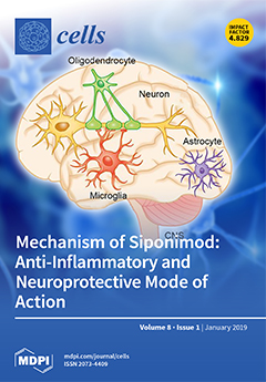
Cover Story (view full-size image): A schematic illustration of the EXPAND trial outcomes is shown in the upper part. Both aspects of multiple sclerosis—inflammation and neurodegeneration—were ameliorated by the spingosine 1 phosphate receptor modulator siponimod/BAF312 (Novartis). The lower part of the figures demonstrates the proposed mode of action of siponimod. Note that following this concept, the anti-inflammatory activity of siponimod is at least in part due to direct interactions with brain cells. Since several studies have demonstrated that degenerative events within the central nervous system can trigger the recruitment of peripheral immune cells into the brain, siponimod-mediated protection of brain damage secondarily results in less severe inflammation. View this paper.
  • Issues are regarded as officially published after their release is announced to the table of contents alert mailing list.
  • You may sign up for e-mail alerts to receive table of contents of newly released issues.
  • PDF is the official format for papers published in both, html and pdf forms. To view the papers in pdf format, click on the "PDF Full-text" link, and use the free Adobe Reader to open them.
Order results
Result details
Section
Select all
Export citation of selected articles as:
13 pages, 2486 KiB  
Article
Crohn’s Disease Patients in Remission Display an Enhanced Intestinal IgM+ B Cell Count in Concert with a Strong Activation of the Intestinal Complement System
by Sophie Preisker, Ann-Kathrin Brethack, Arne Bokemeyer, Dominik Bettenworth, Christian Sina and Stefanie Derer
Cells 2019, 8(1), 78; https://doi.org/10.3390/cells8010078 - 21 Jan 2019
Cited by 8 | Viewed by 6061
Abstract
Inflammatory bowel disease (IBD) is an umbrella term that comprises Crohn’s disease (CD) and ulcerative colitis (UC). Both entities are characterized by a disturbed mucosal immune response and an imbalance of intestinal microbiota composition. The complement system (C) plays a critical role in [...] Read more.
Inflammatory bowel disease (IBD) is an umbrella term that comprises Crohn’s disease (CD) and ulcerative colitis (UC). Both entities are characterized by a disturbed mucosal immune response and an imbalance of intestinal microbiota composition. The complement system (C) plays a critical role in the detection, and clearance of bacteria and dysregulation of single complement components has been linked to IBD. Here, we asked if the C contributes to distinct subtypes of inflammation observed in CD and UC. We performed systematical expression analyses of the intestinal C in IBD patients and controls. Immunohistochemistry or immunoblot experiments were performed to verify qPCR data. Activity of the three activation pathways of C was studied in sera samples. In CD patients a strong upregulation of the C was observed enabling the definition of unique expression patterns being associated either with remission or active disease. These data were reflected by an enhanced C activation in sera and fecal samples. An excessive mucosal presence of immunoglobulin M (IgM) and CR2/CD21 positive B cells in concert with decreased fecal IgA level was identified in CD patients in remission. These findings point to an exacerbated induction of the intestinal C that may potentially be involved in the etiology of CD. Full article
Show Figures

Figure 1

24 pages, 2705 KiB  
Review
Roles of Autophagy-Related Genes in the Pathogenesis of Inflammatory Bowel Disease
by Sup Kim, Hyuk Soo Eun and Eun-Kyeong Jo
Cells 2019, 8(1), 77; https://doi.org/10.3390/cells8010077 - 21 Jan 2019
Cited by 82 | Viewed by 12616
Abstract
Autophagy is an intracellular catabolic process that is essential for a variety of cellular responses. Due to its role in the maintenance of biological homeostasis in conditions of stress, dysregulation or disruption of autophagy may be linked to human diseases such as inflammatory [...] Read more.
Autophagy is an intracellular catabolic process that is essential for a variety of cellular responses. Due to its role in the maintenance of biological homeostasis in conditions of stress, dysregulation or disruption of autophagy may be linked to human diseases such as inflammatory bowel disease (IBD). IBD is a complicated inflammatory colitis disorder; Crohn’s disease and ulcerative colitis are the principal types. Genetic studies have shown the clinical relevance of several autophagy-related genes (ATGs) in the pathogenesis of IBD. Additionally, recent studies using conditional knockout mice have led to a comprehensive understanding of ATGs that affect intestinal inflammation, Paneth cell abnormality and enteric pathogenic infection during colitis. In this review, we discuss the various ATGs involved in macroautophagy and selective autophagy, including ATG16L1, IRGM, LRRK2, ATG7, p62, optineurin and TFEB in the maintenance of intestinal homeostasis. Although advances have been made regarding the involvement of ATGs in maintaining intestinal homeostasis, determining the precise contribution of autophagy has remained elusive. Recent efforts based on direct targeting of ATGs and autophagy will further facilitate the development of new therapeutic opportunities for IBD. Full article
(This article belongs to the Special Issue Autophagy in Tissue Injury and Homeostasis)
Show Figures

Figure 1

20 pages, 1910 KiB  
Review
NUP214 in Leukemia: It’s More than Transport
by Adélia Mendes and Birthe Fahrenkrog
Cells 2019, 8(1), 76; https://doi.org/10.3390/cells8010076 - 21 Jan 2019
Cited by 43 | Viewed by 8597
Abstract
NUP214 is a component of the nuclear pore complex (NPC) with a key role in protein and mRNA nuclear export. Chromosomal translocations involving the NUP214 locus are recurrent in acute leukemia and frequently fuse the C-terminal region of NUP214 with SET and DEK, [...] Read more.
NUP214 is a component of the nuclear pore complex (NPC) with a key role in protein and mRNA nuclear export. Chromosomal translocations involving the NUP214 locus are recurrent in acute leukemia and frequently fuse the C-terminal region of NUP214 with SET and DEK, two chromatin remodeling proteins with roles in transcription regulation. SET-NUP214 and DEK-NUP214 fusion proteins disrupt protein nuclear export by inhibition of the nuclear export receptor CRM1, which results in the aberrant accumulation of CRM1 protein cargoes in the nucleus. SET-NUP214 is primarily associated with acute lymphoblastic leukemia (ALL), whereas DEK-NUP214 exclusively results in acute myeloid leukemia (AML), indicating different leukemogenic driver mechanisms. Secondary mutations in leukemic blasts may contribute to the different leukemia outcomes. Additional layers of complexity arise from the respective functions of SET and DEK in transcription regulation and chromatin remodeling, which may drive malignant hematopoietic transformation more towards ALL or AML. Another, less frequent fusion protein involving the C terminus of NUP214 results in the sequestosome-1 (SQSTM1)-NUP214 chimera, which was detected in ALL. SQSTM1 is a ubiquitin-binding protein required for proper autophagy induction, linking the NUP214 fusion protein to yet another cellular mechanism. The scope of this review is to summarize the general features of NUP214-related leukemia and discuss how distinct chromosomal translocation partners can influence the cellular effects of NUP214 fusion proteins in leukemia. Full article
(This article belongs to the Special Issue Nuclear Transport in Ageing and Diseases)
Show Figures

Figure 1

11 pages, 1864 KiB  
Communication
Development of a Bicistronic Vector for the Expression of a CRISPR/Cas9-mCherry System in Fish Cell Lines
by Sebastian Escobar-Aguirre, Duxan Arancibia, Amanda Escorza, Cristián Bravo, María Estela Andrés, Pedro Zamorano and Víctor Martínez
Cells 2019, 8(1), 75; https://doi.org/10.3390/cells8010075 - 21 Jan 2019
Cited by 18 | Viewed by 8388
Abstract
The clustered regularly interspaced short palindromic repeats/CRISPR-associated protein 9 (CRISPR/Cas9) system has been widely used in animals as an efficient genome editing tool. In fish cells, the technique has been difficult to implement due to the lack of proper vectors that use active [...] Read more.
The clustered regularly interspaced short palindromic repeats/CRISPR-associated protein 9 (CRISPR/Cas9) system has been widely used in animals as an efficient genome editing tool. In fish cells, the technique has been difficult to implement due to the lack of proper vectors that use active promoters to drive the expression of both small guide RNA (sgRNA) and the S. pyogenes Cas9 (spCas9) protein within a single expression platform. Until now, fish cells have been modified using co-transfection of the mRNA of both the sgRNA and the spCas9. In the present study, we describe the optimization of a new vector for the expression of a CRISPR/Cas9 system, designed to edit the genome of fish cell lines, that combines a gene reporter (mCherry), sgRNA, and spCas9 in a single vector, facilitating the study of the efficiency of piscine and non-piscine promoters. A cassette containing the zebrafish U6 RNA III polymerase (U6ZF) promoter was used for the expression of the sgRNA. The new plasmid displayed the expression of spCas9, mCherry, and sgRNA in CHSE/F fish cells. The results demonstrate the functionality of the mammalian promoter and the U6ZF promoter in fish cell lines. This is the first approach aimed at developing a unified genome editing system in fish cells using bicistronic vectors, thus creating a powerful biotechnological platform to study gene function. Full article
Show Figures

Graphical abstract

27 pages, 581 KiB  
Review
Application of Prostate Cancer Models for Preclinical Study: Advantages and Limitations of Cell Lines, Patient-Derived Xenografts, and Three-Dimensional Culture of Patient-Derived Cells
by Takeshi Namekawa, Kazuhiro Ikeda, Kuniko Horie-Inoue and Satoshi Inoue
Cells 2019, 8(1), 74; https://doi.org/10.3390/cells8010074 - 20 Jan 2019
Cited by 132 | Viewed by 21992
Abstract
Various preclinical models have been developed to clarify the pathophysiology of prostate cancer (PCa). Traditional PCa cell lines from clinical metastatic lesions, as exemplified by DU-145, PC-3, and LNCaP cells, are useful tools to define mechanisms underlying tumorigenesis and drug resistance. Cell line-based [...] Read more.
Various preclinical models have been developed to clarify the pathophysiology of prostate cancer (PCa). Traditional PCa cell lines from clinical metastatic lesions, as exemplified by DU-145, PC-3, and LNCaP cells, are useful tools to define mechanisms underlying tumorigenesis and drug resistance. Cell line-based experiments, however, have limitations for preclinical studies because those cells are basically adapted to 2-dimensional monolayer culture conditions, in which the majority of primary PCa cells cannot survive. Recent tissue engineering enables generation of PCa patient-derived xenografts (PDXs) from both primary and metastatic lesions. Compared with fresh PCa tissue transplantation in athymic mice, co-injection of PCa tissues with extracellular matrix in highly immunodeficient mice has remarkably improved the success rate of PDX generation. PDX models have advantages to appropriately recapitulate the molecular diversity, cellular heterogeneity, and histology of original patient tumors. In contrast to PDX models, patient-derived organoid and spheroid PCa models in 3-dimensional culture are more feasible tools for in vitro studies for retaining the characteristics of patient tumors. In this article, we review PCa preclinical model cell lines and their sublines, PDXs, and patient-derived organoid and spheroid models. These PCa models will be applied to the development of new strategies for cancer precision medicine. Full article
Show Figures

Graphical abstract

19 pages, 518 KiB  
Review
Telomere Biology and Human Phenotype
by Kara J. Turner, Vimal Vasu and Darren K. Griffin
Cells 2019, 8(1), 73; https://doi.org/10.3390/cells8010073 - 19 Jan 2019
Cited by 252 | Viewed by 18792
Abstract
Telomeres are nucleoprotein structures that cap the end of each chromosome arm and function to maintain genome stability. The length of telomeres is known to shorten with each cell division and it is well-established that telomere attrition is related to replicative capacity in [...] Read more.
Telomeres are nucleoprotein structures that cap the end of each chromosome arm and function to maintain genome stability. The length of telomeres is known to shorten with each cell division and it is well-established that telomere attrition is related to replicative capacity in vitro. Moreover, telomere loss is also correlated with the process of aging in vivo. In this review, we discuss the mechanisms that lead to telomere shortening and summarise telomere homeostasis in humans throughout a lifetime. In addition, we discuss the available evidence that shows that telomere shortening is related to human aging and the onset of age-related disease. Full article
Show Figures

Graphical abstract

27 pages, 7912 KiB  
Article
Functional Characterization of Ubiquitin-Like Core Autophagy Protein ATG12 in Dictyostelium discoideum
by Sarah Fischer, Ramesh Rijal, Peter Frommolt, Prerana Wagle, Roman Konertz, Jan Faix, Susanne Meßling and Ludwig Eichinger
Cells 2019, 8(1), 72; https://doi.org/10.3390/cells8010072 - 19 Jan 2019
Cited by 19 | Viewed by 5867
Abstract
Autophagy is a highly conserved intracellular degradative pathway that is crucial for cellular homeostasis. During autophagy, the core autophagy protein ATG12 plays, together with ATG5 and ATG16, an essential role in the expansion of the autophagosomal membrane. In this study we analyzed gene [...] Read more.
Autophagy is a highly conserved intracellular degradative pathway that is crucial for cellular homeostasis. During autophagy, the core autophagy protein ATG12 plays, together with ATG5 and ATG16, an essential role in the expansion of the autophagosomal membrane. In this study we analyzed gene replacement mutants of atg12 in Dictyostelium discoideum AX2 wild-type and ATG16‾ cells. RNAseq analysis revealed a strong enrichment of, firstly, autophagy genes among the up-regulated genes and, secondly, genes implicated in cell motility and phagocytosis among the down-regulated genes in the generated ATG12‾, ATG16‾ and ATG12‾/16‾ cells. The mutant strains showed similar defects in fruiting body formation, autolysosome maturation, and cellular viability, implying that ATG12 and ATG16 act as a functional unit in canonical autophagy. In contrast, ablation of ATG16 or of ATG12 and ATG16 resulted in slightly more severe defects in axenic growth, macropinocytosis, and protein homeostasis than ablation of only ATG12, suggesting that ATG16 fulfils an additional function in these processes. Phagocytosis of yeast, spore viability, and maximal cell density were much more affected in ATG12‾/16‾ cells, indicating that both proteins also have cellular functions independent of each other. In summary, we show that ATG12 and ATG16 fulfil autophagy-independent functions in addition to their role in canonical autophagy. Full article
(This article belongs to the Special Issue Proteostasis and Autophagy)
Show Figures

Figure 1

19 pages, 824 KiB  
Review
Prohibitins: A Critical Role in Mitochondrial Functions and Implication in Diseases
by Anna Signorile, Giuseppe Sgaramella, Francesco Bellomo and Domenico De Rasmo
Cells 2019, 8(1), 71; https://doi.org/10.3390/cells8010071 - 18 Jan 2019
Cited by 132 | Viewed by 9614
Abstract
Prohibitin 1 (PHB1) and prohibitin 2 (PHB2) are proteins that are ubiquitously expressed, and are present in the nucleus, cytosol, and mitochondria. Depending on the cellular localization, PHB1 and PHB2 have distinctive functions, but more evidence suggests a critical role within mitochondria. In [...] Read more.
Prohibitin 1 (PHB1) and prohibitin 2 (PHB2) are proteins that are ubiquitously expressed, and are present in the nucleus, cytosol, and mitochondria. Depending on the cellular localization, PHB1 and PHB2 have distinctive functions, but more evidence suggests a critical role within mitochondria. In fact, PHB proteins are highly expressed in cells that heavily depend on mitochondrial function. In mitochondria, these two proteins assemble at the inner membrane to form a supra-macromolecular structure, which works as a scaffold for proteins and lipids regulating mitochondrial metabolism, including bioenergetics, biogenesis, and dynamics in order to determine the cell fate, death, or life. PHB alterations have been found in aging and cancer, as well as neurodegenerative, cardiac, and kidney diseases, in which significant mitochondrial impairments have been observed. The molecular mechanisms by which prohibitins regulate mitochondrial function and their role in pathology are reviewed and discussed herein. Full article
(This article belongs to the Section Cell Signaling)
Show Figures

Figure 1

8 pages, 1832 KiB  
Communication
Possible Mechanisms by which Stefin B could Regulate Proteostasis and Oxidative Stress
by Eva Žerovnik
Cells 2019, 8(1), 70; https://doi.org/10.3390/cells8010070 - 18 Jan 2019
Cited by 4 | Viewed by 4182
Abstract
Human stefin B is a protease inhibitor from the family of cystatins. It was reported that it forms oligomers in cells. We have shown that it has a role in cell’s response to misfolded proteins. We also have shown that its oligomers bind [...] Read more.
Human stefin B is a protease inhibitor from the family of cystatins. It was reported that it forms oligomers in cells. We have shown that it has a role in cell’s response to misfolded proteins. We also have shown that its oligomers bind amyloid-beta (Aβ). Here, we discuss ways, how stefin B could reduce build-up of protein aggregates by other proteins and consequently reduces ROS and, how this might be connected to autophagy. When overexpressed, stefin B forms protein aggregates itself and these protein aggregates induce autophagy. Similarly, cystatin C was shown to bind Aβ and to induce autophagy. It is also suggested how more knowledge about the role of stefin B in a cell’s response to misfolded proteins could be used to modulate progressive myoclonus epilepsy of type 1 EPM1 disease. Full article
(This article belongs to the Special Issue Proteostasis and Autophagy)
Show Figures

Scheme 1

15 pages, 2259 KiB  
Article
Human Lung Cell Pyroptosis Following Traumatic Brain Injury
by Nadine A. Kerr, Juan Pablo de Rivero Vaccari, Oliver Umland, M. Ross Bullock, Gregory E. Conner, W. Dalton Dietrich and Robert W. Keane
Cells 2019, 8(1), 69; https://doi.org/10.3390/cells8010069 - 18 Jan 2019
Cited by 52 | Viewed by 5552
Abstract
Approximately 30% of traumatic brain injured patients suffer from acute lung injury or acute respiratory distress syndrome. Our previous work revealed that extracellular vesicle (EV)-mediated inflammasome signaling plays a crucial role in the pathophysiology of traumatic brain injury (TBI)-induced lung injury. Here, serum-derived [...] Read more.
Approximately 30% of traumatic brain injured patients suffer from acute lung injury or acute respiratory distress syndrome. Our previous work revealed that extracellular vesicle (EV)-mediated inflammasome signaling plays a crucial role in the pathophysiology of traumatic brain injury (TBI)-induced lung injury. Here, serum-derived EVs from severe TBI patients were analyzed for particle size, concentration, origin, and levels of the inflammasome component, an apoptosis-associated speck-like protein containing a caspase-recruiting domain (ASC). Serum ASC levels were analyzed from EV obtained from patients that presented lung injury after TBI and compared them to EV obtained from patients that did not show any signs of lung injury. EVs were co-cultured with lung human microvascular endothelial cells (HMVEC-L) to evaluate inflammasome activation and endothelial cell pyroptosis. TBI patients had a significant increase in the number of serum-derived EVs and levels of ASC. Severe TBI patients with lung injury had a significantly higher level of ASC in serum and serum-derived EVs compared to individuals without lung injury. Only EVs isolated from head trauma patients with gunshot wounds were of neural origin. Delivery of serum-derived EVs to HMVEC-L activated the inflammasome and resulted in endothelial cell pyroptosis. Thus, serum-derived EVs and inflammasome proteins play a critical role in the pathogenesis of TBI-induced lung injury, supporting activation of an EV-mediated neural-respiratory inflammasome axis in TBI-induced lung injury. Full article
(This article belongs to the Special Issue Mechanisms of Inflammasome Activation)
Show Figures

Graphical abstract

14 pages, 2803 KiB  
Article
Retinal Neuron Is More Sensitive to Blue Light-Induced Damage than Glia Cell Due to DNA Double-Strand Breaks
by Pei Chen, Zhipeng Lai, Yihui Wu, Lijun Xu, Xiaoxiao Cai, Jin Qiu, Panyang Yang, Meng Yang, Pan Zhou, Jiejie Zhuang, Jian Ge, Keming Yu and Jing Zhuang
Cells 2019, 8(1), 68; https://doi.org/10.3390/cells8010068 - 18 Jan 2019
Cited by 22 | Viewed by 7026
Abstract
Blue light is a major component of visible light and digital displays. Over-exposure to blue light could cause retinal damage. However, the mechanism of its damage is not well defined. Here, we demonstrate that blue light (900 lux) impairs cell viability and induces [...] Read more.
Blue light is a major component of visible light and digital displays. Over-exposure to blue light could cause retinal damage. However, the mechanism of its damage is not well defined. Here, we demonstrate that blue light (900 lux) impairs cell viability and induces cell apoptosis in retinal neurocytes in vitro. A DNA electrophoresis assay shows severe DNA damage in retinal neurocytes at 2 h after blue light treatment. γ-H2AX foci, a specific marker of DNA double-strand breaks (DSBs), is mainly located in the Map2-posotive neuron other than the glia cell. After assaying the expression level of proteins related to DNA repair, Mre11, Ligase IV and Ku80, we find that Ku80 is up-regulated in retinal neurocytes after blue light treatment. Interestingly, Ku80 is mainly expressed in glia fibrillary acidic protein (GFAP)-positive glia cells. Moreover, following blue light exposure in vivo, DNA DSBs are shown in the ganglion cell layer and only observed in Map2-positive cells. Furthermore, long-term blue light exposure significantly thinned the retina in vivo. Our findings demonstrate that blue light induces DNA DSBs in retinal neurons, and the damage is more pronounced compared to glia cells. Thus, this study provides new insights into the mechanisms of the effect of blue light on the retina. Full article
(This article belongs to the Section Intracellular and Plasma Membranes)
Show Figures

Figure 1

15 pages, 891 KiB  
Review
Yeast to Study Human Purine Metabolism Diseases
by Bertrand Daignan-Fornier and Benoît Pinson
Cells 2019, 8(1), 67; https://doi.org/10.3390/cells8010067 - 17 Jan 2019
Cited by 28 | Viewed by 7490
Abstract
Purine nucleotides are involved in a multitude of cellular processes, and the dysfunction of purine metabolism has drastic physiological and pathological consequences. Accordingly, several genetic disorders associated with defective purine metabolism have been reported. The etiology of these diseases is poorly understood and [...] Read more.
Purine nucleotides are involved in a multitude of cellular processes, and the dysfunction of purine metabolism has drastic physiological and pathological consequences. Accordingly, several genetic disorders associated with defective purine metabolism have been reported. The etiology of these diseases is poorly understood and simple model organisms, such as yeast, have proved valuable to provide a more comprehensive view of the metabolic consequences caused by the identified mutations. In this review, we present results obtained with the yeast Saccharomyces cerevisiae to exemplify how a eukaryotic unicellular organism can offer highly relevant information for identifying the molecular basis of complex human diseases. Overall, purine metabolism illustrates a remarkable conservation of genes, functions and phenotypes between humans and yeast. Full article
Show Figures

Figure 1

20 pages, 2736 KiB  
Review
Adhesion Deregulation in Acute Myeloid Leukaemia
by Alicja M. Gruszka, Debora Valli, Cecilia Restelli and Myriam Alcalay
Cells 2019, 8(1), 66; https://doi.org/10.3390/cells8010066 - 17 Jan 2019
Cited by 39 | Viewed by 6989
Abstract
Cell adhesion is a process through which cells interact with and attach to neighboring cells or matrix using specialized surface cell adhesion molecules (AMs). Adhesion plays an important role in normal haematopoiesis and in acute myeloid leukaemia (AML). AML blasts express many of [...] Read more.
Cell adhesion is a process through which cells interact with and attach to neighboring cells or matrix using specialized surface cell adhesion molecules (AMs). Adhesion plays an important role in normal haematopoiesis and in acute myeloid leukaemia (AML). AML blasts express many of the AMs identified on normal haematopoietic precursors. Differential expression of AMs between normal haematopoietic cells and leukaemic blasts has been documented to a variable extent, likely reflecting the heterogeneity of the disease. AMs govern a variety of processes within the bone marrow (BM), such as migration, homing, and quiescence. AML blasts home to the BM, as the AM-mediated interaction with the niche protects them from chemotherapeutic agents. On the contrary, they detach from the niches and move from the BM into the peripheral blood to colonize other sites, i.e., the spleen and liver, possibly in a process that is reminiscent of epithelial-to-mesenchymal-transition in metastatic solid cancers. The expression of AMs has a prognostic impact and there are ongoing efforts to therapeutically target adhesion in the fight against leukaemia. Full article
(This article belongs to the Special Issue Cell Adhesion Molecules)
Show Figures

Graphical abstract

18 pages, 4061 KiB  
Article
Oxidative Insults and Mitochondrial DNA Mutation Promote Enhanced Autophagy and Mitophagy Compromising Cell Viability in Pluripotent Cell Model of Mitochondrial Disease
by Dar-Shong Lin, Yu-Wen Huang, Che-Sheng Ho, Pi-Lien Hung, Mei-Hsin Hsu, Tuan-Jen Wang, Tsu-Yen Wu, Tsung-Han Lee, Zo-Darr Huang, Po-Chun Chang and Ming-Fu Chiang
Cells 2019, 8(1), 65; https://doi.org/10.3390/cells8010065 - 17 Jan 2019
Cited by 52 | Viewed by 9644
Abstract
Dysfunction of mitochondria causes defects in oxidative phosphorylation system (OXPHOS) and increased production of reactive oxygen species (ROS) triggering the activation of the cell death pathway that underlies the pathogenesis of aging and various diseases. The process of autophagy to degrade damaged cytoplasmic [...] Read more.
Dysfunction of mitochondria causes defects in oxidative phosphorylation system (OXPHOS) and increased production of reactive oxygen species (ROS) triggering the activation of the cell death pathway that underlies the pathogenesis of aging and various diseases. The process of autophagy to degrade damaged cytoplasmic components as well as dysfunctional mitochondria is essential for ensuring cell survival. We analyzed the role of autophagy inpatient-specific induced pluripotent stem (iPS) cells generated from fibroblasts of patients with mitochondrial encephalomyopathy, lactic acidosis, and stroke-like episodes (MELAS) with well-characterized mitochondrial DNA mutations and distinct OXPHOS defects. MELAS iPS cells recapitulated the pathogenesis of MELAS syndrome, and showed an increase of autophagy in comparison with its isogenic normal counterpart, whereas mitophagy is very scarce at the basal condition. Our results indicated that the existence of pathogenic mtDNA alone in mitochondrial disease was not sufficient to elicit the degradation of dysfunctional mitochondria. Nonetheless, oxidative insults induced bulk macroautophagy with the accumulation of autophagosomes and autolysosomes upon marked elevation of ROS, overload of intracellular calcium, and robust depolarization of mitochondrial membrane potential, while mitochondria respiratory function was impaired and widespread mitophagy compromised cell viability. Collectively, our studies provide insights into the dysfunction of autophagy and activation of mitophagy contributing to the pathological mechanism of mitochondrial disease. Full article
(This article belongs to the Special Issue iPS Cells for Disease Modeling)
Show Figures

Figure 1

23 pages, 2225 KiB  
Article
Carnosine Prevents Aβ-Induced Oxidative Stress and Inflammation in Microglial Cells: A Key Role of TGF-β1
by Giuseppe Caruso, Claudia G. Fresta, Nicolò Musso, Mariaconcetta Giambirtone, Margherita Grasso, Simona F. Spampinato, Sara Merlo, Filippo Drago, Giuseppe Lazzarino, Maria A. Sortino, Susan M. Lunte and Filippo Caraci
Cells 2019, 8(1), 64; https://doi.org/10.3390/cells8010064 - 17 Jan 2019
Cited by 90 | Viewed by 9179
Abstract
Carnosine (β-alanyl-L-histidine), a dipeptide, is an endogenous antioxidant widely distributed in excitable tissues like muscles and the brain. Carnosine is involved in cellular defense mechanisms against oxidative stress, including the inhibition of amyloid-beta (Aβ) aggregation and the scavenging of reactive species. Microglia play [...] Read more.
Carnosine (β-alanyl-L-histidine), a dipeptide, is an endogenous antioxidant widely distributed in excitable tissues like muscles and the brain. Carnosine is involved in cellular defense mechanisms against oxidative stress, including the inhibition of amyloid-beta (Aβ) aggregation and the scavenging of reactive species. Microglia play a central role in the pathogenesis of Alzheimer’s disease, promoting neuroinflammation through the secretion of inflammatory mediators and free radicals. However, the effects of carnosine on microglial cells and neuroinflammation are not well understood. In the present work, carnosine was tested for its ability to protect BV-2 microglial cells against oligomeric Aβ1-42-induced oxidative stress and inflammation. Carnosine prevented cell death in BV-2 cells challenged with Aβ oligomers through multiple mechanisms. Specifically, carnosine lowered the oxidative stress by decreasing NO and O2−• intracellular levels as well as the expression of iNOS and Nox enzymes. Carnosine also decreased the secretion of pro-inflammatory cytokines such as IL-1β, simultaneously rescuing IL-10 levels and increasing the expression and the release of TGF-β1. Carnosine also prevented Aβ-induced neurodegeneration in mixed neuronal cultures challenged with Aβ oligomers, and these neuroprotective effects were completely abolished by SB431542, a selective inhibitor of the type-1 TGF-β receptor. Our data suggest a multimodal mechanism of action of carnosine underlying its protective effects on microglial cells against Aβ toxicity with a key role of TGF-β1 in mediating these protective effects. Full article
(This article belongs to the Special Issue TGF-beta/BMP Signaling Pathway)
Show Figures

Graphical abstract

16 pages, 4904 KiB  
Article
Bone Marrow Derived Extracellular Vesicles Activate Osteoclast Differentiation in Traumatic Brain Injury Induced Bone Loss
by Quante Singleton, Kumar Vaibhav, Molly Braun, Chandani Patel, Andrew Khayrullin, Bharati Mendhe, Byung R. Lee, Ravindra Kolhe, Helen Kaiser, Mohamed E. Awad, Tunde Fariyike, Ranya Elsayed, Mohammed Elsalanty, Carlos M. Isales, Yutao Liu, Mark W. Hamrick, Krishnan M. Dhandapani and Sadanand Fulzele
Cells 2019, 8(1), 63; https://doi.org/10.3390/cells8010063 - 17 Jan 2019
Cited by 28 | Viewed by 6913
Abstract
Traumatic brain injury (TBI) is a major source of worldwide morbidity and mortality. Patients suffering from TBI exhibit a higher susceptibility to bone loss and an increased rate of bone fractures; however, the underlying mechanisms remain poorly defined. Herein, we observed significantly lower [...] Read more.
Traumatic brain injury (TBI) is a major source of worldwide morbidity and mortality. Patients suffering from TBI exhibit a higher susceptibility to bone loss and an increased rate of bone fractures; however, the underlying mechanisms remain poorly defined. Herein, we observed significantly lower bone quality and elevated levels of inflammation in bone and bone marrow niche after controlled cortical impact-induced TBI in in vivo CD-1 mice. Further, we identified dysregulated NF-κB signaling, an established mediator of osteoclast differentiation and bone loss, within the bone marrow niche of TBI mice. Ex vivo studies revealed increased osteoclast differentiation in bone marrow-derived cells from TBI mice, as compared to sham injured mice. We also found bone marrow derived extracellular vesicles (EVs) from TBI mice enhanced the colony forming ability and osteoclast differentiation efficacy and activated NF-κB signaling genes in bone marrow-derived cells. Additionally, we showed that miRNA-1224 up-regulated in bone marrow-derived EVs cargo of TBI. Taken together, we provide evidence that TBI-induced inflammatory stress on bone and the bone marrow niche may activate NF-κB leading to accelerated bone loss. Targeted inhibition of these signaling pathways may reverse TBI-induced bone loss and reduce fracture rates. Full article
(This article belongs to the Section Intracellular and Plasma Membranes)
Show Figures

Graphical abstract

15 pages, 581 KiB  
Review
New Insights into the Interplay between Non-Coding RNAs and RNA-Binding Protein HnRNPK in Regulating Cellular Functions
by Yongjie Xu, Wei Wu, Qiu Han, Yaling Wang, Cencen Li, Pengpeng Zhang and Haixia Xu
Cells 2019, 8(1), 62; https://doi.org/10.3390/cells8010062 - 17 Jan 2019
Cited by 56 | Viewed by 8900
Abstract
The emerging data indicates that non-coding RNAs (ncRNAs) epresent more than the “junk sequences” of the genome. Both miRNAs and long non-coding RNAs (lncRNAs) are involved in fundamental biological processes, and their deregulation may lead to oncogenesis and other diseases. As an important [...] Read more.
The emerging data indicates that non-coding RNAs (ncRNAs) epresent more than the “junk sequences” of the genome. Both miRNAs and long non-coding RNAs (lncRNAs) are involved in fundamental biological processes, and their deregulation may lead to oncogenesis and other diseases. As an important RNA-binding protein (RBP), heterogeneous nuclear ribonucleoprotein K (hnRNPK) is known to regulate gene expression through the RNA-binding domain involved in various pathways, such as transcription, splicing, and translation. HnRNPK is a highly conserved gene that is abundantly expressed in mammalian cells. The interaction of hnRNPK and ncRNAs defines the novel way through which ncRNAs affect the expression of protein-coding genes and form autoregulatory feedback loops. This review summarizes the interactions of hnRNPK and ncRNAs in regulating gene expression at transcriptional and post-transcriptional levels or by changing the genomic structure, highlighting their involvement in carcinogenesis, glucose metabolism, stem cell differentiation, virus infection and other cellular functions. Drawing connections between such discoveries might provide novel targets to control the biological outputs of cells in response to different stimuli. Full article
(This article belongs to the Special Issue lncRNA and Cancer)
Show Figures

Figure 1

20 pages, 2410 KiB  
Review
Autophagy in Chronic Kidney Diseases
by Tien-An Lin, Victor Chien-Chia Wu and Chao-Yung Wang
Cells 2019, 8(1), 61; https://doi.org/10.3390/cells8010061 - 16 Jan 2019
Cited by 125 | Viewed by 12844
Abstract
Autophagy is a cellular recycling process involving self-degradation and reconstruction of damaged organelles and proteins. Current evidence suggests that autophagy is critical in kidney physiology and homeostasis. In clinical studies, autophagy activations and inhibitions are linked to acute kidney injuries, chronic kidney diseases, [...] Read more.
Autophagy is a cellular recycling process involving self-degradation and reconstruction of damaged organelles and proteins. Current evidence suggests that autophagy is critical in kidney physiology and homeostasis. In clinical studies, autophagy activations and inhibitions are linked to acute kidney injuries, chronic kidney diseases, diabetic nephropathies, and polycystic kidney diseases. Oxidative stress, inflammation, and mitochondrial dysfunction, which are implicated as important mechanisms underlying many kidney diseases, modulate the autophagy activation and inhibition and lead to cellular recycling dysfunction. Abnormal autophagy function can induce loss of podocytes, damage proximal tubular cells, and glomerulosclerosis. After acute kidney injuries, activated autophagy protects tubular cells from apoptosis and enhances cellular regeneration. Patients with chronic kidney diseases have impaired autophagy that cannot be reversed by hemodialysis. Multiple nephrotoxic medications also alter the autophagy signaling, by which the mechanistic insights of the drugs are revealed, thus providing the unique opportunity to manage the nephrotoxicity of these drugs. In this review, we summarize the current concepts of autophagy and its molecular aspects in different kidney cells pathophysiology. We also discuss the current evidence of autophagy in acute kidney injury, chronic kidney disease, toxic effects of drugs, and aging kidneys. In addition, we examine therapeutic possibilities targeting the autophagy system in kidney diseases. Full article
(This article belongs to the Special Issue Autophagy in Tissue Injury and Homeostasis)
Show Figures

Figure 1

15 pages, 208 KiB  
Editorial
Acknowledgement to Reviewers of Cells in 2018
by Cells Editorial Office
Cells 2019, 8(1), 60; https://doi.org/10.3390/cells8010060 - 16 Jan 2019
Viewed by 2459
Abstract
Rigorous peer-review is the corner-stone of high-quality academic publishing [...] Full article
12 pages, 1254 KiB  
Review
Are Proteinopathy and Oxidative Stress Two Sides of the Same Coin?
by Nihar J. Mehta, Praneet Kaur Marwah and David Njus
Cells 2019, 8(1), 59; https://doi.org/10.3390/cells8010059 - 16 Jan 2019
Cited by 8 | Viewed by 5304
Abstract
Parkinson’s disease, like other neurodegenerative diseases, exhibits two common features: Proteinopathy and oxidative stress, leading to protein aggregation and mitochondrial damage respectively. Because both protein aggregates and dysfunctional mitochondria are eliminated by autophagy, we suggest that inadequate clearance may couple the two phenomena. [...] Read more.
Parkinson’s disease, like other neurodegenerative diseases, exhibits two common features: Proteinopathy and oxidative stress, leading to protein aggregation and mitochondrial damage respectively. Because both protein aggregates and dysfunctional mitochondria are eliminated by autophagy, we suggest that inadequate clearance may couple the two phenomena. If a neuron’s autophagy machinery is overwhelmed, whether by excessive oxidative stress or by excessive protein aggregation, protein aggregates and dysfunctional mitochondria will both accumulate. Parkinson’s disease may provide a unique window into this because there is evidence that both sides contribute. Mutations amplifying the aggregation of α-synuclein are associated with Parkinson’s disease. Likewise, mutations in Parkin and PINK1, proteins involved in mitophagy, suggest that impaired mitochondrial clearance is also a contributing factor. Many have suggested that dopamine oxidation products lead to oxidative stress accounting for the dopaminergic selectivity of the disease. We have presented evidence for the specific involvement of hypochlorite-oxidized cysteinyl-dopamine (HOCD), a redox-cycling benzothiazine derivative. While toxins like 6-hydroxydopamine and 1-methyl-4-phenyl pyridinium (MPP+) have been used to study mitochondrial involvement in Parkinson’s disease, HOCD may provide a more physiologically relevant approach. Understanding the role of mitochondrial dysfunction and oxidative stress in Parkinson’s disease and their relation to α-synuclein proteinopathy is important to gain a full picture of the cause, especially for the great majority of cases which are idiopathic. Full article
(This article belongs to the Special Issue The Molecular and Cellular Basis for Parkinson's Disease)
Show Figures

Figure 1

31 pages, 1860 KiB  
Review
Telomeres in Plants and Humans: Not So Different, Not So Similar
by Petra Procházková Schrumpfová, Miloslava Fojtová and Jiří Fajkus
Cells 2019, 8(1), 58; https://doi.org/10.3390/cells8010058 - 16 Jan 2019
Cited by 39 | Viewed by 10829
Abstract
Parallel research on multiple model organisms shows that while some principles of telomere biology are conserved among all eukaryotic kingdoms, we also find some deviations that reflect different evolutionary paths and life strategies, which may have diversified after the establishment of telomerase as [...] Read more.
Parallel research on multiple model organisms shows that while some principles of telomere biology are conserved among all eukaryotic kingdoms, we also find some deviations that reflect different evolutionary paths and life strategies, which may have diversified after the establishment of telomerase as a primary mechanism for telomere maintenance. Much more than animals, plants have to cope with environmental stressors, including genotoxic factors, due to their sessile lifestyle. This is, in principle, made possible by an increased capacity and efficiency of the molecular systems ensuring maintenance of genome stability, as well as a higher tolerance to genome instability. Furthermore, plant ontogenesis differs from that of animals in which tissue differentiation and telomerase silencing occur during early embryonic development, and the “telomere clock” in somatic cells may act as a preventive measure against carcinogenesis. This does not happen in plants, where growth and ontogenesis occur through the serial division of apical meristems consisting of a small group of stem cells that generate a linear series of cells, which differentiate into an array of cell types that make a shoot and root. Flowers, as generative plant organs, initiate from the shoot apical meristem in mature plants which is incompatible with the human-like developmental telomere shortening. In this review, we discuss differences between human and plant telomere biology and the implications for aging, genome stability, and cell and organism survival. In particular, we provide a comprehensive comparative overview of telomere proteins acting in humans and in Arabidopsis thaliana model plant, and discuss distinct epigenetic features of telomeric chromatin in these species. Full article
(This article belongs to the Section Cell Nuclei: Function, Transport and Receptors)
Show Figures

Figure 1

18 pages, 10034 KiB  
Review
Cellular and Structural Studies of Eukaryotic Cells by Cryo-Electron Tomography
by Miriam Sarah Weber, Matthias Wojtynek and Ohad Medalia
Cells 2019, 8(1), 57; https://doi.org/10.3390/cells8010057 - 16 Jan 2019
Cited by 39 | Viewed by 18528
Abstract
The architecture of protein assemblies and their remodeling during physiological processes is fundamental to cells. Therefore, providing high-resolution snapshots of macromolecular complexes in their native environment is of major importance for understanding the molecular biology of the cell. Cellular structural biology by means [...] Read more.
The architecture of protein assemblies and their remodeling during physiological processes is fundamental to cells. Therefore, providing high-resolution snapshots of macromolecular complexes in their native environment is of major importance for understanding the molecular biology of the cell. Cellular structural biology by means of cryo-electron tomography (cryo-ET) offers unique insights into cellular processes at an unprecedented resolution. Recent technological advances have enabled the detection of single impinging electrons and improved the contrast of electron microscopic imaging, thereby significantly increasing the sensitivity and resolution. Moreover, various sample preparation approaches have paved the way to observe every part of a eukaryotic cell, and even multicellular specimens, under the electron beam. Imaging of macromolecular machineries at high resolution directly within their native environment is thereby becoming reality. In this review, we discuss several sample preparation and labeling techniques that allow the visualization and identification of macromolecular assemblies in situ, and demonstrate how these methods have been used to study eukaryotic cellular landscapes. Full article
(This article belongs to the Special Issue Development and Challenges in Microscopy for Cellular Imaging)
Show Figures

Figure 1

25 pages, 5309 KiB  
Article
Transected Tendon Treated with a New Fibrin Sealant Alone or Associated with Adipose-Derived Stem Cells
by Katleen Frauz, Luis Felipe R. Teodoro, Giane Daniela Carneiro, Fernanda Cristina da Veiga, Danilo Lopes Ferrucci, André Luis Bombeiro, Priscyla Waleska Simões, Lúcia Elvira Alvares, Alexandre Leite R. de Oliveira, Cristina Pontes Vicente, Rui Seabra Ferreira, Benedito Barraviera, Maria Esméria C. do Amaral, Marcelo Augusto M. Esquisatto, Benedicto de Campos Vidal, Edson Rosa Pimentel and Andrea Aparecida de Aro
Cells 2019, 8(1), 56; https://doi.org/10.3390/cells8010056 - 16 Jan 2019
Cited by 21 | Viewed by 5179
Abstract
Tissue engineering and cell-based therapy combine techniques that create biocompatible materials for cell survival, which can improve tendon repair. This study seeks to use a new fibrin sealant (FS) derived from the venom of Crotalus durissus terrificus, a biodegradable three-dimensional scaffolding produced [...] Read more.
Tissue engineering and cell-based therapy combine techniques that create biocompatible materials for cell survival, which can improve tendon repair. This study seeks to use a new fibrin sealant (FS) derived from the venom of Crotalus durissus terrificus, a biodegradable three-dimensional scaffolding produced from animal components only, associated with adipose-derived stem cells (ASC) for application in tendons injuries, considered a common and serious orthopedic problem. Lewis rats had tendons distributed in five groups: normal (N), transected (T), transected and FS (FS) or ASC (ASC) or with FS and ASC (FS + ASC). The in vivo imaging showed higher quantification of transplanted PKH26-labeled ASC in tendons of FS + ASC compared to ASC on the 14th day after transection. A small number of Iba1 labeled macrophages carrying PKH26 signal, probably due to phagocytosis of dead ASC, were observed in tendons of transected groups. ASC up-regulated the Tenomodulin gene expression in the transection region when compared to N, T and FS groups and the expression of TIMP-2 and Scleraxis genes in relation to the N group. FS group presented a greater organization of collagen fibers, followed by FS + ASC and ASC in comparison to N. Tendons from ASC group presented higher hydroxyproline concentration in relation to N and the transected tendons of T, FS and FS + ASC had a higher amount of collagen I and tenomodulin in comparison to N group. Although no marked differences were observed in the other biomechanical parameters, T group had higher value of maximum load compared to the groups ASC and FS + ASC. In conclusion, the FS kept constant the number of transplanted ASC in the transected region until the 14th day after injury. Our data suggest this FS to be a good scaffold for treatment during tendon repair because it was the most effective one regarding tendon organization recovering, followed by the FS treatment associated with ASC and finally by the transplanted ASC on the 21st day. Further investigations in long-term time points of the tendon repair are needed to analyze if the higher tissue organization found with the FS scaffold will improve the biomechanics of the tendons. Full article
(This article belongs to the Special Issue Extracellular Matrix Remodeling)
Show Figures

Figure 1

15 pages, 3300 KiB  
Review
The Ribosome Biogenesis—Cancer Connection
by Marianna Penzo, Lorenzo Montanaro, Davide Treré and Massimo Derenzini
Cells 2019, 8(1), 55; https://doi.org/10.3390/cells8010055 - 15 Jan 2019
Cited by 159 | Viewed by 11499
Abstract
Multifaceted relations link ribosome biogenesis to cancer. Ribosome biogenesis takes place in the nucleolus. Clarifying the mechanisms involved in this nucleolar function and its relationship with cell proliferation: (1) allowed the understanding of the reasons for the nucleolar changes in cancer cells and [...] Read more.
Multifaceted relations link ribosome biogenesis to cancer. Ribosome biogenesis takes place in the nucleolus. Clarifying the mechanisms involved in this nucleolar function and its relationship with cell proliferation: (1) allowed the understanding of the reasons for the nucleolar changes in cancer cells and their exploitation in tumor pathology, (2) defined the importance of the inhibition of ribosome biogenesis in cancer chemotherapy and (3) focused the attention on alterations of ribosome biogenesis in the pathogenesis of cancer. This review summarizes the research milestones regarding these relevant relationships between ribosome biogenesis and cancer. The structure and function of the nucleolus will also be briefly described. Full article
(This article belongs to the Special Issue Nucleolar Organization and Functions in Health and Disease)
Show Figures

Figure 1

10 pages, 641 KiB  
Review
Roles of Telomere Biology in Cell Senescence, Replicative and Chronological Ageing
by Jun Liu, Lihui Wang, Zhiguo Wang and Jun-Ping Liu
Cells 2019, 8(1), 54; https://doi.org/10.3390/cells8010054 - 15 Jan 2019
Cited by 112 | Viewed by 12525
Abstract
Telomeres with G-rich repetitive DNA and particular proteins as special heterochromatin structures at the termini of eukaryotic chromosomes are tightly maintained to safeguard genetic integrity and functionality. Telomerase as a specialized reverse transcriptase uses its intrinsic RNA template to lengthen telomeric G-rich strand [...] Read more.
Telomeres with G-rich repetitive DNA and particular proteins as special heterochromatin structures at the termini of eukaryotic chromosomes are tightly maintained to safeguard genetic integrity and functionality. Telomerase as a specialized reverse transcriptase uses its intrinsic RNA template to lengthen telomeric G-rich strand in yeast and human cells. Cells sense telomere length shortening and respond with cell cycle arrest at a certain size of telomeres referring to the “Hayflick limit.” In addition to regulating the cell replicative senescence, telomere biology plays a fundamental role in regulating the chronological post-mitotic cell ageing. In this review, we summarize the current understandings of telomere regulation of cell replicative and chronological ageing in the pioneer model system Saccharomyces cerevisiae and provide an overview on telomere regulation of animal lifespans. We focus on the mechanisms of survivals by telomere elongation, DNA damage response and environmental factors in the absence of telomerase maintenance of telomeres in the yeast and mammals. Full article
(This article belongs to the Special Issue The Role of Telomere Biology in Aging and Human Disease)
Show Figures

Figure 1

22 pages, 2566 KiB  
Article
Dystrophin Deficiency Leads to Genomic Instability in Human Pluripotent Stem Cells via NO Synthase-Induced Oxidative Stress
by Sarka Jelinkova, Petr Fojtik, Aneta Kohutova, Aleksandra Vilotic, Lenka Marková, Martin Pesl, Tereza Jurakova, Miriama Kruta, Jan Vrbsky, Renata Gaillyova, Iveta Valášková, Ivan Frák, Alain Lacampagne, Giancarlo Forte, Petr Dvorak, Albano C. Meli and Vladimir Rotrekl
Cells 2019, 8(1), 53; https://doi.org/10.3390/cells8010053 - 15 Jan 2019
Cited by 30 | Viewed by 6781
Abstract
Recent data on Duchenne muscular dystrophy (DMD) show myocyte progenitor’s involvement in the disease pathology often leading to the DMD patient’s death. The molecular mechanism underlying stem cell impairment in DMD has not been described. We created dystrophin-deficient human pluripotent stem cell (hPSC) [...] Read more.
Recent data on Duchenne muscular dystrophy (DMD) show myocyte progenitor’s involvement in the disease pathology often leading to the DMD patient’s death. The molecular mechanism underlying stem cell impairment in DMD has not been described. We created dystrophin-deficient human pluripotent stem cell (hPSC) lines by reprogramming cells from two DMD patients, and also by introducing dystrophin mutation into human embryonic stem cells via CRISPR/Cas9. While dystrophin is expressed in healthy hPSC, its deficiency in DMD hPSC lines induces the release of reactive oxygen species (ROS) through dysregulated activity of all three isoforms of nitric oxide synthase (further abrev. as, NOS). NOS-induced ROS release leads to DNA damage and genomic instability in DMD hPSC. We were able to reduce both the ROS release as well as DNA damage to the level of wild-type hPSC by inhibiting NOS activity. Full article
(This article belongs to the Special Issue iPS Cells for Disease Modeling)
Show Figures

Graphical abstract

15 pages, 433 KiB  
Review
Mood Disorders, Accelerated Aging, and Inflammation: Is the Link Hidden in Telomeres?
by Alessio Squassina, Claudia Pisanu and Roberta Vanni
Cells 2019, 8(1), 52; https://doi.org/10.3390/cells8010052 - 15 Jan 2019
Cited by 42 | Viewed by 8037
Abstract
Mood disorders are associated with an increased risk of aging-related diseases, which greatly contribute to the excess morbidity and mortality observed in affected individuals. Clinical and molecular findings also suggest that mood disorders might be characterized by a permanent state of low-grade inflammation. [...] Read more.
Mood disorders are associated with an increased risk of aging-related diseases, which greatly contribute to the excess morbidity and mortality observed in affected individuals. Clinical and molecular findings also suggest that mood disorders might be characterized by a permanent state of low-grade inflammation. At the cellular level, aging translates into telomeres shortening. Intriguingly, inflammation and telomere shortening show a bidirectional association: a pro-inflammatory state seems to contribute to aging and telomere dysfunction, and telomere attrition is able to induce low-grade inflammation. Several independent studies have reported shorter telomere length and increased levels of circulating inflammatory cytokines in mood disorders, suggesting a complex interplay between altered inflammatory–immune responses and telomere dynamics in the etiopathogenesis of these disorders. In this review, we critically discuss studies investigating the role of telomere attrition and inflammation in the pathogenesis and course of mood disorders, and in pharmacological treatments with psychotropic medications. Full article
(This article belongs to the Special Issue The Role of Telomere Biology in Aging and Human Disease)
Show Figures

Graphical abstract

10 pages, 5700 KiB  
Article
Lack of Endogenous Annexin A1 Increases Mast Cell Activation and Exacerbates Experimental Atopic Dermatitis
by Jéssica dos Santos Parisi, Mab Pereira Corrêa and Cristiane Damas Gil
Cells 2019, 8(1), 51; https://doi.org/10.3390/cells8010051 - 15 Jan 2019
Cited by 24 | Viewed by 5028
Abstract
Annexin A1 (AnxA1) is a protein with potent anti-inflammatory actions and an interesting target that has been poorly explored in skin inflammation. This work evaluated the lack of endogenous AnxA1 in the progression of ovalbumin (OVA)-induced atopic dermatitis (AD)-like skin lesions. OVA/Alum-immunized C57BL/6 [...] Read more.
Annexin A1 (AnxA1) is a protein with potent anti-inflammatory actions and an interesting target that has been poorly explored in skin inflammation. This work evaluated the lack of endogenous AnxA1 in the progression of ovalbumin (OVA)-induced atopic dermatitis (AD)-like skin lesions. OVA/Alum-immunized C57BL/6 male wild-type (WT) and AnxA1 null (AnxA1-/-) mice were challenged with drops containing OVA on days 11, 14–18 and 21–24. The AnxA1-/- AD group exhibited skin with intense erythema, erosion and dryness associated with increased skin thickness compared to the AD WT group. The lack of endogenous AnxA1 also increased IgE relative to WT animals, demonstrating exacerbation of the allergic response. Histological analysis revealed intense eosinophilia and mast-cell activation in AD animals, especially in AnxA1-/-. Both AD groups increased skin interleukin (IL)-13 levels, while IL-17A was upregulated in AnxA1-/- lymph nodes and mast cells. High levels of phosphorylated ERK were detected in keratinocytes from AD groups. However, phospho-ERK levels were higher in the AnxA1-/- when compared to the respective control groups. Our results suggest AnxA1 as an important therapeutic target for inflammatory skin diseases. Full article
(This article belongs to the Special Issue Mast Cells in Inflammation and Immunity)
Show Figures

Figure 1

11 pages, 3134 KiB  
Article
Characterization of Somatic Embryogenesis Receptor-Like Kinase 4 as a Negative Regulator of Leaf Senescence in Arabidopsis
by Xiaoxu Li, Salman Ahmad, Akhtar Ali, Cun Guo, Hong Li, Jing Yu, Yan Zhang, Xiaoming Gao and Yongfeng Guo
Cells 2019, 8(1), 50; https://doi.org/10.3390/cells8010050 - 14 Jan 2019
Cited by 23 | Viewed by 5204
Abstract
Leaf senescence is a genetically controlled process that involves the perception of extracellular signals and signal transduction. The receptor-like protein kinases (RLKs) are known to act as an important class of cell surface receptors and are involved in multiple biological processes such as [...] Read more.
Leaf senescence is a genetically controlled process that involves the perception of extracellular signals and signal transduction. The receptor-like protein kinases (RLKs) are known to act as an important class of cell surface receptors and are involved in multiple biological processes such as development and stress responses. The functions of a number of RLK members have been characterized in Arabidopsis and other plant species, but only a limited number of RLK proteins have been reported to be associated with leaf senescence. In the present study, we have characterized the role of the somatic embryogenesis receptor kinase 4 (SERK4) gene in leaf senescence. The expression of SERK4 was up-regulated during leaf senescence and by several abiotic stress treatments in Arabidopsis. The serk4-1 knockout mutant was found to display a significant early leaf senescence phenotype. Furthermore, the results of overexpression analysis and complementary analysis supported the idea that SERK4 acts as a negative regulator in the process of leaf senescence. Full article
(This article belongs to the Section Cell Signaling)
Show Figures

Figure 1

22 pages, 1932 KiB  
Article
Metformin Impairs Glutamine Metabolism and Autophagy in Tumour Cells
by Serena Saladini, Michele Aventaggiato, Federica Barreca, Emanuela Morgante, Luigi Sansone, Matteo A. Russo and Marco Tafani
Cells 2019, 8(1), 49; https://doi.org/10.3390/cells8010049 - 14 Jan 2019
Cited by 27 | Viewed by 6274
Abstract
Metformin has been shown to inhibit glutaminase (GLS) activity and ammonia accumulation thereby reducing the risk of hepatic encephalopathy in type 2 diabetic patients. Since tumour cells are addicted to glutamine and often show an overexpression of glutaminase, we hypothesize that the antitumoral [...] Read more.
Metformin has been shown to inhibit glutaminase (GLS) activity and ammonia accumulation thereby reducing the risk of hepatic encephalopathy in type 2 diabetic patients. Since tumour cells are addicted to glutamine and often show an overexpression of glutaminase, we hypothesize that the antitumoral mechanism of metformin could be ascribed to inhibition of GLS and reduction of ammonia and ammonia-induced autophagy. Our results show that, in different tumour cell lines, micromolar doses of metformin prevent cell growth by reducing glutamate, ammonia accumulation, autophagy markers such as MAP1LC3B-II and GABARAP as well as degradation of long-lived proteins. Reduced autophagy is then accompanied by increased BECN1/BCL2 binding and apoptotic cell death. Interestingly, GLS-silenced cells reproduce the effect of metformin treatment showing reduced MAP1LC3B-II and GABARAP as well as ammonia accumulation. Since metformin is used as adjuvant drug to increase the efficacy of cisplatin-based neoadjuvant chemotherapy, we co-treated tumour cells with micromolar doses of metformin in the presence of cisplatin observing a marked reduction of MAP1LC3B-II and an increase of caspase 3 cleavage. In conclusion, our work demonstrates that the anti-tumoral action of metformin is due to the inhibition of glutaminase and autophagy and could be used to improve the efficacy of chemotherapy. Full article
(This article belongs to the Special Issue Autophagy in Tissue Injury and Homeostasis)
Show Figures

Figure 1

Previous Issue
Next Issue
Back to TopTop