Article

Increased CD16a (FcγRIIIA) Expression in The Tumor Microenvironment of Atypical Neurofibromatous Neoplasms of Uncertain Biologic Potential May Be Associated with Progression from Neurofibromas to Atypical Neurofibromas

1
Department of Pathology, Chungnam National University School of Medicine, Munwha-ro 266, Daejeon 35015, Republic of Korea
2
Department of Computer Science & Engineering, Chungnam National University, Daejeon 34134, Republic of Korea
3
Department of Biochemistry, College of Medicine, Chungnam National University, Daejeon 35015, Republic of Korea
4
Translational Immunology Institute, Chungnam National University College of Medicine, Daejeon 35015, Republic of Korea
5
Brain Korea 21 FOUR Project for Medical Science, Chungnam National University, Daejeon 35015, Republic of Korea
*
Author to whom correspondence should be addressed.
J. Pers. Med. 2023, 13(12), 1720; https://doi.org/10.3390/jpm13121720
Submission received: 24 October 2023 / Revised: 26 November 2023 / Accepted: 14 December 2023 / Published: 17 December 2023
(This article belongs to the Section Clinical Medicine, Cell, and Organism Physiology)

Abstract

:
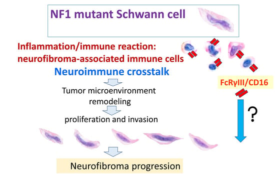
Neurofibroma (NF) is a benign tumor in the peripheral nervous system, but it can infiltrate around structures and cause functional impairment and disfigurement. We incidentally found that the expression of CD16a (Fc gamma receptor IIIA) was increased in NFs compared to in non-neoplastic nerves and hypothesized that CD16 could be relevant to NF progression. We evaluated the expressions of CD16a, CD16b, CD68, TREM2, Galectin-3, S-100, and SOX10 in 38 cases of neurogenic tumors (NF, n = 18; atypical neurofibromatous neoplasm of uncertain biologic potential (ANNUBP), n = 14; and malignant peripheral nerve sheath tumor (MPNST), n = 6) by immunohistochemical staining. In the tumor microenvironment (TME) of the ANNUBPs, CD16a and CD16b expression levels had increased more than in the NFs or MPNSTs. CD68 and Galectin-3 expression levels in the ANNUBPs were higher than in the MPNSTs. Dual immunohistochemical staining showed an overlapping pattern for CD16a and CD68 in TME immune cells. Increased CD16a expression was detected in the ANNUBPs compared to the NFs but decreased with malignant progression. The CD16a overexpression with CD68 positivity in the ANNUBPs potentially reflects that the TME immune modulation could be associated with NF progression to an ANNUBP. Further studies should explore the role of CD16a in immunomodulation for accelerating NF growth.

Graphical Abstract

1. Introduction

Neurofibromas (NFs) are the most common benign peripheral nerve sheath tumors. In the defective function of neurofibromin, NFs actively infiltrate around non-neoplastic structures and occur in numbers, impairing skeletal and neurological functions and cosmetic appearance and pain. NFs are subtyped into localized, diffuse, and plexiform depending on the growth pattern. According to their tumor location, NFs are classified as cutaneous and plexiform. The localized NFs usually occur sporadically as cutaneous or intraneural tumors. The diffuse NFs manifest as plaque-like cutaneous and subcutaneous tumors. The plexiform NFs present irregularly arranged superficial or deep soft tissue tumors [1]. The diffuse or plexiform form is often associated with neurofibromatosis 1 (NF1). NF1, first described by the German pathologist Friedrich von Recklinghausen, is an autosomal-dominant disorder, which refers to autosomal dominant mutations in the NF1 tumor suppressor gene. NF1 is also caused by de novo NF1 mutations. The NF1 gene encodes neurofibromin protein, a GTPase-activating protein that suppresses the Ras signaling pathway. NF1 is one of the most common genetic diseases, with a frequency of occurrence of 1 in 3000 newborns without a racial, gender, or ethnic preference [2].
Peripheral nerve sheath tumors can be one of three major categories based on published pathologic literature: NF, atypical neurofibromatous neoplasm of uncertain biologic potential (ANNUBP), and malignant peripheral nerve sheath tumors (MPNSTs). Some NFs can progress to MPNSTs. The lifetime risk for malignant transformation of NF1 patients has been reported to be 8% to 16%. The category of ANNUBP has been proposed for atypical NFs displaying at least two of the following: less NF architecture, high cellularity, and mitotic figures >1/50 with <3/10 HPFs (1.5 mitoses/mm2). Although more reliable clinical or pathological definitions are lacking, ANNUBP has been proposed as a challenging category between the classical atypical NF and high-grade MPNST [3].
NFs are composed of neoplastic Schwann cells, fibroblasts, mast cells, macrophages, T-cells, and other stromal cells and have a predisposition to disordered cellular components in a unique collagen matrix or mucosubstances, although the cellular composition is somewhat similar to regular peripheral nerve bundles [2]. The tumor cells of NFs have been thought to be the Schwann cell lineage cells, but there is no consensus on their exact cellular origin. Moreover, the role of tumor microenvironment (TME) factors in initiating and progressing NFs has been focused. Studies on the role of individual cell types of neoplastic Schwann cells and non-neoplastic tumor microenvironment stromal cells are insufficient. Understanding the interaction between the Schwann cells and TME components is essential to developing treatment and preventing the progression of NFs [4]. Current researchers have explored mast cell infiltration in NFs as a histologic hallmark of tumorigenesis and progression [4,5]. In addition to mast cells, increased macrophage infiltration in NFs has become a focus of investigations related to NF initiation and tumor growth [6,7,8,9].
While studying the TME immune cells of NFs, we found that the expression of the Fc gamma IIIa receptor/CD16a was increased in tumor stromal cells of diffuse and plexiform NFs. This finding led us to evaluate the expression of CD16a in peripheral nerve tumor lesions along with other immune-related cytokines such as Galectin-3, a triggering receptor expressed on myeloid cells-2 (TREM2) and CD68, which are expressed during axon damage [10].
We hypothesize that the CD16a protein might be involved in NF growth associated with microenvironmental stromal cells. Hence, this study aimed to provide information on CD16a, CD16b, Galectin-3, TREM2, CD68, and SOX10 expression levels in NFs, ANNUBPs, and MPNSTs.

2. Materials and Methods

2.1. Patients and Tissue Samples

The study was approved by the Institutional Review Board of Chungnam National University Sejong Hospital (CNUSH 2022-05-004-001). The requirement for informed consent was waived because this was a retrospective immunohistochemical staining study using formalin-fixed, paraffin-embedded (FFPE) tissues. We collected samples from 38 patients who underwent surgical resection of neurogenic tumors in the subcutis or deep soft tissue between 2002 and 2022 at Chungnam National University Hospital in Daejeon, Republic of Korea, or Chungnam National University Sejong Hospital in Sejong, Republic of Korea. Depending on the growth pattern, NF refers to diffuse NFs that affect the subcutaneous and cutaneous tissue or plexiform NFs that occur in the deep soft tissue. The 38 cases of neurogenic tumors consisted of 18 cases of NF, 14 cases of ANNUBP, and 6 cases of MPNST arising in NFs. The categorization of the peripheral neurogenic tumors was based on the proposed nomenclature for a spectrum of neurofibromatous tumors (https://tumourclassification.iarc.who.int/attachment/33/92/5193 accessed on 9 June 2023) [3]. The designation ANNUBP was given to a condition with at least two of the following four features: nuclear atypia, loss of neurofibroma architecture, hypercellularity, and mitotic activity of <1.5 mitoses/mm2 (<3 mitotic figures per 10 HPFs) [3]. The period of freedom from recurrence (FFR) was defined as the interval months between the surgical resection date and the first recurrence or the last follow-up. Overall survival (OS) was defined as the period of months from initial surgical resection time to the date of death due to any cause. If death, recurrence, or metastasis were not confirmed, we recorded OS or FFR time based on the date the patient was last known to be alive. The clinical and pathological features of all 38 patients are summarized in Table 1.

2.2. Immunohistochemical Staining and Analysis

Immunohistochemical staining of the most representative whole FFPE tissue blocks was performed using a Ventana BenchMark GX automated staining instrument with the OptiView DAB IHC Detection Kit (Ventana Medical Systems, Tucson, AZ, USA). The primary antibodies used in this experiment were a rabbit monoclonal anti-human CD16a antibody (clone SP175, 1:100, ab183354; Abcam, Cambridge, UK), a mouse monoclonal anti-human CD16b antibody (clone 07, 1:100, GTX0207; GeneTex, Irvine, CA, USA), a mouse monoclonal anti-human Galectin-3 antibody (clone 9C4, ready to use, Cat#760-4256; Cell Marque, Rocklin, CA, USA), a mouse monoclonal anti-human CD68 antibody (clone PG-M1, 1:100, M0876; Dako, Glostrup, Denmark), a rabbit polyclonal anti-human TREM2 antibody (1:100, Catalog # PA5-87933; Invitrogen, Waltham, MA, USA), mouse monoclonal S-100 antibody (clone 4C4.9, ready to use, Cat# 7990-2914; Ventana, Tucson, AZ, USA), and a rabbit monoclonal anti-human SOX10 antibody (clone SP267, ready to use, Cat# 760-4968; Cell Marque, Rocklin, CA, USA). Dual immunohistochemical staining was performed using a Dako Omnis automated staining instrument with the Envision FLEX HRP MAGENTA and DAB IHC Detection Kit (Dako, Glostrup, Denmark) [11].
All the immunostained slides were digitally scanned using the PANNORAMIC® 250 Flash III DX (3DHISTECH, Budapest, Hungary). Two pathologists (MKY and KHK) selected representative neoplastic areas to evaluate the expression levels quantitatively. They assessed them using the DensitoQuant module of the QuantCenter 2.2 analysis platform (3DHISTECH, Budapest, Hungary) for CD16a, CD16b, Galectin-3, CD68, and TREM2 and the NuclearQuant module for SOX10, which identified positive pixels and classified them based on stain intensity. The QuantCenter 2.2 analysis platform (3DHISTECH, Budapest, Hungary) classified positive staining intensity into four groups: 0 for negative, 1 for weak, 2 for medium, and 3 for strong. The H (histological)-scores, in the range of 0–300, were calculated by multiplying the percentage of positive pixels with the staining intensity value for each group [12].

2.3. Statistical Analysis

The quantitative variables were compared using the Kruskal–Wallis H test, and the Bonferroni Correction Method was used as the post hoc test. Correlations between the CD16a, CD16b, Galectin-3, CD68, and TREM2 expressions were assessed using Spearman’s correlations and the Wilcoxon signed-rank test. The Mann–Whitney U test and Pearson’s chi-squared test were used to compare the clinical values of NFs and ANNUBPs. Postoperative OS and FFR were determined using univariate Cox regression analyses. A p-value of less than 0.05 was considered to be statistically significant. All statistical analyses were performed using IBM SPSS version 26.0 (SPSS Inc., Chicago, IL, USA).

3. Results

3.1. CD16a and CD16b Expression Levels Were Significantly Elevated in ANNUBPs

The CD16a and CD16b expression levels in the ANNUBPs were significantly higher than in the NFs and MPNSTs (Figure 1 and Table 2). There was a significant positive correlation of the CD68 level with the CD16a and CD16b expression levels in the ANNUBPs (p = 0.007 and p = 0.003, Spearman’s correlations; p = 0.003 and p = 0.001) Wilcoxon signed-rank test. Microscopic examination revealed that in the ANNUBPs, CD16a- and CD68-expressing cells were expected to match each other but not CD16b. The cellular localization of CD16a, CD16b, and CD68 were the cytoplasm. The CD16a expression was apparently elevated in ANNUBPS and rarely showed in the non-neoplastic peripheral nerve bundles (Figure 2). We performed dual immunohistochemical staining for CD16a and SOX10 to determine if the CD16a expression was in the Schwann cells. However, the CD16a expression showed little to no in the Schwann cells but mainly in the TME immune cells (Figure 3). Dual immunohistochemical staining for CD16a and CD68 in the ANNUBPs showed an overlapping pattern for both proteins in the cytoplasm of TME immune cells (Figure 3). CD16a expression and other proteins in the 38 cases of neurogenic tumors (18 cases of NF; 14 cases of ANNUBP; and 6 cases of MPNST arising in neurofibroma) showed no association with a shorter FFR or overall survival periods in univariate Cox proportional hazard regression analysis (Table 3), nor did the 32 patients with NF or ANNUBP. The mean follow-up durations for OS and FFR were 45.55 ± 46.561 and 20.47 ± 46.561 months.

3.2. TREM2 Expression Was Significantly Increased along with CD16b, Galectin-3 and SOX10

TREM2 expression in all 38 cases was positively correlated with CD16b, SOX10, and Galectin-3 (p = 0.033, p = 0.003, and p = 0.029). The cellular localization of TREM2 and Galectin-3 was in the cytoplasm, while SOX10 showed nuclear expression. Galectin-3 expression positively correlated with CD16a, CD16b, CD68, and TREM2 in the 38 cases (p = 0.032, p = 0.044, p = 0.018, and p = 0.003) (Table 4). SOX10 and Galectin-3 expression levels in the ANNUBPs were significantly higher than in the MPNSTs (p = 0.012 and p = 0.002) (Figure 1).

4. Discussion

NFs are composed of neoplastic Schwann cells, fibroblasts, endothelial cells, macrophages, mast cells, other immune cells, as well as TME extracellular matrix, and studies on the role of each cell type are insufficient. The neoplastic Schwann cells and TME immune cells in NFs have different functions from normal Schwann cells or normal stromal cells and show different protein expression patterns from those of non-neoplastic peripheral nerve bundles. The growing feature of NFs is their diffuse infiltration, unlike most other benign tumors. NF progression involves diffuse Schwann cell invasion into its surroundings and unpredictable infiltrative pattern, causing a clinical dilemma. A subset of NFs develop into ANNUBPs or MPNSTs. An ANNUBP is a Schwann cell neoplasm with at least two of the following four characteristics: cytologic atypia, loss of NF architecture, hypercellularity, and mitotic index >1/50 HPFs and <3/10 HPFs. The clinicopathological and genetic definitions of ANNUBPs still need to be outlined, and correlated studies will improve our understanding of malignant transformation in NFs [3]. ANNUBPs are proposed to fall on a spectrum between classic NFs and MPNSTs. Patients with ANNUBP showed an increased risk of transition to MPNST, so ANNUBP suggests a precursor lesion of MPNST, and early detection and removal are essential for patient prognosis [13,14]. The copy number loss at the CNKN2A/B locus is caused by chromosome 9p21.3 deletions presented in ANNUBP. Occasionally, ANNUBP are designated as premalignant tumors, having a low risk of recurrence and no risk of metastasis. The chromosome 9p21.3 deletions, encoding CDKN2A/B, were not identified in NFs [15]. Inactivation of CDKN2A (p16INK4A) and its alternative reading frame p14ARF was identified in MPNSTs. CNKN2A alternative reading frame (Arf), senescence-associated transcript, acts as a gatekeeper tumor suppressor that prevents NF progression to ANNUBP and MPNST by inducing senescence-mediated growth arrest in marked proliferating NF1−/− embryonic Schwann cell progenitors. Inactivating a single allele of Arf in NF1−/− Schwann cells can initiate the development of ANNUBPs and MPNSTs [16]. Further research is needed to establish and determine their biological potential.
We speculated that the tumor-promoting local microenvironment was essential for the neoplastic Schwann cells to infiltrate into the neighboring non-neoplastic tissue. In our results, CD16a was overexpressed in the TMEs of ANNUBPs and NFs but not in non-neoplastic peripheral nerve bundles, with the highest overexpression observed in ANNUBPs. Microscopically, CD16a expression was more pronounced than that of CD16b. CD16a expression was positively correlated with CD68 expression, and double-staining immunohistochemistry for CD16a and CD68 showed that the two proteins’ expression patterns overlapped. We expect CD16a to influence the TME immunomodulatory response associated with the benign peripheral nerve sheath tumor infiltration into local non-neoplastic tissue, particularly by ANNUBPs.
Schwann cells are the prevalent glial cells in the peripheral nervous system. Myelinating Schwann cells form myelin sheaths around axons to accelerate the conduction velocity of nerve impulses. The tumor cells of both cutaneous and plexiform NFs have been thought to originate from the Schwann cell lineage. However, the specific cell type of the Schwann cell lineage in NF formation is controversial. Recently published studies using genetically engineered mouse models have shown the possibility of initiating NF from the early-stage Schwann cells. The developmental stage of Schwann cell lineage characterizes the corresponding different cells, neural crest stem cells, multipotent boundary cap cells, Schwann cell precursors, immature Schwann cells, and myelinating or non-myelinating Schwann cells. The progress of the study of the tumor cell origin of NFs demonstrated the potential cellular origins. Loss of NF1 gene in Hoxb7 lineage-derived cells occurring before the distinct Schwann cell lineages could recapitate cutaneous or plexiform NFs. NF1 gene inactivation in Prss56-positive boundary cap cells progresses to plexiform or diffuse cutaneous NFs, revealing multiple steps involved in the inflammatory processes. Several reports suggest that skin injury can accelerate the development of diffuse cutaneous NFs. NF1-null human-induced pluripotential stem cells in mouse SOX10-expressing cells led to NF formation [17,18,19,20]. In addition to the tumor cells, the TME on NF formation and development is crucial. The interaction between NF tumor cells and their TME is vital to promote ANNUBP or MPNST [21]. This complicated interaction needs further research.
While studying the TME immune cells of NFs, we incidentally found that CD16a expression was increased in the TME of NFs more so than in non-neoplastic peripheral nerve bundles. The fragment crystallizable γ receptors (FcγRs) have been known to express on the surface of various leukocytes and induce FcγR-dependent mechanisms against targets presented by immunoglobulin G (IgG). FcγR has binding specificity for fragment crystallizable (Fc) region of IgG. The human FcγRs are divided into two groups based on their inflammatory function: activating receptors (FcγRI/CD64, FcγRIIA/CD32a, FcγRIIC/CD32c, FcγRIIIA/CD16a, and FcγRIIIB/CD16b) and an inhibitory receptor (FcγRIIB/CD32b). The different FcγR subtypes have specific affinity for each of the IgG: FcγRI, FcγRIIB, and FcγRIIC bind to IgG1 and IgG3 equally.; FcγRIIA binds to IgG1 more strongly than IgG3.; and FcγRIIIA and FcγRIIIB bind to IgG3 more strongly than IgG1 [22,23]. CD16 exists as CD16a and CD16b, which showed a sequence homology >95%. CD16a (FcgRIIIA) is a surface receptor expressed by natural killer cells, macrophages, and monocytes. Some circulating monocytes are involved in clearing antibody-coated targets. It is involved in antibody-dependent cellular cytotoxicity and antibody-dependent cell-mediated phagocytosis and mediating direct natural killer cell cytotoxicity, whereas CD16b is expressed by neutrophils and is not involved in antibody-dependent cellular cytotoxicity [24,25]. A study showed that the homeostatic myeloid cell cluster in the murine peripheral nervous system expresses CD16 [26]. In a study of glioma, CD16-positive macrophages were observed in all the grades of gliomas but were notably lower in high-grade gliomas [27].
In this study, CD16a expression significantly increased in ANNUBPs and decreased in MPNSTs compared to NFs. For NFs to progress to ANNUBPs, CD16a expression may be required for Schwann cells to infiltrate and proliferate into the surrounding tissues; however, we predict that MPNST cells can independently invade non-neoplastic tissues without CD16a and that the CD16a expression in the ANNUBPs might be associated with neuro-inflammatory proliferative processes. We believe the TME immune cells that showed CD16a expression could be a neuro-inflammatory immune reaction involving local tissue homeostasis disruption for NF expansion into the non-neoplastic surrounding tissues.
NFs comprise tumor Schwann cells, fibroblasts, vascular endothelial cells, and various immune cells. Among the immune cells, NF-associated mast cells have been a focus of study in NF tumorigenesis. The mast cell infiltration in NF tumorigenesis is recruited by the chemoattractant stem cell factor. However, high levels of macrophages in NFs, regardless of the presence of stem cell factor, are essential in NF tumorigenesis, suggesting the role of inflammation [6]. Resident macrophages in peripheral nerves and recruited macrophages from outside are activated upon nerve injury [28,29]. In addition, macrophages are expected to have essential functions in NF development and growth because NF maintenance is impaired in macrophage-depleted environments [8,30]. The fluorescence-activated cell-sorted macrophages showed significantly different cytokine gene expression profiles in NFs compared with the non-neoplastic peripheral nervous system [9].
After facial nerve axotomy, a retrograde axonal injury model had increased phagocytic marker (CD16, CD68, and Galectin-3) and TREM2 expression, suggesting increased proliferation and phagocytosis after nerve injury [10,27,31,32,33]. TREMs are a family of activating immune receptors that regulate the inflammatory response. Based on reports of interactions between Schwann cells and TME immune cells in nerve injury or neurogenic diseases, we expected that the neuroinflammatory-related proteins (CD16, CD68, galectin-3, and TREM2) might also be specifically expressed in peripheral neurogenic tumors.
We evaluated the CD16a, CD16b, CD68, TREM2, and Galectin-3 expression levels in NF (n = 18), ANNUBP (n = 14), and MPNST (n = 6). Increased CD16a expression was detected in the ANNUBPs compared to the NFs, but it decreased with malignant progression. CD16a overexpression was positively correlated with CD16b and CD68 expression in the ANNUBPs, indicating that CD16a could affect the TME immune modulation of NF progression to ANNUBP or accelerate NF growth.

5. Conclusions

The present study shows that there was an increase in the CD16a-positive macrophage population in the TME of NFs, which may be associated with an enhanced susceptibility to ANNUBP. The CD16a overexpression in CD68-positive cells in the ANNUBPs potentially reflects that TME immune modulation is involved in NF progression to ANNUBP. Further studies should explore the role of CD16a in immunomodulation and acceleration of NF growth. More knowledge of the roles of CD16a in this process would help the development of potential therapeutic approaches for NF.

Author Contributions

Conceptualization, K.-H.K.; methodology, K.-H.K., Y.J.K., J.-I.P. and M.-K.Y.; formal analysis, K.-H.K. and M.-K.Y.; investigation, K.-H.K. and M.-K.Y.; data curation, K.-H.K., Y.J.K. and M.-K.Y.; writing—original draft preparation, K.-H.K. and M.-K.Y.; writing—review and editing, K.-H.K., Y.J.K., J.-I.P. and M.-K.Y.; supervision, K.-H.K.; project administration, K.-H.K. All authors have read and agreed to the published version of the manuscript.

Funding

This work was supported by research fund of Chungnam National University.

Institutional Review Board Statement

The study obtained ethical approval from the Institutional Review Board of Chungnam National University Sejong Hospital (CNUSH 2022-05-004-001 Dated 7 June 2022).

Informed Consent Statement

The requirement for informed consent was waived because this was a retrospective immunohistochemical staining study using formalin-fixed, paraffin-embedded (FFPE) tissues.

Data Availability Statement

All data generated or analyzed during this study are included in this article. Further enquiries can be directed to the corresponding author.

Acknowledgments

We thank Hanbin Kim and Bogyeong Kim for the excellent technical support and Jimyeong Bae for the coordination of the Chungnam national university sejong hospital. This work was supported by research funds from Chungnam National University.

Conflicts of Interest

The authors declare no conflict of interest.

References

  1. Goldblum, J.P.; Folpe, A.L.; Weiss, S.W. Enzinger and Weiss’s Soft Tissue Tumors; Elsevier: Philadelphia, PA, USA, 2020. [Google Scholar]
  2. Brosseau, J.P.; Liao, C.P.; Le, L.Q. Translating current basic research into future therapies for neurofibromatosis type 1. Br. J. Cancer 2020, 123, 178–186. [Google Scholar] [CrossRef] [PubMed]
  3. Miettinen, M.M.; Antonescu, C.R.; Fletcher, C.D.M.; Kim, A.; Lazar, A.J.; Quezado, M.M.; Reilly, K.M.; Stemmer-Rachamimov, A.; Stewart, D.R.; Viskochil, D.; et al. Histopathologic evaluation of atypical neurofibromatous tumors and their transformation into malignant peripheral nerve sheath tumor in patients with neurofibromatosis 1-a consensus overview. Hum. Pathol. 2017, 67, 1–10. [Google Scholar] [CrossRef] [PubMed]
  4. Brosseau, J.P.; Pichard, D.C.; Legius, E.H.; Wolkenstein, P.; Lavker, R.M.; Blakeley, J.O.; Riccardi, V.M.; Verma, S.K.; Brownell, I.; Le, L.Q. The biology of cutaneous neurofibromas: Consensus recommendations for setting research priorities. Neurology 2018, 91, S14–S20. [Google Scholar] [CrossRef] [PubMed]
  5. Monk, K.R.; Wu, J.; Williams, J.P.; Finney, B.A.; Fitzgerald, M.E.; Filippi, M.D.; Ratner, N. Mast cells can contribute to axon-glial dissociation and fibrosis in peripheral nerve. Neuron Glia Biol. 2007, 3, 233–244. [Google Scholar] [CrossRef] [PubMed]
  6. Liao, C.P.; Booker, R.C.; Brosseau, J.P.; Chen, Z.; Mo, J.; Tchegnon, E.; Wang, Y.; Clapp, D.W.; Le, L.Q. Contributions of inflammation and tumor microenvironment to neurofibroma tumorigenesis. J. Clin. Investig. 2018, 128, 2848–2861. [Google Scholar] [CrossRef] [PubMed]
  7. Noy, R.; Pollard, J.W. Tumor-associated macrophages: From mechanisms to therapy. Immunity 2014, 41, 49–61. [Google Scholar] [CrossRef]
  8. Prada, C.E.; Jousma, E.; Rizvi, T.A.; Wu, J.; Dunn, R.S.; Mayes, D.A.; Cancelas, J.A.; Dombi, E.; Kim, M.O.; West, B.L.; et al. Neurofibroma-associated macrophages play roles in tumor growth and response to pharmacological inhibition. Acta Neuropathol. 2013, 125, 159–168. [Google Scholar] [CrossRef]
  9. Choi, K.; Komurov, K.; Fletcher, J.S.; Jousma, E.; Cancelas, J.A.; Wu, J.; Ratner, N. An inflammatory gene signature distinguishes neurofibroma Schwann cells and macrophages from cells in the normal peripheral nervous system. Sci. Rep. 2017, 7, 43315. [Google Scholar] [CrossRef]
  10. Manich, G.; Gomez-Lopez, A.R.; Almolda, B.; Villacampa, N.; Recasens, M.; Shrivastava, K.; Gonzalez, B.; Castellano, B. Differential Roles of TREM2+ Microglia in Anterograde and Retrograde Axonal Injury Models. Front. Cell. Neurosci. 2020, 14, 567404. [Google Scholar] [CrossRef]
  11. Yeo, M.K.; Kim, S.; Yoo, H.J.; Suh, K.S.; Kim, K.H. HER2 Expression in Peritoneal Dissemination of High-Grade Serous Ovarian Carcinoma: A Comparative Study of Immunohistochemical Reactivity Using Four HER2 Antibodies. J. Clin. Med. 2022, 11, 6963. [Google Scholar] [CrossRef]
  12. Fedchenko, N.; Reifenrath, J. Different approaches for interpretation and reporting of immunohistochemistry analysis results in the bone tissue—A review. Diagn. Pathol. 2014, 9, 221. [Google Scholar] [CrossRef] [PubMed]
  13. Higham, C.S.; Dombi, E.; Rogiers, A.; Bhaumik, S.; Pans, S.; Connor, S.E.J.; Miettinen, M.; Sciot, R.; Tirabosco, R.; Brems, H.; et al. The characteristics of 76 atypical neurofibromas as precursors to neurofibromatosis 1 associated malignant peripheral nerve sheath tumors. Neuro Oncol. 2018, 20, 818–825. [Google Scholar] [CrossRef] [PubMed]
  14. Nelson, C.N.; Dombi, E.; Rosenblum, J.S.; Miettinen, M.M.; Lehky, T.J.; Whitcomb, P.O.; Hayes, C.; Scott, G.; Benzo, S.; Widemann, B.C.; et al. Safe marginal resection of atypical neurofibromas in neurofibromatosis type 1. J. Neurosurg. 2019, 133, 1516–1526. [Google Scholar] [CrossRef] [PubMed]
  15. Beert, E.; Brems, H.; Daniels, B.; De Wever, I.; Van Calenbergh, F.; Schoenaers, J.; Debiec-Rychter, M.; Gevaert, O.; De Raedt, T.; Van Den Bruel, A.; et al. Atypical neurofibromas in neurofibromatosis type 1 are premalignant tumors. Genes Chromosomes Cancer 2011, 50, 1021–1032. [Google Scholar] [CrossRef] [PubMed]
  16. Rhodes, S.D.; He, Y.; Smith, A.; Jiang, L.; Lu, Q.; Mund, J.; Li, X.; Bessler, W.; Qian, S.; Dyer, W.; et al. Cdkn2a (Arf) loss drives NF1-associated atypical neurofibroma and malignant transformation. Hum. Mol. Genet. 2019, 28, 2752–2762. [Google Scholar] [CrossRef]
  17. Ge, L.L.; Xing, M.Y.; Zhang, H.B.; Wang, Z.C. Neurofibroma Development in Neurofibromatosis Type 1: Insights from Cellular Origin and Schwann Cell Lineage Development. Cancers 2022, 14, 4513. [Google Scholar] [CrossRef]
  18. Chen, Z.; Mo, J.; Brosseau, J.P.; Shipman, T.; Wang, Y.; Liao, C.P.; Cooper, J.M.; Allaway, R.J.; Gosline, S.J.C.; Guinney, J.; et al. Spatiotemporal Loss of NF1 in Schwann Cell Lineage Leads to Different Types of Cutaneous Neurofibroma Susceptible to Modification by the Hippo Pathway. Cancer Discov. 2019, 9, 114–129. [Google Scholar] [CrossRef]
  19. Radomska, K.J.; Coulpier, F.; Gresset, A.; Schmitt, A.; Debbiche, A.; Lemoine, S.; Wolkenstein, P.; Vallat, J.M.; Charnay, P.; Topilko, P. Cellular Origin, Tumor Progression, and Pathogenic Mechanisms of Cutaneous Neurofibromas Revealed by Mice with Nf1 Knockout in Boundary Cap Cells. Cancer Discov. 2019, 9, 130–147. [Google Scholar] [CrossRef]
  20. Mo, J.; Anastasaki, C.; Chen, Z.; Shipman, T.; Papke, J.; Yin, K.; Gutmann, D.H.; Le, L.Q. Humanized neurofibroma model from induced pluripotent stem cells delineates tumor pathogenesis and developmental origins. J. Clin. Investig. 2021, 131, e139807. [Google Scholar] [CrossRef]
  21. Jiang, C.; McKay, R.M.; Le, L.Q. Tumorigenesis in neurofibromatosis type 1: Role of the microenvironment. Oncogene 2021, 40, 5781–5787. [Google Scholar] [CrossRef]
  22. Chenoweth, A.M.; Wines, B.D.; Anania, J.C.; Mark Hogarth, P. Harnessing the immune system via FcgammaR function in immune therapy: A pathway to next-gen mAbs. Immunol. Cell Biol. 2020, 98, 287–304. [Google Scholar] [CrossRef] [PubMed]
  23. Barb, A.W. Fc gamma receptor compositional heterogeneity: Considerations for immunotherapy development. J. Biol. Chem. 2021, 296, 100057. [Google Scholar] [CrossRef] [PubMed]
  24. Victor, A.R.; Weigel, C.; Scoville, S.D.; Chan, W.K.; Chatman, K.; Nemer, M.M.; Mao, C.; Young, K.A.; Zhang, J.; Yu, J.; et al. Epigenetic and Posttranscriptional Regulation of CD16 Expression during Human NK Cell Development. J. Immunol. 2018, 200, 565–572. [Google Scholar] [CrossRef]
  25. Yeap, W.H.; Wong, K.L.; Shimasaki, N.; Teo, E.C.; Quek, J.K.; Yong, H.X.; Diong, C.P.; Bertoletti, A.; Linn, Y.C.; Wong, S.C. CD16 is indispensable for antibody-dependent cellular cytotoxicity by human monocytes. Sci. Rep. 2016, 6, 34310. [Google Scholar] [CrossRef] [PubMed]
  26. Wolbert, J.; Li, X.; Heming, M.; Mausberg, A.K.; Akkermann, D.; Frydrychowicz, C.; Fledrich, R.; Groeneweg, L.; Schulz, C.; Stettner, M.; et al. Redefining the heterogeneity of peripheral nerve cells in health and autoimmunity. Proc. Natl. Acad. Sci. USA 2020, 117, 9466–9476. [Google Scholar] [CrossRef]
  27. Ding, P.; Wang, W.; Wang, J.; Yang, Z.; Xue, L. Expression of tumor-associated macrophage in progression of human glioma. Cell Biochem. Biophys. 2014, 70, 1625–1631. [Google Scholar] [CrossRef] [PubMed]
  28. Mueller, M.; Wacker, K.; Ringelstein, E.B.; Hickey, W.F.; Imai, Y.; Kiefer, R. Rapid response of identified resident endoneurial macrophages to nerve injury. Am. J. Pathol. 2001, 159, 2187–2197. [Google Scholar] [CrossRef]
  29. Perego, C.; Fumagalli, S.; Zanier, E.R.; Carlino, E.; Panini, N.; Erba, E.; De Simoni, M.G. Macrophages are essential for maintaining a M2 protective response early after ischemic brain injury. Neurobiol. Dis. 2016, 96, 284–293. [Google Scholar] [CrossRef]
  30. Fletcher, J.S.; Springer, M.G.; Choi, K.; Jousma, E.; Rizvi, T.A.; Dombi, E.; Kim, M.O.; Wu, J.; Ratner, N. STAT3 inhibition reduces macrophage number and tumor growth in neurofibroma. Oncogene 2019, 38, 2876–2884. [Google Scholar] [CrossRef]
  31. Jay, T.R.; von Saucken, V.E.; Landreth, G.E. TREM2 in Neurodegenerative Diseases. Mol. Neurodegener. 2017, 12, 56. [Google Scholar] [CrossRef]
  32. De Picker, L.J.; Victoriano, G.M.; Richards, R.; Gorvett, A.J.; Lyons, S.; Buckland, G.R.; Tofani, T.; Norman, J.L.; Chatelet, D.S.; Nicoll, J.A.R.; et al. Immune environment of the brain in schizophrenia and during the psychotic episode: A human post-mortem study. Brain Behav. Immun. 2021, 97, 319–327. [Google Scholar] [CrossRef] [PubMed]
  33. Tzekova, N.; Heinen, A.; Kury, P. Molecules involved in the crosstalk between immune- and peripheral nerve Schwann cells. J. Clin. Immunol. 2014, 34 (Suppl. 1), S86–S104. [Google Scholar] [CrossRef] [PubMed]
Figure 1. Kruskal–Wallis independent sample test scatter plots of the neurofibroma (NF), atypical neurofibromatous neoplasm of uncertain biologic potential (ANNUBP), and malignant peripheral nerve sheath tumor (MPNST) sample scores including the (A) CD16a, (B) CD16b, (C) CD68, (D) Galectin-3, (E) SOX10, and (F) TREM2 H-score results. p-values were calculated using a Bonferroni error correction. The TREM2 H-Score was not significantly different between NF, ANNUBP, or MPNST samples. The points are marked with red lines.
Jpm 13 01720 g001
Figure 2. (AO) Representative photographs of hematoxylin-eosin staining in neurofibroma (NF) (A), atypical neurofibromatous neoplasm of uncertain biologic potential (ANNUBP) (B), and malignant peripheral nerve sheath tumor (MPNST) (C). Representative immunohistochemical staining images of CD16a (DF), CD68 (GI), Galectin-3 (JL), and SOX10 (MO) in NF (D,G,J,M), ANNUBP (E,H,K,N), and MPNST (F,I,L,O). The immunohistochemical staining of CD16a, CD68, and Galectin-3 exhibited a cytoplasmic expression pattern. The Schwann cells showed nuclear expression of SOX10. The expression of CD16a, CD68, Galectin-3, and SOX10 is higher than in the case s of NF or MPNST (Scale bar, 50 μm, Original magnification, 200).
Jpm 13 01720 g002
Figure 3. (A) Dual immunostaining for SOX10 and CD16a in the ANNUBP: SOX10 is expressed in the nucleus of Schwann cells (brown staining) and CD16a in the cytoplasm of tumor microenvironment immune cells (magenta staining). (B) Single immunostaining for SOX10 (brown color, the same section as (A)). (C) Dual staining immunohistochemistry for CD68 (magenta cytoplasmic stain) and CD16a (brown cytoplasmic stain) in the ANNUBP showed an overlapping pattern. (D) Single immunostaining for CD68 (magenta color, the same section as (C)) (Scale bar, 50 μm, Original magnification, 200).
Jpm 13 01720 g003
Table 1. Summary of 38 patients and tissue samples.
CaseNeurofibroma (NF)ANNUBPMPNSTp
No. (total: 38)18146
Age (mean)54.5056.4347.330.613 *
Sex (F/M)8/109/50/60.265 **
Type (diffuse/plexiform)11/79/51/50.854 **
ANNUBP: atypical neurofibromatous neoplasm of uncertain biologic potential; MPNST: malignant peripheral nerve sheath tumor arising in neurofibroma; diffuse, diffuse growth involving dermis and subcutaneous tissue; plexiform, plexiform growth involving deep soft tissue; *, Mann–Whitney U test (NF vs. ANNUBP); **, Pearson’s chi-squared test (NF vs. ANNUBP).
Table 2. The correlation of the CD16a, CD16b, CD68, Galectin-3, SOX10, and TREM2 H-score results with clinicopathological factors in 38 patients with neurogenic tumors.
Variable (Mean)No.CD16aCD16b CD68Galectin-3SOX10TREM2
Age (rho)380.3190.2330.173−0.1010.365 *0.256
Gender (M-W)38 p = 0.014p = 0.078p = 0.121 p = 0.045 p = 0.294p = 0.399
Female1763.9256.0328.9084.80111.2476.97
Male2123.6428.6118.4657.8190.6562.15
Diagnosis (K-W)38p < 0.001p < 0.001p = 0.001p = 0.002p = 0.011p = 0.263
NF1815.5110.0018.8169.0781.5860.13
ANNUBP1491.3794.3336.6294.37150.7084.95
MPNST64.118.774.6115.1736.0956.96
Type (M-W)38p = 0.246p = 0.736p = 0.826p = 0.018p = 0.051p = 0.311
diffuse2152.2546.2423.8685.88119.4376.50
Plexiform1728.5734.2522.2450.1275.6959.23
NF: neurofibroma; ANNUBP: atypical neurofibroma and atypical neurofibromatous neoplasm of uncertain biologic potential; MPNST: malignant peripheral nerve sheath tumor arising in neurofibroma; diffuse, diffuse growth involving dermis and subcutaneous tissue; plexiform, plexiform growth involving deep soft tissue; Rho: Spearman correlation coefficient; M-W: Mann-Whitney U test; K-W: Kruskal–Wallis test; *: p < 0.05.
Table 3. The results of univariate analysis of disease-free survival and overall survival in 38 patients with neurogenic tumors, including neurofibroma, atypical neurofibromatous neoplasm of uncertain biologic potential, and malignant peripheral nerve sheath tumor arising in neurofibroma (univariate Cox regression analysis).
Disease-Free SurvivalOverall Survival
95% CI 95% CI
HRLowerUpperp ValueHRLowerUpperp Value
CD16a 0.9890.9681.0110.3331.1010.6711.8070.702
CD16b1.0140.9911.0370.2430.9070.5711.4400.678
CD68 0.9960.9661.0270.7871.0560.7961.4000.708
Galectin31.0030.9911.0160.6080.9280.6211.3860.714
SOX100.9930.9861.0010.0850.9760.8911.0680.596
TREM21.0020.9851.0180.8341.1740.9041.5250.229
HR, hazard ratio; CI, confidence interval.
Table 4. The correlation between CD16a, CD16b, CD68, Galectin-3, TREM2, and SOX10 H-score results in 38 patients with neurogenic tumors, including neurofibroma, atypical neurofibromatous neoplasm of uncertain biologic potential and malignant peripheral nerve sheath tumor arising in neurofibroma (Spearman’s correlations).
Spearman’s RhoCD16a CD16b CD68 Galectin3 TREM2 SOX10
CD16a Correlation coefficient1.0000.830 **0.612 **0.349 *0.2450.402 *
Sig. (2-tailed) 0.0000.0000.0320.1380.012
No.383838383838
CD16b Correlation coefficient0.830 **10.0000.733 **0.328 *0.347 *0.337 *
Sig. (2-tailed)0.000 0.0000.0440.0330.038
No.383838383838
CD68 Correlation coefficient0.612 **0.733 **1.0000.382 *0.3060.373 *
Sig. (2-tailed)0.0000.000 0.0180.0620.021
No.383838383838
Galectin3 Correlation coefficient0.349 *0.328 *0.382 *1.0000.468 **0.257
Sig. (2-tailed)0.0320.0440.018 0.0030.119
No.383838383838
TREM2 Correlation coefficient0.2450.347 *0.3060.468 **1.0000.354 *
Sig. (2-tailed)0.1380.0330.0620.003 0.029
No.383838383838
SOX10 Correlation coefficient0.402 *0.337 *0.373 *0.2570.354 *1.000
Sig. (2-tailed)0.0120.0380.0210.1190.029
No.383838383838
**, Correlation is significant at the 0.01 level (2-tailed); *, Correlation is significant at the 0.05 level (2-tailed).
Disclaimer/Publisher’s Note: The statements, opinions and data contained in all publications are solely those of the individual author(s) and contributor(s) and not of MDPI and/or the editor(s). MDPI and/or the editor(s) disclaim responsibility for any injury to people or property resulting from any ideas, methods, instructions or products referred to in the content.