Article

Surface Investigation of Ni81Fe19 Thin Film: Using ARXPS for Thickness Estimation of Oxidation Layers

1
School of Materials and Energy, University of Electronic Science and Technology of China, Chengdu 610054, China
2
Shandong Chunguang Magnetoelectricity Technology Co., Ltd., Linyi 276017, China
*
Author to whom correspondence should be addressed.
Metals 2021, 11(12), 2061; https://doi.org/10.3390/met11122061
Submission received: 29 November 2021 / Revised: 15 December 2021 / Accepted: 16 December 2021 / Published: 20 December 2021
(This article belongs to the Special Issue Advances in Metal-Containing Magnetic Materials)

Abstract

:
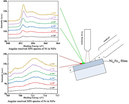
This work demonstrates the dependence between magnetic properties and the thickness of NiFe thin films. More importantly, a quantitative study of the surface composition of NiFe thin film exposed to atmospheric conditions has been carried out employing angle-resolved X-ray photoelectron spectroscopy (ARXPS). In this study, we fabricated Ni81Fe19 (NiFe) thin films on Si (100) substrate using electron beam evaporation and investigated their surface morphologies, magnetic properties, and the thickness of the surface oxide layer. The coexistence of metallic and oxidized species on the surface are suggested by the depth profile of ARXPS spectra. The thickness of the oxidized species, including NiO, Ni(OH)2, Fe2O3, and Fe3O4, are also estimated based on the ARXPS results. This work provides an effective approach to clarify the surface composition, as well as the thickness of the oxide layer of the thin films.

Graphical Abstract

1. Introduction

Microwave magnetic devices, such as circulators, filters, and phase shifters are indispensable components in satellite and mobile communications systems [1,2,3]. Recently, the rapid development in the electronic communication industry has proposed higher requirements for microwave devices including higher operating frequency, lower loss, and higher integration level. As magnetoelectronic devices are the core components of microwave devices [4,5,6], there is an urgent demand to improve their magnetic properties at the high-frequency range. However, most microwave/radio-frequency magnetic components are discrete devices based on bulk materials, and directly reducing the size of high integration often leads to degradation in performance. The progress of chip-type technology is still behind for the perfect integration of magnetic components with existing semiconductor devices. Given the above context, ferromagnetic/anti-ferromagnetic (FM/AF) thin films with a multilayer structure are considered as ideal candidates. The higher saturation magnetization (Ms), permeability (μ), and self-biased ferromagnetic resonance (FMR) frequency of the magnetic thin film make them suitable for high-frequency applications as nanostructured magnetic media and magnetic sensors [7,8,9,10,11,12,13]. Meanwhile, the multilayer structure has been widely used in giant magnetoresistance spin valves based on the exchange bias phenomenon.
The operating frequency, FMR linewidth and the resonance field of periodically arranged FM/AF multilayer structure can easily be affected by the change in thickness of each single layer, as well as the surface morphology. Especially, when the thickness of individual layers falls below the critical thickness, the states and properties of surface and interface dominate the magnetic properties. In the multilayer structure of thin films, the thickness of each single layer can reach down to the level of several nanometers. However, such high accuracy is often susceptible to environmental oxidation, which leads to the change in the effective thickness, as well as the change in composition.
To study the composition of surface and interface as well as the thickness distribution in multilayer structures, atomic force microscopy (AFM) and angle resolved X-ray photoelectron spectroscopy (ARXPS) are commonly applied. Among the previous studies, Yu G.H. et al. [14] studied the relationship between the oxidation states of Ni in NiOx and the exchange bias field Hex of the NiFe film by ARXPS, but the critical thickness of the pinning of NiFe film was not studied. S.S. Sakhonenkov et al. [15] presented an in-depth study of Mo/Si multilayer systems using ARXPS. They reported that a MoSi2 interlayer with a thickness of 0.19 ± 0.05 nm was identified on the Si-on-Mo interface. J. Zemek et al. [16] investigated the surface and in-depth distribution of sp2 and sp3 coordinated carbon atoms in modified diamond-like carbon films. A. Sanchez-Martinez et al. [17] discovered that small amounts of oxidized gallium and metallic arsenic are located at the HfO2/InGaAs interface. Meanwhile, they studied the structure of TiN/HfO2 nanofilms grown on InGaAs substrates by ARXPS. The above evidences have shown ARXPS as a useful tool in characterizing the surface/interface of the thin films. In this study, AFM and ARXPS are used to characterize the surface oxidation layers of the NiFe thin film, which is known for its high permeability while having a relatively high Ms. More importantly, the thickness of each oxidation layer is estimated based on the ARXPS results. The static magnetic properties of the NiFe thin films were also investigated.

2. Experimental Details

In this experiment, Ni81Fe19 thin films were deposited on 5 mm × 5 mm Si (100) substrates by electron beam evaporation (EB-500) in vacuum (≤5.0 × 10⁻⁵ Pa) at 25 °C. The thicknesses of the Ni81Fe19 films are 90, 100, 110, and 120 nm. The growth rate of the film is 0.03 nm/s. The Si (100) substrates were preliminarily cleaned in a sequential bath of acetone, alcohol, and deionized water and dried with ionized dry N2 flux. Once prepared, the samples were stored at room temperature to be oxidized under ambient environment.
The surface morphologies of the NiFe thin films were investigated using an atomic force microscope (AFM, Bruker MultiMode8) with a scanned area of 2 μm × 2 μm. Static magnetic properties of NiFe films were measured using a vibrating sample magnetometer (VSM, Lake Shore 8604) at room temperature. The samples were analyzed using ARXPS (ULVAC-PHI5000 Veraprobe III) with an Al Kα emission source (1486.6 eV). The XPS measurements were conducted at a base pressure of 8 × 10⁻¹⁰ Pa. The pass energy of the spectrometer was set to 69 eV. The energy was calibrated by setting the adventitious C 1s binding energy to 284.8 eV. The chemical depth profile was acquired at six angles (α) of 20°, 35°, 45°, 60°, 75°, and 90°.

3. Results and Discussion

3.1. AFM Analysis and Surface Roughness

Figure 1 illustrates the AFM surface morphology and roughness of the NiFe thin films with a NiFe thicknesses of 100 nm. The surface roughness of the films with different NiFe thicknesses from 90 to 120 nm was 1.31 nm, 1.27 nm, 1.45nm, and 1.67 nm, respectively. The minimum roughness of 1.27 nm is seen for the sample with a thickness of 100 nm, whose average grain size also appears to be the smallest among the samples (see Figure 3a). The experimental results are in good correspondence with a previous report [18], which shows the positive correlation between the average grain size and surface roughness.

3.2. Magnetic Properties of NiFe Films

Figure 2a,b shows the in-plane and out-of-plane magnetic hysteresis loops of NiFe thin films with the thicknesses from 90 to 120 nm. The samples with thickness of 90–110 nm show narrow M-H loops, while the 120-nm-thick sample shows a much wider curve, suggesting a higher coercivity (Hc) [19]. Figure 3 shows the static magnetic properties of the NiFe thin films, including the saturation magnetization (4πMs) and Hc. According to Figure 3a, as the thickness of NiFe thin film increases, the 4πMs is increased. Concomitantly, in-plane (Hc//) and out-of-plane (Hc) coercivity are displayed in Figure 3b. Both Hc// and Hc exhibit a similar trend, which shows a slight reduction upon the increase of thickness from 90 to 110 nm, followed by a dramatic increase as the thickness is further increased to 120 nm. As the Hc is dependent on the quality and defects of the film [20], the dramatic increase of Hc observed in Figure 3b can be attributed to the high roughness of the 120 nm thick sample.

3.3. Oxidation Thickness of NiFe Films

Figure 4 shows the ARXPS spectra of Ni in the NiFe thin film with a thickness of 100 nm from various take-off angles. For clear identification of each peak, as well as to obtain the peak intensity, fitted results for each spectrum are obtained (examples of α = 20° and 90° are shown in Figure 5). The relationship between the detection depth (d), photoelectron take-off grazing angle, and the mean free path of inelastic scattering (λ), is as follows [21].
d = 3 λ sin α
As can be seen in Figure 5, there are several noticeable peaks representing the signals from Ni 2p3/2 (852.3 eV), NiO 2p3/2 (853.3 eV), Ni(OH)2 2p3/2 (856.6 eV), as well as their satellite peaks [22,23,24]. The coexistence of metallic and oxide components is further confirmed by the spectra from O 1s photoemission, which is shown in Figure 6a. The thickness of the oxide film (do) can be calculated according to the ARXPS results through the following equations [25]:
ln ( 1 + R R ) = d o λ 1 sin α
where α is different take-off angles: 20°, 35°, 45°, 60°, 75°, and 90°, and R is the ratio of photoelectron peak intensity from the oxides (Io) to that from nickel (Is):
R = I o I s
In Equation (2), R can be calculated as follows:
R = M o M s ρ s ρ o λ s λ o
where the values of M are the molecular weight of nickel oxides (Mo) and nickel (Ms), values of ρ are the density of nickel oxides (ρo) and nickel (ρs), and values of λ are the mean free path of inelastic scattering for nickel oxides (λo) and nickel (λs). Because the photoelectron kinetic energy of nickel and its oxides are almost the same, thus λo and λs are approximately equal [21]. Table 1 shows the values of density, molecular weight for Ni, NiO, and Ni(OH)2, as well as the calculated R for both nickel oxides.
Table 2 shows the relative peak intensities of nickel (Is) and nickel oxides (Io) at different take-off angles based on the fitting results shown in Figure 5, as well as the calculated ratios between them (R). As α is increased from 20 to 90°, Is(Ni) is seen to increase from ~20 to ~43%, while both Io(NiO) and Io(Ni(OH)2) are reduced from ~46 to ~32% and from ~33 to ~26%, respectively. As the oxidation reactions take place at the top surface of the metal substrate, with a limited traveling distance of photoemission signal, it is natural that the detection depth becomes deeper as the take-off angle becomes closer to 90°. Therefore, one can observe the obvious enhancement in Is(Ni), and slight reduction in Io(NiO) and Io(Ni(OH)2).
According to Equation (2), to calculate the thickness of the oxidation layer, one can plot the results of ln(1 + R/R) against 1/sinα, the slope of which indicates the thickness divided by the mean free path of inelastic scattering (do/λ). Figure 6b shows the results of ln(1 + R/R) against 1/sinα for Ni(OH)2 and NiO. Given that λ = 6 Å [26], the thickness of NiO in NiFe thin film is determined to be 0.2 nm, the thickness of Ni(OH)2 is determined to be 0.3 nm.
As with nickel oxides, the composition and thickness of iron oxides can also be determined. Figure 7 shows the Fe 2p photoemission spectra of partially oxidized NiFe thin film. The signals from each take-off angle can be fitted with Fe 2p3/2 (706.8 eV), Fe2O3 2p3/2 (710.7 eV), and Fe3O4 2p3/2 (709.3 eV) [27,28,29], the results of which are shown in Figure 8. Figure 9a shows the fitted O 1s spectra to further confirm the existence of iron oxides. Table 3 shows the relevant parameters including Is(Fe), Io(Fe2O3), Io(Fe3O4), and the intensity ratios between iron and its oxides. For the same reason mentioned above, Is(Fe) is increased with increasing α, as higher take-off angle results in deeper detection depth.
Table 4 shows the values of density, molecular weight for Fe, Fe3O4, and Fe2O3, as well as the calculated R for both iron oxides. The values in Table 3 and Table 4 are integrated into Equations (2)–(4), and the results of ln(1 + R/R) against 1/sinα for Fe 2p photoemission in the NiFe thin film are shown in Figure 9b. As λ = 7.5 Å [26], the thickness of Fe2O3 layer determined is 0.9 nm and that of Fe3O4 is 0.7 nm. It means that oxidation in the natural environment occurs only in the extreme depth of the surface. To summarize, a schematic illustration is shown in Figure 10 to better demonstrate the multilayer structure of the oxidized NiFe.

4. Conclusions

In summary, NiFe thin films with thickness from 90 to 120 nm were fabricated by electron beam evaporation. The sample with a thickness of 100 nm shows the lowest surface roughness and the smallest grain size. The saturation magnetization of the films is increased from 9891 to 10,300 Gs as the thickness of film increases. The optimum magnetic properties (4πMs = 9930 Gs, Hc = 1.37 Oe) are obtained when the thickness of film is 100 nm. In addition, the coexistence of NiFe and its oxides on the top surface is observed along the probing depth of ARXPS. By fitting the ARXPS spectra and obtaining the relative intensity of each peak, the thickness of NiO, Ni(OH)2, Fe2O3, and Fe3O4, which are 0.3 nm, 0.2 nm, 0.9 nm, and 0.7 nm, respectively, are determined. This work demonstrates the beneficial application of ARXPS in characterizing the oxide layers including their compositions and thicknesses as a quick and damage-free approach, especially for future investigations with even lower film thickness.

Author Contributions

Conceptualization, K.S.; funding acquisition, K.S.; investigation, Z.H., X.S. and X.J.; methodology, Z.H.; software, Y.L. and Y.W.; supervision, C.W., Z.Y., Z.L. (Zhongwen Lan) and K.S.; writing—original draft, Z.H.; writing—review and editing, Z.H. and Z.L. (Ziyu Li). All authors have read and agreed to the published version of the manuscript.

Funding

This present work was financially supported by the National Natural Science Foundation of China under grant No. 52172267 and 51772046.

Institutional Review Board Statement

Not applicable.

Informed Consent Statement

Not applicable.

Data Availability Statement

Not applicable.

Conflicts of Interest

The authors declare that they have no known competing financial interests or personal relationships that could have appeared to influence the work reported in this paper.

References

  1. Su, Z.; Bennett, S.; Hu, B.; Chen, Y.; Harris, V.G. Magnetic and microwave properties of U-type hexaferrite films with high remanence and low ferromagnetic resonance linewidth. J. Appl. Phys. 2014, 115, 17A504. [Google Scholar] [CrossRef]
  2. Belyaev, B.A.; Lemberg, K.V.; Serzhantov, A.M.; Leksikov, A.A.; Bal’va, Y.F. Magnetically tunable resonant phase shifters for UHF band. IEEE Trans. Magn. 2015, 51, 1–5. [Google Scholar] [CrossRef]
  3. Sharma, V.; Khivintsev, Y.; Harward, I.; Kuanr, B.K.; Celinski, Z. Fabrication and characterization of microwave phase shifter in microstrip geometry with Fe film as the frequency tuning element. J. Magn. Magn. Mater. 2019, 489, 165412. [Google Scholar] [CrossRef]
  4. Kuanr, B.K.; Veerakumar, V.; Camley, R.E.; Celinski, Z. Permalloy (NiFe) nanometer square-antidot arrays: Dynamic modes and use as a monolithic microwave band-pass filter. J. Magn. Magn. Mater. 2019, 484, 272–278. [Google Scholar] [CrossRef]
  5. Lu, Q.; Yu, B.; Hu, Z.; He, Y.; Hu, T.; Zhao, Y.; Wang, Z.; Zhou, Z.; Cui, W.; Liu, M. Surface roughness evolution induced low secondary electron yield in carbon coated Ag/Al substrates for space microwave devices. Appl. Surf. Sci. 2020, 501, 144236. [Google Scholar] [CrossRef]
  6. Paiva, D.V.M.; Silva, M.A.S.; Sombra, A.S.B.; Fechine, P.B.A. Properties of the Sr3MoO6 electroceramic for RF/microwave devices. J. Alloys Compd. 2018, 748, 766–773. [Google Scholar] [CrossRef]
  7. Chen, Y.; Fan, X.; Zhou, Y.; Xie, Y.; Wu, J.; Wang, T.; Chui, S.T.; Xiao, J.Q. Designing and tuning magnetic resonance with exchange interaction. Adv. Mater. 2015, 27, 1351–1355. [Google Scholar] [CrossRef]
  8. Belmeguenai, M.; Martin, T.; Woltersdorf, G.; Bayreuther, G.; Baltz, V. Microwave spectroscopy with vector network analyzer for interlayer exchange-coupled symmetrical and asymmetrical NiFe/Ru/NiFe. J. Phys. Condens. Matter. 2008, 20, 345206. [Google Scholar] [CrossRef] [Green Version]
  9. Li, S.; Li, Q.; Xu, J.; Yan, S.; Miao, G.X.; Kang, S.; Dai, Y. Tunable optical mode ferromagnetic resonance in FeCoB/Ru/FeCoB synthetic antiferromagnetic trilayers under uniaxial magnetic anisotropy. Adv. Funct. Mater. 2016, 26, 3738–3744. [Google Scholar] [CrossRef]
  10. Xu, F.; Liao, Z.; Huang, Q.; Phuoc, N.N.; Ong, C.K.; Li, S. Influence of thickness on magnetic properties and microwave characteristics of NiFe/IrMn/NiFe Trilayers. IEEE Trans. Magn. 2011, 47, 3486–3489. [Google Scholar] [CrossRef]
  11. Naik, R.; Kota, C.; Payson, J.S.; Dunifer, G.L. Ferromagnetic-resonance studies of epitaxial Ni, Co, and Fe films grown on Cu(100)/Si(100). Phys. Rev. B 1993, 48, 1008–1013. [Google Scholar] [CrossRef]
  12. Chappert, C.; Le Dang, K.; Beauvillain, P.; Hurdequint, H.; Renard, D. Ferromagnetic resonance studies of very thin cobalt films on a gold substrate. Phys. Rev. B 1986, 34, 3192–3197. [Google Scholar] [CrossRef]
  13. Biondo, A.; Nascimento, V.P.; Lassri, H.; Passamani, E.C.; Morales, M.A.; Mello, A. Structural and magnetic properties of Ni81Fe19/Zr multilayers. J. Magn. Magn. Mater. 2004, 277, 144–152. [Google Scholar] [CrossRef]
  14. Yu, G.; Zhao, H.; Teng, J.; Chai, C.; Zhu, F.; Xia, Y.; Chai, X. XPS Studies of magnetic multilayers. J. Univ. Sci. Technol. B 2001, 8, 210–213. [Google Scholar]
  15. Sakhonenkov, S.S.; Filatova, E.O.; Gaisin, A.U.; Kasatikov, S.A.; Konashuk, A.S. Angle resolved photoelectron spectroscopy as applied to X-ray mirrors: An in-depth study of Mo/Si multilayer systems. Phys. Chem. Chem. Phys. 2019, 21, 25002–250109. [Google Scholar] [CrossRef]
  16. Zemek, J.; Houdkova, J.; Jiricek, P.; Jelinek, M. Surface and in-depth distribution of sp2 and sp3 coordinated carbon atoms in diamond-like carbon films modified by argon ion beam bombardment during growth. Carbon 2018, 134, 71–79. [Google Scholar] [CrossRef]
  17. Sanchez-Martinez, A.; Ceballos-Sanchez, O.; Vazquez-Lepe, M.O.; Duong, T.; Arroyave, R. Diffusion of In and Ga in TiN/HfO2/InGaAs nanofilms. J. Appl. Phys. 2013, 114, 143504. [Google Scholar] [CrossRef]
  18. Dong, B.Z.; Fang, G.J.; Wang, J.F.; Guan, W.J.; Zhao, X.Z. Effect of thickness on structural, electrical, and optical properties of ZnO: Al films deposited by pulsed laser deposition. J. Appl. Phys. 2007, 101, 033713. [Google Scholar] [CrossRef]
  19. Akhter, M.A.; Mapps, D.J.; Ma Tan, Y.Q.; Petford-Long, A.; Doole, R. Thickness and grain-size dependence of the coercivity in permalloy thin films. J. Appl. Phys. 1997, 81, 4122–4124. [Google Scholar] [CrossRef]
  20. Berzins, A.; Smits, J.; Petruhins, A. Characterization of Microscopic ferromagnetic defects in thin films using magnetic microscope based on nitrogen-vacancy centres. Mater. Chem. Phys. 2021, 267, 124617. [Google Scholar] [CrossRef]
  21. John, F.W.; John, W. An Introduction to Surface Analysis by XPS and AES, 1st ed.; East China University of Technology Publisher: Shanghai, China, 2008. [Google Scholar]
  22. Nagarkar, P.V.; Kulkarni, S.K.; Umbach, E. Surface oxidation investigation of Ni36Fe32Cr14P12B6 glass using angle resolved XPS. Appl. Surf. Sci. 1987, 29, 194–222. [Google Scholar] [CrossRef]
  23. Biesinger, M.C.; Payne, B.P.; Grosvenor, A.P.; Lau, L.W.M.; Gerson, A.R.; Smart, R.S.C. Resolving surface chemical states in XPS analysis of first row transition metals, oxides and hydroxides: Cr, Mn, Fe, Co and Ni. Appl. Surf. Sci. 2011, 257, 2717–2730. [Google Scholar] [CrossRef]
  24. Dupin, J.C.; Gonbeau, D.; Vinatier, P.; Levasseur, A. Systematic XPS studies of metal oxides, hydroxides and peroxides. Phys. Chem. Chem. Phys. 2000, 2, 1319–1324. [Google Scholar] [CrossRef]
  25. Wang, J.Q. Introduction to Electron Spectroscopy (XPS/XAES/UPS); National Defense Industry Publisher: Beijing, China, 1992. [Google Scholar]
  26. Cao, Q.X.; Lei, T.M.; Huang, Y.X.; Li, G.F. Fundamentals of Solid Physics; Xidian University Publisher: Xi’an, China, 2008. [Google Scholar]
  27. Yamashita, T.; Hayes, P. Analysis of XPS spectra of Fe2+ and Fe3+ ions in oxide materials. Appl. Surf. Sci. 2008, 254, 2441–2449. [Google Scholar] [CrossRef]
  28. Grosvenor, A.P.; Kobe, B.A.; Biesinger, M.C.; McIntyre, N.S. Investigation of multiplet splitting of Fe 2p XPS spectra and bonding in iron compounds. Surf. Interface Anal. 2004, 36, 1564–1574. [Google Scholar] [CrossRef]
  29. Carver, J.C.; Schweitzer, G.K.; Carlson, T.A. Use of X-ray photoelectron spectroscopy to study bonding in Cr, Mn, Fe, and Co compounds. J. Chem. Phys. 1972, 57, 973–982. [Google Scholar] [CrossRef]
Figure 1. AFM images of NiFe films with a thickness of 100 nm.
Metals 11 02061 g001
Figure 2. The magnetic hysteresis (M-H) loops of NiFe thin films with thickness from 90 to 120 nm: (a) the in-plane magnetic hysteresis loops, (b) the out-of-plane magnetic hysteresis loop.
Metals 11 02061 g002
Figure 3. The static magnetic properties of the Ni81Fe19 thin films with thicknesses from 90 to 120 nm: (a) the saturation magnetization (4πMs) and average grain size (D), (b) the in-plane (Hc//) and out-of-plane (Hc) coercivity.
Metals 11 02061 g003
Figure 4. Spectra of Ni 2p photoemission in the NiFe thin film with a thickness of 100 nm at different take-off angles (α).
Metals 11 02061 g004
Figure 5. ARXPS spectra of Ni 2p photoemission and the fitting results for NiFe thin film with a thickness of 100 nm at different take-off angles: (a) 20° and (b) 90°.
Metals 11 02061 g005
Figure 6. (a) ARXPS spectra of O 1s photoemission and the fitting results, (b) the results of ln(1 + R/R) against 1/sinα for the nickel oxides in NiFe thin film.
Metals 11 02061 g006
Figure 7. Spectra at different take-off angle of Fe 2p in NiFe(100 nm) thin film.
Metals 11 02061 g007
Figure 8. ARXPS spectra of Fe 2p photoemission and the peak fitting results for NiFe thin film with a thickness of 100 nm at different take-off angles of: (a) 20° and (b) 90°.
Metals 11 02061 g008
Figure 9. (a) ARXPS spectra of O 1s photoemission and the fitting results, (b) the results of ln(1 + R/R∞) against 1/sinα for the iron oxides in NiFe thin film.
Metals 11 02061 g009
Figure 10. The schematic diagram of NiFe thin film: (a) unoxidized, (b) oxidized.
Metals 11 02061 g010
Table 1. The values of density, molecular weight, and kinetic energy for Ni, NiO, and Ni(OH)2, as well as R for both nickel oxides.
Materialρ (g/cm3)M (g/mol)RKinetic Energy (eV)
Ni8.959-853.3
NiO6.84750.6853.6
Ni(OH)24.15930.3855.0
Table 2. Relative peak intensities (Is and Io) and the Io/Is ratios for both nickel oxides in the NiFe (100 nm) thin film.
α (°)Is(Ni) (%)Io(NiO) (%)Io(Ni(OH)2) (%)R(NiO) = Io(NiO)/IsR(Ni(OH)2) = Io/Is
2020.3846.1833.442.271.64
3532.5037.9529.551.170.91
4536.7335.3727.900.960.76
6040.3233.2726.410.830.66
7542.0532.2925.660.770.61
9042.5732.0025.440.750.60
Table 3. Relative peak intensities (Is and Io) and the Io/Is ratios for both iron oxides in the NiFe (100 nm) thin film.
α (°)Is(Fe) (%)Io(Fe3O4) (%)Io(Fe2O3) (%)R(Fe3O4) = Io(Fe3O4)/IsR(Fe2O3) = Io(Fe2O3)/Is
202.117.8590.043.7242.67
358.598.0183.410.939.71
4512.497.9279.580.636.37
6016.637.7575.610.474.55
7518.917.6373.460.403.88
9019.637.5972.780.393.71
Table 4. The values of density, molecular weight, and kinetic energy for Fe, Fe2O3, and Fe3O4, as well as R for both iron oxides.
Materialρ (g/cm3)M (g/mol)RKinetic Energy (eV)
Fe7.8656-710.7
Fe2O35.241600.23710.4
Fe3O45.182320.16710.8
Publisher’s Note: MDPI stays neutral with regard to jurisdictional claims in published maps and institutional affiliations.