Next Issue
Volume 10, October
Previous Issue
Volume 10, August


Pathogens, Volume 10, Issue 9 (September 2021) – 154 articles

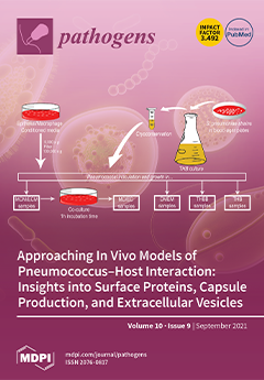
Cover Story (view full-size image): The cell surface of pathogenic organisms plays a critical role in the interaction with the host. The bacterial surface houses numerous molecules that are key in this interplay—e.g., proteins—as well as other structures, such as the capsule or extracellular vesicles that pass through it. Surface molecules and structures change according to the way in which the pathogen interacts with the host, being critical for developing infections. Moreover, knowing the actual composition of the surface in a given situation may be fundamental for finding treatments against infections. In this work, the authors have emulated experimental conditions that approach closer bacterial–host cell interactions, in order to gain insights into changes in surface proteins, capsule, and extracellular vesicles that may affect the infection process. View this paper.
  • Issues are regarded as officially published after their release is announced to the table of contents alert mailing list.
  • You may sign up for e-mail alerts to receive table of contents of newly released issues.
  • PDF is the official format for papers published in both, html and pdf forms. To view the papers in pdf format, click on the "PDF Full-text" link, and use the free Adobe Reader to open them.
Order results
Result details
Section
Select all
Export citation of selected articles as:
15 pages, 2765 KiB  
Article
Covid-19 Infection in India: A Comparative Analysis of the Second Wave with the First Wave
by Arnab Sarkar, Alok Kumar Chakrabarti and Shanta Dutta
Pathogens 2021, 10(9), 1222; https://doi.org/10.3390/pathogens10091222 - 21 Sep 2021
Cited by 65 | Viewed by 8206
Abstract
Coronavirus disease 2019 (COVID-19) is considered as the most dreaded disease that has spread all over the world in the recent past. Despite its outbreak in December 2019–January 2020, a few continents and countries such as India started to experience a significant number [...] Read more.
Coronavirus disease 2019 (COVID-19) is considered as the most dreaded disease that has spread all over the world in the recent past. Despite its outbreak in December 2019–January 2020, a few continents and countries such as India started to experience a significant number of COVID-19-positive cases from March 2020. GISAID clade variation analysis in the period March 2020–February 2021 (period I) and March 2021–first week of April 2021 (period II) showed a rapid variation of SARS-CoV-2 in all continents and India over time. Studying the relationship of patient age or gender with viral clades in these two periods revealed that the population under 10 years of age was the least affected, whereas the 11–60-year-old population was the most affected, irrespective of patient gender and ethnicity. In the first wave, India registered quite a low number of COVID-19-positive cases/million people, but the scenario unexpectedly changed in the second wave, when even over 400,000 confirmed cases/day were reported. Lineage analysis in India showed the emergence of new SARS-CoV-2 variants, i.e., B.1.617.1 and B.1.617.2, during April–May 2021, which might be one of the key reasons for the sudden upsurge of confirmed cases/day. Furthermore, the emergence of the new variants contributed to the shift in infection spread by the G clade of SARS-CoV-2 from 46% in period II to 82.34% by the end of May 2021. Along with the management of the emergence of new variants, few factors viz., lockdown and vaccination were also accountable for controlling the upsurge of new COVID-19 cases throughout the country. Collectively, a comparative analysis of the scenario of the first wave with that of the second wave would suggest policymakers the way to prepare for better management of COVID-19 recurrence or its severity in India and other countries. Full article
(This article belongs to the Section Emerging Pathogens)
Show Figures

Figure 1

17 pages, 2131 KiB  
Article
Prevalence of Vector-Borne Pathogens in Reproductive and Non-Reproductive Tissue Samples from Free-Roaming Domestic Cats in the South Atlantic USA
by Charlotte Manvell, Kelli Ferris, Ricardo Maggi, Edward B. Breitschwerdt and Erin Lashnits
Pathogens 2021, 10(9), 1221; https://doi.org/10.3390/pathogens10091221 - 21 Sep 2021
Cited by 10 | Viewed by 2974
Abstract
Reservoir to multiple species of zoonotic pathogens, free-roaming cats (FRCs) interact with domestic and wild animals, vectors, and humans. To assess the potential for feline vector-borne pathogens to be vertically transmitted, this study surveyed ear tip and reproductive tissues of FRCs from two [...] Read more.
Reservoir to multiple species of zoonotic pathogens, free-roaming cats (FRCs) interact with domestic and wild animals, vectors, and humans. To assess the potential for feline vector-borne pathogens to be vertically transmitted, this study surveyed ear tip and reproductive tissues of FRCs from two locations in the South Atlantic United States for Anaplasma, Bartonella, Ehrlichia, hemotropic Mycoplasma, and Rickettsia species. We collected ovary (n = 72), uterus (n = 54), testicle (n = 74), and ear tip (n = 73) tissue from 73 cats, and fetal (n = 20) and placental (n = 19) tissue from 11 queens. Pathogen DNA was amplified utilizing qPCR, confirmed by sequencing. Cats were more frequently Bartonella henselae positive on reproductive tissues (19%, 14/73) than ear tip (5%, 4/73; p = 0.02). B. henselae was amplified from fetus (20%, 4/20) and placenta samples (11%, 2/19). Bartonella spp. infection was more common in cats from North Carolina (76%, 26/34) than Virginia (13%, 5/39; p < 0.0001). Fourteen percent (10/73) of both ear tip and reproductive tissues were positive for hemotropic Mycoplasma spp. Anaplasma, Ehrlichia, and Rickettsia spp. DNA was not amplified from any cat/tissue. These findings suggest that B. henselae preferentially infected cats’ reproductive tissue and reinforces the importance of investigating the potential for B. henselae vertical transmission or induction of reproductive failure. Full article
(This article belongs to the Special Issue The Evolving Biomedical Importance of Bartonella Species Infections)
Show Figures

Figure 1

9 pages, 1827 KiB  
Hypothesis
The Acari Hypothesis, II: Interspecies Operability of Pattern Recognition Receptors
by Andrew C. Retzinger and Gregory S. Retzinger
Pathogens 2021, 10(9), 1220; https://doi.org/10.3390/pathogens10091220 - 21 Sep 2021
Cited by 3 | Viewed by 3783
Abstract
Hypersensitivity to galactose-α-1,3-galactose (α-gal) is an informative example of a pathologic IgE-mediated process. By way of their saliva, ticks are able to sensitize humans to tick dietary elements that express α-gal. Mites, which along with ticks constitute the phyletic subclass Acari, feed on [...] Read more.
Hypersensitivity to galactose-α-1,3-galactose (α-gal) is an informative example of a pathologic IgE-mediated process. By way of their saliva, ticks are able to sensitize humans to tick dietary elements that express α-gal. Mites, which along with ticks constitute the phyletic subclass Acari, feed on proteinaceous foodstuffs that represent most, if not all, human allergens. Given: (1) the gross nature of the pathophysiological reactions of allergy, especially anaphylaxis, (2) the allergenicity of acarian foodstuffs, and (3) the relatedness of ticks and mites, it has been hypothesized that human-acarian interactions are cardinal to the pathogenesis of allergy. In this report, a means by which such interactions contribute to that pathogenesis is proposed. Full article
(This article belongs to the Section Immunological Responses and Immune Defense Mechanisms)
Show Figures

Figure 1

15 pages, 1688 KiB  
Article
African Swine Fever in Wild Boar (Poland 2020): Passive and Active Surveillance Analysis and Further Perspectives
by Maciej Piotr Frant, Anna Gal-Cisoń, Łukasz Bocian, Anna Ziętek-Barszcz, Krzysztof Niemczuk, Grzegorz Woźniakowski and Anna Szczotka-Bochniarz
Pathogens 2021, 10(9), 1219; https://doi.org/10.3390/pathogens10091219 - 19 Sep 2021
Cited by 13 | Viewed by 3463
Abstract
African swine fever (ASF) is a fatal hemorrhagic disease of wild boar and domestic pigs which has been present in Poland since 2014. By 2020, the ASF virus (ASFV) spread across Central, Eastern and Western Europe (including Germany), and Asian countries (including China, [...] Read more.
African swine fever (ASF) is a fatal hemorrhagic disease of wild boar and domestic pigs which has been present in Poland since 2014. By 2020, the ASF virus (ASFV) spread across Central, Eastern and Western Europe (including Germany), and Asian countries (including China, Vietnam, and South Korea). The national ASF eradication and prevention program includes continuous passive (wild boar found dead and road-killed wild boar) and active (hunted wild boar) surveillance. The main goal of this study was to analyze the dynamic of the spread of ASF in the wild boar population across the territory of Poland in 2020. In that year in Poland, in total 6191 ASF-positive wild boar were declared. Most of them were confirmed in a group of animals found dead. The conducted statistical analysis indicates that the highest chance of obtaining an ASF-positive result in wild boar was during the winter months, from January to March, and in December 2020. Despite the biosecurity measures implemented by holdings of domestic pigs, the disease also occurred in 109 pig farms. The role of ASF surveillance in the wild boar population is crucial to apply more effective and tailored measures of disease control and eradication. The most essential measures to maintain sustainable production of domestic pigs in Poland include effective management of the wild boar population, along with strict implementation of biosecurity measures by domestic pig producers. Full article
(This article belongs to the Special Issue Swine Viral Diseases)
Show Figures

Figure 1

27 pages, 2255 KiB  
Review
Biology and Pathogenesis of SARS-CoV-2: Understandings for Therapeutic Developments against COVID-19
by Homa Nath Sharma, Charity O. D. Latimore and Qiana L. Matthews
Pathogens 2021, 10(9), 1218; https://doi.org/10.3390/pathogens10091218 - 19 Sep 2021
Cited by 5 | Viewed by 9221
Abstract
Coronaviruses are positive sense, single-stranded, enveloped, and non-segmented RNA viruses that belong to the Coronaviridae family within the order Nidovirales and suborder Coronavirinae. Two Alphacoronavirus strains: HCoV-229E and HCoV-NL63 and five Betacoronaviruses: HCoV-HKU1, HCoV-OC43, SARS-CoV, MERS-CoV, and SARS-CoV-2 have so far been recognized [...] Read more.
Coronaviruses are positive sense, single-stranded, enveloped, and non-segmented RNA viruses that belong to the Coronaviridae family within the order Nidovirales and suborder Coronavirinae. Two Alphacoronavirus strains: HCoV-229E and HCoV-NL63 and five Betacoronaviruses: HCoV-HKU1, HCoV-OC43, SARS-CoV, MERS-CoV, and SARS-CoV-2 have so far been recognized as Human Coronaviruses (HCoVs). Coronavirus disease 2019 (COVID-19) caused by SARS-CoV-2 is currently the greatest concern for humanity. Despite the overflow of research on SARS-CoV-2 and other HCoVs published every week, existing knowledge in this area is insufficient for the complete understanding of the viruses and the diseases caused by them. This review is based on the analysis of 210 published works, and it attempts to cover the basic biology of coronaviruses, including the genetic characteristics, life cycle, and host-pathogen interaction, pathogenesis, the antiviral drugs, and vaccines against HCoVs, especially focusing on SARS-CoV-2. Furthermore, we will briefly discuss the potential link between extracellular vesicles (EVs) and SARS-CoV-2/COVID-19 pathophysiology. Full article
(This article belongs to the Special Issue Pathogenesis of Human Coronavirus Infections)
Show Figures

Figure 1

13 pages, 1543 KiB  
Article
A Cranberry Concentrate Decreases Adhesion and Invasion of Escherichia coli (AIEC) LF82 In Vitro
by Derek Zhang, Lynn Verstrepen, Jelle De Medts, Cindy Duysburgh, Pieter Van den Abbeele, Massimo Marzorati and Christina Khoo
Pathogens 2021, 10(9), 1217; https://doi.org/10.3390/pathogens10091217 - 18 Sep 2021
Cited by 3 | Viewed by 3079
Abstract
While many beneficial host–microbiota interactions have been described, imbalanced microbiota in the gut is speculated to contribute to the progression and recurrence of chronic inflammatory diseases such as Crohn’s disease (CD). This in vitro study evaluated the impact of a cranberry concentrate Type [...] Read more.
While many beneficial host–microbiota interactions have been described, imbalanced microbiota in the gut is speculated to contribute to the progression and recurrence of chronic inflammatory diseases such as Crohn’s disease (CD). This in vitro study evaluated the impact of a cranberry concentrate Type M (CTM) on adherent-invasive Escherichia coli (AIEC) LF82, a pathobiont associated with CD. Different stages of pathogenic infection were investigated: (i) colonization of the mucus layer, and (ii) adhesion to and (iii) invasion of the epithelial cells. Following 48 h of fecal batch incubation, 0.5 and 1 mM of CTM significantly altered AIEC LF82 levels in a simulated mucus layer, resulting in a decrease of 50.5% in the untreated blank, down to 43.0% and 11.4%, respectively. At 1 mM of CTM, the significant decrease in the levels of AIEC LF82 coincided with a stimulation of the metabolic activity of the background microbiota. The increased levels of health-associated acetate (+7.9 mM) and propionate levels (+3.5 mM) suggested selective utilization of CTM by host microorganisms. Furthermore, 1 mM of both fermented and unfermented CTM decreased the adhesion and invasion of human-derived epithelial Caco-2 cells by AIEC LF82. Altogether, this exploratory in vitro study demonstrates the prebiotic potential of CTM and supports its antipathogenic effects through direct and/or indirect modulation of the gut microbiome. Full article
(This article belongs to the Collection Current Status of Research on Gut Metabolites and Microbiota)
Show Figures

Figure 1

17 pages, 3632 KiB  
Article
First Characterisation of the Phoma Species Complex on Maize Leaves in Central Europe
by Lucia Ramos Romero, Dagmar Tacke, Birger Koopmann and Andreas von Tiedemann
Pathogens 2021, 10(9), 1216; https://doi.org/10.3390/pathogens10091216 - 18 Sep 2021
Cited by 2 | Viewed by 3937
Abstract
In the last decade, the cultivated area of maize has increased in Central Europe due to its high yield potential and diverse uses for feed and bio-energy. This has led to more intense maize cultivation, with narrowed crop rotations resulting in the increase [...] Read more.
In the last decade, the cultivated area of maize has increased in Central Europe due to its high yield potential and diverse uses for feed and bio-energy. This has led to more intense maize cultivation, with narrowed crop rotations resulting in the increase in maize leaf diseases. During 2012 and 2013, an inventory of maize leaf spot diseases was carried out in various regions in Central Europe. In addition to the major leaf pathogens, isolates of Phoma-like species were obtained from oval to elliptical spots on leaves or found in lesions produced by other leaf pathogens. A total of 16 representative Phoma-like strains were characterised for their pathogenicity on maize leaves, for their morphological characteristics and with a phylogenetic analysis based on multilocus sequence analysis using part of the actin (ACT), calmodulin (CAL), β-tubulin (TUB), internal transcribed spacer (ITS) region of ribosomal DNA and large subunit ribosomal RNA (LSU) genes. The strains were grouped into four clades, and morphological studies supported this classification for most of them. Strains were compared with six reference Phoma-like species strains from the Westerndijk Fungal Biodiversity Institute collection reported to colonise maize. The pathogenic group of strains from our collection (after completion of Koch’s postulates) did not cluster with any of these species, indicating a different and novel Phoma-like species infecting maize leaves. To our knowledge, this is the first study dissecting the Phoma species complex on maize leaves in Central Europe. Full article
Show Figures

Figure 1

4 pages, 509 KiB  
Brief Report
Detection and Stability of SARS-CoV-2 Fragments in Wastewater: Impact of Storage Temperature
by Rudolf Markt, Markus Mayr, Evelyn Peer, Andreas O. Wagner, Nina Lackner and Heribert Insam
Pathogens 2021, 10(9), 1215; https://doi.org/10.3390/pathogens10091215 - 18 Sep 2021
Cited by 24 | Viewed by 3109
Abstract
SARS-CoV-2 wastewater epidemiology suffers from uncertainties concerning sample storage. We show the effect of the storage of wastewater on the detectable SARS-CoV-2 load. Storage at 4 °C for up to 9 days had no significant effect, while storage at −20 °C led to [...] Read more.
SARS-CoV-2 wastewater epidemiology suffers from uncertainties concerning sample storage. We show the effect of the storage of wastewater on the detectable SARS-CoV-2 load. Storage at 4 °C for up to 9 days had no significant effect, while storage at −20 °C led to a significant reduction in gene copy numbers. Full article
(This article belongs to the Special Issue SARS-CoV-2 in the Water Environment)
Show Figures

Figure 1

9 pages, 458 KiB  
Article
Cytauxzoon sp. and Hepatozoon spp. in Domestic Cats: A Preliminary Study in North-Eastern Italy
by Marika Grillini, Giulia Simonato, Cinzia Tessarin, Giorgia Dotto, Donato Traversa, Rudi Cassini, Erica Marchiori and Antonio Frangipane di Regalbono
Pathogens 2021, 10(9), 1214; https://doi.org/10.3390/pathogens10091214 - 18 Sep 2021
Cited by 22 | Viewed by 3105
Abstract
Knowledge on the presence of Cytauxzoon sp. and Hepatozoon spp. in Italy is scant and mostly limited to a few areas of Northern and Southern regions, respectively. The present study updated the current epidemiological scenario by investigating the occurrence of these protozoa in [...] Read more.
Knowledge on the presence of Cytauxzoon sp. and Hepatozoon spp. in Italy is scant and mostly limited to a few areas of Northern and Southern regions, respectively. The present study updated the current epidemiological scenario by investigating the occurrence of these protozoa in domestic cats from three broad regions of North-Eastern Italy. Blood samples from cats at risk of vector-borne diseases were processed by PCR to detect Cytauxzoon and Hepatozoon DNA. Blood smears were observed for haemoparasite inclusions. The influence of cat individual data (e.g., provenance, management, indoor/outdoor lifestyle) on the prevalence of haemoprotozoan infections was statistically evaluated. Among 158 cats, Cytauxzoon and Hepatozoon DNA were detected in 6 (3.8%) and 26 (16.5%) animals, respectively. No Hepatozoon gamonts were detected in blood smears, whereas all Cytauxzoon PCR-positive samples were microscopically positive, though with low levels of parasitaemia. Two species of Hepatozoon were identified, Hepatozoon felis (n = 10) and Hepatozoon silvestris (n = 16). Hepatozoon silvestris prevalence values were significantly (p < 0.05) higher in the region Friuli Venezia Giulia and in stray cats. Cytauxzoon sp. was detected in 6/39 (15.4%) stray cats from Friuli Venezia Giulia (Trieste province). These data add new information on the occurrence of these neglected protozoa in domestic cats’ populations. Full article
(This article belongs to the Special Issue Parasites of the Third Millennium)
Show Figures

Figure 1

6 pages, 234 KiB  
Brief Report
Evaluation of the Ridaquick Rotavirus/Adenovirus Immuno-Chromatographic Assay in Real-Life Situation
by Francis Simo-Fouda, Laetitia Ninove, Léa Luciani, Christine Zandotti, Céline Gazin, Remi N. Charrel and Antoine Nougairède
Pathogens 2021, 10(9), 1213; https://doi.org/10.3390/pathogens10091213 - 18 Sep 2021
Cited by 3 | Viewed by 2192
Abstract
Immunochromatographic tests (ICT) are diagnostics tools providing rapid results without the need for specialized equipment. Our aim was to evaluate retrospectively the rotavirus and adenovirus ICT routinely used in the virology laboratory serving the University Hospital of Marseille, France. From January 2017 to [...] Read more.
Immunochromatographic tests (ICT) are diagnostics tools providing rapid results without the need for specialized equipment. Our aim was to evaluate retrospectively the rotavirus and adenovirus ICT routinely used in the virology laboratory serving the University Hospital of Marseille, France. From January 2017 to March 2020, 715 stool specimens from patients were screened using the Ridaquick Rotavirus/Adenovirus Combi ICT (RR/AC ICT) and a commercially available multiplex PCR detection kit. Rotavirus was detected in 9.2% of specimens by PCR and 7.7% of specimens by RR/AC ICT while adenovirus was detected in 8.5% of specimens by PCR and 2.4% of specimens by RR/AC ICT. The RR/AC ICT parameters for rotavirus were 75.8% sensitivity, 99.2% specificity, 90.9% positive predictive value (PPV) and 97.6% negative predictive value (NPV). The RR/AC ICT parameters for adenovirus were 6.6% sensitivity, 98.0% specificity, 23.5% PPV and 91.8% NPV. While the ICT test may be suitable for rotavirus detection, a PCR–based assay is better adapted for adenovirus detection in stools. Full article
(This article belongs to the Section Viral Pathogens)
15 pages, 2722 KiB  
Article
Optimization of Streptococcus agalactiae Biofilm Culture in a Continuous Flow System for Photoinactivation Studies
by Michal K. Pieranski, Michal Rychlowski and Mariusz Grinholc
Pathogens 2021, 10(9), 1212; https://doi.org/10.3390/pathogens10091212 - 18 Sep 2021
Cited by 5 | Viewed by 3086
Abstract
Streptococcus agalactiae is a relevant cause of neonatal mortality. It can be transferred to infants via the vaginal tract and cause meningitis, pneumonia, arthritis, or sepsis, among other diseases. The cause of therapy ineffectiveness and infection recurrence is the growth of bacteria as [...] Read more.
Streptococcus agalactiae is a relevant cause of neonatal mortality. It can be transferred to infants via the vaginal tract and cause meningitis, pneumonia, arthritis, or sepsis, among other diseases. The cause of therapy ineffectiveness and infection recurrence is the growth of bacteria as biofilms. To date, several research teams have attempted to find a suitable medium for the cultivation of S. agalactiae biofilms. Among others, simulated vaginal fluid has been used; however, biofilm production in this medium has been found to be lower than that in tryptic soy broth. We have previously shown that S. agalactiae can be successfully eradicated by photoinactivation in planktonic culture, but there have been no studies on biofilms. The aim of this study was to optimize S. agalactiae biofilm culture conditions to be used in photoinactivation studies. We compared biofilm production by four strains representing the most common serotypes in four different broth media with crystal violet staining. Then, we evaluated stationary biofilm culture in microtiter plates and biofilm growth in a CDC Biofilm Reactor® (BioSurface Technologies, Bozeman, MT, USA) under continuous flow conditions. Subsequently, we applied Rose Bengal-mediated photoinactivation to both biofilm models. We have shown that photoinactivation is efficient in biofilm eradication and is not cyto/phototoxic to human keratinocytes. We found conditions allowing for stable and repetitive S. agalactiae biofilm growth in continuous flow conditions, which can be successfully utilized in photoinactivation assays and potentially in all other antibacterial studies. Full article
(This article belongs to the Special Issue Group B-Streptococcus (GBS))
Show Figures

Figure 1

18 pages, 2184 KiB  
Article
Development and Validation of Novel PCR Assays for the Diagnosis of Bovine Stephanofilariasis and Detection of Stephanofilaria sp. Nematodes in Vector Flies
by Muhammad Noman Naseem, Ali Raza, Rachel Allavena, Michael McGowan, Jess A. T. Morgan, Constantin Constantinoiu, Ala E. Tabor and Peter James
Pathogens 2021, 10(9), 1211; https://doi.org/10.3390/pathogens10091211 - 17 Sep 2021
Cited by 4 | Viewed by 3596
Abstract
Background: Stephanofilaria spp. nematodes are associated with cutaneous lesions in cattle and other livestock and mammalian wildlife species. In Australia, Haematobia irritans exigua, commonly known as buffalo fly (BF) transmits a well-described but presently unnamed species of Stephanofilaria, which has been speculatively [...] Read more.
Background: Stephanofilaria spp. nematodes are associated with cutaneous lesions in cattle and other livestock and mammalian wildlife species. In Australia, Haematobia irritans exigua, commonly known as buffalo fly (BF) transmits a well-described but presently unnamed species of Stephanofilaria, which has been speculatively implicated in the aetiology of BF lesions. The sensitivity of current techniques for detecting Stephanofilaria spp. in skin lesions and vector species is low, and there is no genomic sequence for any member of the genus Stephanofilaria currently available in sequence databases. Methods: To develop molecular assays for the detection of the Australian Stephanofilaria sp., skin biopsies were collected from freshly slaughtered cattle with typical lesions near the medial canthus. Adult nematodes and microfilariae were isolated from the biopsies using a saline recovery technique. The nematodes were morphologically identified as Stephanofilaria sp. by scanning electron microscopy. DNA was extracted and the internal transcribed spacer 2 (ITS2) region of rDNA, and the cytochrome c oxidase subunit 1 (cox1) region of mtDNA was amplified and sequenced. Stephanofilaria sp. specific polymerase chain reaction (PCR) and qPCR assays (SYBR Green® and TaqMan™) were developed and optimised from the novel ITS2 sequence obtained. The specificity of each assay was confirmed by testing against nematode species Onchocerca gibsoni and Dirofilaria immitis, as well as host (bovine) and BF DNA. Results: Scanning electron microscopy of the anterior and posterior ends of isolated nematodes confirmed Stephanofilaria sp. A phylogenetic analysis of the cox1 sequence demonstrated that this species is most closely related to Thelazia callipaeda, a parasitic nematode that is a common cause of thelaziasis (or eyeworm infestation) in humans, dogs, and cats. Both conventional and qPCR assays specifically amplified DNA from Stephanofilaria sp. Conventional PCR, TaqMan™, and SYBR Green® assays were shown to detect 1 ng, 1 pg, and 100 fg of Stephanofilaria DNA, respectively. Both qPCR assays detected DNA from single Stephanofilaria microfilaria. Conclusion: Molecular diagnostic assays developed in this study showed high specificity and sensitivity for Stephanofilaria sp. DNA. The availability of an accurate and sensitive PCR assay for Stephanofilaria will assist in determining its role in the pathogenesis of cattle skin lesions, as well as in understanding its epidemiological dynamics. This assay may also have application for use in epidemiological studies with other species of Stephanofilaria, most particularly closely related S. stilesi, but this will require confirmation. Full article
(This article belongs to the Special Issue Animal Vector-Borne Diseases)
Show Figures

Graphical abstract

20 pages, 38156 KiB  
Article
Very High Fascioliasis Intensities in Schoolchildren from Nile Delta Governorates, Egypt: The Old World Highest Burdens Found in Lowlands
by M. Victoria Periago, M. Adela Valero, Patricio Artigas, Verónica H. Agramunt, M. Dolores Bargues, Filippo Curtale and Santiago Mas-Coma
Pathogens 2021, 10(9), 1210; https://doi.org/10.3390/pathogens10091210 - 17 Sep 2021
Cited by 14 | Viewed by 2438
Abstract
Quantitative coprological analyses of children were performed in Alexandria and Behera governorates, Egypt, to ascertain whether individual intensities in the Nile Delta lowlands reach high levels as those known in hyperendemic highland areas of Latin America. Analyses focused on subjects presenting intensities higher [...] Read more.
Quantitative coprological analyses of children were performed in Alexandria and Behera governorates, Egypt, to ascertain whether individual intensities in the Nile Delta lowlands reach high levels as those known in hyperendemic highland areas of Latin America. Analyses focused on subjects presenting intensities higher than 400 eggs per gram of faeces (epg), the high burden cut-off according to WHO classification. A total of 96 children were found to shed between 408 and 2304 epg, with arithmetic and geometric means of 699.5 and 629.07 epg, respectively. Intensities found are the highest hitherto recorded in Egypt, and also in the whole Old World. A total of 38 (39.6%) were males and 58 (60.4%) were females, with high intensities according to gender following a negative binomial distribution. The high burden distribution shows a peak in the 7–10 year-old children group, more precocious in females than males. Results showed high burdens in winter to be remarkably higher than those known in summer. The fascioliasis scenario in Egyptian lowlands shows similarities to highlands of Bolivia and Peru. Diagnostic methods, pathogenicity and morbidity in high burdens should be considered. The need for an appropriate quantitative assessment of heavy infected children to avoid post-treatment colic episodes is highlighted. Full article
(This article belongs to the Special Issue Epidemiology of Parasitic Zoonoses)
Show Figures

Figure 1

10 pages, 1070 KiB  
Article
Bartonella Infections in Cats and Cat Fleas in Lithuania
by Miglė Razgūnaitė, Indrė Lipatova, Algimantas Paulauskas, Birutė Karvelienė, Vita Riškevičienė and Jana Radzijevskaja
Pathogens 2021, 10(9), 1209; https://doi.org/10.3390/pathogens10091209 - 17 Sep 2021
Cited by 9 | Viewed by 3169
Abstract
Bartonella are vector-borne parasitic bacteria that cause zoonotic infections in humans. One of the most common infections is cat-scratch disease caused by Bartonella henselae and Bartonella clarridgeiae. Cats are the major reservoir for these two species of bacteria, while cat fleas are [...] Read more.
Bartonella are vector-borne parasitic bacteria that cause zoonotic infections in humans. One of the most common infections is cat-scratch disease caused by Bartonella henselae and Bartonella clarridgeiae. Cats are the major reservoir for these two species of bacteria, while cat fleas are vectors for the transmission of infection agents among cats. The aim of the present study was to investigate the presence of Bartonella infections in stray and pet cats and in cat fleas in Lithuania. Blood samples were taken from 163 cats presented in pet clinics and animal shelters. A total of 102 fleas representing two species, Ctenocephalides felis and Ctenocephalides canis, were collected from 12 owned cats that live both outdoors and indoors. Bartonella DNA in samples was detected using a nested PCR targeting the 16S–23S rRNA intergenic spacer (ITS) region. Bartonella DNA was detected in 4.9% (8/163) of the cats and 29.4% (30/102) of the fleas. Sequence analysis of the ITS region showed that the cats and fleas were infected with B. henselae, B. clarridgeiae and Bartonella sp., closely related to B. schoenbuchensis. This study is the first report on the prevalence and molecular characterization of Bartonella spp. in cats and cat fleas in Lithuania. Full article
(This article belongs to the Special Issue The Evolving Biomedical Importance of Bartonella Species Infections)
Show Figures

Figure 1

21 pages, 9439 KiB  
Article
Evaluation of Inhibitory Activity In Silico of In-House Thiomorpholine Compounds between the ACE2 Receptor and S1 Subunit of SARS-CoV-2 Spike
by Victor H. Vázquez-Valadez, Alejandro Hernández-Serda, Ma. Fernanda Jiménez-Cabiedes, Pablo Aguirre-Vidal, Ingrid González-Tapia, Laura Carreño-Vargas, Yoshio A. Alarcón-López, Andrea Espejel-Fuentes, Pablo Martínez-Soriano, Miguel Lugo Álvarez, Ana María Velázquez-Sánchez, Nathan Marko Markarian, Enrique Angeles and Levon Abrahamyan
Pathogens 2021, 10(9), 1208; https://doi.org/10.3390/pathogens10091208 - 17 Sep 2021
Cited by 1 | Viewed by 4442
Abstract
At the end of 2019, the world was struck by the COVID-19 pandemic, which resulted in dire repercussions of unimaginable proportions. From the beginning, the international scientific community employed several strategies to tackle the spread of this disease. Most notably, these consisted of [...] Read more.
At the end of 2019, the world was struck by the COVID-19 pandemic, which resulted in dire repercussions of unimaginable proportions. From the beginning, the international scientific community employed several strategies to tackle the spread of this disease. Most notably, these consisted of the development of a COVID-19 vaccine and the discovery of antiviral agents through the repositioning of already known drugs with methods such as de novo design. Previously, methylthiomorphic compounds, designed by our group as antihypertensive agents, have been shown to display an affinity with the ACE2 (angiotensin converting enzyme) receptor, a key mechanism required for SARS-CoV-2 (severe acute respiratory syndrome coronavirus 2) entry into target cells. Therefore, the objective of this work consists of evaluating, in silico, the inhibitory activity of these compounds between the ACE2 receptor and the S1 subunit of the SARS-CoV-2 spike protein. Supported by the advances of different research groups on the structure of the coronavirus spike and the interaction of the latter with its receptor, ACE2, we carried out a computational study that examined the effect of in-house designed compounds on the inhibition of said interaction. Our results indicate that the polyphenol LQM322 is one of the candidates that should be considered as a possible anti-COVID-19 agent. Full article
(This article belongs to the Special Issue Animal and Human Coronavirus Infections)
Show Figures

Graphical abstract

25 pages, 7208 KiB  
Article
The Antimicrobial and Antibiofilm In Vitro Activity of Liquid and Vapour Phases of Selected Essential Oils against Staphylococcus aureus
by Malwina Brożyna, Justyna Paleczny, Weronika Kozłowska, Grzegorz Chodaczek, Ruth Dudek-Wicher, Anna Felińczak, Joanna Gołębiewska, Agata Górniak and Adam Junka
Pathogens 2021, 10(9), 1207; https://doi.org/10.3390/pathogens10091207 - 17 Sep 2021
Cited by 16 | Viewed by 4272
Abstract
The high resistance of staphylococcal biofilm against antibiotics and developing resistance against antiseptics induces a search for novel antimicrobial compounds. Due to acknowledged and/or alleged antimicrobial activity of EOs, their application seems to be a promising direction to follow. Nevertheless, the high complexity [...] Read more.
The high resistance of staphylococcal biofilm against antibiotics and developing resistance against antiseptics induces a search for novel antimicrobial compounds. Due to acknowledged and/or alleged antimicrobial activity of EOs, their application seems to be a promising direction to follow. Nevertheless, the high complexity of EOs composition and differences in laboratory protocols of the antimicrobial activity assessment hinders the exact estimation of EOs effectiveness. To overcome these disadvantages, in the present work we analysed the effectiveness of volatile and liquid forms of seven EOs (derived from thyme, tea tree, basil, rosemary, eucalyptus, lavender, and menthol mint) against 16 staphylococcal biofilm-forming strains using cohesive set of in vitro techniques, including gas chromatography–mass spectrometry, inverted Petri dish, modified disk-diffusion assay, microdilution techniques, antibiofilm dressing activity measurement, AntiBioVol protocol, fluorescence/confocal microscopy, and dynamic light scattering. Depending on the requirements of the technique, EOs were applied in emulsified or non-emulsified form. The obtained results revealed that application of different in vitro techniques allows us to get a comprehensive set of data and to gain insight into the analysed phenomena. In the course of our investigation, liquid and volatile fractions of thyme EO displayed the highest antibiofilm activity. Liquid fractions of rosemary oil were the second most active against S. aureus. Vapour phases of tea tree and lavender oils exhibited the weakest anti-staphylococcal activity. The size of emulsified droplets was the lowest for T-EO and the highest for L-EO. Bearing in mind the limitations of the in vitro study, results from presented analysis may be of pivotal meaning for the potential application of thymol as a antimicrobial agent used to fight against staphylococcal biofilm-based infections. Full article
(This article belongs to the Section Bacterial Pathogens)
Show Figures

Figure 1

13 pages, 977 KiB  
Review
Cellular Organelles Involved in Hepatitis E Virus Infection
by Xing Liu, Menghang Wang and Xin Yin
Pathogens 2021, 10(9), 1206; https://doi.org/10.3390/pathogens10091206 - 17 Sep 2021
Cited by 4 | Viewed by 3483
Abstract
Hepatitis E virus (HEV), a major cause of acute hepatitis worldwide, infects approximately 20 million individuals annually. HEV can infect a wide range of mammalian and avian species, and cause frequent zoonotic spillover, increasingly raising public health concerns. To establish a successful infection, [...] Read more.
Hepatitis E virus (HEV), a major cause of acute hepatitis worldwide, infects approximately 20 million individuals annually. HEV can infect a wide range of mammalian and avian species, and cause frequent zoonotic spillover, increasingly raising public health concerns. To establish a successful infection, HEV needs to usurp host machineries to accomplish its life cycle from initial attachment to egress. However, relatively little is known about the HEV life cycle, especially the functional role(s) of cellular organelles and their associated proteins at different stages of HEV infection. Here, we summarize current knowledge regarding the relation of HEV with the different cell organelles during HEV infection. Furthermore, we discuss the underlying mechanisms by which HEV infection is precisely regulated in infected cells and the modification of host cell organelles and their associated proteins upon HEV infection. Full article
Show Figures

Figure 1

47 pages, 2744 KiB  
Review
Mediterranean Aquaculture in a Changing Climate: Temperature Effects on Pathogens and Diseases of Three Farmed Fish Species
by Maria Chiara Cascarano, Orestis Stavrakidis-Zachou, Ivona Mladineo, Kim D. Thompson, Nikos Papandroulakis and Pantelis Katharios
Pathogens 2021, 10(9), 1205; https://doi.org/10.3390/pathogens10091205 - 16 Sep 2021
Cited by 67 | Viewed by 11370
Abstract
Climate change is expected to have a drastic effect on aquaculture worldwide. As we move forward with the agenda to increase and diversify aquaculture production, rising temperatures will have a progressively relevant impact on fish farming, linked to a multitude of issues associated [...] Read more.
Climate change is expected to have a drastic effect on aquaculture worldwide. As we move forward with the agenda to increase and diversify aquaculture production, rising temperatures will have a progressively relevant impact on fish farming, linked to a multitude of issues associated with fish welfare. Temperature affects the physiology of both fish and pathogens, and has the potential to lead to significant increases in disease outbreaks within aquaculture systems, resulting in severe financial impacts. Significant shifts in future temperature regimes are projected for the Mediterranean Sea. We therefore aim to review and discuss the existing knowledge relating to disease outbreaks in the context of climate change in Mediterranean finfish aquaculture. The objective is to describe the effects of temperature on the physiology of both fish and pathogens, and moreover to list and discuss the principal diseases of the three main fish species farmed in the Mediterranean, namely gilthead seabream (Sparus aurata), European seabass (Dicentrarchus labrax), and meagre (Argyrosomus regius). We will attempt to link the pathology of each disease to a specific temperature range, while discussing potential future disease threats associated with the available climate change trends for the Mediterranean Sea. Full article
Show Figures

Figure 1

18 pages, 3252 KiB  
Article
Competing Bioaerosols May Influence the Seasonality of Influenza-Like Illnesses, including COVID-19. The Chicago Experience
by Richa B. Shah, Rachna D. Shah, Damien G. Retzinger, Andrew C. Retzinger, Deborah A. Retzinger and Gregory S. Retzinger
Pathogens 2021, 10(9), 1204; https://doi.org/10.3390/pathogens10091204 - 16 Sep 2021
Cited by 10 | Viewed by 3952
Abstract
Data from Chicago confirm the end of flu season coincides with the beginning of pollen season. More importantly, the end of flu season also coincides with onset of seasonal aerosolization of mold spores. Overall, the data suggest bioaerosols, especially mold spores, compete with [...] Read more.
Data from Chicago confirm the end of flu season coincides with the beginning of pollen season. More importantly, the end of flu season also coincides with onset of seasonal aerosolization of mold spores. Overall, the data suggest bioaerosols, especially mold spores, compete with viruses for a shared receptor, with the periodicity of influenza-like illnesses, including COVID-19, a consequence of seasonal factors that influence aerosolization of competing species. Full article
(This article belongs to the Collection SARS-CoV-2 Infection and COVID-19 Disease)
Show Figures

Graphical abstract

18 pages, 1695 KiB  
Systematic Review
Role of Helicobacter pylori and Other Environmental Factors in the Development of Gastric Dysbiosis
by Uriel Gomez-Ramirez, Pedro Valencia-Mayoral, Sandra Mendoza-Elizalde, Juan Rafael Murillo-Eliosa, Fortino Solórzano Santos, Araceli Contreras-Rodríguez, Gerardo Zúñiga, Pamela Aguilar-Rodea, Verónica Leticia Jiménez-Rojas, Juan Carlos Vigueras Galindo, Marcela Salazar-García and Norma Velázquez-Guadarrama
Pathogens 2021, 10(9), 1203; https://doi.org/10.3390/pathogens10091203 - 16 Sep 2021
Cited by 7 | Viewed by 3980
Abstract
Microbiomes are defined as complex microbial communities, which are mainly composed of bacteria, fungi, and viruses residing in diverse regions of the human body. The human stomach consists of a unique and heterogeneous habitat of microbial communities owing to its anatomical and functional [...] Read more.
Microbiomes are defined as complex microbial communities, which are mainly composed of bacteria, fungi, and viruses residing in diverse regions of the human body. The human stomach consists of a unique and heterogeneous habitat of microbial communities owing to its anatomical and functional characteristics, that allow the optimal growth of characteristic bacteria in this environment. Gastric dysbiosis, which is defined as compositional and functional alterations of the gastric microbiota, can be induced by multiple environmental factors, such as age, diet, multiple antibiotic therapies, proton pump inhibitor abuse, H. pylori status, among others. Although H. pylori colonization has been reported across the world, chronic H. pylori infection may lead to serious consequences; therefore, the infection must be treated. Multiple antibiotic therapy improvements are not always successful because of the lack of adherence to the prescribed antibiotic treatment. However, the abuse of eradication treatments can generate gastric dysbiotic states. Dysbiosis of the gastric microenvironment induces microbial resilience, due to the loss of relevant commensal bacteria and simultaneous colonization by other pathobiont bacteria, which can generate metabolic and physiological changes or even initiate and develop other gastric disorders by non-H. pylori bacteria. This systematic review opens a discussion on the effects of multiple environmental factors on gastric microbial communities. Full article
Show Figures

Figure 1

14 pages, 440 KiB  
Article
Seasonal Variation in Prevalence of Mycoplasma hyopneumoniae and Other Respiratory Pathogens in Peri-Weaned, Post-Weaned, and Fattening Pigs with Clinical Signs of Respiratory Diseases in Belgian and Dutch Pig Herds, Using a Tracheobronchial Swab Sampling Technique, and Their Associations with Local Weather Conditions
by Frédéric A. C. J. Vangroenweghe and Olivier Thas
Pathogens 2021, 10(9), 1202; https://doi.org/10.3390/pathogens10091202 - 16 Sep 2021
Cited by 9 | Viewed by 2986
Abstract
Besides Mycoplasma hyopneumoniae (M. hyopneumoniae), many other viruses and bacteria can concurrently be present in pigs. These pathogens can provoke clinical signs, known as porcine respiratory disease complex (PRDC). A sampling technique on live animals, namely tracheobronchial swab (TBS) sampling, was [...] Read more.
Besides Mycoplasma hyopneumoniae (M. hyopneumoniae), many other viruses and bacteria can concurrently be present in pigs. These pathogens can provoke clinical signs, known as porcine respiratory disease complex (PRDC). A sampling technique on live animals, namely tracheobronchial swab (TBS) sampling, was applied to detect different PRDC pathogens in pigs using PCR. The objective was to determine prevalence of different PRDC pathogens and their variations during different seasons, including correlations with local weather conditions. A total of 974 pig farms and 22,266 pigs were sampled using TBS over a 5-year period. TBS samples were analyzed using mPCR and results were categorized and analyzed according to the season of sampling and local weather data. In samples of peri-weaned and post-weaned piglets, influenza A virus in swine (IAV-S), porcine reproductive and respiratory syndrome virus—European strain (PRRSV1), and M. hyopneumoniae were found as predominant pathogens. In fattening pigs, M. hyopneumoniae, porcine circovirus type 2 (PCV-2) and PRRSV1 were predominant pathogens. Pathogen prevalence in post-weaned and finishing pigs was highest during winter, except for IAV-S and A. pleuropneumoniae, which were more prevalent during autumn. Associations between prevalence of several PRDC pathogens, i.e., M. hyopneumoniae, PCV-2 and PRRSV, and specific weather conditions could be demonstrated. In conclusion, the present study showed that many respiratory pathogens are present during the peri-weaning, post-weaning, and fattening periods, which may complicate the clinical picture of respiratory diseases. Interactions between PRDC pathogens and local weather conditions over the 5-year study period were demonstrated. Full article
Show Figures

Figure 1

26 pages, 6643 KiB  
Review
Filovirus Neutralising Antibodies: Mechanisms of Action and Therapeutic Application
by Alexander Hargreaves, Caolann Brady, Jack Mellors, Tom Tipton, Miles W. Carroll and Stephanie Longet
Pathogens 2021, 10(9), 1201; https://doi.org/10.3390/pathogens10091201 - 16 Sep 2021
Cited by 10 | Viewed by 5278
Abstract
Filoviruses, especially Ebola virus, cause sporadic outbreaks of viral haemorrhagic fever with very high case fatality rates in Africa. The 2013–2016 Ebola epidemic in West Africa provided large survivor cohorts spurring a large number of human studies which showed that specific neutralising antibodies [...] Read more.
Filoviruses, especially Ebola virus, cause sporadic outbreaks of viral haemorrhagic fever with very high case fatality rates in Africa. The 2013–2016 Ebola epidemic in West Africa provided large survivor cohorts spurring a large number of human studies which showed that specific neutralising antibodies played a key role in protection following a natural Ebola virus infection, as part of the overall humoral response and in conjunction with the cellular adaptive response. This review will discuss the studies in survivors and animal models which described protective neutralising antibody response. Their mechanisms of action will be detailed. Furthermore, the importance of neutralising antibodies in antibody-based therapeutics and in vaccine-induced responses will be explained, as well as the strategies to avoid immune escape from neutralising antibodies. Understanding the neutralising antibody response in the context of filoviruses is crucial to furthering our understanding of virus structure and function, in addition to improving current vaccines & antibody-based therapeutics. Full article
(This article belongs to the Special Issue Characterization of Antibody Responses to Virus Infections in Humans)
Show Figures

Figure 1

14 pages, 356 KiB  
Article
Simultaneous Exposure to Angiostrongylus vasorum and Vector-Borne Pathogens in Dogs from Italy
by Simone Morelli, Francesca Gori, Mariasole Colombo, Donato Traversa, Giulia Sarrocco, Giulia Simonato, Chiara Nespeca, Angela Di Cesare, Antonio Frangipane di Regalbono, Fabrizia Veronesi, Ilaria Russi and Manuela Schnyder
Pathogens 2021, 10(9), 1200; https://doi.org/10.3390/pathogens10091200 - 15 Sep 2021
Cited by 7 | Viewed by 2914
Abstract
Several drivers have recently fostered the expansion of Angiostrongylus vasorum throughout Europe, where Vector-Borne Pathogens (VBPs) are also spreading. However, the level of simultaneous risk of infection is still unknown in canine populations. This study evaluated the simultaneous exposure to A. vasorum and [...] Read more.
Several drivers have recently fostered the expansion of Angiostrongylus vasorum throughout Europe, where Vector-Borne Pathogens (VBPs) are also spreading. However, the level of simultaneous risk of infection is still unknown in canine populations. This study evaluated the simultaneous exposure to A. vasorum and major canine VBPs in dogs of Italy. Sera of 294 dogs were subjected to two ELISAs, detecting A. vasorum circulating antigens and antibodies against the parasite, and to the following assays: (i) SNAP® 4DX (IDEXX Laboratories Inc.) detecting Dirofilaria immitis antigens, and antibodies vs. Borrelia burgdorferi, Anaplasma spp. and Ehrlichia spp. and (ii) IFAT for the detection of antibodies vs. Leishmania infantum, Babesia canis and Rickettsia conorii. Twenty-two (7.5%, CI: 4.8–11.1%) and six (2%, CI: 0.7–4.4%) dogs scored positive for circulating A. vasorum antibodies and antigens, respectively. Seventeen dogs (5.8%, CI: 3.4–9.1%) were positive for A. vasorum antibodies + at least one VBP, three (1%, CI: 0.2–3%) for A. vasorum antigen + at least one VBP, while one dog (0.3%, CI: 0.01–1.88%) was positive for A. vasorum antigen + A. vasorum antibodies + B. canis antibodies. These results show that dogs living in different regions of Italy are at risk of simultaneous infections with both A. vasorum and VBPs. Despite the same scenario being likely in other countries of Europe, the current knowledge is scant. Therefore, further studies are warranted to amplify current epizootiological information and to understand whether control programs should be improved. Full article
(This article belongs to the Special Issue Parasites of the Third Millennium)
17 pages, 1651 KiB  
Article
RETRACTED: Canine Morbillivirus from Colombian Lineage Exhibits In Silico and In Vitro Potential to Infect Human Cells
by Santiago Rendon-Marin, Carolina Quintero-Gil, Diego Guerra, Carlos Muskus and Julian Ruiz-Saenz
Pathogens 2021, 10(9), 1199; https://doi.org/10.3390/pathogens10091199 - 15 Sep 2021
Cited by 5 | Viewed by 3393 | Retraction
Abstract
Canine morbillivirus (CDV) is a viral agent that infects domestic dogs and a vast array of wildlife species. It belongs to the Paramyxoviridae family, genus Morbillivirus, which is shared with the Measles virus (MeV). Both viruses employ orthologous cellular receptors, SLAM in mononuclear [...] Read more.
Canine morbillivirus (CDV) is a viral agent that infects domestic dogs and a vast array of wildlife species. It belongs to the Paramyxoviridae family, genus Morbillivirus, which is shared with the Measles virus (MeV). Both viruses employ orthologous cellular receptors, SLAM in mononuclear cells and Nectin-4 in epithelial cells, to enter the cells. Although CDV and MeV hemagglutinin (H) have similar functions in viral pathogenesis and cell tropism, the potential interaction of CDV-H protein with human cellular receptors is still uncertain. Considering that CDV is classified as a multi-host pathogen, the potential risk of CDV transmission to humans has not been fully discarded. In this study, we aimed to evaluate both in silico and in vitro, whether there is a cross-species transmission potential from CDV to humans. To accomplish this, the CDV-H protein belonging to the Colombian lineage was modelled. After model validations, molecular docking and molecular dynamics simulations were carried out between Colombian CDV-H protein and canine and human cellular receptors to determine different aspects of the protein–protein interactions. Moreover, cell lines expressing orthologous cellular receptors, with both reference and wild-type CDV strains, were conducted to determine the CDV cross-species transmission potential from an in vitro model. This in silico and in vitro approach suggests the possibility that CDV interacts with ortholog human SLAM (hSLAM) and human Nectin-4 receptors to infect human cell lines, which could imply a potential cross-species transmission of CDV from dogs to humans. Full article
(This article belongs to the Special Issue Canine Distemper Virus Infection)
Show Figures

Figure 1

18 pages, 12013 KiB  
Article
Complete Genome Sequencing of Leptospira interrogans Isolates from Malaysia Reveals Massive Genome Rearrangement but High Conservation of Virulence-Associated Genes
by Siti Roszilawati Ramli, Boyke Bunk, Cathrin Spröer, Robert Geffers, Michael Jarek, Sabin Bhuju, Marga Goris, Sahlawati Mustakim and Frank Pessler
Pathogens 2021, 10(9), 1198; https://doi.org/10.3390/pathogens10091198 - 15 Sep 2021
Cited by 6 | Viewed by 3700
Abstract
The ability of Leptospirae to persist in environments and animal hosts but to cause clinically highly variable disease in humans has made leptospirosis the most common zoonotic disease. Considering the paucity of data on variation in complete genomes of human pathogenic Leptospirae, we [...] Read more.
The ability of Leptospirae to persist in environments and animal hosts but to cause clinically highly variable disease in humans has made leptospirosis the most common zoonotic disease. Considering the paucity of data on variation in complete genomes of human pathogenic Leptospirae, we have used a combination of Single Molecule Real-Time (SMRT) and Illumina sequencing to obtain complete genome sequences of six human clinical L. interrogans isolates from Malaysia. All six contained the larger (4.28–4.56 Mb) and smaller (0.34–0.395 Mb) chromosome typical of human pathogenic Leptospirae and 0–7 plasmids. Only 24% of the plasmid sequences could be matched to databases. We identified a chromosomal core genome of 3318 coding sequences and strain-specific accessory genomes of 49–179 coding sequences. These sequences enabled detailed genomic strain typing (Genome BLAST Distance Phylogeny, DNA–DNA hybridization, and multi locus sequence typing) and phylogenetic classification (whole-genome SNP genotyping). Even though there was some shared synteny and collinearity across the six genomes, there was evidence of major genome rearrangement, likely driven by horizontal gene transfer and homologous recombination. Mobile genetic elements were identified in all strains in highly varying numbers, including in the rfb locus, which defines serogroups and contributes to immune escape and pathogenesis. On the other hand, there was high conservation of virulence-associated genes including those relating to sialic acid, alginate, and lipid A biosynthesis. These findings suggest (i) that the antigenic variation, adaption to various host environments, and broad spectrum of virulence of L. interrogans are in part due to a high degree of genomic plasticity and (ii) that human pathogenic strains maintain a core set of genes required for virulence. Full article
(This article belongs to the Special Issue Biomarkers for Human and Veterinary Infectious Diseases)
Show Figures

Figure 1

17 pages, 2871 KiB  
Article
Immunobiotic Lactobacilli Improve Resistance of Respiratory Epithelial Cells to SARS-CoV-2 Infection
by Md. Aminul Islam, Leonardo Albarracin, Mikado Tomokiyo, Juan Carlos Valdez, Jacinto Sacur, Maria Guadalupe Vizoso-Pinto, Bruno G. N. Andrade, Rafael R. C. Cuadrat, Haruki Kitazawa and Julio Villena
Pathogens 2021, 10(9), 1197; https://doi.org/10.3390/pathogens10091197 - 15 Sep 2021
Cited by 13 | Viewed by 4077
Abstract
Previously, we reported that immunomodulatory lactobacilli, nasally administered, beneficially regulated the lung antiviral innate immune response induced by Toll-like receptor 3 (TLR3) activation and improved protection against the respiratory pathogens, influenza virus and respiratory syncytial virus in mice. Here, we assessed the immunomodulatory [...] Read more.
Previously, we reported that immunomodulatory lactobacilli, nasally administered, beneficially regulated the lung antiviral innate immune response induced by Toll-like receptor 3 (TLR3) activation and improved protection against the respiratory pathogens, influenza virus and respiratory syncytial virus in mice. Here, we assessed the immunomodulatory effects of viable and non-viable Lactiplantibacillus plantarum strains in human respiratory epithelial cells (Calu-3 cells) and the capacity of these immunobiotic lactobacilli to reduce their susceptibility to the acute respiratory syndrome coronavirus 2 (SARS-CoV-2) infection. Immunobiotic L. plantarum MPL16 and CRL1506 differentially modulated IFN-β, IL-6, CXCL8, CCL5 and CXCL10 production and IFNAR2, DDX58, Mx1 and OAS1 expression in Calu-3 cells stimulated with the TLR3 agonist poly(I:C). Furthermore, the MPL16 and CRL1506 strains increased the resistance of Calu-3 cells to the challenge with SARS-CoV-2. L. plantarum MPL16 induced these beneficial effects more efficiently than the CRL1506 strain. Of note, neither non-viable MPL16 and CRL1506 strains nor the non-immunomodulatory strains L. plantarum CRL1905 and MPL18 could modify the resistance of Calu-3 cells to SARS-CoV-2 infection or the immune response to poly(I:C) challenge. To date, the potential beneficial effects of immunomodulatory probiotics on SARS-CoV-2 infection and COVID-19 outcome have been extrapolated from studies carried out in the context of other viral pathogens. To the best of our knowledge, this is the first demonstration of the ability of immunomodulatory lactobacilli to positively influence the replication of the new coronavirus. Further mechanistic studies and in vivo experiments in animal models of SARS-CoV-2 infection are necessary to identify specific strains of beneficial immunobiotic lactobacilli like L. plantarum MPL16 or CRL1506 for the prevention or treatment of the COVID-19. Full article
(This article belongs to the Collection SARS-CoV-2 Infection and COVID-19 Disease)
Show Figures

Figure 1

12 pages, 998 KiB  
Article
Nanopore Assay Reveals Cell-Type-Dependent Gene Expression of Vesicular Stomatitis Indiana Virus and Differential Host Cell Response
by Balázs Kakuk, András Attila Kiss, Gábor Torma, Zsolt Csabai, István Prazsák, Máté Mizik, Klára Megyeri, Dóra Tombácz and Zsolt Boldogkői
Pathogens 2021, 10(9), 1196; https://doi.org/10.3390/pathogens10091196 - 15 Sep 2021
Cited by 2 | Viewed by 2820
Abstract
Vesicular stomatitis Indiana virus (VSIV) of genus Vesiculovirus, species IndianaVesiculovirus (formerly as Vesicular stomatitis virus, VSV) causes a disease in livestock that is very similar to the foot and mouth disease, thereby an outbreak may lead to significant economic loss. [...] Read more.
Vesicular stomatitis Indiana virus (VSIV) of genus Vesiculovirus, species IndianaVesiculovirus (formerly as Vesicular stomatitis virus, VSV) causes a disease in livestock that is very similar to the foot and mouth disease, thereby an outbreak may lead to significant economic loss. Long-read sequencing (LRS) -based approaches already reveal a hidden complexity of the transcriptomes in several viruses. This technique has been utilized for the sequencing of the VSIV genome, but our study is the first for the application of this technique for the profiling of the VSIV transcriptome. Since LRS is able to sequence full-length RNA molecules, it thereby provides more accurate annotation of the transcriptomes than the traditional short-read sequencing methods. The objectives of this study were to assemble the complete transcriptome of using nanopore sequencing, to ascertain cell-type specificity and dynamics of viral gene expression, and to evaluate host gene expression changes induced by the viral infection. We carried out a time-course analysis of VSIV gene expression in human glioblastoma and primate fibroblast cell lines using a nanopore-based LRS approach and applied both amplified and direct cDNA sequencing (as well as cap-selection) for a fraction of samples. Our investigations revealed that, although the VSIV genome is simple, it generates a relatively complex transcriptomic architecture. In this study, we also demonstrated that VSIV transcripts vary in structure and exhibit differential gene expression patterns in the two examined cell types. Full article
(This article belongs to the Collection Feature Papers in Viral Pathogens)
Show Figures

Figure 1

13 pages, 1165 KiB  
Article
A Molecular Survey on Neglected Gurltia paralysans and Aelurostrongylus abstrusus Infections in Domestic Cats (Felis catus) from Southern Chile
by Natasha Barrios, Marcelo Gómez, Macarena Zanelli, Lisbeth Rojas-Barón, Paulina Sepúlveda-García, Amir Alabí, Melany Adasme, Ananda Müller, Carla Rosenfeld, César González-Lagos, Anja Taubert and Carlos Hermosilla
Pathogens 2021, 10(9), 1195; https://doi.org/10.3390/pathogens10091195 - 15 Sep 2021
Cited by 5 | Viewed by 2639
Abstract
Gurltia paralysans and Aelurostrongylus abstrusus are neglected metastrongyloid nematode species which infect domestic and wild cats in South American countries and in Chile, but no epidemiological studies on concomitant infections have been conducted in Chile so far. The aim of this study was [...] Read more.
Gurltia paralysans and Aelurostrongylus abstrusus are neglected metastrongyloid nematode species which infect domestic and wild cats in South American countries and in Chile, but no epidemiological studies on concomitant infections have been conducted in Chile so far. The aim of this study was not only to evaluate the occurrence of concomitant infections, but also to identify epidemiological risk factors associated with of G. paralysans and A. abstrusus infections in urban domestic cats (Felis catus) from Southern Chile. Blood samples from clinically healthy domestic cats from three cities of Southern Chile—Temuco, Valdivia, and Puerto Montt—were analyzed by an experimental semi-nested PCR protocol. A total of 171 apparently healthy domestic cats in Temuco (n = 68), Valdivia (n = 50), and Puerto Montt (n = 53) were sampled and analyzed. A total of 93 domestic cats (54.4%) were positive for G. paralysans, and 34 (19.9%) were positive for A. abstrusus infections. From those animals, 34 (19.9%) were co-infected. Cats positive with G. paralysans were found in all three cities; 47.2% in Puerto Montt, 48% in Valdivia, and 64.7% in Temuco. Levels of infection for A. abstrusus in the population under study were 4% (Valdivia), 10% (Puerto Montt), and 32.4% (Temuco). The present large-scale epidemiological study confirmed the presence of these neglected nematodes in domestic cat populations in Southern Chile, and described the possible risk factors associated with feline gurltiosis and aelurostrongylosis. Full article
(This article belongs to the Special Issue Paralysis Worm - Gurltia paralysans)
Show Figures

Figure 1

13 pages, 1200 KiB  
Article
Proteomic Analysis of Leishmania donovani Membrane Components Reveals the Role of Activated Protein C Kinase in Host-Parasite Interaction
by Sandeep Verma, Deepak Kumar Deep, Poonam Gautam, Ruchi Singh and Poonam Salotra
Pathogens 2021, 10(9), 1194; https://doi.org/10.3390/pathogens10091194 - 15 Sep 2021
Cited by 2 | Viewed by 3443
Abstract
Visceral leishmaniasis (VL), mainly caused by the Leishmania donovani parasitic infection, constitutes a potentially fatal disease, for which treatment is primarily dependent on chemotherapy. The emergence of a resistant parasite towards current antileishmanial agents and increasing reports of relapses are the major concerns. [...] Read more.
Visceral leishmaniasis (VL), mainly caused by the Leishmania donovani parasitic infection, constitutes a potentially fatal disease, for which treatment is primarily dependent on chemotherapy. The emergence of a resistant parasite towards current antileishmanial agents and increasing reports of relapses are the major concerns. Detailed research on the molecular interaction at the host-parasite interface may provide the identification of the parasite and the host-related factors operating during disease development. Genomic and proteomic studies highlighted several essential secretory and cytosolic proteins that play vital roles during Leishmania pathogenesis. The aim of this study was to identify membrane proteins from the Leishmania donovani parasite and the host macrophage that interact with each other using 2-DE/MALDI-TOF/MS. We identified membrane proteins including activated protein C kinase, peroxidoxin, small myristoylated protein 1 (SMP-1), and cytochrome C oxidase from the parasite, while identifying filamin A interacting protein 1(FILIP1) and β-actin from macrophages. We further investigated parasite replication and persistence within macrophages following the macrophage-amastigote model in the presence or absence of withaferin (WA), an inhibitor of activated C kinase. WA significantly reduced Leishmania donovani replication within host macrophages. This study sheds light on the important interacting proteins for parasite proliferation and virulence, and the establishment of infection within host cells, which can be targeted further to develop a strategy for chemotherapeutic intervention. Full article
(This article belongs to the Special Issue Immune Response in Parasite Infections)
Show Figures

Figure 1

8 pages, 2419 KiB  
Case Report
Potential Role of Hepatozoon canis in a Fatal Systemic Disease in a Puppy
by Andrea De Bonis, Mariasole Colombo, Rossella Terragni, Barbara Bacci, Simone Morelli, Marika Grillini and Massimo Vignoli
Pathogens 2021, 10(9), 1193; https://doi.org/10.3390/pathogens10091193 - 14 Sep 2021
Cited by 10 | Viewed by 3908
Abstract
Canine hepatozoonosis caused by Hepatozoon canis is an emerging disease in Europe. Clinical pictures vary from subclinical to life-threatening and non-specific clinical signs are predominantly reported. A 2-month-old female puppy originating from Southern Italy was adopted and moved to Northern Italy. Then, [...] Read more.
Canine hepatozoonosis caused by Hepatozoon canis is an emerging disease in Europe. Clinical pictures vary from subclinical to life-threatening and non-specific clinical signs are predominantly reported. A 2-month-old female puppy originating from Southern Italy was adopted and moved to Northern Italy. Then, the dog was brought to a local veterinary practice for gastrointestinal signs, migrating lameness and pruritic dermatitis, and then tested positive for Hepatozoon spp. gamonts at the blood smear. After treatment with imidocarb dipropionate and doxycycline, the dog showed an initial clinical improvement. However, gastrointestinal signs recurred, and diffuse superficial pyoderma appeared on the thoracolumbar region, along with fever, lethargy, and weight loss. Eight months from the first onset of clinical signs, the dog was referred to a veterinary clinic and subjected to complete blood count, urine and fecal analysis, along with abdominal ultrasonography, whole-body CT and gastroduodenal endoscopy. Skin biopsies and blood samples were subjected to a PCR-coupled sequencing protocol, which scored both positive for H. canis. Alterations were consistent with a pre-existing cholangiohepatitis and multiple acquired extrahepatic shunts secondary to portal hypertension. The dog was euthanatized due to a clinical worsening two months later. The potential role of H. canis in the systemic disease observed, clinic-pathological findings and epizootiological implications are discussed. Full article
(This article belongs to the Special Issue Parasites of the Third Millennium)
Show Figures

Figure 1

Previous Issue
Next Issue
Back to TopTop