Article

Atomic-Layer-Deposition-Made Very Thin Layer of Al2O3, Improves the Young’s Modulus of Graphene

Institute of Physics, University of Tartu, 50411 Tartu, Estonia
*
Author to whom correspondence should be addressed.
Appl. Sci. 2022, 12(5), 2491; https://doi.org/10.3390/app12052491
Submission received: 28 January 2022 / Revised: 22 February 2022 / Accepted: 25 February 2022 / Published: 27 February 2022
(This article belongs to the Topic Advances and Applications of 2D Materials)

Abstract

:
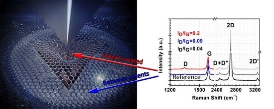
Nanostructures with graphene make them highly promising for nanoelectronics, memristor devices, nanosensors and electrodes for energy storage. In some devices the mechanical properties of graphene are important. Therefore, nanoindentation has been used to measure the mechanical properties of polycrystalline graphene in a nanostructure containing metal oxide and graphene. In this study the graphene was transferred, prior to the deposition of the metal oxide overlayers, to the Si/SiO2 substrate were SiO2 thickness was 300 nm. The atomic layer deposition (ALD) process for making a very thin film of Al2O3 (thickness comparable with graphene) was applied to improve the elasticity of graphene. For the alumina film the Al(CH3)3 and H2O were used as the precursors. According to the micro-Raman analysis, after the Al2O3 deposition process, the G-and 2D-bands of graphene slightly broadened but the overall quality did not change (D-band was mostly absent). The chosen process did not decrease the graphene quality and the improvement in elastic modulus is significant. In case the load was 10 mN, the Young’s modulus of Si/SiO2/Graphene nanostructure was 96 GPa and after 5 ALD cycles of Al2O3 on graphene (Si/SiO2/Graphene/Al2O3) it increased up to 125 GPa. Our work highlights the correlation between nanoindentation and defects appearance in graphene.

Graphical Abstract

1. Introduction

The demand of new devices is increasing, and in several cases, the graphene was used as one component (or layer) in nanostructures, for example graphene-based biosensors [1] and gas sensors [2,3], battery technology [4,5], graphene field-effect transistors [6] and graphene based memristor devices [7,8]. It has been noticed that a combination of conductive materials as graphene and relatively better insulating materials (typically metal-oxides, high-k), if deposited alternately, can provide decrement in the forming voltage to the level of switching voltages, also mechanical flexibility and transparency. The new generation nanodevices are very thin and the mechanical properties of graphene should be taken into consideration. Especially, if these devices must be flexible, then Young’s modulus of graphene and the materials surrounded should be addressed.
High-k dielectrics layers on graphene are commonly used for building semiconductor nanodevices (containing graphene), because they are electrically and thermally insulating and can be easily up for wafer-scale fabrication. There are many methods of depositing dielectrics onto graphene, such as sputtering [9], a pulsed laser deposition [3] and atomic layer deposition (ALD) [7,8,10,11,12], but there are not many works for learning the mechanical properties of those devices. There are several theoretical works where graphene nanoindentation has been studied [13,14] and molecular dynamics used for modelling Young’s modulus [15,16,17], but the maximum load has been small and graphene has not been located inside the nanostructure. Graphene-metal nanolaminates have been investigated using molecular dynamics [18]. It was deduced that graphene inclusions could increase modulus and strength approximately 90–100%. Other theoretical calculations have indicated that during nanoindentation the generated stresses are the highest at the tip of the indenter and the amplitude of the stress reduces outwards the site along the graphene structure [19]. Obviously, the graphene layer inside a nanodevice affects the nanostructure strength and the elastic modulus, but it has not been widely studied in literature up to now.
In this study, we are measuring the effect of graphene to Young’s modulus and observing how the atomic layer deposited (ALD) thin film affects the graphene quality and Young’s modulus thereof. The graphene transferred onto a Si/SiO2 substrate (as a typical substrate for nanodevices) and a thin layer film was made with ALD in Al(CH3)3 and H2O process (also well-known and widely used). We show that even the few cycles will improve the elastic properties of graphene, even though the deposition of thin film on graphene slightly decreases the quality of graphene.

2. Materials and Methods

Graphene was grown on commercial 25 µm thick polycrystalline copper foils (99.5%, Alfa Aesar, Haverhill, MA, USA) in an in-house built chemical vapor deposition (CVD) reactor. The foil was annealed, prior to the graphene deposition, at 1000 °C in Ar/H2 (99.999%, Linde Gas, Estonia) flow for 60 min, and then additionally exposed to the mixture of 10% CH4 (99.999%, Linde Gas, Estonia) in Ar at 1000 °C for 120 min. Then, the foil was cooled down in an Ar flow. Graphene/Cu foil (20 mm × 40 mm) was cut into 5 mm × 5 mm pieces and graphene was transferred onto 300 nm thick 10 mm × 10 mm Si/SiO2 substrates by using a wet chemical transferring process described in a publication by T.Kahro et al. [7].
The Al2O3 thin film was deposited from Al(CH3)3 (TMA) and H2O in a commercial PicosunTM R-200 (Picosun Oy, Finland) Advanced ALD system. TMA was evaporated at 22 °C, and 5 cycles with pulse times 0.3–4–0.3–6 s (TMA-N2-H2O-N2, respectively) were made at room temperature. For reference the thick Al2O3 film was deposited on graphene with 300 ALD cycles with the same cycle times at 300 °C. The deposition substrate was Si with 300 nm of amorphous SiO2 on top of it.
The surface morphology was characterized by using a scanning electron microscope (SEM) FEI Helios NanoLab 600 (FEI) in a high-resolution mode (10 kV, 86 pA) and composition was performed using an energy-dispersive X-ray (EDX) spectrometer INCA Energy 350 (Oxford Instruments, UK), attached to the SEM Helios NanoLab 600 (FEI)). A diffractometer SmartLabTM (Rigaku, Japan) with a rotating Cu anode (λ = 0.15406 nm) working at 8.1 kW was used for X-ray reflectivity (XRR) measurements for evaluating the thicknesses of the layers. A structural characterization of graphene-based nanostructures was performed by using a micro-Raman spectroscopic system (Renishaw, UK) in Via at an excitation wavelength of 514 nm and the silicon reference was used for calibration. The spectral resolution reached, approximately, 1.5–2 cm−1.
The mechanical properties (hardness and Young’s modulus) were studied with the nanoindentation method by using the Hysitron Triboindenter TI980 ((Eden Prairie, MN, USA) implementing a Berkovich’s diamond tip. The samples were measured in continuous stiffness mode with maximum applied loads of 0.25, 0.5, 2, 5 and 10 mN. Nine nanoindentation measurements were carried out, each with maximum loads with the total of 45 indentation events on each sample, while the distances between single indentation sites were 10 μm. The calibrated nanoindentation displacement depth was from 5 to 200 nm. The calibration regions for nanoindentation measurements are shown in Figure 1. The occurrence of pile-up, i.e., the vertical redistribution of the material around the tip under load during nanoindentation measurements was investigated by implementing the scanning probe microscopy (SPM) with the same equipment. Gwyddion 2.56 software was used to analyze the SPM images.

3. Results

Figure 2 represents the SEM images of the samples with graphene before and after five ALD cycles of TMA and H2O. The typical morphology of a single polycrystalline graphene layer on Si/SiO2 surface presented in Figure 2 (left panel) and the small grains of the deposited Al2O3 on graphene edges and in defect places are clearly recognizable in Figure 2 (right panel) and the amount of Al on those samples is very low (Table 1). The thickness of the five cycles of Al2O3, estimated by XRR, is 3.6 Å and the density 1.21 (g/cm3), which indicates the emergence of a very sparse material layer but can still be a model by XRR. The thickness of graphene on Si/SiO2 substrate was 0.6 nm.
The micro-Raman spectra of transferred graphene showed two main bands at ~1590 cm−1 for G- and at ~2690 cm−1 for 2D-band and which is characteristic of slightly p-doped graphene [20]. The D-band was mostly absent, the intensity ratio of D- and G-band ID/IG was less than 0.04 (Figure 3), indicating a good (structural) quality of graphene [21].
After a deposition of five cycles Al2O3 the main bands of graphene had the same average positions values (the difference was less than 1 cm−1) as compared to the pristine graphene. On the other hand, the G-band broadened up to 15% and the 2D-band up to 10% and the intensity ratio of D- and G-band increased up to 25% (Table 2) as compared to pristine graphene. The broadening of the bands after the ALD process may also be associated with doping [20], or correlated with higher amplitudes of charge fluctuations [22] or be caused by nanometer-scale strain variations [23].
In the case of five cycles of Al2O3 layer coated graphene, the G-band was well decomposed into three bands noted X, G and D’ (Figure 3 inset). A similar splitting of the G-band has been reported in the case when graphene was transferred onto the thicker (20–30 nm) HfO2 coated Si/SiO2 substrates [24].
The averaged elastic modulus and hardness of the substrate without graphene (Si/SiO2) and the modulus covered with the single polycrystalline graphene layer (Si/SiO2/G) are presented in Figure 4. At higher displacements (190 nm) the difference in Young’s modulus in these areas was up to 20%, yet only 6% for hardness. The averaged elastic modulus of the substrate without graphene was around 74.1 with a standard deviation of 0.1 GPa and hardness 9.0 (0.1) GPa. This resembles closely to the calibration standard, which was also amorphous SiO2 (Figure 1). The substrate with a graphene possessed an averaged modulus of 93 (1) and hardness of 9.5 (0.1) GPa at a tip displacement of 190 nm. Five ALD cycles of Al2O3 increased the elastic modulus and hardness of the sample (not shown) and the thin Al2O3 layer induced a rise of modulus with a displacement up to 110 GPa. Hardness was increased to 10 GPa. However, the differences between the samples with and without graphene diminished.
For comparison the reference sample with a thicker layer of Al2O3 (thickness by XRR was 64 nm) increased the modulus near the surface, but it decreased to around 90 GPa, as the substrate started to influence the measurement. Hardness was similar to the previously described samples (9 GPa). There was no significant difference between the areas with and without graphene.
Figure 5 compares the depth of indents made at different indentation loads. The substrate, graphene, thin Al2O3 on the substrate and on the graphene. The depth was acquired from a single SPM image for an indent, one of which is also shown in Figure 4. The substrate had the deepest indents at any given load. At the lowest and highest loads, the samples with graphene and Al2O3 had similar indent depths, however, in the case of intermediate loads some samples resisted plastic deformation more than others. At the maximum indentation load of 5 mN the indent on the Si/SiO2/G/Al2O3 sample was 28.4 nm, which is 24.6 nm shallower than the indent on the graphene-covered substrate. The sample with only five cycles Al2O3 on substrate (without an underlaying graphene) had a slightly deeper indent of 58 nm and the depth of the indent on the Si/SiO2 substrate was 73 nm.
Figure 6 compares the projected area of the indents made at different indentation loads for the same samples. The approximated area of the indent was found from a SPM image of the indent with the Gwyddion 2.56 software. It can be seen that the lateral size of the indents was comparable for all surfaces at low indentation loads. At 250 and 500 µN the indent was not discernible for the samples with graphene. However, with the increasing maximum load the size of the indents increased for both the substrate and the graphene-covered substrate similarly, while in comparison the indents on surfaces with Al2O3 were smaller in size. No occurrence of pile-up during nanoindentation measurements was detected for any of the samples.

4. Discussion

The graphene layer increased the Young’s modulus and hardness of the samples with the indentation depth (Figure 4). As similar trend has been measured before by Zhang et al. for the graphene-SiO2-Si system with nanoindentation [13]. As the deformation area expanded under the tip with higher loads, the number of C-C bonds participating in the deformation was multiplied, which probably increased the resistance to the tip. The indents on graphene covered substrate were shallower compared to the uncovered areas, yet the lateral sizes of the indents were similar. It is possible, the indenter tip penetrated both samples to similar depths, but the graphene layer recovered elastically to a much shallower shape. This would also explain why the graphene layer had a greater impact on Young’s modulus than hardness.
The relationship between the maximum averaged elastic modulus and loading force is presented in Table 3. The micro-Raman measurements were made on indents and between them to assess the quality of graphene (Figure 7). The change in D-band is clearly visible and we can see the relationship between the loading force and amount of defect rise (Table 3, D-band Area). As we can see, the loading force 10 mN was the increase of the amount of the defects and graphene is broken in the inset place. Despite this, the crystallinity of the graphene remains, i.e., the intensity of the 2D-band was only decreased by 18% compared to the graphene between the indents.
Sha et al. [25] found that the grain boundaries (and other defects) in polycrystalline graphene might decrease the mechanical strength of the material. However, it was shown by Lee et al. [26] that the mechanical properties of the grain boundaries of the polycrystalline graphene might be dependent on the production processes and the possibility of grain boundaries possessing similar mechanical strength to pristine graphene. In the present work, at the maximum load of 10 mN, the area influenced by the nanoindentation measurement could have been large enough to reach the grain boundaries where the graphene layer might have been weaker and defective (Figure 7 and Table 3). Wrinkles and bilayers have also been reported to lower the mechanical strength of graphene [25]. All these defects in polycrystalline graphene have been shown to make the graphene more ductile [14,26], and at the highest load the graphene could start to plastically deform until a fracture at the grain boundaries, leading to deeper indents.
For the polycrystalline graphene layer there might exist an optimal deformation size, at which it may possess the ability to recover the deformation during nanoindentation. Pristine graphene or polycrystalline graphene with mechanically stronger grain boundaries could increase Young’s modulus and hardness further at higher loads. This may at the same time make the material more brittle. In our case it seems that the very thin amorphous Al2O3 layer could possess sufficient mechanical stiffness to cause a steady increase in the modulus with the displacement in Figure 8, acting similarly to the graphene layer. Namely, the elastic modulus of a nanomaterial can become size-dependent due to the surface effects, when one or more dimensions of the material is under 10 nm [27,28,29]. The 3.6 nm thick Al2O3 film might have possessed higher modulus than the 64 nm thick Al2O3 film used as reference.
For all samples where ALD was used to cover graphene, there was no significant difference in mechanical properties for multilayer systems with graphene, compared to those without graphene. The additional ALD cycling was found to increase the defects in graphene layer which could have hindered its mechanical resilience. Higher brittleness of the top ALD ceramic layer was accompanied with the brittleness of graphene, because the crack propagation in the graphene layer was enhanced as well.

5. Conclusions

Chemical vapor deposited graphene layers were successfully transferred into Si/SiO2 substrate. After that some samples were treated with Al2O3 in ALD process, where 5 cycles of TMA and H2O were applied at room temperature. After deposition the small grains of Al2O3 were visible in SEM images. According to the micro-Raman analysis, after the Al2O3 deposition process, the G- and 2D-bands of graphene slightly broadened but the overall quality did not change (D-band was mostly absent). Despite this, the process slightly decreased the graphene quality and increased the count of defects. The micro-Raman probing allowed one to conclude that after five ALD cycles on graphene the (averaged) FWHM values for 2D-bands (Γ2D) changed from 34 up to 37.5 cm−1, but those five cycles increased the elastic properties of the sample. Young’s modulus increased from 93 and hardness from 9.5 GPa up to 110 GPa and 10 GPa, respectively.
Our measurements revealed that the elastic modulus of a coated CVD graphene is better with that of pristine graphene. Comparing to the approaches for measuring the mechanical property by using different load forces on the samples Si/SiO2/G and Si/SiO2/G/Al2O3, the improvement in Young’s modulus, 96 versus 125 GPa (at max. load 10 mN) was achieved. This study establishes that large-area CVD-graphene, being coated with thin film made by ALD, is a strengthened material, that would be suitable for flexible electronics and components of nanodevices.

Author Contributions

Conceptualization, A.T.; methodology, T.K. and T.J.; validation, A.T. and H.-M.P.; investigation, H.-M.P. and T.K.; resources, A.T.; writing—original draft preparation, A.T.; writing—review and editing, H.-M.P., T.K. and T.J.; visualization, T.K.; project administration, A.T.; funding acquisition, A.T. All authors have read and agreed to the published version of the manuscript.

Funding

The present study was partially funded by the European Regional Development Fund project “Emerging orders in quantum and nanomaterials” (TK134) and Estonian Research Agency (PRG4).

Institutional Review Board Statement

Not applicable.

Informed Consent Statement

Not applicable.

Data Availability Statement

Not applicable.

Acknowledgments

This work was supported by European Regional Development Fund project “Centre of nanomaterials technologies and research” (NAMUR+, Project No. 2014-2020.4.01.16-0123). The authors are grateful to Aivar Tarre in assistance for deposition of films.

Conflicts of Interest

The authors declare no conflict of interest.

References

  1. Ali, M.M.; Mitchell, J.J.; Burwell, G.; Rejnhard, K.; Jenkins, C.A.; Ahmadi, E.D.; Sharma, S.; Guy, O.J. Application of Molecular Vapour Deposited Al2O3 for Graphene-Based Biosensor Passivation and Improvements in Graphene Device Homogeneity. Nanomaterials 2021, 11, 2121. [Google Scholar] [CrossRef]
  2. Hassani, N.; Rashidi, R.; Miloševic, M.V.; Neek-Amal, M. Evaluating gas permeance through graphene nanopores and porous 2D-membranes: A generalized approach. Carbon Trends 2021, 5, 100086. [Google Scholar] [CrossRef]
  3. Kodu, M.; Berholts, A.; Kahro, T.; Eriksson, J.; Yakimova, R.; Avarmaa, T.; Renge, I.; Alles, H.; Jaaniso, R. Graphene-Based Ammonia Sensors Functionalised with Sub-Monolayer V2O5: A Comparative Study of Chemical Vapour Deposited and Epitaxial Graphene. Sensors 2019, 19, 951. [Google Scholar] [CrossRef] [PubMed] [Green Version]
  4. Son, I.H.; Park, J.H.; Park, S.; Park, K.; Han, S.; Shin, J.; Doo, S.-G.; Hwang, Y.; Chang, H.; Choi, J.W. Graphene balls for lithium rechargeable batteries with fast charging and high volumetric energy densities. Nat. Commun. 2017, 8, 1561. [Google Scholar] [CrossRef] [PubMed]
  5. Hussain, S.; Kongi, N.; Treshchalov, A.; Kahro, T.; Rähn, M.; Merisalu, M.; Tamm, A.; Sammelselg, V.; Tammeveski, K. Enhanced oxygen reduction reaction activity and durability of Pt nanoparticles deposited on graphene-coated alumina nanofibres. Nanoscale Adv. 2021, 3, 2261–2268. [Google Scholar] [CrossRef]
  6. Xia, F.; Farmer, D.B.; Lin, Y.-M.; Avouris, P. Graphene Field-Effect Transistors with High On/Off Current Ratio and Large Transport Band Gap at Room Temperature. Nano Lett. 2010, 10, 715–718. [Google Scholar] [CrossRef] [Green Version]
  7. Kahro, T.; Tarre, A.; Käämbre, T.; Piirsoo, H.-M.; Kozlova, J.; Ritslaid, P.; Kasikov, A.; Jõgiaas, T.; Vinuesa, G.; Dueñas, S.; et al. Hafnium Oxide/Graphene/Hafnium Oxide-Stacked Nanostructures as Resistive Switching Media. CS Appl. Nano Mater. 2021, 4, 5152–5163. [Google Scholar] [CrossRef]
  8. Kahro, T.; Castán, H.; Dueñas, S.; Merisalu, J.; Kozlova, J.; Jõgiaas, T.; Piirsoo, H.-M.; Kasikov, A.; Ritslaid, P.; Mändar, H.; et al. Structure and behavior of ZrO2-graphene-ZrO2 stacks. J. Vac. Sci. Technol. A 2020, 38, 063411. [Google Scholar] [CrossRef]
  9. Qiu, X.P.; Shin, Y.J.; Niu, J.; Kulothungasagaran, N.; Kalon, G.; Qiu, C.; Yu, T.; Yang, H. Disorder-free sputtering method on graphene. AIP Adv. 2012, 2, 032121. [Google Scholar] [CrossRef]
  10. Vervuurt, R.H.J.; Kessels, W.M.M.; Bol, A. Atomic Layer Deposition for Graphene Device Integration. Adv. Mater. Interfaces 2017, 4, 1700232. [Google Scholar] [CrossRef] [Green Version]
  11. Tamm, A.; Kozlova, J.; Aarik, L.; Aidla, A.; Lu, J.; Kiisler, A.-A.; Kasikov, A.; Ritslaid, P.; Mändar, H.; Hultman, L.; et al. Atomic layer deposition of ZrO2 for graphene-based multilayer structures: In situ and ex situ characterization of growth process. Phys. Status Solidi A 2013, 211, 397–402. [Google Scholar] [CrossRef]
  12. Jõgiaas, T.; Tarre, A.; Mändar, H.; Kozlova, J.; Tamm, A. Nanoindentation of Chromium Oxide Possessing Superior Hardness among Atomic-Layer-Deposited Oxides. Nanomaterials 2022, 12, 82. [Google Scholar] [CrossRef] [PubMed]
  13. Zhang, Y.; Pan, C. Measurements of mechanical properties and number of layers of graphene from nano-indentation. Diam. Relat. Mater. 2012, 24, 1–5. [Google Scholar] [CrossRef]
  14. Daniels, C.; Horning, A.; Phillips, A.; Massote, D.V.P.; Liang, L.; Bullard, Z.; Sumpter, B.G.; Meunier, V. Elastic, plastic, and fracture mechanisms in graphene materials. J. Phys. Condens. Matter 2015, 27, 373002. [Google Scholar] [CrossRef]
  15. Rabiei, M.; Palevicius, A.; Dashti, A.; Nasiri, S.; Monshi, A.; Vilkauskas, A.; Janusas, G. Measurement Modulus of Elasticity Related to the Atomic Density of Planes in Unit Cell of Crystal Lattices. Materials 2020, 13, 4380. [Google Scholar] [CrossRef]
  16. Rabiei, M.; Palevicius, A.; Nasiri, S.; Dashti, A.; Vilkauskas, A.; Janusas, G. Relationship between Young’s Modulus and Planar Density of Unit Cell, Super Cells (2 × 2 × 2), Symmetry Cells of Perovskite (CaTiO3). Lattice Mater. 2021, 14, 1258. [Google Scholar] [CrossRef]
  17. Rezaei, R. Tensile mechanical characteristics and deformation mechanism of metal-graphene nanolayered composites. Comput. Mat. Sci. 2018, 151, 181–188. [Google Scholar] [CrossRef]
  18. Fan, Y.-C.; Wu, C.-D.; Fang, T.-H.; Chen, T.-H. Thermal relaxation and deformation of indented graphene. Comput. Mat. Sci. 2013, 79, 105–109. [Google Scholar] [CrossRef]
  19. Tan, X.; Wu, J.; Zhang, K.; Peng, X.; Sun, L.; Zhong, J. Nanoindentation models and Young’s modulus of monolayer graphene: A molecular dynamics study. Appl. Phys. Lett. 2013, 102, 071908. [Google Scholar] [CrossRef]
  20. Beams, R.; Cançado, L.G.; Novotny, L. Raman characterization of defects and dopants in graphene. J. Phys. Condens. Matter 2015, 27, 083002. [Google Scholar] [CrossRef]
  21. Ferrari, A.C.; Basko, D.M. Raman spectroscopy as a versatile tool for studying the properties of graphene. Nat. Nanotechnol. 2013, 8, 235–246. [Google Scholar] [CrossRef] [PubMed] [Green Version]
  22. Wang, Q.H.; Jin, Z.; Kim, K.K.; Hilmer, A.J.; Paulus, G.L.C.; Shih, C.-J.; Ham, M.-H.; Sanchez-Yamagishi, J.D.; Watanabe, K.; Taniguchi, T.; et al. Understanding and controlling the substrate effect on graphene electron-transfer chemistry via reactivity imprint lithography. Nat. Chem. 2012, 4, 724–732. [Google Scholar] [CrossRef] [PubMed]
  23. Neumann, C.; Reichardt, S.; Venezuela, P.; Drögeler, M.; Banszerus, L.; Schmitz, M.; Watanabe, K.; Taniguchi, T.; Mauri, F.; Beschoten, B.; et al. Raman spectroscopy as probe of nanometre-scale strain variations in graphene. Nat. Commun. 2015, 6, 8429. [Google Scholar] [CrossRef] [PubMed] [Green Version]
  24. Maad, B.; Durnez, A.; ·Ajlani, H.; Madouri, A.; Oueslati, M.; ·Meftah, A. Modulation of electron transfer in Si/SiO2/HfO2/Graphene by the HfO2 thickness. Appl. Phys. A 2020, 126, 754. [Google Scholar] [CrossRef]
  25. Sha, Z.D.; Wan, Q.; Pei, Q.X.; Quek, S.S.; Liu, Z.S.; Zhang, Y.W.; Shenoy, V.B. On the failure load and mechanism of polycrystalline graphene by nanoindentation. Sci. Rep. 2014, 4, 7437. [Google Scholar] [CrossRef] [PubMed] [Green Version]
  26. Lee, G.-H.; Cooper, R.C.; An, S.J.; Lee, S.; van der Zande, A.; Petrone, N.; Hammerberg, A.G.; Lee, C.; Crawford, B.; Oliver, W.; et al. High-Strength Chemical-Vapor–Deposited Graphene and Grain Boundaries. Science 2013, 340, 1073–1076. [Google Scholar] [CrossRef]
  27. Hobi, E.; Pontes, R.B., Jr.; Fazzio, A.; da Silva, A.J.R. Formation of atomic carbon chains from graphene nanoribbons. Phys. Rev. B 2010, 81, 201406. [Google Scholar] [CrossRef] [Green Version]
  28. Peikolainen, A.-L.; Uibu, M.; Kozlova, J.; Mändar, H.; Tamm, A.; Aablo, A. Carbon xerogel from 5-methylresorcinol-formaldehyde gel: The controllability of structural properties. Carbon Trends 2021, 3, 100037. [Google Scholar] [CrossRef]
  29. Tamm, A.; Piirsoo, H.-M.; Jõgiaas, T.; Tarre, T.; Link, J.; Stern, R.; Kukli, K. Mechanical and Magnetic Properties of Double Layered Nanostructures of Tin and Zirconium Oxides Grown by Atomic Layer Deposition. Nanomaterials 2021, 11, 1633. [Google Scholar] [CrossRef]
Figure 1. Nanoindentation tip calibration results for reduced modulus (y axis on the left) and hardness (y axis on the right) as data points compared to the reference values 69.6 and 9.25 GPa, respectively, from the quartz glass presented with horizontal lines. The x axis indicates the depth of the measurement—the displacement of the tip. The displacement region where the measured values match with the properties of the fused quartz glass can be regarded as a trustworthy range.
Applsci 12 02491 g001
Figure 2. Scanning microscope images—(a) pristine graphene on substrate (sample Si/SiO2/G) and (b) graphene with 5 cycles TMA and H2O (sample Si/SiO2/G/Al2O3).
Applsci 12 02491 g002
Figure 3. Raman spectra of Si/SiO2/G (red line) and Si/SiO2/G/Al2O3 (black line) structures. Normalized by G-band according to G-band intensities. Inset: Raman spectra fitting (in accord with Lorentz function) in the range from 1300 to 1700 cm−1 for G-band after Al2O3 deposition process.
Applsci 12 02491 g003
Figure 4. (a) Averaged elastic modulus and (b) hardness for pure Si/SiO2 substrate and with graphene (sample Si/SiO2/G). Applied maximum force was 10 mN.
Applsci 12 02491 g004
Figure 5. Depth of indents on Si/SiO2 substrate, substrate with a graphene, Si/SiO2 substrate with a thin Al2O3 layer and sample Si/SiO2/G/Al2O3. Maximum applied indentation loads were 0.25, 0.5, 2, 5 and 10 mN. Shown is the scanning probe microscopy image of an indent made with 5 mN on the surface of the substrate.
Applsci 12 02491 g005
Figure 6. Approximate projected area of indents on Si/SiO2 substrate, substrate with a graphene, Si/SiO2 substrate with a thin Al2O3 layer and sample Si/SiO2/G/Al2O3. Maximum applied indentation loads were 0.25, 0.5, 2, 5 and 10 mN. The scanning probe microscopy image of an indent made with 10 mN on the surface of the sample Si/SiO2/G/Al2O3 with a mask is illustrating the indent area.
Applsci 12 02491 g006
Figure 7. Micro-Raman spectra of graphene-based nanostructures Si/SiO2/G, indents made with loading force 10 mN.
Applsci 12 02491 g007
Figure 8. (a) Average elastic modulus of nanostructure Si/SiO2/G and (b) Si/SiO2/G/Al2O3 with different load forces.
Applsci 12 02491 g008
Table 1. Elemental composition of samples made (wt%) as determined by EDX analysis. (All elements analyzed (normalized) and all result in weight%).
EDX Spectrum COAlSi
Si/SiO2/G1.6933.190.065.12
Si/SiO2/G/Al2O31.0555.960.4942.50
Table 2. Raman data of graphene-containing structures, fitted in accord with using a Lorentz for G band and a Voigt function for 2D band. The average Raman shift values reflect the effects of substrate and dielectric layers on graphene. The parameters shown express the positions of graphene and 2D bands (ωG and ω2D, respectively), and FWHM values for G- and 2D-bands (ΓG and Γ2D, respectively).
SampleωG (cm−1)ΓG (cm−1)ω2D (cm−1)Γ2D (cm−1)ID/IG
Si/SiO2/G1588.515.52690.034.00.04
Si/SiO2/G/Al2O31587.518.02689.537.50.05
Ref, Si/SiO2/G/Al2O31594322707621.3
Table 3. This is a table of the relationship between maximum load and amount of defect (area of D band) rose (D-band area fitted by Lorenz).
SampleMax. Load (mN)D-Band Area Measured on HoleD-Band Area
between Holes
Max. Ave. Young’s Modulus (Gpa)
Si/SiO2/G1012,3666578.596.0
5.05825176793
2.05653251488
0.55425300680
Si/SiO2/G/Al2O31062203967125
555663967107
26529156895
Publisher’s Note: MDPI stays neutral with regard to jurisdictional claims in published maps and institutional affiliations.