Article

Characterization of Cysteine Cathepsin Expression in the Central Nervous System of Aged Wild-Type and Cathepsin-Deficient Mice

1
Department of Life Sciences and Chemistry, Jacobs University Bremen, Campus Ring 1, D-29759 Bremen, Germany
2
Fraunhofer-Institute for Translational Medicine and Pharmacology ITMP and Fraunhofer Cluster of Excellence Immune-Mediated Diseases CIMD, 60596 Frankfurt, Germany
*
Author to whom correspondence should be addressed.
These authors contributed equally to this work.
Present Address: Department of Pediatric Hematology and Oncology, Center for Pediatrics, University Hospital Bonn, Venusberg-Campus 1, D-53127 Bonn, Germany.
Appl. Sci. 2022, 12(5), 2608; https://doi.org/10.3390/app12052608
Submission received: 19 January 2022 / Revised: 24 February 2022 / Accepted: 28 February 2022 / Published: 3 March 2022

Abstract

:

Featured Application

Endolysosomal cathepsin expression and activities are differentially regulated in the cortex, striatum, hippocampus, and cerebellum of mice upon aging and/or cathepsin deficiencies.

Abstract

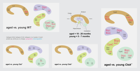
The association of cathepsin proteases in neurobiology is increasingly recognized. Our previous studies indicated that cathepsin-K-deficient (Ctsk−/−) mice have learning and memory impairments. Alterations in cathepsin expression are known to result in compensatory changes in levels of related cathepsins. To gain insight into the therapeutic usefulness of cathepsin inhibitors in aging individuals with osteoporosis or neurodegenerative diseases, we studied for variations in cathepsin expression and activity in aged (18–20 months) versus young (5–7 months) wild-type (WT) and cathepsin-deficient mice brains. There were age-dependent increases in cathepsin B, D, and L and cystatin C protein levels in various brain regions, mainly of WT and Ctsk−/− mice. This corresponded with changes in activity levels of cathepsins B and L, but not cathepsin D. In contrast, very little age-dependent variation was observed in cathepsin-B- and cathepsin-L-deficient mouse brain, especially at the protein level. The observed alterations in cathepsin protein amounts and activity are likely contributing to changes in important aging-related processes such as autophagy. In addition, the results provide insight into the potential impact of cathepsin inhibitor therapy in aged individuals, as well as in long-term use of cathepsin inhibitor therapy.

Graphical Abstract

1. Introduction

Aspartic and cysteine cathepsins are important enzymes of the endolysosomal system and are vital biological regulators with diverse fundamental roles in protein processing and degradation [1,2,3]. Cathepsins also have specific functions in protein processing such as in endocrine regulation [4]. Of special interest is the bone remodeling ability of cathepsin K that has staged its prominence as a targeted therapy for osteoporosis [5,6,7]. Moreover, there has been interest in the targeting of cysteine cathepsins, in particular for the treatment of cancer [8] and Alzheimer’s disease (AD) [8,9,10].
It is accepted that endolysosomes, and the hydrolases they contain, change during aging, also in the central nervous system (CNS), although the outcomes of such changes are not yet fully understood. After the first biochemical and preclinical studies, a major expansion of research occurred in the last 20 years, which was facilitated by the development of cathepsin-deficient and/or other murine disease models and by using new approaches [2,9,10,11,12,13,14,15]. This research field led to a major advancement in our understanding of the molecular mechanisms underlying physiological and degenerative processes entailing cathepsin functions in the brain, beyond other organs.
Cathepsins B, D, K, and L are essential for the central nervous system (CNS). While cathepsin-B- (Ctsb−/−) and cathepsin-L-deficient (Ctsl−/−) mice are viable, double cathepsin B/L-deficient mouse pups die between 2 and 4 weeks due to brain atrophy and massive apoptosis of brain cells [16], suggesting that cathepsins B and L have overlapping neurological roles. Cathepsin D knockout mice are also lethal due to a severe neurological phenotype [17]. In contrast, cathepsin-K-deficient (Ctsk−/−) mice show milder afflictions, in that impairments of memory formation and neurobehavior were observed in comparison with wild-type (WT) controls [18]. At the molecular level, there are alterations in expression of important molecules such as synaptotagmin, glial fibrillary acidic protein (GFAP), cyclic nucleotide phosphodiesterase, and tyrosine hydroxylase in the Ctsk−/− murine model [18].
Hence, the exact role of cathepsins in the CNS is yet to be fully elucidated. For instance, while cathepsins have been reported to play a role in increasing neurodegenerative protein aggregate formation [9,19,20], there is also mounting evidence that cathepsins can have neuroprotective functions [21,22,23], probably through aggregate-clearance mechanisms as part of their involvement in the autophagy–lysosomal system [23,24]. Cathepsin B, for example, is associated with neuroinflammatory response and oxidative stress in inflammatory brain diseases and brain aging, where it is found to be upregulated particularly in microglia [11]. Furthermore, cathepsin D plays an important role in neuronal cell homeostasis because of its proteolytic action towards target proteins such as α-synuclein, apolipoprotein E, huntingtin, lipofuscin, or tau, namely proteins involved in neurological disorders, including Huntington’s and Parkinson’s disease [3,25]. Cathepsins are also associated with inflammatory neurological diseases like Niemann–Pick type C (NPC) disease and neuronal ceroid lipofuscinosis (NCL) [3,26,27].
Cathepsins are relevant in many biological systems and processes, and this poses therapeutic considerations. For instance, a highly effective cathepsin K inhibitor entered clinical trials for the treatment of osteoporosis (for review, see [6]). However, it was argued that oral administration of cathepsin K inhibitors for osteoporosis could result in off-target CNS effects [5,18] or alterations in immune function [28]. Indeed, it was because of such off-target effects that the highly efficient bone resorption inhibitor Odanacatib was withdrawn from otherwise successful clinical trials as late as in phase III due to cerebrovascular events [6]. Since the downregulation of one cathepsin often leads to compensatory upregulation of related cathepsins [1], another important therapeutic consideration is to ask, whether the use of cathepsin inhibitors is likely to affect cathepsin balance due to compensation. On the one hand, if the activity of one cathepsin is downregulated using an inhibitor, upregulation of another cathepsin with similar or overlapping functions may offer a degree of protection [4,29]. On the other hand, possibilities remain for overcompensatory effects due to upregulation of cathepsins with slightly different functions. Thus, more comprehensive knowledge on the individual biological effects of cathepsins is invaluable in therapeutic targeting.
In order to understand the possible effects of cathepsin inhibitor therapy on CNS in particular, and to gain an in-depth understanding of the potential of cathepsin inhibitor therapeutics in the treatment of aging-related diseases, this study aimed to examine alterations in cathepsin expression and activity in the aging brain of mice. Using young and aged WT mice, as well as cathepsin-deficient (Ctsb−/−, Ctsl−/−, Ctsk−/−) mice of the same age groups, various brain regions were studied for cathepsin localization using immunofluorescence confocal laser scanning microscopy, proteolytic activity assays, and immunoblot in order to elucidate morphological, biochemical, and functional variations. The endogenous inhibitor of cysteine cathepsins, cystatin C, was included in this study to better understand the balancing of proteolytic with antiproteolytic factors upon aging and/or loss of cysteine cathepsin function. We hypothesized that there would be alterations in aged compared to young CNS cathepsin levels, and that deficiency in one cathepsin could lead to compensatory upregulation of other cathepsins that could be further exacerbated in aging.

2. Materials and Methods

2.1. Animals

All studies were performed on male cathepsin-deficient or WT C57Bl/6J mice. The Cts−/− mice were housed under standard conditions, with a 12 h/12 h light/dark cycle with lights out at 07:00 p.m. and ad libitum water and food in the animal facility of Jacobs University Bremen, Germany. Backcrossing to a congenic C57Bl/6J background was performed at least over 8 generations for Ctsk−/− mice. Generation of the founder cathepsin-deficient mice at University of Göttingen, Germany, and the genotyping methods are described elsewhere [7]. The procedures have been described for the generation of Ctsb−/− [30,31] and Ctsl−/− mice [32].

2.2. Tissue Sampling

Brain samples were collected from young (5–7 months old) and aged (18–20 months old) male WT C57Bl/6J mice and from mice deficient for cathepsins B, K, or L. The mice were perfused with 0.9% NaCl supplemented with 200 IU heparin (Braun Melsungen AG, Melsungen, Germany). Harvested whole brains were separated into two hemispheres, of which one was fixed with 4% PFA in 200 mM HEPES, pH 7.4, and further processed for cryosectioning as described before [18]. The other hemisphere was immediately dissected on ice and the cortex, hippocampus, cerebellum, and striatum combined with the mesencephalon and diencephalon (collectively referred to in this paper as striatum) were separated and snap-frozen in liquid nitrogen.

2.3. SDS-PAGE, Immunoblotting, and Densitometry

Protein extraction. A Potter S homogenizer (1000 rpm for 2 min; Sartorius, Göttingen, Germany) was used to homogenize tissue samples in ice-cold PBS containing 0.5% Triton X-100. The resulting homogenates were placed in a rotary mixer for 40 min at 4 °C for further extraction. Subsequent centrifugation was performed at 10,000× g and 4 °C for 10 min. Afterwards, the supernatants were removed and stored at −20 °C.
Gel electrophoresis. The Neuhoff assay was used to specify the protein content of all samples [33]. Tissue lysates were normalized to equal amounts of protein and boiled in sample buffer consisting of 10 mM Tris-HCl, pH 7.6, 0.5% (wt/vol) SDS, 25 mM DTT, 10% (wt/vol) glycerol, and 25 μg/mL bromophenol blue. Proteins were separated on 12.5% acrylamide gels.
Immunoblotting and immunodetection. Separated proteins were semidry blotted onto nitrocellulose membrane. Blocking with 5% milk powder in PBS containing 0.3% Tween-20 (PBST) was performed overnight at 4 °C. Primary antibodies, goat anti-mouse cathepsin B (GT15047, Neuromics, through Acris Antibodies, Herford, Germany), rabbit anti-human cathepsin D (IM16, Calbiochem-Novabiochem GmbH, Bad Soden, Germany), goat anti-mouse cathepsin L (GT15049, Neuromics), and rabbit anti-mouse cystatin C (Dr. Magnus Abrahamson, Lund, Sweden) were applied for 1.5 h at room temperature (RT). The appropriate secondary antibodies, either goat anti-rabbit IgG (4050-05, Southern Biotech, Birmingham, AL, USA) or rabbit anti-goat IgG (6160-05; Southern Biotech), were applied for 1 h at RT. Commonly used housekeeping proteins are altered in aged compared to young tissue due to large age-related differences in protein turnover [34,35]. Protein loading was therefore verified by Ponceau staining of blots and Coomassie-stained gels. Immunoreactions were visualized by enhanced chemiluminescence on CL-XPosure film (Pierce through Perbio Science Europe, Bonn, Germany) or on Hyperfilm MP (GE Healthcare Europe GmbH, Munich, Germany).

2.4. Cathepsin Activity Assays

Cathepsins B and L. Enzyme activity assays were performed as described [18]. Tissue extracts containing 10 μg of total protein were mixed with 0.1% Brij35 and reactivated for 5 min at 40 °C with reaction buffer (88 mM KH2PO4, 12 mM Na2HPO4, 1 mM Na2-EDTA, 2 mM freshly prepared L-cysteine, pH 6.0; all final concentrations). For controls, run in parallel, the reaction buffer included 10 μM of the cysteine protease inhibitor E64. After reactivation, samples and controls were incubated with 5 μM cathepsin B substrate N-benzyloxycarbonyl-arginyl-arginine-7-amido-4-methylcoumarin (Z-Arg-Arg-AMC; Bachem Distribution Services GmbH, Weil am Rhein, Germany) or with 5 μM cathepsin L substrate N-benzyloxycarbonyl-phenylalanyl-arginine-7-amido-4-methylcoumarin (Z-Phe-Arg-AMC; Bachem Distribution Services GmbH) plus 1.5 μM CA-074 cathepsin-B-specific inhibitor (Merck Biosciences GmbH, Darmstadt, Germany) at pH 5.5 for 10 min at 40 °C and 800 rpm. Reactions were terminated by adding stop solution containing 100 mM sodium chloroacetate, 30 mM sodium acetate, and 70 mM acetic acid, pH 4.3, and fluorescence was measured with a Tecan GENios Reader (Tecan Deutschland GmbH, Crailsheim, Germany), using excitation/emission wavelengths of 360/465 nm.
Cathepsin D. Activity was tested as previously described [18] by incubating tissue extracts with 200 μM cathepsin D substrate 7-methoxycoumarin-4-yl-acetyl-glycyl-lysyl-prolyl-isoleucyl-phenylalanyl-phenylalanyl-arginyl-leucyl-lysine (2,4-dinitrophenyl)-D-arginine (MOCAc-Gly-Lys-Pro-Ile-Leu-Phe~Phe-Arg-Leu-Lys (Dnp)-D-Arg-NH2); Merck Biosciences GmbH) in 50 mM sodium acetate (pH 4.0) for 10 min at 40 °C and 800 rpm. Controls were incubated in addition with 1 µM pepstatin A (Sigma-Aldrich Chemie GmbH, Taufkirchen, Germany). Reactions were stopped by adding 5% trichloroacetic acid, and fluorescence was measured using excitation/emission wavelengths of 328/393 nm.
Analysis. Assays were performed twice generally in duplicate. All assays were performed with n = 4 mice for each genotype. Relative fluorescence units (RFU) of controls were subtracted from the values for the corresponding samples. In order to allow averaging over several assays, RFU values were normalized using the averages of young wild-type control measurements within a given assay for each brain region.

2.5. Immunohistochemistry

Brain hemispheres were cut horizontally on a cryostat (Leica CM1900, Leica Microsystems) into 16 μm serial sections and washed prior to the staining procedure in calcium and magnesium-free (CMF)-PBS at 4 °C as previously described [18]. Upon blocking of nonspecific binding sites with 3% bovine serum albumin (Albumin Fraction V, Roth, Karlsruhe, Germany) including 0.3% Triton X-100 in CMF-PBS, respectively, primary antibodies were applied on brain sections overnight at 4 °C in a moisturized chamber. Goat anti-mouse cathepsin B (GT15047, Neuromics, through Acris Antibodies, Herford, Germany), goat anti-mouse cathepsin L (GT15049, Neuromics), rabbit anti-human cathepsin D (IM16, Calbiochem-Novabiochem GmbH, Bad Soden, Germany), and mouse anti-NeuN (Millipore, Schwalbach am Taunus, Germany) were used. Secondary antibodies, rabbit anti-goat IgG F(ab’)2 fragment conjugated with Alexa 488 (A21222, Invitrogen through Molecular Probes, Karlsruhe, Germany), or goat anti-mouse IgG conjugated with Alexa 546 (A11018, Invitrogen through Molecular Probes, Karlsruhe, Germany), or goat anti-rabbit IgG conjugated with Alexa 488 (A11034, Invitrogen) were applied for 4 h along with DRAQ5™ as a nuclear marker (Biostatus Ltd., Shepshed Leicestershire, UK). Sections were mounted with Mowiol embedding medium (33% glycerol, 14% Mowiol/Elvanol in 200 mM Tris-HCl, pH 8.5, Hoechst, Frankfurt, Germany), imaged with a Zeiss LSM 510 META laser-scanning microscope (Carl Zeiss GmbH, Oberkochen, Germany) and analyzed using LSM 5 software (version 3.2; Carl Zeiss, Jena, Germany).

2.6. Densitometry Analysis and Statistical Evaluations

TINA software (Raytest, Straubenhardt, Germany) was used for densitometry. All data are shown as mean ± standard errors. Differences among groups were analyzed using Student’s t-test using GraphPad Prism 5.04 (GraphPad, San Diego, CA, USA).

3. Results

3.1. Localization of Cathepsins B, D, and L in Mouse Brain

To look for age-dependent alterations in cathepsin localization in mouse CNS, brain sections of WT young (5–7 months old) and aged (18–20 months old) mice were stained for cathepsins B, D, or L, the neuronal marker NeuN, and the nuclear marker Draq 5™. Cathepsin-B-, D-, and L-positive staining was detected in neurons and non-neuronal brain cells in a vesicular pattern around nuclei (Figure 1, Figure 2 and Figure 3). In the cortex and striatum, cathepsin staining was sparse and distributed mostly around the cellular nuclei in a vesicular pattern (Figure 1A–D, Figure 2A–D and Figure 3A–D). This was particularly evident in NeuN-positive neurons, but was also observed for some NeuN-negative cells. In the hippocampus, cells of the dentate gyrus (DG) showed intense accumulation of cathepsin-B- and L-positive vesicles (Figure 1E,F and Figure 2E,F). In the cerebellum, there was intense cathepsin staining in neurons and Purkinje cells of the granular layer, while neuronal cells of the molecular layer were sparsely stained (Figure 1G,H, Figure 2G,H and Figure 3G,H). Overall, both young and aged brain samples showed similar patterns of cathepsin distribution, suggesting that there were no age-dependent alterations in cathepsin localization.

3.2. Cathepsin B, D and L Protein and Activity Levels in WT and Cathepsin-Deficient Mouse Brain

Immunoblot studies and activity assays were performed with cathepsins B, D, and L in various brain regions of young and aged WT and Ctsb−/−, Ctsk−/−, and Ctsl−/− mice (Figure 4, Figure 5 and Figure 6). Negligible cathepsin B protein and activity levels were detected in all Ctsb−/− samples, which served as appropriate controls (Figure 4). Most notably, cathepsin B protein levels were increased in all brain regions of aged Ctsk−/− mice compared to young Ctsk−/− animals (Figure 4A–D). This finding corresponded with an observed increase in the activity levels of cathepsin B in all brain regions of aged compared to young Ctsk−/− mice (Figure 4E–H). Similarly, in aged WT mice, there were also significant increases in cathepsin B activity in the striatum, hippocampus, and cerebellum (Figure 4F–H), although these differences were not significantly detected at the protein level (Figure 4A–D). However, the immunostainings also showed a marked increase in cathepsin B in aged mice in all four brain regions (Figure 1). In the cerebellum of aged Ctsl−/− mice, cathepsin B activity levels were slightly, but significantly, elevated compared to young Ctsl−/− mice (Figure 4H). When comparing among genotypes, the elevated levels of cathepsin B protein in aged Ctsk−/− striatum and hippocampus was significantly higher compared to age-matched WT controls (Figure 4B,C). In addition, significantly higher levels of cathepsin B protein were detected in young Ctsk−/− and Ctsl−/− striatum compared to WT controls (Figure 4B), and in the cerebellum, cathepsin B protein levels were also elevated in young Ctsl−/− mice compared to WT controls (Figure 4D). These observations could indicate compensatory upregulation of cathepsin B protein in the absence of cathepsins K and L.
However, these alterations in cathepsin B protein were not necessarily reflected at the activity level. In the cortex, cathepsin B activity was significantly lower in aged and young Ctsk−/− and Ctsl−/− mice compared to WT controls (Figure 4E), while the decrease at the protein level did not reach significance. In the striatum, contrary to the generally observed increases at the protein level, cathepsin B activity was lower in young Ctsk−/− and Ctsl−/− mice and aged Ctsl−/− compared to WT controls (Figure 4F). Hippocampal cathepsin B activity was also lower in young and aged Ctsl−/− mice compared to WT controls (Figure 4G). In the cerebellum, cathepsin B activity was lower in aged Ctsk−/− and Ctsl−/− mice compared to WT controls, and similar to the protein level, activity was significantly increased in the young Ctsl−/− mice compared to the young WT controls (Figure 4H). Thus, in general, and despite the increased cathepsin B protein levels detected by immunoblot, it appeared that ablation of cathepsins K and L resulted in an overall decrease in cathepsin B activity.
Cathepsin D protein levels were elevated in aged compared to young WT in the cortex, striatum, and cerebellum; the same was observed in aged Ctsk−/− mice (Figure 5A–D), reflecting the general pattern observed for Cathepsin B protein in these genotypes. In contrast, the cathepsin D protein levels in the Ctsb−/− and Ctsl−/− mice were unaffected with age (Figure 5A–D). Furthermore, activity of cathepsin D was generally stable with age in all the different genotypes (Figure 5E–H), with only a significant age-dependent increase in cathepsin D activity observed in Ctsl−/− hippocampus (Figure 5G). Few differences were seen when comparing different genotypes for cathepsin D protein and activity. In aged striatum of Ctsb−/− and Ctsl−/− cathepsin D protein levels were significantly lower, whereas cathepsin D protein levels in aged Ctsk−/− mice were increased compared to aged WT controls (Figure 5B); none of these differences were detected at the activity level (Figure 5F). In the hippocampus, cathepsin D protein levels were elevated in aged Ctsk−/− mice compared to WT controls (Figure 5C), and cathepsin D activity was decreased in the hippocampus of young Ctsl−/− mice compared to WT controls (Figure 5G). In the cerebellum, a significant increase in cathepsin D protein levels was observed in young and aged Ctsb−/− mice and a decrease in aged Ctsk−/− compared to age-matched WT controls (Figure 5D). Cathepsin D cerebellar activity was increased in both young and aged Ctsl−/− mice compared to WT controls (Figure 5H). Overall, while age and/or ablation of cathepsins B, K, and L may have resulted in increased cathepsin D protein, cathepsin D activity remained relatively unaffected.
Negligible cathepsin L activity and protein were detected in all Ctsl−/− samples, which served as experimental controls (Figure 6). Cathepsin L protein levels were increased in all brain regions except cortex of aged WT compared to young WT mice (Figure 6A–D), corresponding to an age-dependent increase in cathepsin L activity in WT hippocampus (Figure 6G). In Ctsk−/− mice, cathepsin L protein levels were observed to be age-dependently elevated in the hippocampus and cerebellum (Figure 6C,D), and activity was age-dependently elevated in the cortex (Figure 6E) in contrast to almost equal protein levels. The only alteration in Ctsb−/− mice was a slight age-dependent decrease in cathepsin L protein in the cortex (Figure 6A). When comparing cathepsin L protein levels among different genotypes, it was observed that cathepsin L protein levels were increased in both young and aged Ctsb−/− striatum (Figure 6B) and in young cerebellum (Figure 6D) compared to age-matched WT controls (Figure 6B), suggesting that there may be upregulation of cathepsin L protein in these brain regions to compensate for the lack of cathepsin B. In contrast, hippocampal cathepsin L protein levels were decreased in aged Ctsb−/− and Ctsk−/− mice compared to young WT animals (Figure 6C). At the activity level, cathepsin L activity was significantly decreased in the cerebellum of aged Ctsb−/− mice compared to aged WT animals (Figure 6H), but no other significant variations in activity were detected with the absence of cathepsins B or K. Thus, similar to cathepsin D, while age and/or ablation of cathepsins B, K, and L may have resulted in variations to cathepsin L protein, activity remained relatively unaffected.

3.3. Cystatin C Protein Levels in WT and Cathepsin-Deficient Mouse Brain

Immunoblot studies were also performed to determine protein amounts of the endogenous cysteine cathepsin inhibitor cystatin C in various brain regions of young and aged WT and Ctsb−/−, Ctsk−/−, and Ctsl−/− mice (Figure 7). Differences in protein levels in the cortex were rather seen between different genotypes, whereas aging seemed to have less effect on the cystatin C protein levels. Only in Ctsl−/− mice did we see an increase in cystatin C levels in the cortex in aged mice. In the striatum, an increase in cystatin C levels was observed in Ctsk−/− aged mice. The other genotypes did not show alterations in cystatin C levels with aging. In the striatum of Ctsl−/− mice, significantly more cystatin C was detected over WT controls. In the hippocampus, an age-dependent increase in cystatin C protein was detected in WT and Ctsk−/− mice, while significant differences were also observed comparing WT and Ctsb−/− and Ctsl−/− mice. In the cerebellum, an increase in cystatin C was detected in aged WT mice and a decrease in aged Ctsb−/− mice. A higher cystatin C protein level was observed in all three cathepsin-deficient young animals compared to young WT mice. Thus, both aging and loss of specific cathepsins affected protein levels of cystatin C in different brain regions.

4. Discussion

To our knowledge, this is the first report examining alterations of cysteine cathepsin and cystatin C levels in aging brain from both WT and cathepsin-deficient (Ctsb−/−, Ctsl−/−, and Ctsk−/−) mice. Overall, age-dependent increases in cathepsin protein levels were detected particularly in WT and Ctsk−/− brain regions (see Graphical Abstract). This corresponded in part to an increase in enzyme activity, as for cathepsin B protein and activity, which were both elevated in Ctsk−/− mice upon aging. However, in many cases, while cathepsin protein was age-dependently increased, there were no significant changes in activity levels, particularly for cathepsin D activity, which remained relatively stable with age among the different genotypes. Localization of cathepsins B, D, and L expression confirmed their vesicular staining pattern in neuronal and non-neuronal cells in both young and aged brain.

4.1. The Proteolytic Network of the Rodent Brain upon Aging

Alterations in proteolytic networks involved in the aging CNS are generally not fully understood, particularly in relation to cathepsins. However, the endolysosomal system of neurons comprises cathepsins among other proteases, which are capable of initiating and executing cell death programs not only in certain pathologic states but also during aging [14]. Because cathepsin proteases are clearly important in processing and/or in degrading neuronal proteins, they may thus exert either neuroprotective or harmful roles [14].
Early studies injecting lysosomotropic drugs into the brain of young rats resulted in morphological features that are otherwise only seen in aged brain [14,36]. The alterations caused by lysosomotropic drug injections included accumulation of lipofuscin and decline in dopamine receptors, suggesting that endolysosomal enzymes are likely involved [14,36]. Relevant to our findings of the present study, a previous investigation of others has reported that cathepsin B levels and activity were higher in aged compared to young neostriatum in rat brain, and detected age-related increased cathepsin D protein levels in different brain regions [37]. Some care is needed in the interpretation of the enzyme activity assays of this particular report, because cathepsins S and X/Z were described only later, such that specific activity assay designs are needed to distinguish them from related cysteine cathepsins and verify the data. Another study focusing on hippocampal cathepsin B activity reported no significant change for aged (16–20 months) compared to young mice (1–3 and 7–8 months) [21], which correlates with our findings. Using a transgenic mouse model that results in increased amyloid beta (Aß) production, the same study found that cathepsin B hippocampal activity was higher with increased Aß levels in young but not in aged mice, which may suggest that upregulation of cathepsin B in this model could be a protective response to a stimulus that declines with aging [21]. Collectively, the results mentioned above and those of this study suggest a connection between variations in endolysosomal proteolysis by cathepsins and development of CNS aging-related diseases. Thus, activation of endolysosomal cathepsins in aging could either be a compensatory effect or a regenerative attempt to rescue cells [38].
Cystatin C was also inspected in this study. We reasoned that cystatin C as an endogenous antiproteolytic factor has the ability to balance cysteine cathepsin activity. This was indeed reported in, e.g., thyroid tissue of differently aged WT and mice deficient in the G-protein-coupled receptor Taar1 [39]. In the present study, increases in cystatin C protein levels were observed upon aging in the hippocampus of WT and Ctsk−/− mice. Hence, increased cystatin C could potentially counteract the increased cathepsin B activities in the hippocampus of Ctsk−/− mice. However, cystatin C levels remained largely unaffected in the cortex, striatum, and hippocampus of Ctsb−/− mice upon aging, while it was downregulated in the cerebellum (see Figure 7). We conclude that the upregulated cathepsin amounts are not counter balanced by an upregulation of the cystatin C inhibitor. Therefore, the role of cystatin C remains elusive in the brain of cathepsin-deficient mice.
However, cystatin C is recognized as a neuroprotective protein in AD [40,41], and it is considered in recent therapeutic strategies aiming at drug delivery to the brain [42,43,44]. Cystatin C is present in all body fluids [45], and it passes the blood–brain barrier (BBB), which is why its blood concentrations are indicative of the permeability of the BBB [46] among other tissue barriers or kidney function. Whether the changes in cystatin C protein amounts are also indicative of different permeabilities of the blood–brain and the blood–cerebrospinal fluid barriers upon aging or cathepsin deficiencies in mice is not known at this point and must be investigated further. It is interesting to note in this context that cathepsin K is particularly highly expressed in cells of the choroid plexus of mice [18]. This is of special interest because most changes in cysteine cathepsin protein or activity levels were observed in the Ctsk−/− mice (this study). Hence, future studies must aim at investigating structural and functional changes of the blood–cerebrospinal fluid barrier of Ctsk−/− mice in comparison with WT and upon aging.
Several studies also showed age-dependent changes in aspartic cathepsin D expression and/or activity in human and rat brain tissue (summarized in [14]). In Wistar rats, cathepsin D activity was significantly increased with aging in the cerebellum, cerebrum, hippocampus, and pons [47]. This agrees partly with our findings of this study, because we found an increase in cathepsin D in the cerebellum, the cortex, and the striatum of WT mice. Another study evaluated the distribution of cathepsin D activity (based on hemoglobin degradation) in 50 different areas of the CNS of adult and aged humans, revealing that the alterations in cathepsin D’s proteolytic activity were less pronounced with age in the human brain than in the rat brain [48]. On this basis, a possible relationship was suggested between activation of cathepsin D and senile plaques formation in AD and aging [49].

4.2. Potential Contributions of Microglia and Autophagy

Another source of excessive cathepsins amounts in the aged brain stems from activated microglia [50]. Microglia activation is understood as an integral part of neuroinflammation that accompanies numerous neurodegenerative disorders, including AD. Moreover, alterations of cathepsin expression during brain aging were proposed as a possible mechanism underlying microglia priming in brain aging [14,51,52]. By way of example, cathepsin B was suggested to be involved in NALP3 inflammasome-mediated generation of interleukin 1-β in response to Aβ [53].
The general pattern of cathepsin protein upregulation in aged compared to young animals’ tissues may also relate to their important roles in autophagy, a vital process of protein recycling and turnover for removal of accumulated cellular damaging factors, which declines with age [54,55,56]. However, an increase in cathepsin levels in aged brain, particularly if in conjunction with endolysosomal leakage, could conversely lead to activation of apoptotic pathways [54,57]. We can neither rule out nor support that the age-dependent upregulation of cathepsin expression in mouse brain, in particular of WT and Ctsk−/− mice, found in this study is a response to an increased requirement for autophagy. Hence, for future studies, it will be interesting to determine whether autophagy is enhanced in brain tissue of any of the mouse models used in this study.

4.3. Changes in Wild-Type and Cathepsin-K-Deficient Mouse Brain Tissues upon Aging Differ from Those in Cathepsin B and L Deficiency

Interestingly, in Ctsk−/− samples, an age-dependent increase in protein expression was generally pronounced for cathepsins B, D, and L in all brain regions, with the exceptions of cathepsin D in the hippocampus and cathepsin L in cortex and striatum (see Figure 4, Figure 5 and Figure 6, see also Graphical Abstract); this phenomenon was observed to a lesser extent in WT mice. Thus, it is possible that cathepsins B, D, and L are upregulated in Ctsk−/− brain to compensate for the lack of cathepsin K in these particular brain regions upon aging. Further studies are needed to support this hypothesis, for instance by treating WT mice with Odanacatib to pharmacologically knock-down cathepsin K activity. Such investigations would allow drawing conclusions on the possible effects of long-term use of cathepsin K inhibitors upon aging and whether such an intervention would result in alterations of redundant and/or related cysteine cathepsins in the CNS. In keeping with this notion, partial reduction of cathepsin K activity by an inhibitor is likely to have fewer effects than those observed in Ctsk−/− mice, which are lacking cathepsin K function completely from development onwards.
When comparing across genotypes, genetic ablation of cathepsins B, L, and K often led to increased cathepsin protein expression compared to WT, though activity was frequently unchanged or even decreased (this study). Again, this may be in an attempt to upregulate cathepsin protein to compensate for the lack of another cathepsin. In contrast to WT and Ctsk−/−, the mouse brain regions of Ctsl−/− and Ctsb−/− showed fewer or more subtle age-dependent variations in cathepsin expression, especially at the protein level. This may mean that homeostatic processes involving cathepsins in the aging CNS are less extensive in Ctsl−/− and Ctsb−/− compared to WT and Ctsk−/− brain regions.

5. Conclusions and Perspectives

In conclusion, the present and previous studies demonstrate that alterations in levels of cathepsin activity may not correspond to drastic changes in tissue morphology and/or protein homeostasis [1,18,58]. However, the results highlight once more the importance of examining both protease protein amounts and their activity levels in biological systems to better understand and predict the effectiveness of inhibitor therapeutics. Overall, this study has identified variations in the cathepsin proteolytic network in the CNS of aging mice, and the findings provide insights into regional differences of the somewhat redundant cysteine cathepsin protease network. An understanding of the various aspects of cathepsin dynamics will lead to more effective application in inhibitor therapy for diseases with higher incidences in aging populations.

Author Contributions

Conceptualization, D.M.T.Y., S.D., S.J. and K.B.; Data curation, D.M.T.Y., S.D., S.J. and K.B.; Formal analysis, D.M.T.Y., S.D., S.J. and K.B.; Funding acquisition, D.M.T.Y., S.D. and K.B.; Investigation, D.M.T.Y., S.D., M.B.M., V.S., M.R. and S.J.; Methodology, D.M.T.Y., S.D., M.B.M., V.S., M.R. and S.J.; Project administration, D.M.T.Y., S.D. and K.B.; Resources, K.B.; Supervision, D.M.T.Y. and K.B.; Validation, D.M.T.Y. and S.D.; Visualization, D.M.T.Y., S.D., M.B.M., V.S., M.R. and S.J.; Writing—original draft, D.M.T.Y., S.D. and K.B.; Writing—review and editing, D.M.T.Y., S.D., M.B.M., V.S., M.R., S.J. and K.B. All authors have read and agreed to the published version of the manuscript.

Funding

This research was funded by a grant from the Deutscher Akademischer Austausch Dienst (DAAD), DAAD-A9/09/97458, to D.M.T.Y. and by Jacobs University Bremen, who provided a stipend to S.D., and Projects 2140/90140 and 6113/90140 to K.B., as well as the Research Centers AgeAct and MOLIFE at Jacobs University Bremen to D.M.T.Y., K.B., S.D. and K.B.

Institutional Review Board Statement

Animal studies were carried out in compliance with the institutional guidelines in S1 laboratories of Jacobs University Bremen (Senatorin für Gesundheit, Frauen und Verbraucherschutz der Hansestadt Bremen, Bremen, Germany, Az. 513-30-00/2-15-32 including K.B. as project leader, and Az. 0515_2040_15 to K.B. authorizing animal breeding, housing, and sacrifice of genetically engineered mice).

Informed Consent Statement

This article does not contain any studies with human participants performed by any of the authors.

Data Availability Statement

All data are included in the manuscript. Note that the data of this manuscript are partially included in chapter 4.6 of the dissertation of S.D. that is publicly available at http://nbn-resolving.org/urn:nbn:de:101:1-201305294530 (accessed on 12 January 2022).

Acknowledgments

The authors are grateful to Thomas Ströhlein (Jacobs University Bremen) for his excellent assistance in animal keeping. We are grateful to Alaa A.I. Al-Hashimi (Jacobs University Bremen) for helpful comments. The authors are grateful to Paul Saftig, Institute of Biochemistry, Christian-Albrechts-University Kiel, Germany, for providing the cathepsin-K-deficient mouse model, and to Christoph Peters and Thomas Reinheckel, Institute for Molecular Medicine and Cell Research, Albert-Ludwigs-University Freiburg, Germany, for providing the cathepsin-B- and L-deficient mouse models.

Conflicts of Interest

The authors declare no conflict of interest. The funders had no role in the design of the study; in the collection, analyses, or interpretation of data; in the writing of the manuscript; or in the decision to publish the results.

References

  1. Brix, K.; Dunkhorst, A.; Mayer, K.; Jordans, S. Cysteine cathepsins: Cellular roadmap to different functions. Biochimie 2008, 90, 194–207. [Google Scholar] [CrossRef] [PubMed]
  2. Reiser, J.; Adair, B.; Reinheckel, T. Specialized roles for cysteine cathepsins in health and disease. J. Clin. Investig. 2010, 120, 3421–3431. [Google Scholar] [CrossRef] [PubMed] [Green Version]
  3. Yadati, T.; Houben, T.; Bitorina, A.; Shiri-Sverdlov, R. The Ins and Outs of Cathepsins: Physiological Function and Role in Disease Management. Cells 2020, 9, 1679. [Google Scholar] [CrossRef] [PubMed]
  4. Friedrichs, B.; Tepel, C.; Reinheckel, T.; Deussing, J.; von Figura, K.; Herzog, V.; Peters, C.; Saftig, P.; Brix, K. Thyroid functions of mouse cathepsins B, K, and L. J. Clin. Investig. 2003, 111, 1733–1745. [Google Scholar] [CrossRef] [Green Version]
  5. Dauth, S.; Arampatzidou, M.; Rehders, M.; Yu, D.; Führer, D.; Brix, K. Thyroid Cathepsin K: Roles in Physiology and Thyroid Disease. Clin. Rev. Bone Miner. Metab. 2011, 9, 94–106. [Google Scholar] [CrossRef]
  6. Drake, M.T.; Clarke, B.L.; Oursler, M.J.; Khosla, S. Cathepsin K Inhibitors for Osteoporosis: Biology, Potential Clinical Utility, and Lessons Learned. Endocr. Rev. 2017, 38, 325–350. [Google Scholar] [CrossRef] [Green Version]
  7. Saftig, P.; Hunziker, E.; Wehmeyer, O.; Jones, S.; Boyde, A.; Rommerskirch, W.; Moritz, J.D.; Schu, P.; von Figura, K. Impaired osteoclastic bone resorption leads to osteopetrosis in cathepsin-K-deficient mice. Proc. Natl. Acad. Sci. USA 1998, 95, 13453–13458. [Google Scholar] [CrossRef] [Green Version]
  8. Palermo, C.; Joyce, J.A. Cysteine cathepsin proteases as pharmacological targets in cancer. Trends Pharmacol. Sci. 2008, 29, 22–28. [Google Scholar] [CrossRef]
  9. Hook, V.; Toneff, T.; Bogyo, M.; Greenbaum, D.; Medzihradszky, K.F.; Neveu, J.; Lane, W.; Hook, G.; Reisine, T. Inhibition of cathepsin B reduces beta-amyloid production in regulated secretory vesicles of neuronal chromaffin cells: Evidence for cathepsin B as a candidate beta-secretase of Alzheimer’s disease. Biol. Chem. 2005, 386, 931–940. [Google Scholar] [CrossRef]
  10. Hook, V.; Yoon, M.; Mosier, C.; Ito, G.; Podvin, S.; Head, B.P.; Rissman, R.; O’Donoghue, A.J.; Hook, G. Cathepsin B in neurodegeneration of Alzheimer’s disease, traumatic brain injury, and related brain disorders. Biochim. Biophys. Acta Proteins Proteom. 2020, 1868, 140428. [Google Scholar] [CrossRef]
  11. Nakanishi, H. Microglial cathepsin B as a key driver of inflammatory brain diseases and brain aging. Neural Regen. Res. 2020, 15, 25–29. [Google Scholar] [CrossRef] [PubMed]
  12. Reinheckel, T.; Deussing, J.; Roth, W.; Peters, C. Towards specific functions of lysosomal cysteine peptidases: Phenotypes of mice deficient for cathepsin B or cathepsin L. Biol. Chem. 2001, 382, 735–741. [Google Scholar] [CrossRef] [PubMed]
  13. Sevenich, L.; Joyce, J.A. Pericellular proteolysis in cancer. Genes Dev. 2014, 28, 2331–2347. [Google Scholar] [CrossRef] [PubMed] [Green Version]
  14. Stoka, V.; Turk, V.; Turk, B. Lysosomal cathepsins and their regulation in aging and neurodegeneration. Ageing Res. Rev. 2016, 32, 22–37. [Google Scholar] [CrossRef]
  15. Vasiljeva, O.; Reinheckel, T.; Peters, C.; Turk, D.; Turk, V.; Turk, B. Emerging roles of cysteine cathepsins in disease and their potential as drug targets. Curr. Pharm. Des. 2007, 13, 387–403. [Google Scholar] [CrossRef]
  16. Felbor, U.; Kessler, B.; Mothes, W.; Goebel, H.H.; Ploegh, H.L.; Bronson, R.T.; Olsen, B.R. Neuronal loss and brain atrophy in mice lacking cathepsins B and L. Proc. Natl. Acad. Sci. USA 2002, 99, 7883–7888. [Google Scholar] [CrossRef] [Green Version]
  17. Koike, M.; Nakanishi, H.; Saftig, P.; Ezaki, J.; Isahara, K.; Ohsawa, Y.; Schulz-Schaeffer, W.; Watanabe, T.; Waguri, S.; Kametaka, S.; et al. Cathepsin D deficiency induces lysosomal storage with ceroid lipofuscin in mouse CNS neurons. J. Neurosci. 2000, 20, 6898–6906. [Google Scholar] [CrossRef]
  18. Dauth, S.; Sîrbulescu, R.F.; Jordans, S.; Rehders, M.; Avena, L.; Oswald, J.; Lerchl, A.; Saftig, P.; Brix, K. Cathepsin K deficiency in mice induces structural and metabolic changes in the central nervous system that are associated with learning and memory deficits. BMC Neurosci. 2011, 12, 74. [Google Scholar] [CrossRef] [Green Version]
  19. Bednarski, E.; Lynch, G. Cytosolic proteolysis of tau by cathepsin D in hippocampus following suppression of cathepsins B and L. J. Neurochem. 1996, 67, 1846–1855. [Google Scholar] [CrossRef]
  20. Kenessey, A.; Nacharaju, P.; Ko, L.W.; Yen, S.H. Degradation of tau by lysosomal enzyme cathepsin D: Implication for Alzheimer neurofibrillary degeneration. J. Neurochem. 1997, 69, 2026–2038. [Google Scholar] [CrossRef]
  21. Mueller-Steiner, S.; Zhou, Y.; Arai, H.; Roberson, E.D.; Sun, B.; Chen, J.; Wang, X.; Yu, G.; Esposito, L.; Mucke, L.; et al. Antiamyloidogenic and neuroprotective functions of cathepsin B: Implications for Alzheimer’s disease. Neuron 2006, 51, 703–714. [Google Scholar] [CrossRef] [PubMed] [Green Version]
  22. Sun, B.; Zhou, Y.; Halabisky, B.; Lo, I.; Cho, S.H.; Mueller-Steiner, S.; Devidze, N.; Wang, X.; Grubb, A.; Gan, L. Cystatin C-cathepsin B axis regulates amyloid beta levels and associated neuronal deficits in an animal model of Alzheimer’s disease. Neuron 2008, 60, 247–257. [Google Scholar] [CrossRef] [PubMed] [Green Version]
  23. Wang, D.; Zaitsev, S.; Taylor, G.; d’Azzo, A.; Bonten, E. Protective protein/cathepsin A rescues N-glycosylation defects in neuraminidase-1. Biochim. Biophys. Acta 2009, 1790, 275–282. [Google Scholar] [CrossRef] [PubMed] [Green Version]
  24. Nixon, R.A.; Cataldo, A.M. Lysosomal system pathways: Genes to neurodegeneration in Alzheimer’s disease. J. Alzheimers Dis. 2006, 9, 277–289. [Google Scholar] [CrossRef]
  25. Vidoni, C.; Follo, C.; Savino, M.; Melone, M.A.; Isidoro, C. The Role of Cathepsin D in the Pathogenesis of Human Neurodegenerative Disorders. Med. Res. Rev. 2016, 36, 845–870. [Google Scholar] [CrossRef]
  26. Cermak, S.; Kosicek, M.; Mladenovic-Djordjevic, A.; Smiljanic, K.; Kanazir, S.; Hecimovic, S. Loss of Cathepsin B and L Leads to Lysosomal Dysfunction, NPC-Like Cholesterol Sequestration and Accumulation of the Key Alzheimer’s Proteins. PLoS ONE 2016, 11, e0167428. [Google Scholar] [CrossRef] [Green Version]
  27. Babar, K.M.; Naz, F.; Hassan, A.M.; Nadeem, S.; Nabeela, A.H.; Rabia, M.; Maryam, M.; Amin, A.; Zaira, A.; Zerwa, S.; et al. Role and Molecular Mechanisms of Lysosomes and Cathepsins in Neuropathology and Aging: New Insights. J. Neurodegener. Disord. 2021, 4, 113–121. [Google Scholar]
  28. Asagiri, M.; Hirai, T.; Kunigami, T.; Kamano, S.; Gober, H.J.; Okamoto, K.; Nishikawa, K.; Latz, E.; Golenbock, D.T.; Aoki, K.; et al. Cathepsin K-dependent toll-like receptor 9 signaling revealed in experimental arthritis. Science 2008, 319, 624–627. [Google Scholar] [CrossRef]
  29. Sevenich, L.; Pennacchio, L.A.; Peters, C.; Reinheckel, T. Human cathepsin L rescues the neurodegeneration and lethality in cathepsin B/L double-deficient mice. Biol. Chem. 2006, 387, 885–891. [Google Scholar] [CrossRef] [Green Version]
  30. Deussing, J.; Roth, W.; Saftig, P.; Peters, C.; Ploegh, H.L.; Villadangos, J.A. Cathepsins B and D are dispensable for major histocompatibility complex class II-mediated antigen presentation. Proc. Natl. Acad. Sci. USA 1998, 95, 4516–4521. [Google Scholar] [CrossRef] [Green Version]
  31. Halangk, W.; Lerch, M.M.; Brandt-Nedelev, B.; Roth, W.; Ruthenbuerger, M.; Reinheckel, T.; Domschke, W.; Lippert, H.; Peters, C.; Deussing, J. Role of cathepsin B in intracellular trypsinogen activation and the onset of acute pancreatitis. J. Clin. Investig. 2000, 106, 773–781. [Google Scholar] [CrossRef] [PubMed] [Green Version]
  32. Roth, W.; Deussing, J.; Botchkarev, V.A.; Pauly-Evers, M.; Saftig, P.; Hafner, A.; Schmidt, P.; Schmahl, W.; Scherer, J.; Anton-Lamprecht, I.; et al. Cathepsin L deficiency as molecular defect of furless: Hyperproliferation of keratinocytes and pertubation of hair follicle cycling. Faseb J. 2000, 14, 2075–2086. [Google Scholar] [CrossRef] [PubMed]
  33. Neuhoff, V.; Philipp, K.; Zimmer, H.G.; Mesecke, S. A simple, versatile, sensitive and volume-independent method for quantitative protein determination which is independent of other external influences. Hoppe Seylers Z Physiol. Chem. 1979, 360, 1657–1670. [Google Scholar] [CrossRef]
  34. Boda, E.; Pini, A.; Hoxha, E.; Parolisi, R.; Tempia, F. Selection of reference genes for quantitative real-time RT-PCR studies in mouse brain. J. Mol. Neurosci. 2009, 37, 238–253. [Google Scholar] [CrossRef]
  35. Tanic, N.; Perovic, M.; Mladenovic, A.; Ruzdijic, S.; Kanazir, S. Effects of aging, dietary restriction and glucocorticoid treatment on housekeeping gene expression in rat cortex and hippocampus-evaluation by real time RT-PCR. J. Mol. Neurosci. 2007, 32, 38–46. [Google Scholar] [CrossRef] [PubMed]
  36. Ivy, G.O.; Schottler, F.; Wenzel, J.; Baudry, M.; Lynch, G. Inhibitors of Lysosomal Enzymes: Accumulation of Lipofuscin-Like Dense Bodies in the Brain. Science 1984, 226, 985–987. [Google Scholar] [CrossRef] [PubMed]
  37. Nakanishi, H.; Tominaga, K.; Amano, T.; Hirotsu, I.; Inoue, T.; Yamamoto, K. Age-Related Changes in Activities and Localizations of Cathepsins D, E, B, and L in the Rat Brain Tissues. Exp. Neurol. 1994, 126, 119–128. [Google Scholar] [CrossRef]
  38. Nixon, R.A.; Cataldo, A.M. The lysosomal system in neuronal cell death: A review. Ann. N. Y. Acad. Sci. 1993, 679, 87–109. [Google Scholar] [CrossRef]
  39. Qatato, M.; Szumska, J.; Skripnik, V.; Rijntjes, E.; Köhrle, J.; Brix, K. Canonical TSH Regulation of Cathepsin-Mediated Thyroglobulin Processing in the Thyroid Gland of Male Mice Requires Taar1 Expression. Front. Pharmacol. 2018, 9, 221. [Google Scholar] [CrossRef] [Green Version]
  40. Kaur, G.; Levy, E. Cystatin C in Alzheimer’s disease. Front. Mol. Neurosci. 2012, 5, 79. [Google Scholar] [CrossRef] [Green Version]
  41. Mathews, P.M.; Levy, E. Cystatin C in aging and in Alzheimer’s disease. Ageing Res. Rev. 2016, 32, 38–50. [Google Scholar] [CrossRef] [PubMed] [Green Version]
  42. Bang, S.; Song, J.K.; Shin, S.W.; Lee, K.H. Human serum albumin fusion protein as therapeutics for targeting amyloid beta in Alzheimer’s diseases. Neurosci. Lett. 2022, 767, 136298. [Google Scholar] [CrossRef] [PubMed]
  43. Banks, W.A. From blood-brain barrier to blood-brain interface: New opportunities for CNS drug delivery. Nat. Rev. Drug Discov. 2016, 15, 275–292. [Google Scholar] [CrossRef] [PubMed]
  44. Levy, E. Cystatin C: A potential target for Alzheimer’s treatment. Expert Rev. Neurother. 2008, 8, 687–689. [Google Scholar] [CrossRef] [PubMed]
  45. Kos, J.; Werle, B.; Lah, T.; Brunner, N. Cysteine proteinases and their inhibitors in extracellular fluids: Markers for diagnosis and prognosis in cancer. Int. J. Biol. Markers 2000, 15, 84–89. [Google Scholar] [CrossRef]
  46. Abbott, N.J.; Patabendige, A.A.; Dolman, D.E.; Yusof, S.R.; Begley, D.J. Structure and function of the blood-brain barrier. Neurobiol. Dis. 2010, 37, 13–25. [Google Scholar] [CrossRef]
  47. Nakamura, Y.; Takeda, M.; Suzuki, H.; Morita, H.; Tada, K.; Hariguchi, S.; Nishimura, T. Age-dependent change in activities of lysosomal enzymes in rat brain. Mech. Ageing Dev. 1989, 50, 215–225. [Google Scholar] [CrossRef]
  48. Banay-Schwartz, M.; DeGuzman, T.; Kenessey, A.; Palkovits, M.; Lajtha, A. The distribution of cathepsin D activity in adult and aging human brain regions. J. Neurochem. 1992, 58, 2207–2211. [Google Scholar] [CrossRef]
  49. Haas, U.; Sparks, D.L. Cortical cathepsin D activity and immunolocalization in Alzheimer disease, critical coronary artery disease, and aging. Mol. Chem. Neuropathol. 1996, 29, 1–14. [Google Scholar] [CrossRef]
  50. von Bernhardi, R.; Eugenín-von Bernhardi, L.; Eugenín, J. Microglial cell dysregulation in brain aging and neurodegeneration. Front. Aging Neurosci. 2015, 7, 124. [Google Scholar] [CrossRef] [Green Version]
  51. Nakanishi, H. Microglial functions and proteases. Mol. Neurobiol. 2003, 27, 163–176. [Google Scholar] [CrossRef]
  52. Nakanishi, H.; Wu, Z. Microglia-aging: Roles of microglial lysosome- and mitochondria-derived reactive oxygen species in brain aging. Behav. Brain Res. 2009, 201, 1–7. [Google Scholar] [CrossRef] [PubMed]
  53. Halle, A.; Hornung, V.; Petzold, G.C.; Stewart, C.R.; Monks, B.G.; Reinheckel, T.; Fitzgerald, K.A.; Latz, E.; Moore, K.J.; Golenbock, D.T. The NALP3 inflammasome is involved in the innate immune response to amyloid-beta. Nat. Immunol. 2008, 9, 857–865. [Google Scholar] [CrossRef] [PubMed] [Green Version]
  54. Nixon, R.A.; Yang, D.S.; Lee, J.H. Neurodegenerative lysosomal disorders: A continuum from development to late age. Autophagy 2008, 4, 590–599. [Google Scholar] [CrossRef] [PubMed] [Green Version]
  55. Reznick, A.Z.; Gershon, D. The effect of age on the protein degradation system in the nematode Turbatrix aceti. Mech. Ageing Dev. 1979, 11, 403–415. [Google Scholar] [CrossRef]
  56. Venugopalan, V.; Al-Hashimi, A.; Rehders, M.; Golchert, J.; Reinecke, V.; Homuth, G.; Völker, U.; Manirajah, M.; Touzani, A.; Weber, J.; et al. The Thyroid Hormone Transporter Mct8 Restricts Cathepsin-Mediated Thyroglobulin Processing in Male Mice through Thyroid Auto-Regulatory Mechanisms That Encompass Autophagy. Int. J. Mol. Sci. 2021, 22, 462. [Google Scholar] [CrossRef]
  57. Kroemer, G.; Jäättelä, M. Lysosomes and autophagy in cell death control. Nat. Rev. Cancer 2005, 5, 886–897. [Google Scholar] [CrossRef]
  58. Dauth, S.; Rakov, H.; Sîrbulescu, R.F.; Ilieş, I.; Weber, J.; Batbajar Dugershaw, B.; Braun, D.; Rehders, M.; Wirth, E.K.; Führer, D.; et al. Function of Cathepsin K in the Central Nervous System of Male Mice is Independent of Its Role in the Thyroid Gland. Cell. Mol. Neurobiol. 2020, 40, 695–710. [Google Scholar] [CrossRef]
Figure 1. Distribution and localization of cathepsin B in the CNS of young and aged WT mice. (AH) Cathepsin B localization in CNS of WT young (A,C,E,G, 5–7 months old) and aged (B,D,F,H, 18–20 months old) mice using confocal scanning microscopy (green, cathepsin B; blue, Draq5™ nuclear stain; red, NeuN neuronal marker). Boxed areas in (AD) are magnified in the respective inserts (top right). Cathepsin B was localized in a vesicular pattern (arrows) in neuronal cells and other brain cells in cortex (A,B), striatum (C,D), hippocampus (E,F), and cerebellum (G,H). Scale bars: 50 µm.
Applsci 12 02608 g001
Figure 2. Distribution and localization of cathepsin D in the CNS of young and aged WT mice. (AH) Cathepsin D localization in CNS of WT young (A,C,E,G, 5–7 months old) and aged (B,D,F,H, 18–20 months old) mice using confocal scanning microscopy (green, cathepsin D; blue, Draq5™ nuclear stain; red, NeuN neuronal marker). Like cathepsin B, cathepsin D was localized in a vesicular pattern (arrows) in neuronal cells and other brain cells in cortex (A,B), striatum (C,D), hippocampus (E,F), and cerebellum (G,H). Scale bars: 50 µm.
Applsci 12 02608 g002
Figure 3. Distribution and localization of cathepsin L in the CNS of young and aged WT mice. (AH) Cathepsin L localization in CNS of WT young (A,C,E,G, 5–7 months old) and aged (B,D,F,H, 18–20 months old) mice using confocal scanning microscopy (green, cathepsin L; blue, Draq5™ nuclear stain; red, NeuN neuronal marker). Boxed areas in (AD) are magnified in the respective inserts (top right). Like cathepsin B, cathepsin L was localized in a vesicular pattern (arrows) in neuronal cells and other brain cells in cortex (A,B), striatum (C,D), and hippocampus (E,F), and cerebellum (G,H). Scale bars: 50 µm.
Applsci 12 02608 g003
Figure 4. Status of cathepsin B protein and activity levels in the CNS of young and aged cathepsin-deficient mice compared to WT controls. (AD) Densitometry analysis of immunoblots prepared from lysates of cerebral cortex, striatum, hippocampus, and cerebellum of young (white bars) and aged (gray bars) WT, Ctsb−/−, Ctsk−/−, and Ctsl−/− mice as indicated. Representative immunoblots are shown in the lower panels; lanes represent separate individuals. Coomassie-stained gels serve as a loading control. There were age-dependent increases in cathepsin B protein in all brain regions of aged Ctsk−/− animals. (EH) Cathepsin B activity (measured in relative fluorescence units (RFU)), as determined by cleavage of Z-Arg-Arg-AMC at pH 6.0. Cathepsin B activity was increased in all brain regions of aged Ctsk−/− mice, as well as in striatum, hippocampus, and cerebellum of aged WT mice. Levels of significance between different ages are denoted as * or # for p < 0.05; ** or ## for p < 0.01; *** or ### for p < 0.001.
Applsci 12 02608 g004
Figure 5. Status of cathepsin D protein and activity levels in the CNS of young and aged cathepsin-deficient mice compared to WT controls. (AD) Densitometry analysis of immunoblots prepared from lysates of cerebral cortex, striatum, hippocampus, and cerebellum of young (white bars) and aged (gray bars) WT, Ctsb−/−, Ctsk−/−, and Ctsl−/− mice as indicated. Representative immunoblots are shown in the lower panels; lanes represent separate individuals. Coomassie-stained gels serve as a loading control. There were age-dependent increases in cathepsin D protein in cortex, striatum, and cerebellum of aged Ctsk−/− and WT animals. (EH) Cathepsin D activity (measured in relative fluorescence units (RFU)), as determined by cleavage of MOCAc-Gly-Lys-Pro-Ile-Leu-Phe~Phe-Arg-Leu-Lys (Dnp)-D-Arg-NH2 at pH 4.0. Cathepsin D activity was increased in the hippocampus of aged Ctsl−/− mice. Levels of significance between different ages are denoted as * or # for p < 0.05; ** or ## for p < 0.01; *** or ### for p < 0.001.
Applsci 12 02608 g005
Figure 6. Status of cathepsin L protein and activity levels in the CNS of young and aged cathepsin-deficient mice compared to WT controls. (AD) Densitometry analysis of immunoblots prepared from lysates of cerebral cortex, striatum, hippocampus, and cerebellum of young (white bars) and aged (gray bars) WT, Ctsb−/−, Ctsk−/−, and Ctsl−/− mice as indicated. Representative immunoblots are shown in the lower panels; lanes represent separate individuals. Coomassie-stained gels serve as a loading control. There were age-dependent increases in cathepsin L protein in striatum, hippocampus, and cerebellum of aged WT animals and in hippocampus and cerebellum of aged Ctsk−/− mice. (EH) Cathepsin L activity (measured in relative fluorescence units (RFU)), as determined by cleavage of Z-Phe-Arg-AMC at pH 5.5. Cathepsin L activity was increased in the cortex of aged Ctsk−/− mice. Levels of significance between different ages are denoted as * or # for p < 0.05; ** or ## for p < 0.01; *** or ### for p < 0.001.
Applsci 12 02608 g006
Figure 7. Status of cystatin C protein levels in the CNS of young and aged cathepsin-deficient mice compared to WT controls. (AD) Densitometry analysis of immunoblots prepared from lysates of cerebral cortex, striatum, hippocampus, and cerebellum of young (white bars) and aged (gray bars) WT, Ctsb−/−, Ctsk−/−, and Ctsl−/− mice as indicated. Representative immunoblots are shown in the lower panels; lanes represent separate individuals. Coomassie-stained gels serve as a loading control. There were age-dependent decreases in cystatin C protein in cerebellum of Ctsb−/− mice and increases in striatum and hippocampus of aged Ctsk−/− mice, in cerebellum of aged Ctsl−/− mice, and in hippocampus of aged WT animals. Levels of significance between different ages are denoted as * or # for p < 0.05; ** or ## for p < 0.01; *** or ### for p < 0.001.
Applsci 12 02608 g007
Publisher’s Note: MDPI stays neutral with regard to jurisdictional claims in published maps and institutional affiliations.