Next Issue
Volume 10, April
Previous Issue
Volume 10, February


Brain Sci., Volume 10, Issue 3 (March 2020) – 63 articles

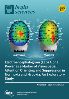
Cover Story (view full-size image): While EEG alpha desynchronization has been related to orienting of visuospatial attention, its synchronization would index an inhibition of attentional processes. Research has indicated that alpha is also affected by a deficient brain oxygenation (hypoxia). In this study, alpha power during attention alerting, cued and uncued orienting, and control functions in different workload conditions was monitored both in normoxia and hypoxia in healthy students. Independent of oxygenation level, alpha desynchronization was maximal during uncued attentional orienting, whereas hypoxia enhanced alpha synchronization over the right-sided occipitoparietal areas. We interpreted such right-sided modulation as a marker of the suppression of attentional processing induced by hypoxia. View this paper.
  • Issues are regarded as officially published after their release is announced to the table of contents alert mailing list.
  • You may sign up for e-mail alerts to receive table of contents of newly released issues.
  • PDF is the official format for papers published in both, html and pdf forms. To view the papers in pdf format, click on the "PDF Full-text" link, and use the free Adobe Reader to open them.
Order results
Result details
Section
Select all
Export citation of selected articles as:
15 pages, 5895 KiB  
Article
A Simple Pattern of Movement Is Not Able to Inhibit Experimental Pain in FM Patients and Controls: An sLORETA Study
by Eleonora Gentile, Katia Ricci, Eleonora Vecchio, Giuseppe Libro, Marianna Delussi, Antonio Casas-Barragàn and Marina de Tommaso
Brain Sci. 2020, 10(3), 190; https://doi.org/10.3390/brainsci10030190 - 24 Mar 2020
Cited by 4 | Viewed by 3838
Abstract
Motor cortex activation seems to induce an analgesic effect on pain that would be different between patients with fibromyalgia (FM) and control subjects. This study was conducted to analyze the changes of the laser-evoked potentials (LEPs) induced during a finger tapping task in [...] Read more.
Motor cortex activation seems to induce an analgesic effect on pain that would be different between patients with fibromyalgia (FM) and control subjects. This study was conducted to analyze the changes of the laser-evoked potentials (LEPs) induced during a finger tapping task in the FM patients and the controls employing a multi-dipolar analysis according to Standardized low resolution brain electromagnetic tomography (sLORETA) method. The LEPs from 38 FM patients and 21 controls were analyzed. The LEPs were recorded while subjects performed a slow and a fast finger tapping task. We confirmed that the difference between N1, N2 and P2 wave amplitudes between conditions and groups was not significant. In control subjects, the fast finger tapping task induced a modification of cortical source activation in the main areas processing laser stimulation from the moving hand independently from the movement speed. In summary, a simple and repetitive movement is not able to induce consistent inhibition of experimental pain evoked by the moving and the not moving hand in each group. It could interfere with LEP sources within the limbic area at least in control subjects, without inhibit cortical responses or explain the different pattern of motor and pain interaction in FM patients. Full article
(This article belongs to the Special Issue Emerging Treatment Approaches for Chronic Pain Syndromes)
Show Figures

Figure 1

16 pages, 3102 KiB  
Article
Olfactory Memory in Depression: State and Trait Differences between Bipolar and Unipolar Disorders
by François Kazour, Sami Richa, Chantale Abi Char, Boriana Atanasova and Wissam El-Hage
Brain Sci. 2020, 10(3), 189; https://doi.org/10.3390/brainsci10030189 - 24 Mar 2020
Cited by 7 | Viewed by 3890
Abstract
Background: Changes in olfactory recognition memory may constitute sensory markers in depression. Significant differences may exist between unipolar and bipolar depression. Our study compares olfactory memory between control, unipolar, and bipolar patients in depressed and euthymic states in order to identify potential markers [...] Read more.
Background: Changes in olfactory recognition memory may constitute sensory markers in depression. Significant differences may exist between unipolar and bipolar depression. Our study compares olfactory memory between control, unipolar, and bipolar patients in depressed and euthymic states in order to identify potential markers of depression. Methods: 176 participants were recruited in 5 groups: depressed bipolar (DB), euthymic bipolar (EB), depressed unipolar (DU), euthymic unipolar (EU), and controls (HC). The participants had a standardized clinical and olfactory assessment (olfactory memory, evaluation of pleasantness, intensity, familiarity, and emotional aspect of smells). Results: DU, DB, and EU patients had a deficit in olfactory memory compared to HC. DB patients had lower capacity to recognize new odors. DB and DU patients had more limited detection of unfamiliar odors than HC. DB patients rated odors as less pleasant compared to the other groups. All groups had lower hedonic ratings than HC. DB patients had lower emotional ratings than EU patients. Conclusions: Olfactory memory is impaired in depressive states, thus constituting a state marker of depression. Impairments in olfactory memory persist after remission of bipolar depression, thus constituting a possible trait marker of bipolarity. Hedonic rating differentiates unipolar from bipolar depression. This is the first study that identifies a sensory marker differentiating between unipolar and bipolar depression. Full article
(This article belongs to the Special Issue Olfaction as a Marker for Psychiatric and Neurological Diseases)
Show Figures

Figure 1

14 pages, 1137 KiB  
Article
The Reaction Switching Produces A Greater Bias to Prepotent Response than Reaction Inhibition
by Kirill Fadeev, Tatyana Alikovskaia, Alexey Tumyalis, Alexey Smirnov and Kirill Golokhvast
Brain Sci. 2020, 10(3), 188; https://doi.org/10.3390/brainsci10030188 - 24 Mar 2020
Cited by 3 | Viewed by 3451
Abstract
There is a discussion about common or various mechanisms of response inhibition and response switching. To understand these mechanisms, we used a modified Go/NoGo task with three stimulus categories. The subjects were instructed to press a button in response to frequent Go stimuli, [...] Read more.
There is a discussion about common or various mechanisms of response inhibition and response switching. To understand these mechanisms, we used a modified Go/NoGo task with three stimulus categories. The subjects were instructed to press a button in response to frequent Go stimuli, press another button in response to rare Go stimuli and hold any motor response following the presentation of NoGo stimuli. The results showed a decrease in reaction time for frequent Go, following both categories of rare stimuli and the decrease was greater following rare Go. Also, the total number of errors did not differ between Go and NoGo, however, a greater bias of error rate towards frequent Go stimuli was found for rare Go compared to NoGo. Finally, positive correlations were found between the increase in reaction time for rare Go compared to frequent Go and the number of errors for both rare Go and rare NoGo. Together, these results indicate that both rare Go and NoGo stimuli required to inhibit the prepotent response, but rare Go in comparison to NoGo stimuli also evoked a conflict between prepotent and alternative responses, which is expressed in greater response bias toward frequent Go. Full article
(This article belongs to the Section Systems Neuroscience)
Show Figures

Figure 1

6 pages, 529 KiB  
Case Report
Valproic Acid-Induced Hyperammonemic Encephalopathy in a Patient with Bipolar Disorder: A Case Report
by Meng-Yu Wu, Fang-Yu Chang, Jian-Yu Ke, Chien-Sheng Chen, Po-Chen Lin and Tzong-Shi Wang
Brain Sci. 2020, 10(3), 187; https://doi.org/10.3390/brainsci10030187 - 24 Mar 2020
Cited by 9 | Viewed by 5276
Abstract
Valproic acid (VPA) is widely used to control various seizure disorders and psychiatric disorders. Valproic acid-induced hyperammonemic encephalopathy (VHE) is a rare but dangerous complication of VPA-induced toxicity. For this case report, several risk factors were identified, including young age, polytherapy regimens, VPA [...] Read more.
Valproic acid (VPA) is widely used to control various seizure disorders and psychiatric disorders. Valproic acid-induced hyperammonemic encephalopathy (VHE) is a rare but dangerous complication of VPA-induced toxicity. For this case report, several risk factors were identified, including young age, polytherapy regimens, VPA overdose, poor liver function, and carnitine deficiency. The detailed mechanisms of VHE remained unclear. Hyperammonemia may be caused by hypocarnitinemia, leading to imbalanced VPA metabolism. VHE may initially cause gastrointestinal symptoms, followed by a decreased level of consciousness and seizure. Early diagnosis of VHE is important for physicians for the timely reversal of VHE by discontinuing administration of VPA and administering lactulose or levocarnitine. Here, we describe a patient with a bipolar disorder who presented with VHE after receiving a strict vegetarian diet in our hospital. We recommend that VHE be included in the differential diagnosis of patients with high serum VPA levels and strictly vegetarian diets, especially those presenting with acute gastrointestinal symptoms. Full article
Show Figures

Figure 1

13 pages, 1449 KiB  
Article
Effects of Repetitive Transcranial Magnetic Stimulation (rTMS) Combined with Aerobic Exercise on the Recovery of Motor Function in Ischemic Stroke Rat Model
by Juanxiu Cui, Cuk-Seong Kim, Yeongwook Kim, Min Kyun Sohn and Sungju Jee
Brain Sci. 2020, 10(3), 186; https://doi.org/10.3390/brainsci10030186 - 23 Mar 2020
Cited by 19 | Viewed by 4375
Abstract
The therapeutic benefits of repetitive transcranial magnetic stimulation (rTMS) combined with rehabilitation therapy on recovery after stroke have not been fully elucidated. This study aimed to explore the therapeutic effects of rTMS followed by aerobic exercise on neuroplasticity and recovery of motor function [...] Read more.
The therapeutic benefits of repetitive transcranial magnetic stimulation (rTMS) combined with rehabilitation therapy on recovery after stroke have not been fully elucidated. This study aimed to explore the therapeutic effects of rTMS followed by aerobic exercise on neuroplasticity and recovery of motor function in a rat model of permanent middle cerebral artery occlusion (MCAO). Rats were randomized into sham operation (N = 10, sham op), MCAO (N = 10, control group), rTMS (N = 10, MCAO and rTMS therapy), and combination groups (N = 10, MCAO and combination therapy). High-frequency rTMS (10 Hz) was applied on the ipsilesional forepaw motor cortex, and aerobic exercise training on the rotarod was performed for two weeks. The rotarod and Garcia tests were conducted to evaluate changes in behavioral function. Motor evoked potentials (MEPs) were used to evaluate electrophysiological changes. Stroke severity was assessed using infarction volume measurement. Neuronal recovery was explored with western blot for brain-derived neurotrophic factor (BDNF) pathway proteins. Compared with control therapy, combination therapy was significantly more effective than rTMS therapy for improving function on the rotarod test (p = 0.08), Garcia test (p = 0.001), and MEP amplitude (p = 0.001) In conclusion, combination therapy may be a potential treatment to promote recovery of motor function and neuroplasticity in stroke patients. Full article
(This article belongs to the Special Issue Stroke Treatments and Therapies)
Show Figures

Figure 1

12 pages, 964 KiB  
Protocol
Effects of Transcranial Direct Current Stimulation on Hand Dexterity in Multiple Sclerosis: A Design for a Randomized Controlled Trial
by Samar S. Ayache, Naji Riachi, Rechdi Ahdab and Moussa A. Chalah
Brain Sci. 2020, 10(3), 185; https://doi.org/10.3390/brainsci10030185 - 23 Mar 2020
Cited by 4 | Viewed by 3473
Abstract
Background: Cerebellar and motor tracts are frequently impaired in multiple sclerosis (MS). Altered hand dexterity constitutes a challenge in clinical practice, since medical treatment shows very limited benefits in this domain. Cerebellar control is made via several cerebellocortical pathways, of which the most [...] Read more.
Background: Cerebellar and motor tracts are frequently impaired in multiple sclerosis (MS). Altered hand dexterity constitutes a challenge in clinical practice, since medical treatment shows very limited benefits in this domain. Cerebellar control is made via several cerebellocortical pathways, of which the most studied one links the cerebellum to the contralateral motor cortex via the contralateral ventro-intermediate nucleus of the thalamus influencing the corticospinal outputs. Modulating the activity of the cerebellum or of the motor cortex could be of help. Method: The main interest here is to evaluate the efficacy of transcranial direct current stimulation (tDCS), a noninvasive brain stimulation technique, in treating altered dexterity in MS. Forty-eight patients will be recruited in a randomized, double-blind, sham-controlled, and crossover study. They will randomly undergo one of the three interventions: anodal tDCS over the primary motor area, cathodal tDCS over the cerebellum, or sham. Each block consists of five consecutive daily sessions with direct current (2 mA), lasting 20 min each. The primary outcome will be the improvement in manual dexterity according to the change in the time required to complete the nine-hole pegboard task. Secondary outcomes will include fatigue, pain, spasticity, and mood. Patients’ safety and satisfaction will be rated. Discussion: Due to its cost-effective, safe, and easy-to-use profile, motor or cerebellar tDCS may constitute a potential tool that might improve dexterity in MS patients and therefore ameliorate their quality of life. Full article
(This article belongs to the Special Issue Novel therapies for Multiple Sclerosis)
Show Figures

Figure 1

12 pages, 1226 KiB  
Article
A Two-Stage Screening Approach with I-TC and Q-CHAT to Identify Toddlers at Risk for Autism Spectrum Disorder within the Italian Public Health System
by Raffaella Devescovi, Lorenzo Monasta, Maura Bin, Giulia Bresciani, Alice Mancini, Marco Carrozzi and Costanza Colombi
Brain Sci. 2020, 10(3), 184; https://doi.org/10.3390/brainsci10030184 - 23 Mar 2020
Cited by 10 | Viewed by 4588
Abstract
Standardized screening programs ensure that children are monitored for early signs of autism spectrum disorder (ASD) in order to promote earlier diagnosis and intervention. The aim of this study is to identify early signs of atypical development consistent with ASD or other developmental [...] Read more.
Standardized screening programs ensure that children are monitored for early signs of autism spectrum disorder (ASD) in order to promote earlier diagnosis and intervention. The aim of this study is to identify early signs of atypical development consistent with ASD or other developmental disorders in a population of 224 low-risk toddlers through a two-stage screening approach applied at 12 and 18 months of age. We adopted two screening tools combined: 1. the Communication and Symbolic Behavior Scales Developmental Profile (CSBS DP) Infant–Toddler Checklist (I-TC) and 2. The Quantitative Checklist for Autism in Toddlers (Q-CHAT). We assessed their sensitivity and specificity related to the diagnostic outcome at 36 months. The results showed that autistic signs can be detected as early as the first year even through a few questions extrapolated from both screeners and that our model could be used as a screening procedure in the Italian public health system. Full article
(This article belongs to the Special Issue Advances in Autism Research)
Show Figures

Graphical abstract

13 pages, 1439 KiB  
Review
Overcoming Alzheimer’s Disease Stigma by Leveraging Artificial Intelligence and Blockchain Technologies
by Alexander Pilozzi and Xudong Huang
Brain Sci. 2020, 10(3), 183; https://doi.org/10.3390/brainsci10030183 - 23 Mar 2020
Cited by 18 | Viewed by 5693
Abstract
Alzheimer’s disease (AD) imposes a considerable burden on those diagnosed. Faced with a neurodegenerative decline for which there is no effective cure or prevention method, sufferers of the disease are subject to judgement, both self-imposed and otherwise, that can have a great deal [...] Read more.
Alzheimer’s disease (AD) imposes a considerable burden on those diagnosed. Faced with a neurodegenerative decline for which there is no effective cure or prevention method, sufferers of the disease are subject to judgement, both self-imposed and otherwise, that can have a great deal of effect on their lives. The burden of this stigma is more than just psychological, as reluctance to face an AD diagnosis can lead people to avoid early diagnosis, treatment, and research opportunities that may be beneficial to them, and that may help progress towards fighting AD and its progression. In this review, we discuss how recent advents in information technology may be employed to help fight this stigma. Using artificial intelligence (AI) technologies, specifically natural language processing (NLP), to classify the sentiment and tone of texts, such as those of online posts on various social media sites, has proven to be an effective tool for assessing the opinions of the general public on certain topics. These tools can be used to analyze the public stigma surrounding AD. Additionally, there is much concern among individuals that an AD diagnosis, or evidence of pre-clinical AD such as a biomarker or imaging test results, may wind up unintentionally disclosed to an entity that may discriminate against them. The lackluster security record of many medical institutions justifies this fear to an extent. Adopting more secure and decentralized methods of data transfer and storage, and giving patients enhanced ability to control their own data, such as a blockchain-based method, may help to alleviate some of these fears. Full article
(This article belongs to the Special Issue Dementia and Cognitive Ageing)
Show Figures

Graphical abstract

18 pages, 1984 KiB  
Review
Keeping in Touch with Mental Health: The Orienting Reflex and Behavioral Outcomes from Calatonia
by Anita Ribeiro Blanchard and William Edgar Comfort
Brain Sci. 2020, 10(3), 182; https://doi.org/10.3390/brainsci10030182 - 22 Mar 2020
Cited by 2 | Viewed by 9125
Abstract
Physical and psychological therapy based on touch has been gradually integrated into broader mental health settings in the past two decades, evolving from a variety of psychodynamic, neurobiological and trauma-based approaches, as well as Eastern and spiritual philosophies and other integrative and converging [...] Read more.
Physical and psychological therapy based on touch has been gradually integrated into broader mental health settings in the past two decades, evolving from a variety of psychodynamic, neurobiological and trauma-based approaches, as well as Eastern and spiritual philosophies and other integrative and converging systems. Nevertheless, with the exception of a limited number of well-known massage therapy techniques, only a few structured protocols of touch therapy have been standardized and researched to date. This article describes a well-defined protocol of touch therapy in the context of psychotherapy—the Calatonia technique—which engages the orienting reflex. The orienting reflex hypothesis is explored here as one of the elements of this technique that helps to decrease states of hypervigilance and chronic startle reactivity (startle and defensive reflexes) and restore positive motivational and appetitive states. Full article
Show Figures

Figure 1

15 pages, 560 KiB  
Article
Multi-View Based Multi-Model Learning for MCI Diagnosis
by Ping Cao, Jie Gao and Zuping Zhang
Brain Sci. 2020, 10(3), 181; https://doi.org/10.3390/brainsci10030181 - 20 Mar 2020
Cited by 10 | Viewed by 4106
Abstract
Mild cognitive impairment (MCI) is the early stage of Alzheimer’s disease (AD). Automatic diagnosis of MCI by magnetic resonance imaging (MRI) images has been the focus of research in recent years. Furthermore, deep learning models based on 2D view and 3D view have [...] Read more.
Mild cognitive impairment (MCI) is the early stage of Alzheimer’s disease (AD). Automatic diagnosis of MCI by magnetic resonance imaging (MRI) images has been the focus of research in recent years. Furthermore, deep learning models based on 2D view and 3D view have been widely used in the diagnosis of MCI. The deep learning architecture can capture anatomical changes in the brain from MRI scans to extract the underlying features of brain disease. In this paper, we propose a multi-view based multi-model (MVMM) learning framework, which effectively combines the local information of 2D images with the global information of 3D images. First, we select some 2D slices from MRI images and extract the features representing 2D local information. Then, we combine them with the features representing 3D global information learned from 3D images to train the MVMM learning framework. We evaluate our model on the Alzheimer’s Disease Neuroimaging Initiative (ADNI) database. The experimental results show that our proposed model can effectively recognize MCI through MRI images (accuracy of 87.50% for MCI/HC and accuracy of 83.18% for MCI/AD). Full article
(This article belongs to the Special Issue Early Diagnosis and Intervention in Alzheimer's Disease)
Show Figures

Figure 1

31 pages, 681 KiB  
Review
Systematic Review of Level 1 and Level 2 Screening Tools for Autism Spectrum Disorders in Toddlers
by Serena Petrocchi, Annalisa Levante and Flavia Lecciso
Brain Sci. 2020, 10(3), 180; https://doi.org/10.3390/brainsci10030180 - 19 Mar 2020
Cited by 45 | Viewed by 8339
Abstract
The present study provides a systematic review of level 1 and level 2 screening tools for the early detection of autism under 24 months of age and an evaluation of the psychometric and measurement properties of their studies. Methods: Seven databases (e.g., Scopus, [...] Read more.
The present study provides a systematic review of level 1 and level 2 screening tools for the early detection of autism under 24 months of age and an evaluation of the psychometric and measurement properties of their studies. Methods: Seven databases (e.g., Scopus, EBSCOhost Research Database) were screened and experts in the autism spectrum disorders (ASD) field were questioned; Preferred Reporting Items for Systematic review and Meta-Analysis (PRISMA) guidelines and Consensus-based Standard for the selection of health Measurement INstruments (COSMIN) checklist were applied. Results: the study included 52 papers and 16 measures; most of them were questionnaires, and the Modified-CHecklist for Autism in Toddler (M-CHAT) was the most extensively tested. The measures’ strengths (analytical evaluation of methodological quality according to COSMIN) and limitations (in term of Negative Predictive Value, Positive Predictive Value, sensitivity, and specificity) were described; the quality of the studies, assessed with the application of the COSMIN checklist, highlighted the necessity of further validation studies for all the measures. According to COSMIN results, the M-CHAT, First Years Inventory (FYI), and Quantitative-CHecklist for Autism in Toddler (Q-CHAT) seem to be promising measures that may be applied systematically by health professionals in the future. Full article
(This article belongs to the Special Issue Advances in Autism Research)
Show Figures

Figure 1

10 pages, 1053 KiB  
Article
Aspirin Resistance Affects Medium-Term Recurrent Vascular Events after Cerebrovascular Incidents: A Three-Year Follow-up Study
by Adam Wiśniewski, Karolina Filipska, Joanna Sikora and Grzegorz Kozera
Brain Sci. 2020, 10(3), 179; https://doi.org/10.3390/brainsci10030179 - 19 Mar 2020
Cited by 13 | Viewed by 3099
Abstract
Background: The aim of this prospective, a three-year follow-up study, was to establish the role of high on-treatment platelet reactivity (HTPR) in predicting the recurrence of vascular events in patients after cerebrovascular incidents, particularly in the aspect of stroke etiology. Methods: [...] Read more.
Background: The aim of this prospective, a three-year follow-up study, was to establish the role of high on-treatment platelet reactivity (HTPR) in predicting the recurrence of vascular events in patients after cerebrovascular incidents, particularly in the aspect of stroke etiology. Methods: The study included 101 subjects with non-embolic cerebral ischemia (69 patients with ischemic stroke and 32 patients with transient ischemic attack) treated with 150 mg of acetylsalicylic acid (aspirin) a day. The platelet reactivity was tested in the first 24 h after the onset of cerebral ischemia by impedance aggregometry. Recurrent vascular events, including recurrent ischemic stroke, transient ischemic attack, myocardial infarction, systemic embolism, or sudden death of vascular reason, were assessed 36 months after the onset of cerebral ischemia. Results: Recurrent vascular events occurred between 3 and 9 months after onset in 8.5% of all subjects; in the HTPR subgroup, recurrent vascular events occurred in 17.9%; in the normal on-treatment platelet reactivity (NTPR) subgroup, they occurred in 4.6%. We did not notice early or long-term recurrent events. Aspirin resistant subjects had a significantly higher risk of recurrent vascular events than did aspirin sensitive subjects (Odds ratio (OR) = 4.57, 95% Confidence interval (CI) 1.00–20.64; p = 0.0486). Cox proportional hazard models showed that large-vessel disease (Hazard ratio (HR) 12.04, 95% CI 2.43–59.72; p = 0.0023) and high on-treatment platelet reactivity (HR 4.28, 95% CI 1.02–17.93; p = 0.0465) were independent predictors of recurrent vascular events. Conclusion: Aspirin resistance in the acute phase of cerebral ischemia was associated with a higher risk of recurrent medium-term vascular events, coexisting with large-vessel etiology of stroke. Platelet function-guided personalized antiplatelet treatment should be considered for patients with recurrent strokes, especially when due to large-vessel disease. Full article
(This article belongs to the Special Issue Stroke Treatments and Therapies)
Show Figures

Figure 1

11 pages, 1445 KiB  
Article
Insomnia Might Influence the Thickness of Choroid, Retinal Nerve Fiber and Inner Plexiform Layer
by Cigdem Sahbaz, Ahmet Elbay, Mine Ozcelik and Hakan Ozdemir
Brain Sci. 2020, 10(3), 178; https://doi.org/10.3390/brainsci10030178 - 19 Mar 2020
Cited by 11 | Viewed by 3508
Abstract
Sleep may play a fundamental role in retinal regulation and the degree of retinal variables. However, no clinical study has investigated optical coherence tomography (OCT) parameters in patients with primary insomnia. All participants were evaluated with the insomnia severity index (ISI) and the [...] Read more.
Sleep may play a fundamental role in retinal regulation and the degree of retinal variables. However, no clinical study has investigated optical coherence tomography (OCT) parameters in patients with primary insomnia. All participants were evaluated with the insomnia severity index (ISI) and the Pittsburgh sleep quality index (PSQI). The retinal nerve fiber layer (RNFL), ganglion cell layer (GC), inner plexiform layer (IPL), macula and choroidal (CH) thickness were compared between 52 drug-naïve patients with primary insomnia and 45 age-gender-BMI-smoke status matched healthy controls (HC). The patients with primary insomnia differed from the HC regarding RNFL-Global (p = 0.024) and RNFL-Nasal inferior (p = 0.010); IPL-Temporal (p < 0.001), IPL-Nasal (p < 0.001); CH-Global (p < 0.001), CH-Temporal (p = 0.004), CH-Nasal (p < 0.001), and CH-Fovea (p = 0.019). ISI correlated with RNFL-Global and RNFL-Nasal inferior. The regression analysis revealed that ISI was the significant predictor for the thickness of RNFL- Nasal inferior (p = 0.020), RNFL-Global (p = 0.031), and CH-Nasal (p = 0.035) in patients with primary insomnia. Sleep disorders are seen commonly in patients with psychiatric, including ocular diseases. Adjusting the effect of insomnia can help to clarify the consistency in findings of OCT. Full article
(This article belongs to the Special Issue Insomnia: Beyond Hyperarousal)
Show Figures

Figure 1

7 pages, 1300 KiB  
Case Report
Injury of Corticospinal Tract in a Patient with Subarachnoid Hemorrhage as Determined by Diffusion Tensor Tractography: A Case Report
by Chan-Hyuk Park, Hyeong Ryu, Chang-Hwan Kim, Kyung-Lim Joa, Myeong-Ok Kim and Han-Young Jung
Brain Sci. 2020, 10(3), 177; https://doi.org/10.3390/brainsci10030177 - 19 Mar 2020
Cited by 6 | Viewed by 4115
Abstract
We report diffusion tensor tractography (DTT) of the corticospinal tract (CST) in a patient with paresis of all four limbs following subarachnoid hemorrhage (SAH) with intraventricular hemorrhage (IVH) after the rupture of an anterior communicating artery (ACoA) aneurysm rupture. The 73-year-old female was [...] Read more.
We report diffusion tensor tractography (DTT) of the corticospinal tract (CST) in a patient with paresis of all four limbs following subarachnoid hemorrhage (SAH) with intraventricular hemorrhage (IVH) after the rupture of an anterior communicating artery (ACoA) aneurysm rupture. The 73-year-old female was admitted to our emergency room in a semi-comatose mental state. After coil embolization—an acute SAH treatment—she was transferred to our rehabilitation department with motor weakness development, two weeks after SAH. Upon admission, she was alert but she complained of motor weakness (upper limbs: MRC 3/5, and lower limbs: MRC 1/5). Four weeks after onset, DTT showed that the bilateral CSTs failed to reach the cerebral cortex. The left CST demonstrated a wide spread of fibers within the corona radiata as well as significantly lower tract volume (TV) and higher fractional anisotropy (FA) as well as mean diffusivity (MD) compared to the controls. On the other hand, the right CST shifted to the posterior region at the corona radiata, and MD values of the right CST were significantly higher when compared to the controls. Changes in both CSTs were attributed to vasogenic edema and compression caused by untreated hydrocephalus. We demonstrate in this case, two different pathophysiological entitles, contributing to this patient’s motor weakness after SAH. Full article
Show Figures

Figure 1

17 pages, 290 KiB  
Review
A Spanish Consensus on the Use of Safinamide for Parkinson’s Disease in Clinical Practice
by Javier Pagonabarraga, José Matías Arbelo, Francisco Grandas, Maria-Rosario Luquin, Pablo Martínez Martín, Mari Cruz Rodríguez-Oroz, Francesc Valldeoriola and Jaime Kulisevsky
Brain Sci. 2020, 10(3), 176; https://doi.org/10.3390/brainsci10030176 - 18 Mar 2020
Cited by 7 | Viewed by 3841
Abstract
Safinamide is an approved drug for the treatment of motor fluctuations in Parkinson’s disease (PD). Scarce data are available on its use in clinical practice. A group of Spanish movement disorders specialists was convened to review the use of safinamide across different clinical [...] Read more.
Safinamide is an approved drug for the treatment of motor fluctuations in Parkinson’s disease (PD). Scarce data are available on its use in clinical practice. A group of Spanish movement disorders specialists was convened to review the use of safinamide across different clinical scenarios that may guide neurologists in clinical practice. Eight specialists with recognized expertise in PD management elaborated the statements based on available evidence in the literature and on their clinical experience. The RAND/UCLA method was carried, with final conclusions accepted after a 2-round modified Delphi process. Higher level of agreement between panellists was reached for the following statements. Safinamide significantly improves mean daily OFF time without troublesome dyskinesias. Adjunctive treatment with safinamide is associated with motor improvements in patients with mid-to-late PD. The efficacy of safinamide on motor fluctuations is maintained at long-term, with no increase over time in dyskinesias severity. The clinical benefits of safinamide on pain and depression remain unclear. Safinamide presents a similar incidence of adverse events compared with placebo. The efficacy and safety of safinamide shown in the pivotal clinical trials are reproduced in clinical practice, with improvement of parkinsonian symptoms, decrease of daily OFF time, control of dyskinesias at the long term, and good tolerability and safety. Full article
(This article belongs to the Special Issue Frontiers in Parkinson’s Disease (PD))
31 pages, 1994 KiB  
Review
The Contribution of Functional Magnetic Resonance Imaging to the Understanding of the Effects of Acute Physical Exercise on Cognition
by Fabian Herold, Norman Aye, Nico Lehmann, Marco Taubert and Notger G. Müller
Brain Sci. 2020, 10(3), 175; https://doi.org/10.3390/brainsci10030175 - 18 Mar 2020
Cited by 37 | Viewed by 7508
Abstract
The fact that a single bout of acute physical exercise has a positive impact on cognition is well-established in the literature, but the neural correlates that underlie these cognitive improvements are not well understood. Here, the use of neuroimaging techniques, such as functional [...] Read more.
The fact that a single bout of acute physical exercise has a positive impact on cognition is well-established in the literature, but the neural correlates that underlie these cognitive improvements are not well understood. Here, the use of neuroimaging techniques, such as functional magnetic resonance imaging (fMRI), offers great potential, which is just starting to be recognized. This review aims at providing an overview of those studies that used fMRI to investigate the effects of acute physical exercises on cerebral hemodynamics and cognition. To this end, a systematic literature survey was conducted by two independent reviewers across five electronic databases. The search returned 668 studies, of which 14 studies met the inclusion criteria and were analyzed in this systematic review. Although the findings of the reviewed studies suggest that acute physical exercise (e.g., cycling) leads to profound changes in functional brain activation, the small number of available studies and the great variability in the study protocols limits the conclusions that can be drawn with certainty. In order to overcome these limitations, new, more well-designed trials are needed that (i) use a more rigorous study design, (ii) apply more sophisticated filter methods in fMRI data analysis, (iii) describe the applied processing steps of fMRI data analysis in more detail, and (iv) provide a more precise exercise prescription. Full article
(This article belongs to the Special Issue Exercising against Age-Effects on the Brain)
Show Figures

Figure 1

14 pages, 2256 KiB  
Review
Locomotor Coordination, Visual Perception and Head Stability during Running
by Joseph Hamill, Jongil Lim and Richard van Emmerik
Brain Sci. 2020, 10(3), 174; https://doi.org/10.3390/brainsci10030174 - 18 Mar 2020
Cited by 9 | Viewed by 6442
Abstract
Perception and action are coupled such that information from the perceptual system is related to the dynamics of action in order to regulate behavior adaptively. Using running as a model of a cyclic behavior, this coupling involves a continuous, cyclic relationship between the [...] Read more.
Perception and action are coupled such that information from the perceptual system is related to the dynamics of action in order to regulate behavior adaptively. Using running as a model of a cyclic behavior, this coupling involves a continuous, cyclic relationship between the runner’s perception of the environment and the necessary adjustments of the body that ultimately result in a stable pattern of behavior. The purpose of this paper is to illustrate how individuals relate visual perception to rhythmic locomotor coordination patterns in conditions during which foot–ground collisions and visual task demands are altered. We review the findings of studies conducted to illustrate how humans change their behavior to maintain head stability during running with and without various degrees of visual challenge from the environment. Finally, we show that the human body adapts specific segment/joint configuration and coordination patterns to maintain head stability, both in the lower extremity and upper body segments, together with an increase in coordinative variability. These results indicate that in human locomotion, under higher speed (running) and visual task demands, systematic adaptations occur in the rhythmic coupling between the perceptual and movement systems. Full article
(This article belongs to the Special Issue Rhythmic Motor Pattern Generation)
Show Figures

Figure 1

15 pages, 373 KiB  
Review
Neurobiological Trajectories Involving Social Isolation in PTSD: A Systematic Review
by Ilias I Vlachos, Charalambos Papageorgiou and Maria Margariti
Brain Sci. 2020, 10(3), 173; https://doi.org/10.3390/brainsci10030173 - 18 Mar 2020
Cited by 21 | Viewed by 8581
Abstract
Social isolation (SI) stress has been recognized as a major risk factor of morbidity in humans and animals, exerting damaging effects at the physical and mental health levels. Posttraumatic stress disorder (PTSD), on the other hand, occurs as a result of experiencing serious, [...] Read more.
Social isolation (SI) stress has been recognized as a major risk factor of morbidity in humans and animals, exerting damaging effects at the physical and mental health levels. Posttraumatic stress disorder (PTSD), on the other hand, occurs as a result of experiencing serious, life-threatening, traumatic events and involves involuntary re-experiencing trauma (intrusion), avoidance symptoms, and distortions of cognition and emotional arousal. The literature shows that PTSD is affected by genetic predisposition and triggers a large neurocircuitry involving the amygdala, insula, hippocampus, anterior cingulate- and prefrontal-cortex, and affects the function of the neuroendocrine and immune systems. Social isolation seems to influence the predisposition, onset and outcome of PTSD in humans, whereas it constitutes a valid model of the disorder in animals. According to the PRISMA (preferred reporting items for systematic reviews and meta-analyses) protocol, we systematically reviewed all original studies involving the neurobiological trajectories between SI and PTSD published till July 2019 (database: PubMed/Medline). Out of 274 studies, 10 met the inclusion criteria. We present the results of the retrieved studies in terms of hypothalamic-pituitary-adrenal (HPA)-axis and endocannabinoid system function, immune reactions, neuroplasticity, novel pharmacological targets, and shortening of telomere length, which confirm a synergistic effect on a neurobiological level between the two entities. Full article
Show Figures

Figure 1

15 pages, 447 KiB  
Article
Design Fluency in Children with ADHD and Comorbid Disorders
by Anaïs Fournier, Bruno Gauthier, Marie-Claude Guay and Véronique Parent
Brain Sci. 2020, 10(3), 172; https://doi.org/10.3390/brainsci10030172 - 17 Mar 2020
Cited by 6 | Viewed by 4150
Abstract
Background: Attention deficit/hyperactivity disorder (ADHD) is often associated with frontal executive impairment in children. Oppositional defiant disorder (ODD) and anxiety disorders (AD) frequently accompany ADHD, but the impact of these comorbid disorders on cognition remains elusive. The five-point test (FPT), a design fluency [...] Read more.
Background: Attention deficit/hyperactivity disorder (ADHD) is often associated with frontal executive impairment in children. Oppositional defiant disorder (ODD) and anxiety disorders (AD) frequently accompany ADHD, but the impact of these comorbid disorders on cognition remains elusive. The five-point test (FPT), a design fluency task, has been shown to be sensitive to neurological damage, specifically to frontal lobe lesions in patients with brain injuries. The purpose of this study was to compare the performances of neurotypical children with that of children with ADHD, ADHD-ODD, and ADHD-AD on the FPT in order to examine whether these groups could be distinguished from one another based on their cognitive profile. Methods: A total of 111 children aged 8 to 11 years old participated in the study. Six measures from the FPT were used to characterize their performance. Results: Statistically significant differences between groups were observed for five of the six FPT measures. Essentially, children with ADHD-ODD made more repeated designs than the three other groups (control p > 0.001, ADHD p = 0.008, ADHD-AD p = 0.008), while children with ADHD-AD produced fewer total and correct designs than the control and ADHD groups (p = 0.009). Conclusions: This suggests that comorbidities have an additive impact on the cognitive profile of children with ADHD. Design fluency may be a sensitive measure for capturing the subtle cognitive deficits that are likely to be involved in these disorders. Full article
Show Figures

Figure 1

6 pages, 1490 KiB  
Case Report
Neisseria meningitidis Induced Fatal Waterhouse–Friderichsen Syndrome in a Patient Presenting With Disseminated Intravascular Coagulation and Multiple Organ Failure
by Meng-Yu Wu, Chien-Sheng Chen, Chih-Yi Tsay, Giou-Teng Yiang, Jian-Yu Ke and Po-Chen Lin
Brain Sci. 2020, 10(3), 171; https://doi.org/10.3390/brainsci10030171 - 17 Mar 2020
Cited by 6 | Viewed by 10211
Abstract
Neisseria meningitidis-induced acute systemic meningococcal disease is an emergency and a fatal condition that has a high mortality rate. In patients with a fulminant infection, a maculopapular petechial eruption, purpura fulminans, or an ecchymotic lesion are worrisome signs reflecting disseminated intravascular coagulation [...] Read more.
Neisseria meningitidis-induced acute systemic meningococcal disease is an emergency and a fatal condition that has a high mortality rate. In patients with a fulminant infection, a maculopapular petechial eruption, purpura fulminans, or an ecchymotic lesion are worrisome signs reflecting disseminated intravascular coagulation (DIC) and hint at Waterhouse–Friderichsen syndrome (WFS). Here, we describe a rare case of a patient with a fulminant Neisseria meningitidis-induced acute systemic meningococcal disease presenting with high-grade fever without meningitis symptoms. Fatal septicemia with DIC and multiple organ failure was noted. WFS was chiefly suspected. We highlight the clinical features and pathogenesis of Neisseria meningitidis-induced meningococcemia and WFS. We propose that they should be kept in mind, especially in patients presenting with a petechial eruption and purpura fulminans. Full article
(This article belongs to the Special Issue Neurocritical Care and Cerebrovascular Health)
Show Figures

Figure 1

9 pages, 236 KiB  
Opinion
Acquisition of Ownership Illusion with Self-Disownership in Neurological Patients
by Mariella Pazzaglia, Anna Maria Giannini and Francesca Federico
Brain Sci. 2020, 10(3), 170; https://doi.org/10.3390/brainsci10030170 - 15 Mar 2020
Cited by 9 | Viewed by 3832
Abstract
The multisensory regions in frontoparietal cortices play a crucial role in the sense of body and self. Disrupting this sense may lead to a feeling of disembodiment, or more generally, a sense of disownership. Experimentally, this altered consciousness disappears during illusory own-body perceptions, [...] Read more.
The multisensory regions in frontoparietal cortices play a crucial role in the sense of body and self. Disrupting this sense may lead to a feeling of disembodiment, or more generally, a sense of disownership. Experimentally, this altered consciousness disappears during illusory own-body perceptions, increasing the intensity of perceived ownership for an external virtual limb. In many clinical conditions, particularly in individuals with a discontinuous or absent sense of bodily awareness, the brain may effortlessly create a convincing feeling of body ownership over a surrogate body or body part. The immediate visual input dominates the current bodily state and induces rapid plastic adaptation that reconfigures the dynamics of bodily representation, allowing the brain to acquire an alternative sense of body and self. Investigating strategies to deconstruct the lack of a normal sense of bodily ownership, especially after a neurological injury, may aid the selection of appropriate clinical treatment. Full article
(This article belongs to the Special Issue The Role of Body in Brain Plasticity)
16 pages, 648 KiB  
Review
Early Life Stress and Pediatric Posttraumatic Stress Disorder
by Panagiota Pervanidou, Gerasimos Makris, George Chrousos and Agorastos Agorastos
Brain Sci. 2020, 10(3), 169; https://doi.org/10.3390/brainsci10030169 - 14 Mar 2020
Cited by 27 | Viewed by 8425
Abstract
Traumatic stress exposure during critical periods of development may have essential and long-lasting effects on the physical and mental health of individuals. Two thirds of youth are exposed to potentially traumatic experiences by the age of 17, and approximately 5% of adolescents meet [...] Read more.
Traumatic stress exposure during critical periods of development may have essential and long-lasting effects on the physical and mental health of individuals. Two thirds of youth are exposed to potentially traumatic experiences by the age of 17, and approximately 5% of adolescents meet lifetime criteria for posttraumatic stress disorder (PTSD). The role of the stress system is the maintenance of homeostasis in the presence of real/perceived and acute/chronic stressors. Early-life stress (ELS) has an impact on neuronal brain networks involved in stress reactions, and could exert a programming effect on glucocorticoid signaling. Studies on pediatric PTSD reveal diverse neuroendocrine responses to adverse events and related long-term neuroendocrine and epigenetic alterations. Neuroendocrine, neuroimaging, and genetic studies in children with PTSD and ELS experiences are crucial in understanding risk and resilience factors, and also the natural history of PTSD. Full article
Show Figures

Figure 1

21 pages, 1022 KiB  
Review
No Longer Underappreciated: The Emerging Concept of Astrocyte Heterogeneity in Neuroscience
by Francisco Pestana, Gabriela Edwards-Faret, T. Grant Belgard, Araks Martirosyan and Matthew G. Holt
Brain Sci. 2020, 10(3), 168; https://doi.org/10.3390/brainsci10030168 - 13 Mar 2020
Cited by 66 | Viewed by 12237
Abstract
Astrocytes are ubiquitous in the central nervous system (CNS). These cells possess thousands of individual processes, which extend out into the neuropil, interacting with neurons, other glia and blood vessels. Paralleling the wide diversity of their interactions, astrocytes have been reported to play [...] Read more.
Astrocytes are ubiquitous in the central nervous system (CNS). These cells possess thousands of individual processes, which extend out into the neuropil, interacting with neurons, other glia and blood vessels. Paralleling the wide diversity of their interactions, astrocytes have been reported to play key roles in supporting CNS structure, metabolism, blood-brain-barrier formation and control of vascular blood flow, axon guidance, synapse formation and modulation of synaptic transmission. Traditionally, astrocytes have been studied as a homogenous group of cells. However, recent studies have uncovered a surprising degree of heterogeneity in their development and function, in both the healthy and diseased brain. A better understanding of astrocyte heterogeneity is urgently needed to understand normal brain function, as well as the role of astrocytes in response to injury and disease. Full article
(This article belongs to the Special Issue Understanding the Molecular Diversity of Astrocytes)
Show Figures

Figure 1

11 pages, 673 KiB  
Opinion
Exposure Therapy for Post-Traumatic Stress Disorder: Factors of Limited Success and Possible Alternative Treatment
by Sara Markowitz and Michael Fanselow
Brain Sci. 2020, 10(3), 167; https://doi.org/10.3390/brainsci10030167 - 13 Mar 2020
Cited by 14 | Viewed by 5958
Abstract
Recent research indicates that there is mixed success in using exposure therapies on patients with post-traumatic stress disorder (PTSD). Our study argues that there are two major reasons for this: The first is that there are nonassociative aspects of PTSD, such as hyperactive [...] Read more.
Recent research indicates that there is mixed success in using exposure therapies on patients with post-traumatic stress disorder (PTSD). Our study argues that there are two major reasons for this: The first is that there are nonassociative aspects of PTSD, such as hyperactive amygdala activity, that cannot be attenuated using the exposure therapy; The second is that exposure therapy is conceptualized from the theoretical framework of Pavlovian fear extinction, which we know is heavily context dependent. Thus, reducing fear response in a therapist’s office does not guarantee reduced response in other situations. This study also discusses work relating to the role of the hippocampus in context encoding, and how these findings can be beneficial for improving exposure therapies. Full article
(This article belongs to the Special Issue Neurobiology of Fear: From Basic Mechanisms to Therapeutic Approaches)
Show Figures

Figure 1

14 pages, 2165 KiB  
Article
Exploratory Analysis of iPSCS-Derived Neuronal Cells as Predictors of Diagnosis and Treatment of Alzheimer Disease
by Eugenio Cavalli, Giuseppe Battaglia, Maria Sofia Basile, Valeria Bruno, Maria Cristina Petralia, Salvo Danilo Lombardo, Manuela Pennisi, Reni Kalfin, Lyubka Tancheva, Paolo Fagone, Ferdinando Nicoletti and Katia Mangano
Brain Sci. 2020, 10(3), 166; https://doi.org/10.3390/brainsci10030166 - 13 Mar 2020
Cited by 14 | Viewed by 4550
Abstract
Alzheimer’s disease (AD) represents the most common neurodegenerative disorder, with 47 million affected people worldwide. Current treatment strategies are aimed at reducing the symptoms and do slow down the progression of the disease, but inevitably fail in the long-term. Induced pluripotent stem cells [...] Read more.
Alzheimer’s disease (AD) represents the most common neurodegenerative disorder, with 47 million affected people worldwide. Current treatment strategies are aimed at reducing the symptoms and do slow down the progression of the disease, but inevitably fail in the long-term. Induced pluripotent stem cells (iPSCs)-derived neuronal cells from AD patients have proven to be a reliable model for AD pathogenesis. Here, we have conducted an in silico analysis aimed at identifying pathogenic gene-expression profiles and novel drug candidates. The GSE117589 microarray dataset was used for the identification of Differentially Expressed Genes (DEGs) between iPSC-derived neuronal progenitor (NP) cells and neurons from AD patients and healthy donors. The Discriminant Analysis Module (DAM) algorithm was used for the identification of biomarkers of disease. Drugs with anti-signature gene perturbation profiles were identified using the L1000FWD software. DAM analysis was used to identify a list of potential biomarkers among the DEGs, able to discriminate AD patients from healthy people. Finally, anti-signature perturbation analysis identified potential anti-AD drugs. This study set the basis for the investigation of potential novel pharmacological strategies for AD. Furthermore, a subset of genes for the early diagnosis of AD is proposed. Full article
(This article belongs to the Special Issue Stem Cell Therapy in Neurodegenerative Diseases)
Show Figures

Figure 1

30 pages, 1696 KiB  
Review
Pre-School Teachers’ Knowledge, Belief, Identification Skills, and Self-Efficacy in Identifying Autism Spectrum Disorder (ASD): A Conceptual Framework to Identify Children with ASD
by Sahar Taresh, Nor Aniza Ahmad, Samsilah Roslan, Aini Marina Ma’rof and Sumaia Zaid
Brain Sci. 2020, 10(3), 165; https://doi.org/10.3390/brainsci10030165 - 13 Mar 2020
Cited by 14 | Viewed by 10114
Abstract
Recently, the identification and detection of children with autism spectrum disorder (ASD) has become an essential issue under ASD intervention services. The high percentage of ASD among children requires preschool teachers to recognizse children’s abnormal development and identify them at an early stage, [...] Read more.
Recently, the identification and detection of children with autism spectrum disorder (ASD) has become an essential issue under ASD intervention services. The high percentage of ASD among children requires preschool teachers to recognizse children’s abnormal development and identify them at an early stage, followed by referral to specialists. Therefore, this identification calls for a specific ability among preschool teachers, identified as knowledge, belief, identification skills, and self-efficacy (KBISSE). This conceptual framework aims to utilize the current literature to present a discussion on preschool teachers’ KBISSE in identifying children with ASD and making decisions to refer children suspected with ASD to specialists. The conceptual framework is discussed based on social cognitive theory (SCT) and the health belief model (HBM). The conceptual framework emphasizes the need for preschool teachers to be educated in ASD via an educational module that could increase teachers’ self-efficacy in identifying children with ASD. Besides, knowledge in ASD, belief in ASD, and identification skills are also necessary variables for building the educational module. The educational module is useful for guiding future research on preschool teachers’ identification of children with any disability, one of which is ASD, and subsequent specialist referral at an early stage. Full article
(This article belongs to the Special Issue Advances in Autism Research)
Show Figures

Figure 1

11 pages, 234 KiB  
Article
Association between Exposure to Air Pollution and Total Gray Matter and Total White Matter Volumes in Adults: A Cross-Sectional Study
by Lance D. Erickson, Shawn D. Gale, Jacqueline E. Anderson, Bruce L. Brown and Dawson W. Hedges
Brain Sci. 2020, 10(3), 164; https://doi.org/10.3390/brainsci10030164 - 13 Mar 2020
Cited by 24 | Viewed by 4160
Abstract
Total brain gray-matter and white-matter volumes can be indicators of overall brain health. Among the factors associated with gray-matter and white-matter volumes is exposure to air pollution. Using data from the UK Biobank, we sought to determine associations between several components of air [...] Read more.
Total brain gray-matter and white-matter volumes can be indicators of overall brain health. Among the factors associated with gray-matter and white-matter volumes is exposure to air pollution. Using data from the UK Biobank, we sought to determine associations between several components of air pollution—PM2.5, PM2.5–10, PM10, nitrogen dioxide, and nitrogen oxides—and total gray-matter and total white-matter volumes in multivariable regression models in a large sample of adults. We found significant inverse associations between PM2.5 concentration and total white-matter volume and between PM2.5, PM2.5–10, PM10, nitrogen dioxide, and nitrogen oxide concentrations and total gray-matter volume in models adjusted for age, sex, body-mass index, self-assessment of overall health, frequency of alcohol use, smoking status, educational attainment, and income. These findings of pollutant-associated decreases in total gray-matter and total white-matter volumes are in the context of mean PM2.5 concentrations near the upper limit of the World Health Organization’s recommendations. Similarly, mean PM10 concentrations were below the recommended upper limit, and nitrogen dioxide concentration was slightly above. Still, there are many areas in the world with much higher concentrations of these pollutants, which could be associated with larger effects. If replicated, these findings suggest that air pollution could be a risk factor for neurodegeneration. Full article
18 pages, 292 KiB  
Opinion
The Neurochemistry of Autism
by Rosa Marotta, Maria C. Risoleo, Giovanni Messina, Lucia Parisi, Marco Carotenuto, Luigi Vetri and Michele Roccella
Brain Sci. 2020, 10(3), 163; https://doi.org/10.3390/brainsci10030163 - 13 Mar 2020
Cited by 153 | Viewed by 21877
Abstract
Autism spectrum disorder (ASD) refers to complex neurobehavioral and neurodevelopmental conditions characterized by impaired social interaction and communication, restricted and repetitive patterns of behavior or interests, and altered sensory processing. Environmental, immunological, genetic, and epigenetic factors are implicated in the pathophysiology of autism [...] Read more.
Autism spectrum disorder (ASD) refers to complex neurobehavioral and neurodevelopmental conditions characterized by impaired social interaction and communication, restricted and repetitive patterns of behavior or interests, and altered sensory processing. Environmental, immunological, genetic, and epigenetic factors are implicated in the pathophysiology of autism and provoke the occurrence of neuroanatomical and neurochemical events relatively early in the development of the central nervous system. Many neurochemical pathways are involved in determining ASD; however, how these complex networks interact and cause the onset of the core symptoms of autism remains unclear. Further studies on neurochemical alterations in autism are necessary to clarify the early neurodevelopmental variations behind the enormous heterogeneity of autism spectrum disorder, and therefore lead to new approaches for the treatment and prevention of autism. In this review, we aim to delineate the state-of-the-art main research findings about the neurochemical alterations in autism etiology, and focuses on gamma aminobutyric acid (GABA) and glutamate, serotonin, dopamine, N-acetyl aspartate, oxytocin and arginine-vasopressin, melatonin, vitamin D, orexin, endogenous opioids, and acetylcholine. We also aim to suggest a possible related therapeutic approach that could improve the quality of ASD interventions. Over one hundred references were collected through electronic database searching in Medline and EMBASE (Ovid), Scopus (Elsevier), ERIC (Proquest), PubMed, and the Web of Science (ISI). Full article
(This article belongs to the Special Issue Recent Advances in Neurodevelopmental Disorders)
15 pages, 1032 KiB  
Article
Joint Neuropsychological Assessment through Coma/Near Coma and Level of Cognitive Functioning Assessment Scales Reduces Negative Findings in Pediatric Disorders of Consciousness
by Erika Molteni, Katia Colombo, Valentina Pastore, Susanna Galbiati, Monica Recla, Federica Locatelli, Sara Galbiati, Claudia Fedeli and Sandra Strazzer
Brain Sci. 2020, 10(3), 162; https://doi.org/10.3390/brainsci10030162 - 12 Mar 2020
Cited by 17 | Viewed by 5091
Abstract
The present study aimed to: (a) characterize the emergence to a conscious state (CS) in a sample of children and adolescents with severe brain injury during the post-acute rehabilitation and through two different neuropsychological assessment tools: the Rappaport Coma/Near Coma Scale (CNCS) and [...] Read more.
The present study aimed to: (a) characterize the emergence to a conscious state (CS) in a sample of children and adolescents with severe brain injury during the post-acute rehabilitation and through two different neuropsychological assessment tools: the Rappaport Coma/Near Coma Scale (CNCS) and Level of Cognitive Functioning Assessment Scale (LOCFAS); (b) compare the evolution in patients with brain lesions due to traumatic and non-traumatic etiologies; and (c) describe the relationship between the emergence to a CS and some relevant clinical variables. In this observational prospective longitudinal study, 92 consecutive patients were recruited. Inclusion criteria were severe disorders of consciousness (DOC), Glasgow Coma Scale (GCS) score ≤8 at insult, age 0 to 18 years, and direct admission to inpatient rehabilitation from acute care. The main outcome measures were CNCS and LOCFAS, both administered three and six months after injury. The cohort globally shifted towards milder DOC over time, moving from overall ‘moderate/near coma’ at three months to ‘near/no coma’ at six months post-injury. The shift was captured by both CNCS and LOCFAS. CNCS differentiated levels of coma at best, while LOCFAS was superior in characterizing the emergence from coma. Agreement between scales was fair, and reduced negative findings at less than 10%. Patients with traumatic brain injury (TBI) vs. non-traumatic brain injury (NTBI) were older and had neurosurgical intervention more frequently. No relation between age and the level of consciousness was found overall. Concurrent administration of CNCS and LOCFAS reduced the rate of false negatives and better detected signs of arousal and awareness. This provides indication to administer both tools to increase measurement precision. Full article
(This article belongs to the Special Issue Recent Advances in the Study of Altered State of Consciousness)
Show Figures

Figure 1

9 pages, 430 KiB  
Article
Efficacy of Dronabinol for Acute Pain Management in Adults with Traumatic Injury: Study Protocol of A Randomized Controlled Trial
by Claire Swartwood, Kristin Salottolo, Robert Madayag and David Bar-Or
Brain Sci. 2020, 10(3), 161; https://doi.org/10.3390/brainsci10030161 - 12 Mar 2020
Cited by 5 | Viewed by 3983
Abstract
Delta-9-tetrahydrocannabinol (Δ9-THC) and other cannabinoids present in cannabis (marijuana) have been shown to affect the normal inhibitory pathways that influence nociception in humans. The potential benefits of cannabinoids as an analgesic are likely greatest in hyperalgesic and inflammatory states, suggesting a [...] Read more.
Delta-9-tetrahydrocannabinol (Δ9-THC) and other cannabinoids present in cannabis (marijuana) have been shown to affect the normal inhibitory pathways that influence nociception in humans. The potential benefits of cannabinoids as an analgesic are likely greatest in hyperalgesic and inflammatory states, suggesting a role as a therapeutic agent for treating acute pain following injury. Dronabinol is a licensed form of Δ9-THC. The primary objective of this single center randomized controlled trial is to evaluate the efficacy of adjunctive dronabinol versus control (systemic analgesics only, no dronabinol) for reducing opioid consumption in adults with traumatic injury. Study inclusion is based on high baseline utilization of opioids ≥50 morphine equivalents (mg) within 24 h of admission for adults aged 18–65 years with traumatic injury. There is a 48-hour screening period followed by a 48-hour treatment period after randomization. A total of 122 patients will be randomized 1:1 across 2 study arms: adjunctive dronabinol versus control (standard of care using systemic analgesics, no adjunctive dronabinol). Patients randomized to the dronabinol arm should receive their first dose within 12 h of randomization, with a dose range of 5 mg up to 30 mg daily in divided doses, in addition to systemic analgesics as needed for pain. The primary efficacy endpoint is a change in opioid consumption (morphine equivalents), assessed post-randomization (48 h after randomization) minus pre-randomization (24 h prior to randomization). This is the first randomized trial to investigate whether adjunctive dronabinol is effective in reducing opioid consumption in acute pain management of traumatic injury. Trial Registration: ClinicalTrials.gov Identifier: NCT03928015. Full article
(This article belongs to the Special Issue Cannabis: Neuropsychiatry and Its Effects on Brain and Behavior)
Show Figures

Figure 1

Previous Issue
Next Issue
Back to TopTop