Next Issue
Volume 14, June
Previous Issue
Volume 14, April


J. Funct. Biomater., Volume 14, Issue 5 (May 2023) – 51 articles

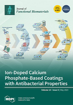
Cover Story (view full-size image): Ion-substituted calcium phosphate (CP) coatings have been extensively studied as promising materials for biomedical implants, due to their ability to enhance biocompatibility, osteoconductivity, and bone formation. This systematic review aims to provide a comprehensive analysis of the current state of the art in ion-doped CP-based coatings for orthopaedic and dental implant applications. Specifically, the effects of ion addition on the physicochemical, mechanical, and biological properties of CP coatings was evaluated, along with the contribution and additional effects (in a separate or a synergistic way) of different components used together with ion-doped CP for advanced composite coatings. In the final part, the effects of antibacterial coatings on specific bacteria strains are reported. View this paper
  • Issues are regarded as officially published after their release is announced to the table of contents alert mailing list.
  • You may sign up for e-mail alerts to receive table of contents of newly released issues.
  • PDF is the official format for papers published in both, html and pdf forms. To view the papers in pdf format, click on the "PDF Full-text" link, and use the free Adobe Reader to open them.
Order results
Result details
Section
Select all
Export citation of selected articles as:
32 pages, 9799 KiB  
Review
Recent Advancements in Electrospun Chitin and Chitosan Nanofibers for Bone Tissue Engineering Applications
by S Shree Ganesh, Ramprasad Anushikaa, Venkadesan Sri Swetha Victoria, Krishnaraj Lavanya, Abinaya Shanmugavadivu and Nagarajan Selvamurugan
J. Funct. Biomater. 2023, 14(5), 288; https://doi.org/10.3390/jfb14050288 - 22 May 2023
Cited by 13 | Viewed by 2995
Abstract
Treatment of large segmental bone loss caused by fractures, osteomyelitis, and non-union results in expenses of around USD 300,000 per case. Moreover, the worst-case scenario results in amputation in 10% to 14.5% of cases. Biomaterials, cells, and regulatory elements are employed in bone [...] Read more.
Treatment of large segmental bone loss caused by fractures, osteomyelitis, and non-union results in expenses of around USD 300,000 per case. Moreover, the worst-case scenario results in amputation in 10% to 14.5% of cases. Biomaterials, cells, and regulatory elements are employed in bone tissue engineering (BTE) to create biosynthetic bone grafts with effective functionalization that can aid in the restoration of such fractured bones, preventing amputation and alleviating expenses. Chitin (CT) and chitosan (CS) are two of the most prevalent natural biopolymers utilized in the fields of biomaterials and BTE. To offer the structural and biochemical cues for augmenting bone formation, CT and CS can be employed alone or in combination with other biomaterials in the form of nanofibers (NFs). When compared with several fabrication methods available to produce scaffolds, electrospinning is regarded as superior since it enables the development of nanostructured scaffolds utilizing biopolymers. Electrospun nanofibers (ENFs) offer unique characteristics, including morphological resemblance to the extracellular matrix, high surface-area-to-volume ratio, permeability, porosity, and stability. This review elaborates on the recent strategies employed utilizing CT and CS ENFs and their biocomposites in BTE. We also summarize their implementation in supporting and delivering an osteogenic response to treat critical bone defects and their perspectives on rejuvenation. The CT- and CS-based ENF composite biomaterials show promise as potential constructions for bone tissue creation. Full article
(This article belongs to the Special Issue Biomedical Applications of Chitin and Chitosan-II)
Show Figures

Figure 1

18 pages, 3117 KiB  
Review
Surface Coatings of Dental Implants: A Review
by Angelo Michele Inchingolo, Giuseppina Malcangi, Laura Ferrante, Gaetano Del Vecchio, Fabio Viapiano, Alessio Danilo Inchingolo, Antonio Mancini, Ciro Annicchiarico, Francesco Inchingolo, Gianna Dipalma, Elio Minetti, Andrea Palermo and Assunta Patano
J. Funct. Biomater. 2023, 14(5), 287; https://doi.org/10.3390/jfb14050287 - 22 May 2023
Cited by 23 | Viewed by 4645
Abstract
Replacement of missing teeth is possible using biocompatible devices such as endosseous implants. This study aims to analyze and recognize the best characteristics of different implant surfaces that ensure good peri-implant tissue healing and thus clinical success over time. The present review was [...] Read more.
Replacement of missing teeth is possible using biocompatible devices such as endosseous implants. This study aims to analyze and recognize the best characteristics of different implant surfaces that ensure good peri-implant tissue healing and thus clinical success over time. The present review was performed on the recent literature concerning endosseous implants made of titanium, a material most frequently used because of its mechanical, physical, and chemical characteristics. Thanks to its low bioactivity, titanium exhibits slow osseointegration. Implant surfaces are treated so that cells do not reject the surface as a foreign material and accept it as fully biocompatible. Analysis of different types of implant surface coatings was performed in order to identify ideal surfaces that improve osseointegration, epithelial attachment to the implant site, and overall peri-implant health. This study shows that the implant surface, with different adhesion, proliferation, and spreading capabilities of osteoblastic and epithelial cells, influences the cells involved in anchorage. Implant surfaces must have antibacterial capabilities to prevent peri-implant disease. Research still needs to improve implant material to minimize clinical failure. Full article
Show Figures

Figure 1

27 pages, 1719 KiB  
Review
A Review of the Application of Natural and Synthetic Scaffolds in Bone Regeneration
by Sok Kuan Wong, Michelle Min Fang Yee, Kok-Yong Chin and Soelaiman Ima-Nirwana
J. Funct. Biomater. 2023, 14(5), 286; https://doi.org/10.3390/jfb14050286 - 20 May 2023
Cited by 20 | Viewed by 4456
Abstract
The management of bone defects is complicated by the presence of clinical conditions, such as critical-sized defects created by high-energy trauma, tumour resection, infection, and skeletal abnormalities, whereby the bone regeneration capacity is compromised. A bone scaffold is a three-dimensional structure matrix serving [...] Read more.
The management of bone defects is complicated by the presence of clinical conditions, such as critical-sized defects created by high-energy trauma, tumour resection, infection, and skeletal abnormalities, whereby the bone regeneration capacity is compromised. A bone scaffold is a three-dimensional structure matrix serving as a template to be implanted into the defects to promote vascularisation, growth factor recruitment, osteogenesis, osteoconduction, and mechanical support. This review aims to summarise the types and applications of natural and synthetic scaffolds currently adopted in bone tissue engineering. The merits and caveats of natural and synthetic scaffolds will be discussed. A naturally derived bone scaffold offers a microenvironment closer to in vivo conditions after decellularisation and demineralisation, exhibiting excellent bioactivity, biocompatibility, and osteogenic properties. Meanwhile, an artificially produced bone scaffold allows for scalability and consistency with minimal risk of disease transmission. The combination of different materials to form scaffolds, along with bone cell seeding, biochemical cue incorporation, and bioactive molecule functionalisation, can provide additional or improved scaffold properties, allowing for a faster bone repair rate in bone injuries. This is the direction for future research in the field of bone growth and repair. Full article
(This article belongs to the Special Issue Bone Tissue Engineering: Material Design and Applications)
Show Figures

Figure 1

18 pages, 2020 KiB  
Review
The Use of Warm Air for Solvent Evaporation in Adhesive Dentistry: A Meta-Analysis of In Vitro Studies
by Rim Bourgi, Louis Hardan, Carlos Enrique Cuevas-Suárez, Francesco Scavello, Davide Mancino, Naji Kharouf and Youssef Haikel
J. Funct. Biomater. 2023, 14(5), 285; https://doi.org/10.3390/jfb14050285 - 20 May 2023
Cited by 3 | Viewed by 2514
Abstract
Any excess solvent from dental adhesive systems must be eliminated prior to material photopolymerization. For this purpose, numerous approaches have been proposed, including the use of a warm air stream. This study aimed to investigate the effect of different temperatures of warm air [...] Read more.
Any excess solvent from dental adhesive systems must be eliminated prior to material photopolymerization. For this purpose, numerous approaches have been proposed, including the use of a warm air stream. This study aimed to investigate the effect of different temperatures of warm air blowing used for solvent evaporation on the bond strength of resin-based materials to dental and nondental substrates. Two different reviewers screened the literature in diverse electronic databases. In vitro studies recording the effect of warm air blowing to evaporate solvents of adhesive systems on the bond strength of resin-based materials to direct and indirect substrates were included. A total of 6626 articles were retrieved from all databases. From this, 28 articles were included in the qualitative analysis, and 27 remained for the quantitative analysis. The results of the meta-analysis for etch-and-rinse adhesives revealed that the use of warm air for solvent evaporation was statistically significantly higher (p = 0.005). For self-etch adhesives and silane-based materials, this effect was observed too (p < 0.001). The use of a warm air stream for solvent evaporation enhanced the bonding performance of alcohol-/water-based adhesive systems for dentin. This effect seems to be similar when a silane coupling agent is submitted to a heat treatment before the cementation of a glass-based ceramic. Full article
Show Figures

Figure 1

16 pages, 15156 KiB  
Article
Cytotoxicity Induced by Black Phosphorus Nanosheets in Vascular Endothelial Cells via Oxidative Stress and Apoptosis Activation
by Hao Dong, Yin Wen, Jiating Lin, Xianxian Zhuang, Ruoting Xian, Ping Li and Shaobing Li
J. Funct. Biomater. 2023, 14(5), 284; https://doi.org/10.3390/jfb14050284 - 20 May 2023
Cited by 3 | Viewed by 1800
Abstract
Black phosphorus (BP), an emerging two-dimensional material with unique optical, thermoelectric, and mechanical properties, has been proposed as bioactive material for tissue engineering. However, its toxic effects on physiological systems remain obscure. The present study investigated the cytotoxicity of BP to vascular endothelial [...] Read more.
Black phosphorus (BP), an emerging two-dimensional material with unique optical, thermoelectric, and mechanical properties, has been proposed as bioactive material for tissue engineering. However, its toxic effects on physiological systems remain obscure. The present study investigated the cytotoxicity of BP to vascular endothelial cells. BP nanosheets (BPNSs) with a diameter of 230 nm were fabricated via a classical liquid-phase exfoliation method. Human umbilical vein endothelial cells (HUVECs) were used to determine the cytotoxicity induced by BPNSs (0.31–80 μg/mL). When the concentrations were over 2.5 μg/mL, BPNSs adversely affected the cytoskeleton and cell migration. Furthermore, BPNSs caused mitochondrial dysfunction and generated excessive intercellular reactive oxygen species (ROS) at tested concentrations after 24 h. BPNSs could influence the expression of apoptosis-related genes, including the P53 and BCL-2 family, resulting in the apoptosis of HUVECs. Therefore, the viability and function of HUVECs were adversely influenced by the concentration of BPNSs over 2.5 μg/mL. These findings provide significant information for the potential applications of BP in tissue engineering. Full article
Show Figures

Figure 1

15 pages, 5173 KiB  
Article
Addition of Resolvins D1 or E1 to Collagen Membranes Mitigates Their Resorption in Diabetic Rats
by Michal Almogy, Ofer Moses, Nathan Schiffmann, Evgeny Weinberg, Carlos E. Nemcovsky and Miron Weinreb
J. Funct. Biomater. 2023, 14(5), 283; https://doi.org/10.3390/jfb14050283 - 19 May 2023
Viewed by 1442
Abstract
Uncontrolled diabetes is characterized by aberrant inflammatory reactions and increased collagenolysis. We have reported that it accelerates the degradation of implanted collagen membranes (CM), thus compromising their function in regenerative procedures. In recent years, a group of physiological anti-inflammatory agents called specialized pro-resolving [...] Read more.
Uncontrolled diabetes is characterized by aberrant inflammatory reactions and increased collagenolysis. We have reported that it accelerates the degradation of implanted collagen membranes (CM), thus compromising their function in regenerative procedures. In recent years, a group of physiological anti-inflammatory agents called specialized pro-resolving lipid mediators (SPMs) have been tested as a treatment for various inflammatory conditions, either systemically or locally, via medical devices. Yet, no study has tested their effect on the fate of the biodegradable material itself. Here, we measured the in vitro release over time of 100 or 800 ng resolvin D1 (RvD1) incorporated into CM discs. In vivo, diabetes was induced in rats with streptozotocin, while buffer-injected (normoglycemic) rats served as controls. Resolvins (100 or 800 ng of RvD1 or RvE1) were added to biotin-labeled CM discs, which were implanted sub-periosteally over the calvaria of rats. Membrane thickness, density, and uniformity were determined by quantitative histology after 3 weeks. In vitro, significant amounts of RvD1 were released over 1–8 days, depending on the amount loaded. In vivo, CMs from diabetic animals were thinner, more porous, and more variable in thickness and density. The addition of RvD1 or RvE1 improved their regularity, increased their density, and reduced their invasion by the host tissue significantly. We conclude that addition of resolvins to biodegradable medical devices can protect them from excessive degradation in systemic conditions characterized by high degree of collagenolysis. Full article
(This article belongs to the Special Issue Biomaterials for Oral Reconstructive Treatment)
Show Figures

Figure 1

13 pages, 1332 KiB  
Systematic Review
Cone-Beam Computed Tomography and Histological Findings for Socket Preservation Techniques Using Different Grafting Materials: A Systematic Review
by Marwa Madi, Ibrahim Almindil, Maria Alrassasi, Doha Alramadan, Osama Zakaria and Adel S Alagl
J. Funct. Biomater. 2023, 14(5), 282; https://doi.org/10.3390/jfb14050282 - 18 May 2023
Cited by 3 | Viewed by 1740
Abstract
Objective: Socket preservation techniques have been used to maintain the ridge dimension following tooth extraction. The materials used influence the quality and quantity of newly formed bone. Therefore, the aim of this article was to systematically review the literature reporting both histological and [...] Read more.
Objective: Socket preservation techniques have been used to maintain the ridge dimension following tooth extraction. The materials used influence the quality and quantity of newly formed bone. Therefore, the aim of this article was to systematically review the literature reporting both histological and radiographic outcomes of socket preservation techniques after tooth extraction in human subjects. Material and method: A systematic electronic search was performed in the electronic databases. English language clinical studies that were published between 2017 and 2022 and included both histological and radiographic findings for the test and control groups. Our primary search produced 848 articles, and of these, 215 were duplicate studies. A total of 72 articles were then eligible for full-text reading. Results: The review included eight studies that met its inclusion criteria. Three outcomes were compared in the included studies. The percentage of newly formed bone ranged from 21.34 ± 9.14% to more than 50% of new bone formation. The materials that showed more than 50% of newly formed bone formation were demineralized dentin graft, platelet-rich fibrin, freeze-dried bone allograft, corticocancellous porcine, and autogenous bone. Four Studies did not report the percentage of the residual graft materials, while those who reported showed a variable range of a minimum 1.5% to more than 25%. One study did not report the changes in horizontal width at the follow-up period, while other studies ranged from 0.6 mm to 10 mm. Conclusion: Socket preservation represents an efficient technique to preserve the ridge contour with satisfactory newly formed bone in the augmented site and maintaining the vertical and horizontal dimensions of the ridge. Full article
Show Figures

Figure 1

13 pages, 12948 KiB  
Article
Photobiomodulation and Inorganic Bovine Bone in Guided Bone Regeneration: Histomorphometric Analysis in Rats
by Nicole Rosa de Freitas, Luísa Belluco Guerrini, Luis Augusto Esper, Michyele Cristhiane Sbrana, Caroline Chepernate Vieira dos Santos and Ana Lúcia Pompéia Fraga de Almeida
J. Funct. Biomater. 2023, 14(5), 281; https://doi.org/10.3390/jfb14050281 - 18 May 2023
Cited by 4 | Viewed by 1625
Abstract
The objective of this study was to evaluate the efficacy of photobiomodulation in the bone regeneration of critical-sized defects (CSD) filled with inorganic bovine bone associated or not with collagen membranes. The study has been conducted on 40 critical defects in the calvaria [...] Read more.
The objective of this study was to evaluate the efficacy of photobiomodulation in the bone regeneration of critical-sized defects (CSD) filled with inorganic bovine bone associated or not with collagen membranes. The study has been conducted on 40 critical defects in the calvaria of male rats, divided into four experimental groups (n = 10): (1) DBBM (deproteinized bovine bone mineral); (2) GBR (DBBM+collagen membrane); (3) DBBM+P (DBBM+photobiomodulation); and (4) GBR+P (GBR+photobiomodulation). At 30 days postoperative, the animals were euthanized, and after the tissue had been processed, histological, histometric, and statistical analyses were performed. The analyses have taken into account newly formed bone area (NBA), linear bone extension (LBE), and residual particle area (RPA) as variables. The Kruskal-Wallis test has been performed, followed by the Dwass-Steel-Critchlow-Fligner test for comparison between groups (p < 0.05). When the DBBM+P group was compared to the DBBM group, it was possible to observe significant statistical differences in all the variables analyzed (p < 0.05). The application of photobiomodulation in guided bone regeneration (GBR+P) has shown a decrease in the median value for the RPA variable (26.8) when compared to the GBR group (32.4), with a significant statistical difference; however, for NBA and LBE, the therapy has not provided significant results. Full article
(This article belongs to the Special Issue Feature Papers in Bone Biomaterials)
Show Figures

Figure 1

11 pages, 2522 KiB  
Article
Development of Salmon Sperm DNA/Regenerated Silk Bio-Based Films for Biomedical Studies on Human Keratinocyte HaCaT Cells under Solar Spectrum
by Maria Rachele Ceccarini, Francesca Ripanti, Veronica Raggi, Alessandro Paciaroni, Caterina Petrillo, Lucia Comez, Kevin Donato, Matteo Bertelli, Tommaso Beccari and Luca Valentini
J. Funct. Biomater. 2023, 14(5), 280; https://doi.org/10.3390/jfb14050280 - 18 May 2023
Cited by 5 | Viewed by 2486
Abstract
In this study, we fabricated adhesive patches from silkworm-regenerated silk and DNA to safeguard human skin from the sun’s rays. The patches are realized by exploiting the dissolution of silk fibers (e.g., silk fibroin (SF)) and salmon sperm DNA in formic acid and [...] Read more.
In this study, we fabricated adhesive patches from silkworm-regenerated silk and DNA to safeguard human skin from the sun’s rays. The patches are realized by exploiting the dissolution of silk fibers (e.g., silk fibroin (SF)) and salmon sperm DNA in formic acid and CaCl2 solutions. Infrared spectroscopy is used to investigate the conformational transition of SF when combined with DNA; the results indicated that the addition of DNA provides an increase in the SF crystallinity. UV–Visible absorption and circular dichroism spectroscopy showed strong absorption in the UV region and the presence of B-form of DNA once dispersed in the SF matrix, respectively. Water absorption measurements as well as thermal dependence of water sorption and thermal analysis, suggested the stability of the fabricated patches. Biological results on cellular viability (MTT assay) of keratinocyte HaCaT cells after exposures to the solar spectrum showed that both SF and SF/DNA patches are photo-protective by increasing the cellular viability of keratinocytes after UV component exposure. Overall, these SF/DNA patches promise applications in wound dressing for practical biomedical purposes. Full article
(This article belongs to the Topic Advanced Functional Materials for Regenerative Medicine)
Show Figures

Figure 1

13 pages, 3068 KiB  
Article
An Eco-Friendly Process to Extract Hydroxyapatite from Sheep Bones for Regenerative Medicine: Structural, Morphologic and Electrical Studies
by Sílvia Rodrigues Gavinho, Mehmet Bozdag, Cevriye Kalkandelen, Joana Soares Regadas, Suresh Kumar Jakka, Oguzhan Gunduz, Faik Nuzhet Oktar and Manuel Pedro Fernandes Graça
J. Funct. Biomater. 2023, 14(5), 279; https://doi.org/10.3390/jfb14050279 - 17 May 2023
Cited by 6 | Viewed by 1876
Abstract
Hydroxyapatite (HA) promotes excellent bone regeneration in bone-tissue engineering, due to its similarity to bone mineral and its ability to connect to living tissues. These factors promote the osteointegration process. This process can be enhanced by the presence of electrical charges, stored in [...] Read more.
Hydroxyapatite (HA) promotes excellent bone regeneration in bone-tissue engineering, due to its similarity to bone mineral and its ability to connect to living tissues. These factors promote the osteointegration process. This process can be enhanced by the presence of electrical charges, stored in the HA. Furthermore, several ions can be added to the HA structure to promote specific biological responses, such as magnesium ions. The main objective of this work was to extract hydroxyapatite from sheep femur bones and to study their structural and electrical properties by adding different amounts of magnesium oxide. The thermal and structural characterizations were performed using DTA, XRD, density, Raman spectroscopy and FTIR analysis. The morphology was studied using SEM, and the electrical measurements were registered as a function of frequency and temperature. Results show that: (i) an increase of MgO amount indicates that the solubility of MgO is below 5%wt for heat treatments at 600 °C; (ii) the rise of MgO content increases the capacity for electrical charge storage; (iii) sheep hydroxyapatite presents itself as a natural source of hydroxyapatite, environmentally sustainable and low cost, and promising for applications in regenerative medicine. Full article
Show Figures

Figure 1

24 pages, 5203 KiB  
Article
Ellagic Acid Inclusion Complex-Loaded Hydrogels as an Efficient Controlled Release System: Design, Fabrication and In Vitro Evaluation
by Chengqun Yu, Abid Naeem, Yali Liu and Yongmei Guan
J. Funct. Biomater. 2023, 14(5), 278; https://doi.org/10.3390/jfb14050278 - 16 May 2023
Cited by 6 | Viewed by 2433
Abstract
Oxidants play a crucial role in the development of oxidative stress, which is linked to disease progression. Ellagic acid is an effective antioxidant with applications in the treatment and prevention of several diseases, since it neutralizes free radicals and reduces oxidative stress. However, [...] Read more.
Oxidants play a crucial role in the development of oxidative stress, which is linked to disease progression. Ellagic acid is an effective antioxidant with applications in the treatment and prevention of several diseases, since it neutralizes free radicals and reduces oxidative stress. However, it has limited application due to its poor solubility and oral bioavailability. Since ellagic acid is hydrophobic, it is difficult to load it directly into hydrogels for controlled release applications. Therefore, the purpose of this study was to first prepare inclusion complexes of ellagic acid (EA) with hydroxypropyl-β-cyclodextrin and then load them into carbopol-934-grafted-2-acrylamido-2-methyl-1-propane sulfonic acid (CP-g-AMPS) hydrogels for orally controlled drug delivery. Fourier transform infrared spectroscopy (FTIR), X-ray diffraction (XRD), scanning electron microscopy (SEM), thermogravimetric analysis (TGA), and differential scanning calorimetry (DSC) were used to validate ellagic acid inclusion complexes and hydrogels. There was slightly higher swelling and drug release at pH 1.2 (42.20% and 92.13%) than at pH 7.4 (31.61% and 77.28%), respectively. Hydrogels had high porosity (88.90%) and biodegradation (9.2% per week in phosphate-buffered saline). Hydrogels were tested for their antioxidant properties in vitro against 2,2-diphenyl-1-picrylhydrazyl (DPPH) and 2,2′-azino-bis (3-ethylbenzothiazoline-6-sulfonic acid) (ABTS). Additionally, the antibacterial activity of hydrogels was demonstrated against Gram-positive bacterial strains (Staphylococcus aureus and Escherichia coli) and Gram-negative bacterial strains (Pseudomonas aeruginosa). Full article
(This article belongs to the Special Issue State of the Art in Biomaterials for Drug Delivery)
Show Figures

Figure 1

20 pages, 9416 KiB  
Article
Combined Porous-Monolithic TiNi Materials Surface-Modified with Electron Beam for New-Generation Rib Endoprostheses
by Anastasiia V. Shabalina, Sergey G. Anikeev, Sergei A. Kulinich, Nadezhda V. Artyukhova, Vitaly A. Vlasov, Maria I. Kaftaranova, Valentina N. Hodorenko, Evgeny V. Yakovlev, Evgeny A. Pesterev, Anna V. Lukyanenko, Mikhail N. Volochaev, Sofiya Pakholkina, Oibek Mamazakirov, Victor V. Stolyarov, Anatolii V. Mokshin and Victor E. Gunther
J. Funct. Biomater. 2023, 14(5), 277; https://doi.org/10.3390/jfb14050277 - 15 May 2023
Cited by 1 | Viewed by 1590
Abstract
TiNi alloys are very widely used materials in implant fabrication. When applied in rib replacement, they are required to be manufactured as combined porous-monolithic structures, ideally with a thin, porous part well-adhered to its monolithic substrate. Additionally, good biocompatibility, high corrosion resistance and [...] Read more.
TiNi alloys are very widely used materials in implant fabrication. When applied in rib replacement, they are required to be manufactured as combined porous-monolithic structures, ideally with a thin, porous part well-adhered to its monolithic substrate. Additionally, good biocompatibility, high corrosion resistance and mechanical durability are also highly demanded. So far, all these parameters have not been achieved in one material, which is why an active search in the field is still underway. In the present study, we prepared new porous-monolithic TiNi materials by sintering a TiNi powder (0–100 µm) on monolithic TiNi plates, followed by surface modification with a high-current pulsed electron beam. The obtained materials were evaluated by a set of surface and phase analysis methods, after which their corrosion resistance and biocompatibility (hemolysis, cytotoxicity, and cell viability) were evaluated. Finally, cell growth tests were conducted. In comparison with flat TiNi monoliths, the newly developed materials were found to have better corrosion resistance, also demonstrating good biocompatibility and potential for cell growth on their surface. Thus, the newly developed porous-on-monolith TiNi materials with different surface porosity and morphology showed promise as potential new-generation implants for use in rib endoprostheses. Full article
(This article belongs to the Section Bone Biomaterials)
Show Figures

Figure 1

14 pages, 422 KiB  
Review
In Vitro Evaluation of Lithium Disilicate Endocrowns and Post and Core Crowns—A Systematic Review
by Zeeshan Qamar, Ahmed Mohammed Saad Alghamdi, Naji Khaled Bin Haydarah, Abdulateef Ahmed Balateef, Ahmed Aydhah Alamoudi, Munther Amer Abumismar, Ankita Mathur and Giuseppe Minervini
J. Funct. Biomater. 2023, 14(5), 276; https://doi.org/10.3390/jfb14050276 - 14 May 2023
Cited by 6 | Viewed by 2344
Abstract
The aim of this systematic review was to summarize the results of the studies that have compared the physical and mechanical properties of lithium disilicate (LDS) endocrowns constructed for posterior teeth to those retained by post-and-core retention systems. The review was conducted following [...] Read more.
The aim of this systematic review was to summarize the results of the studies that have compared the physical and mechanical properties of lithium disilicate (LDS) endocrowns constructed for posterior teeth to those retained by post-and-core retention systems. The review was conducted following the PRISMA guidelines. The electronic search process was conducted on PubMed-Medline, Scopus, Embase and ISI Web of Knowledge (WoS) from the earliest available date till 31 January 2023. Additionally, the studies were assessed for their overall quality and risk of bias using the Quality Assessment Tool For In Vitro Studies (the QUIN). The initial search resulted in 291 articles, out of which, only 10 studies met the eligibility criteria. In all studies LDS endocrowns were compared with various kinds of endodontic posts and crowns made from other materials. There were no definite pattern or trends observed in the fracture strengths of tested specimens. There was no predilection observed in failure patters among the experimental specimens. No predilection was observed in the fracture strengths of LDS endocrowns when compared to post-and-core crowns. Furthermore, no differences in failure patterns could be observed when both types of restorations were compared. The authors propose standardized testing of endocrowns against post-and-core crowns in future studies. In conclusion, long-term clinical trials are advocated to compare the survival, failure and complication rates of LDS endocrowns and post-and-core restorations. Full article
Show Figures

Figure 1

17 pages, 5187 KiB  
Article
Physical Characteristics and Biocompatibility of 3D-Printed Polylactic-Co-Glycolic Acid Membranes Used for Guided Bone Regeneration
by Sidabhat Petposri, Nuttawut Thuaksuban, Supanee Buranadham, Trin Suwanrat, Winita Punyodom and Woraporn Supphaprasitt
J. Funct. Biomater. 2023, 14(5), 275; https://doi.org/10.3390/jfb14050275 - 14 May 2023
Cited by 3 | Viewed by 1665
Abstract
Bioresorbable polymeric membranes for guided bone regeneration (GBR) were fabricated using the three-dimensional printing technique. Membranes made of polylactic-co-glycolic acid (PLGA), which consist of lactic acid (LA) and glycolic acid in ratios of 10:90 (group A) and 70:30 (group B), were compared. Their [...] Read more.
Bioresorbable polymeric membranes for guided bone regeneration (GBR) were fabricated using the three-dimensional printing technique. Membranes made of polylactic-co-glycolic acid (PLGA), which consist of lactic acid (LA) and glycolic acid in ratios of 10:90 (group A) and 70:30 (group B), were compared. Their physical characteristics including architecture, surface wettability, mechanical properties, and degradability were compared in vitro, and their biocompatibilities were compared in vitro and in vivo. The results demonstrated that the membranes of group B had mechanical strength and could support the proliferation of fibroblasts and osteoblasts significantly better than those of group A (p < 0.05). The degradation rate in Group B was significantly lower than that in Group A, but they significantly produced less acidic environment (p < 0.05). In vivo, the membranes of group B were compared with the commercially available collagen membranes (group C). The amount of newly formed bone of rat’s calvarial defects covered with the membranes of group C was stable after week 2, whereas that of group B increased over time. At week 8, the new bone volumes in group B were greater than those in group C (p > 0.05). In conclusion, the physical and biological properties of the PLGA membrane (LA:GA, 70:30) were suitable for GBR. Full article
Show Figures

Figure 1

18 pages, 1318 KiB  
Review
Cellular Alterations in Carbohydrate and Lipid Metabolism Due to Interactions with Nanomaterials
by Ana Martín-Pardillos and Pilar Martin-Duque
J. Funct. Biomater. 2023, 14(5), 274; https://doi.org/10.3390/jfb14050274 - 14 May 2023
Cited by 5 | Viewed by 2797
Abstract
Nanoparticles (NPs) have unique physicochemical properties that are useful for a broad range of biomedical and industrial applications; nevertheless, increasing concern exists about their biosafety. This review aims to focus on the implications of nanoparticles in cellular metabolism and their outcomes. In particular, [...] Read more.
Nanoparticles (NPs) have unique physicochemical properties that are useful for a broad range of biomedical and industrial applications; nevertheless, increasing concern exists about their biosafety. This review aims to focus on the implications of nanoparticles in cellular metabolism and their outcomes. In particular, some NPs have the ability to modify glucose and lipid metabolism, and this feature is especially interesting to treat diabetes and obesity and to target cancer cells. However, the lack of specificity to reach target cells and the toxicological evaluation of nontargeted cells can potentially induce detrimental side effects, closely related to inflammation and oxidative stress. Therefore, identifying the metabolic alterations caused by NPs, independent of their application, is highly needed. To our knowledge, this increase would lead to the improvement and safer use with a reduced toxicity, increasing the number of available NPs for diagnosis and treatment of human diseases. Full article
(This article belongs to the Special Issue Micro and Nanocarriers for Biomedicine)
Show Figures

Figure 1

17 pages, 1876 KiB  
Review
The Role of Natural Compounds in Optimizing Contemporary Dental Treatment—Current Status and Future Trends
by Dana Gabriela Budala, Maria-Alexandra Martu, George-Alexandru Maftei, Diana Antonela Diaconu-Popa, Vlad Danila and Ionut Luchian
J. Funct. Biomater. 2023, 14(5), 273; https://doi.org/10.3390/jfb14050273 - 14 May 2023
Cited by 11 | Viewed by 3179
Abstract
For a long period of time, natural remedies were the only ailment available for a multitude of diseases, and they have proven effective even after the emergence of modern medicine. Due to their extremely high prevalence, oral and dental disorders and anomalies are [...] Read more.
For a long period of time, natural remedies were the only ailment available for a multitude of diseases, and they have proven effective even after the emergence of modern medicine. Due to their extremely high prevalence, oral and dental disorders and anomalies are recognized as major public health concerns. Herbal medicine is the practice of using plants with therapeutic characteristics for the purpose of disease prevention and treatment. Herbal agents have made a significant entry into oral care products in recent years, complementing traditional treatment procedures due to their intriguing physicochemical and therapeutic properties. There has been a resurgence of interest in natural products because of recent updates, technological advancements, and unmet expectations from current strategies. Approximately eighty percent of the world’s population uses natural remedies, especially in poorer nations. When conventional treatments have failed, it may make sense to use natural drugs for the treatment of pathologic oral dental disorders, as they are readily available, inexpensive, and have few negative effects. The purpose of this article is to provide a comprehensive analysis of the benefits and applications of natural biomaterials in dentistry, to gather relevant information from the medical literature with an eye toward its practical applicability, and make suggestions for the directions for future study. Full article
(This article belongs to the Special Issue Biomaterials in Medical Diagnosis and Treatment)
Show Figures

Figure 1

13 pages, 5863 KiB  
Article
Dentin, Dentin Graft, and Bone Graft: Microscopic and Spectroscopic Analysis
by Elio Minetti, Andrea Palermo, Giuseppina Malcangi, Alessio Danilo Inchingolo, Antonio Mancini, Gianna Dipalma, Francesco Inchingolo, Assunta Patano and Angelo Michele Inchingolo
J. Funct. Biomater. 2023, 14(5), 272; https://doi.org/10.3390/jfb14050272 - 13 May 2023
Cited by 9 | Viewed by 2463
Abstract
Background: The use of the human dentin matrix could serve as an alternative to autologous, allogenic, and xenogeneic bone grafts. Since 1967, when the osteoinductive characteristics of autogenous demineralized dentin matrix were revealed, autologous tooth grafts have been advocated. The tooth is very [...] Read more.
Background: The use of the human dentin matrix could serve as an alternative to autologous, allogenic, and xenogeneic bone grafts. Since 1967, when the osteoinductive characteristics of autogenous demineralized dentin matrix were revealed, autologous tooth grafts have been advocated. The tooth is very similar to the bone and contains many growth factors. The purpose of the present study is to evaluate the similarities and differences between the three samples (dentin, demineralized dentin, and alveolar cortical bone) with the aim of demonstrating that the demineralized dentin can be considered in regenerative surgery as an alternative to the autologous bone. Methods: This in vitro study analyzed the biochemical characterizations of 11 dentin granules (Group A), 11 demineralized using the Tooth Transformer (Group B), and dentin granules and 11 cortical bone granules (Group C) using scanning electron microscopy (SEM) and energy dispersive spectroscopy (EDS) to evaluate mineral content. Atomic percentages of C (carbon), O (oxygen), Ca (calcium), and P (phosphorus) were individually analyzed and compared by the statistical t-test. Results: The significant p-value (p < 0.05) between group A and group C indicated that these two groups were not significantly similar, while the non-significant result (p > 0.05) obtained between group B and group C indicated that these two groups are similar. Conclusions: The findings support that the hypothesis that the demineralization process can lead to the dentin being remarkably similar to the natural bone in terms of their surface chemical composition. The demineralized dentin can therefore be considered an alternative to the autologous bone in regenerative surgery. Full article
Show Figures

Figure 1

14 pages, 3029 KiB  
Article
Fabrication of Biomedical Ti-Zr-Nb by Reducing Metal Oxides with Calcium Hydride
by Sergey Yudin, Ivan Alimov, Sergey Volodko, Alexander Gurianov, Galina Markova, Anatoly Kasimtsev, Tatiana Sviridova, Darya Permyakova, Evgeny Evstratov, Vladimir Cheverikin and Dmitry Moskovskikh
J. Funct. Biomater. 2023, 14(5), 271; https://doi.org/10.3390/jfb14050271 - 13 May 2023
Cited by 4 | Viewed by 1683
Abstract
In the present study, a powder of Ti-18Zr-15Nb biomedical alloy with spongy morphology and with more than 95% vol. of β-Ti was obtained by reducing the constituent oxides with calcium hydride. The influence of the synthesis temperature, the exposure time, and the density [...] Read more.
In the present study, a powder of Ti-18Zr-15Nb biomedical alloy with spongy morphology and with more than 95% vol. of β-Ti was obtained by reducing the constituent oxides with calcium hydride. The influence of the synthesis temperature, the exposure time, and the density of the charge (TiO2 + ZrO2 + Nb2O5 + CaH2) on the mechanism and kinetics of the calcium hydride synthesis of the Ti-18Zr-15Nb β-alloy was studied. Temperature and exposure time were established as crucial parameters with the help of regression analysis. Moreover, the correlation between the homogeneity of the powder obtained and the lattice microstrain of β-Ti is shown. As a result, temperatures above 1200 °C and an exposure time longer than 12 h are required to obtain a Ti-18Zr-15Nb powder with a single β-phase structure and uniformly distributed elements. The analysis of β-phase growth kinetics revealed that the formation of β-Ti occurs due to the solid-state diffusion interaction between Ti, Nb, and Zr under the calcium hydride reduction of TiO2 + ZrO2 + Nb2O5, and the spongy morphology of reduced α-Ti is inherited by the β-phase. Thus, the results obtained provide a promising approach for manufacturing biocompatible porous implants from β-Ti alloys that are believed to be attractive candidates for biomedical applications. Moreover, the current study develops and deepens the theory and practical aspects of the metallothermic synthesis of metallic materials and can be compelling to specialists in powder metallurgy. Full article
Show Figures

Figure 1

17 pages, 4498 KiB  
Article
Engineered Cross-Linked Silane with Urea Polymer Thin Durable Coatings onto Polymeric Films for Controlled Antiviral Release of Activated Chlorine and Essential Oils
by Elisheva Sasson, Omer Agazani, Eyal Malka, Meital Reches and Shlomo Margel
J. Funct. Biomater. 2023, 14(5), 270; https://doi.org/10.3390/jfb14050270 - 12 May 2023
Cited by 3 | Viewed by 1949
Abstract
In March 2020, the World Health Organization announced a pandemic attributed to SARS-CoV-2, a novel beta-coronavirus, which spread widely from China. As a result, the need for antiviral surfaces has increased significantly. Here, the preparation and characterization of new antiviral coatings on polycarbonate [...] Read more.
In March 2020, the World Health Organization announced a pandemic attributed to SARS-CoV-2, a novel beta-coronavirus, which spread widely from China. As a result, the need for antiviral surfaces has increased significantly. Here, the preparation and characterization of new antiviral coatings on polycarbonate (PC) for controlled release of activated chlorine (Cl+) and thymol separately and combined are described. Thin coatings were prepared by polymerization of 1-[3-(trimethoxysilyl)propyl] urea (TMSPU) in ethanol/water basic solution by modified Stöber polymerization, followed by spreading the formed dispersion onto surface-oxidized PC film using a Mayer rod with appropriate thickness. Activated Cl-releasing coating was prepared by chlorination of the PC/SiO2-urea film with NaOCl through the urea amide groups to form a Cl-amine derivatized coating. Thymol releasing coating was prepared by linking thymol to TMSPU or its polymer via hydrogen bonds between thymol hydroxyl and urea amide groups. The activity towards T4 bacteriophage and canine coronavirus (CCV) was measured. PC/SiO2-urea-thymol enhanced bacteriophage persistence, while PC/SiO2-urea-Cl reduced its amount by 84%. Temperature-dependent release is presented. Surprisingly, the combination of thymol and chlorine had an improved antiviral activity, reducing the amount of both viruses by four orders of magnitude, indicating synergistic activity. For CCV, coating with only thymol was inactive, while SiO2-urea-Cl reduced it below a detectable level. Full article
(This article belongs to the Topic Advanced Manufacturing and Surface Technology)
Show Figures

Figure 1

24 pages, 2628 KiB  
Review
Recent Advances in Tissue-Engineered Cardiac Scaffolds—The Progress and Gap in Mimicking Native Myocardium Mechanical Behaviors
by Somayeh Baghersad, Abinaya Sathish Kumar, Matt J. Kipper, Ketul Popat and Zhijie Wang
J. Funct. Biomater. 2023, 14(5), 269; https://doi.org/10.3390/jfb14050269 - 12 May 2023
Cited by 6 | Viewed by 4082
Abstract
Heart failure is the leading cause of death in the US and worldwide. Despite modern therapy, challenges remain to rescue the damaged organ that contains cells with a very low proliferation rate after birth. Developments in tissue engineering and regeneration offer new tools [...] Read more.
Heart failure is the leading cause of death in the US and worldwide. Despite modern therapy, challenges remain to rescue the damaged organ that contains cells with a very low proliferation rate after birth. Developments in tissue engineering and regeneration offer new tools to investigate the pathology of cardiac diseases and develop therapeutic strategies for heart failure patients. Tissue -engineered cardiac scaffolds should be designed to provide structural, biochemical, mechanical, and/or electrical properties similar to native myocardium tissues. This review primarily focuses on the mechanical behaviors of cardiac scaffolds and their significance in cardiac research. Specifically, we summarize the recent development of synthetic (including hydrogel) scaffolds that have achieved various types of mechanical behavior—nonlinear elasticity, anisotropy, and viscoelasticity—all of which are characteristic of the myocardium and heart valves. For each type of mechanical behavior, we review the current fabrication methods to enable the biomimetic mechanical behavior, the advantages and limitations of the existing scaffolds, and how the mechanical environment affects biological responses and/or treatment outcomes for cardiac diseases. Lastly, we discuss the remaining challenges in this field and suggestions for future directions to improve our understanding of mechanical control over cardiac function and inspire better regenerative therapies for myocardial restoration. Full article
Show Figures

Figure 1

16 pages, 4029 KiB  
Article
Effect of Whitening Toothpastes with Different Active Agents on the Abrasive Wear of Dentin Following Tooth Brushing Simulation
by Dimitrios Dionysopoulos, Spyros Papageorgiou, Constantinos Papadopoulos, Sotiria Davidopoulou, Avraam Konstantinidis and Kosmas Tolidis
J. Funct. Biomater. 2023, 14(5), 268; https://doi.org/10.3390/jfb14050268 - 12 May 2023
Cited by 5 | Viewed by 3858
Abstract
The aim of this research was to evaluate the abrasive dentin wear that can be induced by three commercial whitening toothpastes following a tooth-brushing simulation (TBS) corresponding to a three-month period. Sixty human canines were selected, and the roots were separated from the [...] Read more.
The aim of this research was to evaluate the abrasive dentin wear that can be induced by three commercial whitening toothpastes following a tooth-brushing simulation (TBS) corresponding to a three-month period. Sixty human canines were selected, and the roots were separated from the crowns. Then the roots were randomly divided into six groups (n = 10) and were submitted to TBS using the following slurries: Group 1—deionized water (RDA = 5); Group 2—ISO dentifrice slurry (RDA = 100); Group 3—a regular toothpaste (RDA = 70); Group 4—a charcoal-containing whitening toothpaste; Group 5—a whitening toothpaste containing blue covasorb and hydrated silica; and Group 6—a whitening toothpaste containing microsilica. Following TBS, surface loss and surface roughness changes were evaluated using confocal microscopy. Additionally, surface morphology and mineral content changes were observed using scanning electron microscopy and energy-dispersive X-ray spectroscopy. The deionized water group presented the lowest surface loss (p < 0.05), while the charcoal-containing toothpaste presented the highest surface loss, followed by ISO dentifrice slurry (p < 0.001). Blue-covasorb-containing and regular toothpastes did not present statistically significant differences (p = 0.245), and neither didmicrosilica-containing toothpaste or ISO dentifrice slurry (p = 0.112). The surface height parameters and surface morphology changes of the experimental groups followed the surface loss patterns, while no differences were detected in mineral content after TBS.Although the charcoal-containing toothpaste exhibited the highest abrasive wear to dentin, according to ISO 11609, all the tested toothpastes exhibited appropriate abrasive behavior towards dentin. Full article
(This article belongs to the Section Dental Biomaterials)
Show Figures

Figure 1

17 pages, 3419 KiB  
Article
The Clinical Potential of 3D-Printed Crowns Reinforced with Zirconia and Glass Silica Microfillers
by Abdullah Alshamrani, Abdulaziz Alhotan, Ahmed Owais and Ayman Ellakwa
J. Funct. Biomater. 2023, 14(5), 267; https://doi.org/10.3390/jfb14050267 - 11 May 2023
Cited by 6 | Viewed by 3373
Abstract
The development of 3D-printed crown resin materials with improved mechanical and physical properties is an area of growing interest in dentistry. This study aimed to develop a 3D-printed crown resin material modified with zirconia glass (ZG) and glass silica (GS) microfillers to enhance [...] Read more.
The development of 3D-printed crown resin materials with improved mechanical and physical properties is an area of growing interest in dentistry. This study aimed to develop a 3D-printed crown resin material modified with zirconia glass (ZG) and glass silica (GS) microfillers to enhance overall mechanical and physical properties. A total of 125 specimens were created and divided into five groups: control unmodified resin, 5% either ZG or GS reinforced 3D-printed resin, and 10% either ZG or GS reinforced 3D-printed resin. The fracture resistance, surface roughness, and translucency parameter were measured, and fractured crowns were studied under a scanning electron microscope. The results showed that 3D-printed parts that were strengthened with ZG and GS microfillers demonstrated comparable mechanical performance to unmodified crown resin but resulted in greater surface roughness, and only the group that contained 5% ZG showed an increase in translucency. However, it should be noted that increased surface roughness may impact the aesthetics of the crowns, and further optimisation of microfillers concentrations may be necessary. These findings suggest that the newly developed dental-based resins that incorporate microfillers could be suitable for clinical applications, but further studies are necessary to optimise the nanoparticle concentrations and investigate their long-term clinical outcomes. Full article
Show Figures

Figure 1

19 pages, 1359 KiB  
Review
Wood as Possible Renewable Material for Bone Implants—Literature Review
by Vadims Nefjodovs, Laura Andze, Martins Andzs, Inese Filipova, Ramunas Tupciauskas, Linda Vecbiskena and Martins Kapickis
J. Funct. Biomater. 2023, 14(5), 266; https://doi.org/10.3390/jfb14050266 - 10 May 2023
Cited by 3 | Viewed by 3826
Abstract
Bone fractures and bone defects affect millions of people every year. Metal implants for bone fracture fixation and autologous bone for defect reconstruction are used extensively in treatment of these pathologies. Simultaneously, alternative, sustainable, and biocompatible materials are being researched to improve existing [...] Read more.
Bone fractures and bone defects affect millions of people every year. Metal implants for bone fracture fixation and autologous bone for defect reconstruction are used extensively in treatment of these pathologies. Simultaneously, alternative, sustainable, and biocompatible materials are being researched to improve existing practice. Wood as a biomaterial for bone repair has not been considered until the last 50 years. Even nowadays there is not much research on solid wood as a biomaterial in bone implants. A few species of wood have been investigated. Different techniques of wood preparation have been proposed. Simple pre-treatments such as boiling in water or preheating of ash, birch and juniper woods have been used initially. Later researchers have tried using carbonized wood and wood derived cellulose scaffold. Manufacturing implants from carbonized wood and cellulose requires more extensive wood processing—heat above 800 °C and chemicals to extract cellulose. Carbonized wood and cellulose scaffolds can be combined with other materials, such as silicon carbide, hydroxyapatite, and bioactive glass to improve biocompatibility and mechanical durability. Throughout the publications wood implants have provided good biocompatibility and osteoconductivity thanks to wood’s porous structure. Full article
(This article belongs to the Special Issue Bone Regeneration and Repair Materials)
Show Figures

Figure 1

16 pages, 6037 KiB  
Article
In Vivo and In Vitro Investigation of a Novel Gelatin/Sodium Polyacrylate Composite Hemostatic Sponge for Topical Bleeding
by Nusrat Jahan, Md Sowaib Ibne Mahbub, Byong-Taek Lee and Sang Ho Bae
J. Funct. Biomater. 2023, 14(5), 265; https://doi.org/10.3390/jfb14050265 - 10 May 2023
Cited by 4 | Viewed by 3519
Abstract
Designing a functional and efficient blood-clotting agent is a major challenge. In this research, hemostatic scaffolds (GSp) were prepared from the superabsorbent, inter-crosslinked polymer sodium polyacrylate (Sp) bound to a natural protein gelatin (G) loaded with thrombin (Th) by a cost-effective freeze-drying method. [...] Read more.
Designing a functional and efficient blood-clotting agent is a major challenge. In this research, hemostatic scaffolds (GSp) were prepared from the superabsorbent, inter-crosslinked polymer sodium polyacrylate (Sp) bound to a natural protein gelatin (G) loaded with thrombin (Th) by a cost-effective freeze-drying method. Five compositions were grafted (GSp0.0, Gsp0.1, GSp0.2, GSp0.3, GSp0.3-Th) where the concentration of Sp varied but the ratios of G remained the same. The fundamental physical characteristics that increased the amounts of Sp with G gave synergistic effects after interacting with thrombin. Due to the presence of superabsorbent polymer (SAP) swelling capacities in GSp0.3 and GSp0.3-Th surge forward 6265% and 6948%, respectively. Pore sizes became uniform and larger (ranging ≤ 300 μm) and well-interconnected. The water-contact angle declined in GSp0.3 and GSp0.3-Th to 75.73 ± 1.097 and 75.33 ± 0.8342 degrees, respectively, thus increasing hydrophilicity. The pH difference was found to be insignificant as well. In addition, an evaluation of the scaffold in in vitro biocompatibility with the L929 cell line showed cell viability >80%, so the samples were nontoxic and produced a favorable environment for cell proliferation. The composite GSp0.3-Th revealed the lowest HR (%) (2.601%), and the in vivo blood-clotting time (s) and blood loss (gm) supported hemostasis. Overall, the results showed that a novel GSp0.3-Th scaffold can be a potential candidate as a hemostatic agent. Full article
Show Figures

Graphical abstract

11 pages, 2588 KiB  
Article
Microleakage Evaluation of Temporary Restorations Used in Endodontic Treatment—An Ex Vivo Study
by Siri Paulo, Ana Margarida Abrantes, Mariana Xavier, Ana Filipa Brito, Ricardo Teixo, Ana Sofia Coelho, Anabela Paula, Eunice Carrilho, Maria Filomena Botelho, Carlos Miguel Marto and Manuel Marques Ferreira
J. Funct. Biomater. 2023, 14(5), 264; https://doi.org/10.3390/jfb14050264 - 9 May 2023
Cited by 1 | Viewed by 3278
Abstract
(1) Background: Coronal microleakage can lead to endodontic treatment failure. This study aimed to compare the sealing ability of different temporary restorative materials used during endodontic treatment. (2) Methods: Eighty sheep incisors were collected, uniformized in length, and access cavities were performed, except [...] Read more.
(1) Background: Coronal microleakage can lead to endodontic treatment failure. This study aimed to compare the sealing ability of different temporary restorative materials used during endodontic treatment. (2) Methods: Eighty sheep incisors were collected, uniformized in length, and access cavities were performed, except for in the negative control group, where the teeth were left intact. The teeth were divided into six different groups. In the positive control group, the access cavity was made and left empty. In the experimental groups, access cavities were restored with three different temporary materials (IRM®, Ketac™ Silver, and Cavit™) and with a definitive restorative material (Filtek Supreme™). The teeth were submitted to thermocycling, and two and four weeks later, they were infiltrated with 99mTcNaO4, and nuclear medicine imaging was performed. (3) Results: Filtek Supreme™ obtained the lowest infiltration values. Regarding the temporary materials, at two weeks, Ketac™ Silver presented the lowest infiltration, followed by IRM®, whereas Cavit™ presented the highest infiltration. At four weeks, Ketac™ Silver remained with the lowest values, whereas Cavit™ decreased the infiltration, comparable to IRM®. (4) Conclusion: Regarding temporary materials, Ketac™ Silver had the lowest infiltration at 2 and 4 weeks, whereas the highest infiltration was found in the Cavit™ group at two weeks and in the IRM® group at 4 weeks. Full article
(This article belongs to the Special Issue Advanced Materials for Clinical Endodontic Applications)
Show Figures

Figure 1

15 pages, 3692 KiB  
Article
Direct-Writing Electrospun Functionalized Scaffolds for Periodontal Regeneration: In Vitro Studies
by Laura Bourdon, Nina Attik, Liza Belkessam, Charlène Chevalier, Colin Bousige, Arnaud Brioude and Vincent Salles
J. Funct. Biomater. 2023, 14(5), 263; https://doi.org/10.3390/jfb14050263 - 9 May 2023
Cited by 3 | Viewed by 2451
Abstract
Multiphasic scaffolds that combine different architectural, physical, and biological properties are the best option for the regeneration of complex tissues such as the periodontium. Current developed scaffolds generally lack architectural accuracy and rely on multistep manufacturing, which is difficult to implement for clinical [...] Read more.
Multiphasic scaffolds that combine different architectural, physical, and biological properties are the best option for the regeneration of complex tissues such as the periodontium. Current developed scaffolds generally lack architectural accuracy and rely on multistep manufacturing, which is difficult to implement for clinical applications. In this context, direct-writing electrospinning (DWE) represents a promising and rapid technique for developing thin 3D scaffolds with controlled architecture. The current study aimed to elaborate a biphasic scaffold using DWE based on two polycaprolactone solutions with interesting properties for bone and cement regeneration. One of the two scaffold parts contained hydroxyapatite nanoparticles (HAP) and the other contained the cementum protein 1 (CEMP1). After morphological characterizations, the elaborated scaffolds were assessed regarding periodontal ligament (PDL) cells in terms of cell proliferation, colonization, and mineralization ability. The results demonstrated that both HAP- and CEMP1-functionalized scaffolds were colonized by PDL cells and enhanced mineralization ability compared to unfunctionalized scaffolds, as revealed by alizarin red staining and OPN protein fluorescent expression. Taken together, the current data highlighted the potential of functional and organized scaffolds to stimulate bone and cementum regeneration. Moreover, DWE could be used to develop smart scaffolds with the ability to spatially control cellular orientation with suitable cellular activity at the micrometer scale, thereby enhancing periodontal and other complex tissue regeneration. Full article
(This article belongs to the Special Issue Advanced Biomaterials for Periodontal Regeneration)
Show Figures

Figure 1

13 pages, 3160 KiB  
Article
Collarless Polished Tapered Stems of Identical Shape Provide Differing Outcomes for Stainless Steel and Cobalt Chrome: A Biomechanical Study
by Ayumi Kaneuji, Mingliang Chen, Eiji Takahashi, Noriyuki Takano, Makoto Fukui, Daisuke Soma, Yoshiyuki Tachi, Yugo Orita, Toru Ichiseki and Norio Kawahara
J. Funct. Biomater. 2023, 14(5), 262; https://doi.org/10.3390/jfb14050262 - 9 May 2023
Cited by 3 | Viewed by 1984
Abstract
Cemented polished tapered femoral stems (PTS) made of cobalt–chrome alloy (CoCr) are a known risk factor for periprosthetic fracture (PPF). The mechanical differences between CoCr-PTS and stainless-steel (SUS) PTS were investigated. CoCr stems having the same shape and surface roughness as the SUS [...] Read more.
Cemented polished tapered femoral stems (PTS) made of cobalt–chrome alloy (CoCr) are a known risk factor for periprosthetic fracture (PPF). The mechanical differences between CoCr-PTS and stainless-steel (SUS) PTS were investigated. CoCr stems having the same shape and surface roughness as the SUS Exeter® stem were manufactured and dynamic loading tests were performed on three each. Stem subsidence and the compressive force at the bone–cement interface were recorded. Tantalum balls were injected into the cement, and their movement was tracked to indicate cement movement. Stem motions in the cement were greater for the CoCr stems than for the SUS stems. In addition, although we found a significant positive correlation between stem subsidence and compressive force in all stems, CoCr stems generated a compressive force over three times higher than SUS stems at the bone–cement interface with the same stem subsidence (p < 0.01). The final stem subsidence amount and final force were greater in the CoCr group (p < 0.01), and the ratio of tantalum ball vertical distance to stem subsidence was significantly smaller for CoCr than for SUS (p < 0.01). CoCr stems appear to move more easily in cement than SUS stems, which might contribute to the increased occurrence of PPF with the use of CoCr-PTS. Full article
Show Figures

Figure 1

15 pages, 3575 KiB  
Article
Safety and Osteointegration of Titanium Screws Coated with a Fibroblast Growth Factor-2–Calcium Phosphate Composite Layer in Non-Human Primates: A Pilot Study
by Yukei Matsumoto, Hirotaka Mutsuzaki, Yuki Hara, Katsuya Nagashima, Eriko Okano, Yohei Yanagisawa, Hiroshi Noguchi, Tadashi Sankai and Masashi Yamazaki
J. Funct. Biomater. 2023, 14(5), 261; https://doi.org/10.3390/jfb14050261 - 8 May 2023
Cited by 3 | Viewed by 1768
Abstract
Spinal instrumentation surgery for older patients with osteoporosis is increasing. Implant loosening may occur due to inappropriate fixation in osteoporotic bone. Developing implants that achieve stable surgical results, even in osteoporotic bone, can reduce re-operation, lower medical costs, and maintain the physical status [...] Read more.
Spinal instrumentation surgery for older patients with osteoporosis is increasing. Implant loosening may occur due to inappropriate fixation in osteoporotic bone. Developing implants that achieve stable surgical results, even in osteoporotic bone, can reduce re-operation, lower medical costs, and maintain the physical status of older patients. Fibroblast growth factor-2 (FGF-2) promotes bone formation; thus, coating pedicle screws with an FGF-2–calcium phosphate (FGF-CP) composite layer is hypothesized to enhance osteointegration in spinal implants. We designed a long-term implantation pilot study that estimated the safety and bone-forming efficacy of pedicle screws coated with an FGF-CP composite layer in cynomolgus monkeys. Titanium alloy screws, either uncoated (controls) or aseptically coated with an FGF-CP composite layer, were implanted in the vertebral bodies of six female adult cynomolgus monkeys (three monkeys per group) for 85 days. Physiological, histological, and radiographic investigations were performed. There were no serious adverse events, and no radiolucent areas were observed around the screws in either group. The bone apposition rate in the intraosseous region was significantly higher in the FGF-CP group than in the controls. Moreover, as analyzed by Weibull plots, the bone formation rate of the FGF-CP group exhibited a significantly higher regression line slope than the control group. These results demonstrated that there was significantly less risk of impaired osteointegration in the FGF-CP group. Our pilot study suggests that FGF-CP-coated implants could promote osteointegration, be safe, and reduce the probability of screw loosening. Full article
Show Figures

Figure 1

12 pages, 2657 KiB  
Article
Self-Assembling Peptide RADA16 Nanofiber Scaffold Hydrogel-Wrapped Concentrated Growth Factors in Osteogenesis of MC3T3
by Renjie Yang, Jiali Chen, Dingjie Wang, Yichen Xu and Guomin Ou
J. Funct. Biomater. 2023, 14(5), 260; https://doi.org/10.3390/jfb14050260 - 8 May 2023
Cited by 4 | Viewed by 2123
Abstract
Concentrated growth factors (CGFs) are widely used in surgery with bone grafting, but the release of growth factors from CGFs is rapid. RADA16, a self-assembling peptide, can form a scaffold that is similar to the extracellular matrix. Based on the properties of RADA16 [...] Read more.
Concentrated growth factors (CGFs) are widely used in surgery with bone grafting, but the release of growth factors from CGFs is rapid. RADA16, a self-assembling peptide, can form a scaffold that is similar to the extracellular matrix. Based on the properties of RADA16 and CGF, we hypothesized that the RADA16 nanofiber scaffold hydrogel could enhance the function of CGFs and that the RADA16 nanofiber scaffold hydrogel-wrapped CGFs (RADA16-CGFs) would perform a good osteoinductive function. This study aimed to investigate the osteoinductive function of RADA16-CGFs. Scanning electron microscopy, rheometry, and ELISA were performed, and MC3T3-E1 cells were used to test cell adhesion, cytotoxicity, and mineralization after administration with RADA16-CGFs. We found that RADA16 endowed with the sustained release of growth factors from CGFs, which can help maximize the function of CGFs in osteoinduction. The application of the atoxic RADA16 nanofiber scaffold hydrogel with CGFs can be a new therapeutic strategy for the treatment of alveolar bone loss and other problems that require bone regeneration. Full article
Show Figures

Figure 1

11 pages, 3987 KiB  
Communication
CaSiO3-HAp Metal-Reinforced Biocomposite Ceramics for Bone Tissue Engineering
by Evgeniy K. Papynov, Oleg O. Shichalin, Anton A. Belov, Igor Yu Buravlev, Vitaly Yu Mayorov, Alexander N. Fedorets, Anastasiya A. Buravleva, Alexey O. Lembikov, Danila V. Gritsuk, Olesya V. Kapustina and Zlata E. Kornakova
J. Funct. Biomater. 2023, 14(5), 259; https://doi.org/10.3390/jfb14050259 - 8 May 2023
Cited by 8 | Viewed by 2223
Abstract
Reconstructive and regenerative bone surgery is based on the use of high-tech biocompatible implants needed to restore the functions of the musculoskeletal system of patients. Ti6Al4V is one of the most widely used titanium alloys for a variety of applications where low density [...] Read more.
Reconstructive and regenerative bone surgery is based on the use of high-tech biocompatible implants needed to restore the functions of the musculoskeletal system of patients. Ti6Al4V is one of the most widely used titanium alloys for a variety of applications where low density and excellent corrosion resistance are required, including biomechanical applications (implants and prostheses). Calcium silicate or wollastonite (CaSiO3) and calcium hydroxyapatite (HAp) is a bioceramic material used in biomedicine due to its bioactive properties, which can potentially be used for bone repair. In this regard, the research investigates the possibility of using spark plasma sintering technology to obtain new CaSiO3-HAp biocomposite ceramics reinforced with a Ti6Al4V titanium alloy matrix obtained by additive manufacturing. The phase and elemental compositions, structure, and morphology of the initial CaSiO3-HAp powder and its ceramic metal biocomposite were studied by X-ray fluorescence, scanning electron microscopy, energy-dispersive X-ray spectroscopy, and Brunauer–Emmett–Teller analysis methods. The spark plasma sintering technology was shown to be efficient for the consolidation of CaSiO3-HAp powder in volume with a Ti6Al4V reinforcing matrix to obtain a ceramic metal biocomposite of an integral form. Vickers microhardness values were determined for the alloy and bioceramics (~500 and 560 HV, respectively), as well as for their interface area (~640 HV). An assessment of the critical stress intensity factor KIc (crack resistance) was performed. The research result is new and represents a prospect for the creation of high-tech implant products for regenerative bone surgery. Full article
(This article belongs to the Special Issue Titanium-Based Implants: Advances in Materials and Applications)
Show Figures

Figure 1

Previous Issue
Next Issue
Back to TopTop