Next Issue
Volume 9, December
Previous Issue
Volume 9, October


Metabolites, Volume 9, Issue 11 (November 2019) – 39 articles

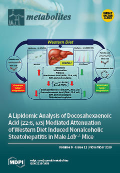
Cover Story (view full-size image): We used a lipidomic approach to identify potential lipid mediators of docosahexaenoic acid [DHA]-mediated attenuation of Western diet (WD)-induced nonalcoholic steatohepatitis (NASH). WD-induced NASH in Ldlr - /- mice increases hepatic steatosis, inflammation, fibrosis, membrane arachidonic acid (ARA) content, and formation of ARA-derived pro-inflammatory oxylipins and decreases C20-22 ω3 PUFA-derived anti-inflammatory oxylipins. DHA supplementation of the WD reversed all of these NASH features. While hepatic pro-inflammatory ARA-derived oxylipins were associated with NASH, DHA-mediated attenuation of NASH was associated with increased hepatic C20-22 ω3 PUFA and anti-inflammatory C20-22 ω3 PUFA-derived oxylipins. Hepatic content of ω3 and ω6 PUFA-derived oxylipins may be a good predictor of liver health. View this paper.
  • Issues are regarded as officially published after their release is announced to the table of contents alert mailing list.
  • You may sign up for e-mail alerts to receive table of contents of newly released issues.
  • PDF is the official format for papers published in both, html and pdf forms. To view the papers in pdf format, click on the "PDF Full-text" link, and use the free Adobe Reader to open them.
Order results
Result details
Section
Select all
Export citation of selected articles as:
15 pages, 2952 KiB  
Article
A New Classification Method of Metastatic Cancers Using a 1H-NMR-Based Approach: A Study Case of Melanoma, Breast, and Prostate Cancer Cell Lines
by Corentin Schepkens, Matthieu Dallons, Jonas Dehairs, Ali Talebi, Jérôme Jeandriens, Lise-Marie Drossart, Guillaume Auquier, Vanessa Tagliatti, Johannes V. Swinnen and Jean-Marie Colet
Metabolites 2019, 9(11), 281; https://doi.org/10.3390/metabo9110281 - 17 Nov 2019
Cited by 5 | Viewed by 5094
Abstract
In this study, metastatic melanoma, breast, and prostate cancer cell lines were analyzed using a 1H-NMR-based approach in order to investigate common features and differences of aggressive cancers metabolomes. For that purpose, 1H-NMR spectra of both cellular extracts and culture media [...] Read more.
In this study, metastatic melanoma, breast, and prostate cancer cell lines were analyzed using a 1H-NMR-based approach in order to investigate common features and differences of aggressive cancers metabolomes. For that purpose, 1H-NMR spectra of both cellular extracts and culture media were combined with multivariate data analysis, bringing to light no less than 20 discriminant metabolites able to separate the metastatic metabolomes. The supervised approach succeeded in classifying the metastatic cell lines depending on their glucose metabolism, more glycolysis-oriented in the BRAF proto-oncogene mutated cell lines compared to the others. Other adaptive metabolic features also contributed to the classification, such as the increased total choline content (tCho), UDP-GlcNAc detection, and various changes in the glucose-related metabolites tree, giving additional information about the metastatic metabolome status and direction. Finally, common metabolic features detected via 1H-NMR in the studied cancer cell lines are discussed, identifying the glycolytic pathway, Kennedy’s pathway, and the glutaminolysis as potential and common targets in metastasis, opening up new avenues to cure cancer. Full article
(This article belongs to the Special Issue Cancer Metabolomics 2019)
Show Figures

Graphical abstract

14 pages, 1062 KiB  
Article
Effects of Aging and Methionine Restriction on Rat Kidney Metabolome
by Irene Pradas, Mariona Jové, Rosanna Cabré, Victoria Ayala, Natalia Mota-Martorell and Reinald Pamplona
Metabolites 2019, 9(11), 280; https://doi.org/10.3390/metabo9110280 - 14 Nov 2019
Cited by 15 | Viewed by 3541
Abstract
Methionine restriction (MetR) in animal models extends maximum longevity and seems to promote renoprotection by attenuating kidney injury. MetR has also been proven to affect several metabolic pathways including lipid metabolism. However, there is a lack of studies about the effect of MetR [...] Read more.
Methionine restriction (MetR) in animal models extends maximum longevity and seems to promote renoprotection by attenuating kidney injury. MetR has also been proven to affect several metabolic pathways including lipid metabolism. However, there is a lack of studies about the effect of MetR at old age on the kidney metabolome. In view of this, a mass spectrometry-based high-throughput metabolomic and lipidomic profiling was undertaken of renal cortex samples of three groups of male rats—An 8-month-old Adult group, a 26-month-old Aged group, and a MetR group that also comprised of 26-month-old rats but were subjected to an 80% MetR diet for 7 weeks. Additionally, markers of mitochondrial stress and protein oxidative damage were analyzed by mass spectrometry. Our results showed minor changes during aging in the renal cortex metabolome, with less than 59 differential metabolites between the Adult and Aged groups, which represents about 4% of changes in the kidney metabolome. Among the compounds identified are glycerolipids and lipid species derived from arachidonic acid metabolism. MetR at old age preferentially induces lipid changes affecting glycerophospholipids, docosanoids, and eicosanoids. No significant differences were observed between the experimental groups in the markers of mitochondrial stress and tissue protein damage. More than rejuvenation, MetR seems to induce a metabolic reprogramming. Full article
Show Figures

Figure 1

17 pages, 2550 KiB  
Article
Multi-Organ NMR Metabolomics to Assess In Vivo Overall Metabolic Impact of Cisplatin in Mice
by Tatiana J. Carneiro, Rita Araújo, Martin Vojtek, Salomé Gonçalves-Monteiro, Carmen Diniz, Ana L.M. Batista de Carvalho, Maria Paula M. Marques and Ana M. Gil
Metabolites 2019, 9(11), 279; https://doi.org/10.3390/metabo9110279 - 13 Nov 2019
Cited by 14 | Viewed by 4100
Abstract
This work describes, to our knowledge, the first NMR metabolomics analysis of mice kidney, liver, and breast tissue in response to cisplatin exposure, in search of early metabolic signatures of cisplatin biotoxicity. Balb/c mice were exposed to a single 3.5 mg/kg dose of [...] Read more.
This work describes, to our knowledge, the first NMR metabolomics analysis of mice kidney, liver, and breast tissue in response to cisplatin exposure, in search of early metabolic signatures of cisplatin biotoxicity. Balb/c mice were exposed to a single 3.5 mg/kg dose of cisplatin and then euthanized; organs (kidney, liver, breast tissue) were collected at 1, 12, and 48 h. Polar tissue extracts were analyzed by NMR spectroscopy, and the resulting spectra were studied by multivariate and univariate analyses. The results enabled the identification of the most significant deviant metabolite levels at each time point, and for each tissue type, and showed that the largest metabolic impact occurs for kidney, as early as 1 h post-injection. Kidney tissue showed a marked depletion in several amino acids, comprised in an overall 13-metabolites signature. The highest number of changes in all tissues was noted at 12 h, although many of those recovered to control levels at 48 h, with the exception of some persistently deviant tissue-specific metabolites, thus enabling the identification of relatively longer-term effects of cDDP. This work reports, for the first time, early (1–48 h) concomitant effects of cDDP in kidney, liver, and breast tissue metabolism, thus contributing to the understanding of multi-organ cDDP biotoxicity. Full article
(This article belongs to the Special Issue Metabolomics in the Study of Disease)
Show Figures

Figure 1

12 pages, 2436 KiB  
Communication
Historical Biobanks in Breast Cancer Metabolomics— Challenges and Opportunities
by Torfinn S. Madssen, Maria D. Cao, Arne V. Pladsen, Lars Ottestad, Kristine K. Sahlberg, Tone F. Bathen and Guro F. Giskeødegård
Metabolites 2019, 9(11), 278; https://doi.org/10.3390/metabo9110278 - 13 Nov 2019
Cited by 4 | Viewed by 2926
Abstract
Background: Metabolomic characterization of tumours can potentially improve prediction of cancer prognosis and treatment response. Here, we describe efforts to validate previous metabolomic findings using a historical cohort of breast cancer patients and discuss challenges with using older biobanks collected with non-standardized sampling [...] Read more.
Background: Metabolomic characterization of tumours can potentially improve prediction of cancer prognosis and treatment response. Here, we describe efforts to validate previous metabolomic findings using a historical cohort of breast cancer patients and discuss challenges with using older biobanks collected with non-standardized sampling procedures. Methods: In total, 100 primary breast cancer samples were analysed by high-resolution magic angle spinning magnetic resonance spectroscopy (HR MAS MRS) and subsequently examined by histology. Metabolomic profiles were related to the presence of cancer tissue, hormone receptor status, T-stage, N-stage, and survival. RNA integrity number (RIN) and metabolomic profiles were compared with an ongoing breast cancer biobank. Results: The 100 samples had a median RIN of 4.3, while the ongoing biobank had a significantly higher median RIN of 6.3 (p = 5.86 × 10⁻⁷). A low RIN was associated with changes in choline-containing metabolites and creatine, and the samples in the older biobank showed metabolic differences previously associated with tissue degradation. The association between metabolomic profile and oestrogen receptor status was in accordance with previous findings, however, with a lower classification accuracy. Conclusions: Our findings highlight the importance of standardized biobanking procedures in breast cancer metabolomics studies. Full article
(This article belongs to the Special Issue Metabolomics in the Study of Disease)
Show Figures

Figure 1

13 pages, 2371 KiB  
Article
Evaluation of Dried Blood Spot Sampling for Clinical Metabolomics: Effects of Different Papers and Sample Storage Stability
by Oxana P. Trifonova, Dmitri L. Maslov, Elena E. Balashova and Petr G. Lokhov
Metabolites 2019, 9(11), 277; https://doi.org/10.3390/metabo9110277 - 12 Nov 2019
Cited by 36 | Viewed by 5487
Abstract
The dried blood spot (DBS) sampling has a lot of advantages in comparison with the “standard” venous blood collecting, such as small collection volume, painless and easy sample collection with minimal training required, stable and transportable at ambient temperatures, etc. The aim of [...] Read more.
The dried blood spot (DBS) sampling has a lot of advantages in comparison with the “standard” venous blood collecting, such as small collection volume, painless and easy sample collection with minimal training required, stable and transportable at ambient temperatures, etc. The aim of this study was to determine the comparability of four different types of DBS sampling (HemaSpot™-HF Blood Collection Device, Whatman® 903 Protein Saver Snap Apart Card, card ImmunoHealth™, and glass fiber strip ImmunoHealth™) for analysis of the global metabolites profile. All the samples were collected from the same person at the same time and stored at room temperature for four weeks in order to exclude all possible deviations deriving from biological variances and to evaluate sample storage stability. Metabolome profiling by direct injection of a deproteinized capillary blood DBS sample into an electrospray ion source of a hybrid quadrupole time-of-flight mass spectrometer was used. Differences in the metabolomics profile were found between the different DBS collection materials, especially for ImmunoHealth™ card and ImmunoHealth™ glass fiber strip. However, our results indicate that the analytical performance of all tested DBS sampling materials showed consistent results overall detected metabolites and no dramatic changes between them in the metabolic composition during the storage time. Full article
Show Figures

Figure 1

14 pages, 443 KiB  
Review
Metabolomics Applied to the Study of Extracellular Vesicles
by Charles Williams, Mari Palviainen, Niels-Christian Reichardt, Pia R.-M. Siljander and Juan M. Falcón-Pérez
Metabolites 2019, 9(11), 276; https://doi.org/10.3390/metabo9110276 - 12 Nov 2019
Cited by 71 | Viewed by 7244
Abstract
Cell-secreted extracellular vesicles (EVs) have rapidly gained prominence as sources of biomarkers for non-invasive biopsies, owing to their ubiquity across human biofluids and physiological stability. There are many characterisation studies directed towards their protein, nucleic acid, lipid and glycan content, but more recently [...] Read more.
Cell-secreted extracellular vesicles (EVs) have rapidly gained prominence as sources of biomarkers for non-invasive biopsies, owing to their ubiquity across human biofluids and physiological stability. There are many characterisation studies directed towards their protein, nucleic acid, lipid and glycan content, but more recently the metabolomic analysis of EV content has also gained traction. Several EV metabolite biomarker candidates have been identified across a range of diseases, including liver disease and cancers of the prostate and pancreas. Beyond clinical applications, metabolomics has also elucidated possible mechanisms of action underlying EV function, such as the arginase-mediated relaxation of pulmonary arteries or the delivery of nutrients to tumours by vesicles. However, whilst the value of EV metabolomics is clear, there are challenges inherent to working with these entities—particularly in relation to sample production and preparation. The biomolecular composition of EVs is known to change drastically depending on the isolation method used, and recent evidence has demonstrated that changes in cell culture systems impact upon the metabolome of the resulting EVs. This review aims to collect recent advances in the EV metabolomics field whilst also introducing researchers interested in this area to practical pitfalls in applying metabolomics to EV studies. Full article
(This article belongs to the Special Issue Sample Preparation in Metabolomics)
Show Figures

Figure 1

12 pages, 1490 KiB  
Article
Urea Cycle Related Amino Acids Measured in Dried Bloodspots Enable Long-Term In Vivo Monitoring and Therapeutic Adjustment
by Julien Baruteau, Youssef Khalil, Stephanie Grunewald, Marta Zancolli, Anupam Chakrapani, Maureen Cleary, James Davison, Emma Footitt, Simon N. Waddington, Paul Gissen and Philippa Mills
Metabolites 2019, 9(11), 275; https://doi.org/10.3390/metabo9110275 - 12 Nov 2019
Cited by 3 | Viewed by 3559
Abstract
Background: Dried bloodspots are easy to collect and to transport to assess various metabolites, such as amino acids. Dried bloodspots are routinely used for diagnosis and monitoring of some inherited metabolic diseases. Methods: Measurement of amino acids from dried blood spots by liquid [...] Read more.
Background: Dried bloodspots are easy to collect and to transport to assess various metabolites, such as amino acids. Dried bloodspots are routinely used for diagnosis and monitoring of some inherited metabolic diseases. Methods: Measurement of amino acids from dried blood spots by liquid chromatography-tandem mass spectrometry. Results: We describe a novel rapid method to measure underivatised urea cycle related amino acids. Application of this method enabled accurate monitoring of these amino acids to assess the efficacy of therapies in argininosuccinate lyase deficient mice and monitoring of these metabolites in patients with urea cycle defects. Conclusion: Measuring urea cycle related amino acids in urea cycle defects from dried blood spots is a reliable tool in animal research and will be of benefit in the clinic, facilitating optimisation of protein-restricted diet and preventing amino acid deprivation. Full article
(This article belongs to the Special Issue Genetic Metabolic Diagnostics)
Show Figures

Figure 1

14 pages, 2181 KiB  
Article
Metabolomics Distinguishes DOCK8 Deficiency from Atopic Dermatitis: Towards a Biomarker Discovery
by Minnie Jacob, Xinyun Gu, Xian Luo, Hamoud Al-Mousa, Rand Arnaout, Bandar Al-Saud, Andreas L. Lopata, Liang Li, Majed Dasouki and Anas M. Abdel Rahman
Metabolites 2019, 9(11), 274; https://doi.org/10.3390/metabo9110274 - 12 Nov 2019
Cited by 21 | Viewed by 4658
Abstract
Bi-allelic mutations in the dedicator of cytokinesis 8 (DOCK8) are responsible for a rare autosomal recessive primary combined immunodeficiency syndrome, characterized by atopic dermatitis, elevated serum Immunoglobulin E (IgE) levels, recurrent severe cutaneous viral infections, autoimmunity, and predisposition to malignancy. The [...] Read more.
Bi-allelic mutations in the dedicator of cytokinesis 8 (DOCK8) are responsible for a rare autosomal recessive primary combined immunodeficiency syndrome, characterized by atopic dermatitis, elevated serum Immunoglobulin E (IgE) levels, recurrent severe cutaneous viral infections, autoimmunity, and predisposition to malignancy. The molecular link between DOCK8 deficiency and atopic skin inflammation remains unknown. Severe atopic dermatitis (AD) and DOCK8 deficiency share some clinical symptoms, including eczema, eosinophilia, and increased serum IgE levels. Increased serum IgE levels are characteristic of, but not specific to allergic diseases. Herein, we aimed to study the metabolomic profiles of DOCK8-deficient and AD patients for potential disease-specific biomarkers using chemical isotope labeling liquid chromatography-mass spectrometry (CIL LC-MS). Serum samples were collected from DOCK8-deficient (n = 10) and AD (n = 9) patients. Metabolomics profiling using CIL LC-MS was performed on patient samples and compared to unrelated healthy controls (n = 33). Seven metabolites were positively identified, distinguishing DOCK8-deficient from AD patients. Aspartic acid and 3-hydroxyanthranillic acid (3HAA, a tryptophan degradation pathway intermediate) were up-regulated in DOCK8 deficiency, whereas hypotaurine, leucyl-phenylalanine, glycyl-phenylalanine, and guanosine were down-regulated. Hypotaurine, 3-hydroxyanthranillic acid, and glycyl-phenylalanine were identified as potential biomarkers specific to DOCK8 deficiency. Aspartate availability has been recently implicated as a limiting metabolite for tumour growth and 3HAA; furthermore, other tryptophan metabolism pathway-related molecules have been considered as potential novel targets for cancer therapy. Taken together, perturbations in tryptophan degradation and increased availability of aspartate suggest a link of DOCK8 deficiency to oncogenesis. Additionally, perturbations in taurine and dipeptides metabolism suggest altered antixidation and cell signaling states in DOCK8 deficiency. Further studies examining the mechanisms underlying these observations are necessary. Full article
Show Figures

Figure 1

9 pages, 1662 KiB  
Article
Production of a New Cyclic Depsipeptide by the Culture Broth of Staphylococcus sp. Isolated from Corallina officinalis L.
by Reda F. A. Abdelhameed, Sameh S. Elhady, Ahmad O. Noor, Diena M. Almasri, Alaa A. Bagalagel, Galal T. Maatooq, Amgad I. M. Khedr and Koji Yamada
Metabolites 2019, 9(11), 273; https://doi.org/10.3390/metabo9110273 - 11 Nov 2019
Cited by 6 | Viewed by 2470
Abstract
A new cyclic depsipeptide (1) has been isolated from culture broth of Staphylococcus sp. (No. P-100826-4-6) derived from Corallina officinalis L., together with the known compounds indol-3-carboxylic acid (2), 1,5-dideoxy-3-C-methyl arabinitol (3), thymine (4), uracil [...] Read more.
A new cyclic depsipeptide (1) has been isolated from culture broth of Staphylococcus sp. (No. P-100826-4-6) derived from Corallina officinalis L., together with the known compounds indol-3-carboxylic acid (2), 1,5-dideoxy-3-C-methyl arabinitol (3), thymine (4), uracil (5), cyclo (L-pro-L-omet) (6) and macrolactin B (7). The structure of (1) was established to be cyclo (2α, 3-diaminopropoinc acid-L-Asn-3-β-hydroxy-5-methyl-tetradecanoic acid-L-Leu1-L-Asp-L-Val-L-Leu2-L-Leu3) by extensive spectroscopic techniques including 1H NMR, 13C NMR, 1H‒1H COSY, HMBC, HSQC, NOESY, and HRFABMS. The antimicrobial activities of compounds 1–7 were evaluated. Compounds 1–5, and 7 showed moderate antimicrobial activity while compound 6 exhibited a potent antimicrobial and antifungal activities. Full article
Show Figures

Figure 1

14 pages, 255 KiB  
Review
A New Perspective on the Health Benefits of Moderate Beer Consumption: Involvement of the Gut Microbiota
by Mar Quesada-Molina, Araceli Muñoz-Garach, Francisco J. Tinahones and Isabel Moreno-Indias
Metabolites 2019, 9(11), 272; https://doi.org/10.3390/metabo9110272 - 9 Nov 2019
Cited by 29 | Viewed by 7335
Abstract
Beer is the most widely consumed fermented beverage in the world. A moderate consumption of beer has been related to important healthy outcomes, although the mechanisms have not been fully understood. Beer contains only a few raw ingredients but transformations that occur during [...] Read more.
Beer is the most widely consumed fermented beverage in the world. A moderate consumption of beer has been related to important healthy outcomes, although the mechanisms have not been fully understood. Beer contains only a few raw ingredients but transformations that occur during the brewing process turn beer into a beverage that is enriched in micronutrients. Beer also contains an important number of phenolic compounds and it could be considered to be a source of dietary polyphenols. On the other hand, gut microbiota is now attracting special attention due to its metabolic effects and as because polyphenols are known to interact with gut microbiota. Among others, ferulic acid, xanthohumol, catechins, epicatechins, proanthocyanidins, quercetin, and rutin are some of the beer polyphenols that have been related to microbiota. However, scarce literature exists about the effects of moderate beer consumption on gut microbiota. In this review, we focus on the relationship between beer polyphenols and gut microbiota, with special emphasis on the health outcomes. Full article
(This article belongs to the Special Issue Metabolic Health and Weight)
30 pages, 6559 KiB  
Article
Caucasian Gentiana Species: Untargeted LC-MS Metabolic Profiling, Antioxidant and Digestive Enzyme Inhibiting Activity of Six Plants
by Daniil N. Olennikov, Aydan I. Gadimli, Javanshir I. Isaev, Nina I. Kashchenko, Alexey S. Prokopyev, Tatyana N. Kataeva, Nadezhda K. Chirikova and Cecile Vennos
Metabolites 2019, 9(11), 271; https://doi.org/10.3390/metabo9110271 - 7 Nov 2019
Cited by 58 | Viewed by 5789
Abstract
The members of Gentiana genus are widely distributed in the Caucasus region where they are used as phytoremedies, but they still have not been studied for their chemical composition and bioactivity. High-performance liquid chromatography with diode array and electrospray triple quadrupole mass detection [...] Read more.
The members of Gentiana genus are widely distributed in the Caucasus region where they are used as phytoremedies, but they still have not been studied for their chemical composition and bioactivity. High-performance liquid chromatography with diode array and electrospray triple quadrupole mass detection (HPLC-DAD-ESI-QQQ-MS) was used to investigate metabolites of herb and roots of six gentians (Gentiana asclepiadea, G. cruciata, G. gelida, G. paradoxa, G. pneumonanthe, G. septemfida) grown in the Caucasus. In total, 137 compounds were found including three carbohydrates, 71 iridoid glycosides (mostly loganic acid), loganin, swertiamarin, gentiopicroside and sweroside derivatives, 40 flavones C-, O-, C,O-glycosides (such as luteolin, apigenin, chrysoeriol, and acacetin derivatives), two phenolic O-glycosides, five hydroxycinnamates, eight xanthones, and seven triterpene glycosides. Most of these compounds were identified in gentian samples for the first time. Quantitative differences were found in levels of seven iridoid glycosides, nine glycosylflavones, and two xanthones obtained by HPLC-DAD assay. The gentian extracts were evaluated for their radical-scavenging properties against DPPH and superoxide anion radicals, lipid peroxidation inhibition, and α-amylase/α-glycosidase inhibition. The herb extracts showed higher activity than root extracts. Positive correlations were found between the content of quantified phenolics and antioxidant and digestive enzymes inhibiting activity. The findings presented in our work suggest that the Caucasian gentians are a good source of bioactive phytocompounds with antioxidant and antidiabetic potential. Full article
(This article belongs to the Special Issue Plant Metabolomics)
Show Figures

Figure 1

21 pages, 6236 KiB  
Article
Metabolomics Test Materials for Quality Control: A Study of a Urine Materials Suite
by Daniel W. Bearden, David A. Sheen, Yamil Simón-Manso, Bruce A. Benner, Jr., Werickson F. C. Rocha, Niksa Blonder, Katrice A. Lippa, Richard D. Beger, Laura K. Schnackenberg, Jinchun Sun, Khyati Y. Mehta, Amrita K. Cheema, Haiwei Gu, Ramesh Marupaka, G. A. Nagana Gowda and Daniel Raftery
Metabolites 2019, 9(11), 270; https://doi.org/10.3390/metabo9110270 - 7 Nov 2019
Cited by 13 | Viewed by 4910
Abstract
There is a lack of experimental reference materials and standards for metabolomics measurements, such as urine, plasma, and other human fluid samples. Reasons include difficulties with supply, distribution, and dissemination of information about the materials. Additionally, there is a long lead time because [...] Read more.
There is a lack of experimental reference materials and standards for metabolomics measurements, such as urine, plasma, and other human fluid samples. Reasons include difficulties with supply, distribution, and dissemination of information about the materials. Additionally, there is a long lead time because reference materials need their compositions to be fully characterized with uncertainty, a labor-intensive process for material containing thousands of relevant compounds. Furthermore, data analysis can be hampered by different methods using different software by different vendors. In this work, we propose an alternative implementation of reference materials. Instead of characterizing biological materials based on their composition, we propose using untargeted metabolomic data such as nuclear magnetic resonance (NMR) or gas and liquid chromatography-mass spectrometry (GC-MS and LC-MS) profiles. The profiles are then distributed with the material accompanying the certificate, so that researchers can compare their own metabolomic measurements with the reference profiles. To demonstrate this approach, we conducted an interlaboratory study (ILS) in which seven National Institute of Standards and Technology (NIST) urine Standard Reference Material®s (SRM®s) were distributed to participants, who then returned the metabolomic data to us. We then implemented chemometric methods to analyze the data together to estimate the uncertainties in the current measurement techniques. The participants identified similar patterns in the profiles that distinguished the seven samples. Even when the number of spectral features is substantially different between platforms, a collective analysis still shows significant overlap that allows reliable comparison between participants. Our results show that a urine suite such as that used in this ILS could be employed for testing and harmonization among different platforms. A limited quantity of test materials will be made available for researchers who are willing to repeat the protocols presented here and contribute their data. Full article
Show Figures

Figure 1

17 pages, 3910 KiB  
Article
Untargeted Urinary 1H NMR-Based Metabolomic Pattern as a Potential Platform in Breast Cancer Detection
by Catarina L. Silva, Ana Olival, Rosa Perestrelo, Pedro Silva, Helena Tomás and José S. Câmara
Metabolites 2019, 9(11), 269; https://doi.org/10.3390/metabo9110269 - 7 Nov 2019
Cited by 28 | Viewed by 4892
Abstract
Breast cancer (BC) remains the second leading cause of death among women worldwide. An emerging approach based on the identification of endogenous metabolites (EMs) and the establishment of the metabolomic fingerprint of biological fluids constitutes a new frontier in medical diagnostics and a [...] Read more.
Breast cancer (BC) remains the second leading cause of death among women worldwide. An emerging approach based on the identification of endogenous metabolites (EMs) and the establishment of the metabolomic fingerprint of biological fluids constitutes a new frontier in medical diagnostics and a promising strategy to differentiate cancer patients from healthy individuals. In this work we aimed to establish the urinary metabolomic patterns from 40 BC patients and 38 healthy controls (CTL) using proton nuclear magnetic resonance spectroscopy (1H-NMR) as a powerful approach to identify a set of BC-specific metabolites which might be employed in the diagnosis of BC. Orthogonal partial least squares-discriminant analysis (OPLS-DA) was applied to a 1H-NMR processed data matrix. Metabolomic patterns distinguished BC from CTL urine samples, suggesting a unique metabolite profile for each investigated group. A total of 10 metabolites exhibited the highest contribution towards discriminating BC patients from healthy controls (variable importance in projection (VIP) >1, p < 0.05). The discrimination efficiency and accuracy of the urinary EMs were ascertained by receiver operating characteristic curve (ROC) analysis that allowed the identification of some metabolites with the highest sensitivities and specificities to discriminate BC patients from healthy controls (e.g. creatine, glycine, trimethylamine N-oxide, and serine). The metabolomic pathway analysis indicated several metabolism pathway disruptions, including amino acid and carbohydrate metabolisms, in BC patients, namely, glycine and butanoate metabolisms. The obtained results support the high throughput potential of NMR-based urinary metabolomics patterns in discriminating BC patients from CTL. Further investigations could unravel novel mechanistic insights into disease pathophysiology, monitor disease recurrence, and predict patient response towards therapy. Full article
(This article belongs to the Special Issue Cancer Metabolomics 2019)
Show Figures

Graphical abstract

11 pages, 3626 KiB  
Article
Study of the Differentially Expressed Genes in the Pomacea canaliculata Transcriptome after Treatment with Pedunsaponin A
by Chunping Yang, Tianxing Lv, Yangyang Zhang, Bin Wang, Xiaomin Zhao, Min Zhang, Guoshu Gong, Xiaoli Chang, Guizhou Yue, Xiaoyan Qiu, Liya Luo and Huabao Chen
Metabolites 2019, 9(11), 268; https://doi.org/10.3390/metabo9110268 - 6 Nov 2019
Cited by 2 | Viewed by 2586
Abstract
Transcriptomes, genomes, and proteomes have played important roles in the search for drug targets. To determine the molluscicidal mechanism of pedunsaponin A against Pomacea canaliculata, RNA-seq technology was adopted to analyze the differentially expressed genes (DEGs) in the P. canaliculata transcriptome after [...] Read more.
Transcriptomes, genomes, and proteomes have played important roles in the search for drug targets. To determine the molluscicidal mechanism of pedunsaponin A against Pomacea canaliculata, RNA-seq technology was adopted to analyze the differentially expressed genes (DEGs) in the P. canaliculata transcriptome after treatment with pedunsaponin A. As a result, 533 DEGs were identified, among which 255 genes were significantly upregulated and 278 genes were significantly downregulated. According to the analysis of Gene Ontology (GO) functions, we found that the DEGs were significantly enriched in the viral life cycle, UDP-glucose 4-epimerase activity, guanylate cyclase activity, the cyclic guanosine monophosphate (cGMP) biosynthetic process, and the cGMP metabolic process. The Kyoto Encyclopedia of Genes and Genomes (KEGG) pathway results showed that the DEGs were mainly involved in the hedgehog signaling pathway, phagosome, cytosolic DNA-sensing pathway, retinoic acid-inducible gene I like (RIG-I-like) receptor signaling pathway, bacterial secretion system, and nuclear factor-kappa B (NF-kappa B) signaling pathway. The above results indicated that pedunsaponin A causes a metabolic disorder, anomalous opening of membrane ion channels, and an imbalance in osmotic pressure between the interior and exterior of cells, eventually resulting in the death of cells involved in immune defense and influencing the immune response of P. canaliculata. Full article
Show Figures

Graphical abstract

13 pages, 627 KiB  
Review
Intrauterine Growth Restriction: New Insight from the Metabolomic Approach
by Elena Priante, Giovanna Verlato, Giuseppe Giordano, Matteo Stocchero, Silvia Visentin, Veronica Mardegan and Eugenio Baraldi
Metabolites 2019, 9(11), 267; https://doi.org/10.3390/metabo9110267 - 6 Nov 2019
Cited by 40 | Viewed by 5984
Abstract
Recognizing intrauterine growth restriction (IUGR) is a matter of great concern because this condition can significantly affect the newborn’s short- and long-term health. Ever since the first suggestion of the “thrifty phenotype hypothesis” in the last decade of the 20th century, a number [...] Read more.
Recognizing intrauterine growth restriction (IUGR) is a matter of great concern because this condition can significantly affect the newborn’s short- and long-term health. Ever since the first suggestion of the “thrifty phenotype hypothesis” in the last decade of the 20th century, a number of studies have confirmed the association between low birth weight and cardiometabolic syndrome later in life. During intrauterine life, the growth-restricted fetus makes a number of hemodynamic, metabolic, and hormonal adjustments to cope with the adverse uterine environment, and these changes may become permanent and irreversible. Despite advances in our knowledge of IUGR newborns, biomarkers capable of identifying this condition early on, and stratifying its severity both pre- and postnatally, are still lacking. We are also still unsure about these babies’ trajectory of postnatal growth and their specific nutritional requirements with a view to preventing, or at least limiting, long-term complications. In this setting, untargeted metabolomics—a relatively new field of ‘-omics’ research—can be a good way to investigate the metabolic perturbations typically associated with IUGR. The aim of this narrative review is to provide a general overview of the pathophysiological and clinical aspects of IUGR, focusing on evidence emerging from metabolomic studies. Though still only preliminary, the reports emerging so far suggest an “early” pattern of glucose intolerance, insulin resistance, catabolite accumulation, and altered amino acid metabolism in IUGR neonates. Further, larger studies are needed to confirm these results and judge their applicability to clinical practice. Full article
(This article belongs to the Special Issue Applications of Metabolomics in Maternal and Child Health)
Show Figures

Figure 1

12 pages, 2667 KiB  
Article
Heat Stress-Induced Metabolic Remodeling in Saccharomyces cerevisiae
by Daqiang Pan, Nils Wiedemann and Bernd Kammerer
Metabolites 2019, 9(11), 266; https://doi.org/10.3390/metabo9110266 - 5 Nov 2019
Cited by 15 | Viewed by 4349
Abstract
Yeast cells respond to heat stress by remodeling their gene expression, resulting in the changes of the corresponding proteins and metabolites. Compared to the intensively investigated transcriptome and proteome, the metabolic response to heat stress is not sufficiently characterized. Mitochondria have been recognized [...] Read more.
Yeast cells respond to heat stress by remodeling their gene expression, resulting in the changes of the corresponding proteins and metabolites. Compared to the intensively investigated transcriptome and proteome, the metabolic response to heat stress is not sufficiently characterized. Mitochondria have been recognized to play an essential role in heat stress tolerance. Given the compartmentalization of the cell, it is not clear if the heat stress-induced metabolic response occurs in mitochondria or in the cytosol. Therefore, a compartment-specific metabolite analysis was performed to analyze the heat stress-induced metabolic response in mitochondria and the cytoplasm. In this work, the isolated mitochondria and the cytoplasm of yeast cells grown at permissive temperature and cells adapting to heat stress were subjected to mass spectrometry-based metabolomics. Over a hundred metabolites could be identified, covering amino acid metabolism, energy metabolism, arginine metabolism, purine and pyrimidine metabolism, and others. Highly accumulated citrulline and reduced arginine suggested remodeled arginine metabolism. A stable isotope-labeled experiment was performed to analyze the heat stress-induced metabolic remodeling of the arginine metabolism, identifying activated de novo ornithine biosynthesis to support arginine and spermidine synthesis. The short-term increased spermidine and trehalose suggest their important roles as heat stress markers. These data provide metabolic clues of heat stress-induced metabolic remodeling, which helps in understanding the heat stress response. Full article
(This article belongs to the Special Issue Metabolomics 2019)
Show Figures

Graphical abstract

10 pages, 586 KiB  
Article
Exposure of HepaRG Cells to Sodium Saccharin Underpins the Importance of Including Non-Hepatotoxic Compounds When Investigating Toxicological Modes of Action Using Metabolomics
by Matthias Cuykx, Charlie Beirnaert, Robim Marcelino Rodrigues, Kris Laukens, Tamara Vanhaecke and Adrian Covaci
Metabolites 2019, 9(11), 265; https://doi.org/10.3390/metabo9110265 - 4 Nov 2019
Cited by 1 | Viewed by 2915
Abstract
Metabolites represent the most downstream information of the cellular organisation. Hence, metabolomics experiments are extremely valuable to unravel the endogenous pathways involved in a toxicological mode of action. However, every external stimulus can introduce alterations in the cell homeostasis, thereby obscuring the involved [...] Read more.
Metabolites represent the most downstream information of the cellular organisation. Hence, metabolomics experiments are extremely valuable to unravel the endogenous pathways involved in a toxicological mode of action. However, every external stimulus can introduce alterations in the cell homeostasis, thereby obscuring the involved endogenous pathways, biasing the interpretation of the results. Here we report on sodium saccharin, which is considered to be not hepatotoxic and therefore can serve as a reference compound to detect metabolic alterations that are not related to liver toxicity. Exposure of HepaRG cells to high levels of sodium saccharin (>10 mM) induced cell death, probably due to an increase in the osmotic pressure. Yet, a low number (n = 15) of significantly altered metabolites were also observed in the lipidome, including a slight decrease in phospholipids and an increase in triacylglycerols, upon daily exposure to 5 mM sodium saccharin for 72 h. The observation that a non-hepatotoxic compound can affect the metabolome underpins the importance of correct experimental design and data interpretation when investigating toxicological modes of action via metabolomics. Full article
(This article belongs to the Special Issue Metabolism and Metabolomics of Liver in Health and Disease)
Show Figures

Figure 1

21 pages, 3214 KiB  
Article
Seasonal Variations and Interspecific Differences in Metabolomes of Freshwater Fish Tissues: Quantitative Metabolomic Profiles of Lenses and Gills
by Yuri P. Tsentalovich, Vadim V. Yanshole, Lyudmila V. Yanshole, Ekaterina A. Zelentsova, Arsenty D. Melnikov and Renad Z. Sagdeev
Metabolites 2019, 9(11), 264; https://doi.org/10.3390/metabo9110264 - 2 Nov 2019
Cited by 21 | Viewed by 3355
Abstract
This work represents the first comprehensive report on quantitative metabolomic composition of tissues of pike-perch (Sander lucioperca) and Siberian roach (Rutilus rutilus lacustris). The total of 68 most abundant metabolites are identified and quantified in the fish lenses and [...] Read more.
This work represents the first comprehensive report on quantitative metabolomic composition of tissues of pike-perch (Sander lucioperca) and Siberian roach (Rutilus rutilus lacustris). The total of 68 most abundant metabolites are identified and quantified in the fish lenses and gills by the combination of LC-MS and NMR. It is shown that the concentrations of some compounds in the lens are much higher than that in the gills; that indicates the importance of these metabolites for the adaptation to the specific living conditions and maintaining the homeostasis of the fish lens. The lens metabolome undergoes significant seasonal changes due to the variations of dissolved oxygen level and fish feeding activity. The most season-affected metabolites are osmolytes and antioxidants, and the most affected metabolic pathway is the histidine pathway. In late autumn, the major lens osmolytes are N-acetyl-histidine and threonine phosphoethanolamine (Thr-PETA), while in winter the highest concentrations were observed for serine phosphoethanolamine (Ser-PETA) and myo-inositol. The presence of Thr-PETA and Ser-PETA in fish tissues and their role in cell osmotic protection are reported for the first time. The obtained concentrations can be used as baseline levels for studying the influence of environmental factors on fish health. Full article
Show Figures

Graphical abstract

18 pages, 3581 KiB  
Article
Long-Term Exposure of Psoralen and Isopsoralen Induced Hepatotoxicity and Serum Metabolites Profiles Changes in Female Rats
by Yingli Yu, Pengli Wang, Ruili Yu, Jiaxi Lu, Miaomiao Jiang and Kun Zhou
Metabolites 2019, 9(11), 263; https://doi.org/10.3390/metabo9110263 - 2 Nov 2019
Cited by 35 | Viewed by 3215
Abstract
Pre-clinical safety evaluation of traditional medicines is imperative because of the universality of drug-induced adverse reactions. Psoralen and isopsoralen are the major active molecules and quality-control components of a traditional herbal medicine which is popularly used in Asia, Fructus Psoraleae. The purpose [...] Read more.
Pre-clinical safety evaluation of traditional medicines is imperative because of the universality of drug-induced adverse reactions. Psoralen and isopsoralen are the major active molecules and quality-control components of a traditional herbal medicine which is popularly used in Asia, Fructus Psoraleae. The purpose of this study is to assess the long-term effects of psoralen and isopsoralen with low levels on the biochemical parameters and metabolic profiles of rats. Three doses (14, 28, and 56 mg/kg) of psoralen and one dose (28 mg/kg) of isopsoralen were administered to rats over 12 weeks. Blood and selected tissue samples were collected and analyzed for hematology, serum biochemistry, and histopathology. Metabolic changes in serum samples were detected via proton nuclear magnetic resonance (1H-NMR) spectroscopy. We found that psoralen significantly changed the visceral coefficients, blood biochemical parameters, and histopathology, and isopsoralen extra influenced the hematological index. Moreover, psoralen induced remarkable elevations of forvaline, isoleucine, isobutyrate, alanine, acetone, pyruvate, glutamine, citrate, unsaturated lipids, choline, creatine, phenylalanine, and 4-hydroxybenzoate, and significant reductions of ethanol and dimethyl sulfone. Isopsoralen only induced a few remarkable changes of metabolites. These results suggest that chronic exposure to low-level of psoralen causes a disturbance in alanine metabolism, glutamate metabolism, urea cycle, glucose-alanine cycle, ammonia recycling, glycine, and serine metabolism pathways. Psoralen and isopsoralen showed different toxicity characteristics to the rats. Full article
Show Figures

Graphical abstract

9 pages, 594 KiB  
Article
Relationship between Metabolomics Profile of Perilymph in Cochlear-Implanted Patients and Duration of Hearing Loss
by Thuy-Trân Trinh, Hélène Blasco, Patrick Emond, Christian Andres, Antoine Lefevre, Emmanuel Lescanne and David Bakhos
Metabolites 2019, 9(11), 262; https://doi.org/10.3390/metabo9110262 - 1 Nov 2019
Cited by 14 | Viewed by 2600
Abstract
Perilymph metabolomic analysis is an emerging innovative strategy to improve our knowledge of physiopathology in sensorineural hearing loss. This study aims to develop a metabolomic profile of human perilymph with which to evaluate the relationship between metabolome and the duration of hearing loss. [...] Read more.
Perilymph metabolomic analysis is an emerging innovative strategy to improve our knowledge of physiopathology in sensorineural hearing loss. This study aims to develop a metabolomic profile of human perilymph with which to evaluate the relationship between metabolome and the duration of hearing loss. Inclusion criteria were eligibility for cochlear implantation and easy access to the round window during surgery; patients with residual acoustic hearing in the ear to be implanted were excluded. Human perilymph was sampled from 19 subjects during cochlear implantation surgery. The perilymph analysis was performed by Liquid Chromatography−High-Resolution Mass and data were analyzed by supervised multivariate analysis based on Partial Least-Squares Discriminant Analysis and univariate analysis. Samples were grouped according to their median duration of hearing loss. We included the age of patients as a covariate in our models. Statistical analysis and pathways evaluation were performed using Metaboanalyst. Nineteen samples of human perilymph were analyzed, and a total of 106 different metabolites were identified. Metabolomic profiles were significantly different for subjects with ≤12 or >12 years of hearing loss, highlighting the following discriminant compounds: N-acetylneuraminate, glutaric acid, cystine, 2-methylpropanoate, butanoate and xanthine. As expected, the age of patients was also one of the main discriminant parameters. Metabolic signatures were observed for duration of hearing loss. These findings are promising steps towards illuminating the pathophysiological pathways associated with etiologies of sensorineural hearing loss, and hold open the possibilities of further explorations into the mechanisms of sensorineural hearing loss using metabolomic analysis. Full article
Show Figures

Figure 1

24 pages, 2372 KiB  
Article
Metabolic Profiling of Human Plasma and Urine, Targeting Tryptophan, Tyrosine and Branched Chain Amino Acid Pathways
by Andrea Anesi, Josep Rubert, Kolade Oluwagbemigun, Ximena Orozco-Ruiz, Ute Nöthlings, Monique M.B. Breteler and Fulvio Mattivi
Metabolites 2019, 9(11), 261; https://doi.org/10.3390/metabo9110261 - 1 Nov 2019
Cited by 50 | Viewed by 8104
Abstract
Tryptophan and tyrosine metabolism has a major effect on human health, and disorders have been associated with the development of several pathologies. Recently, gut microbial metabolism was found to be important for maintaining correct physiology. Here, we describe the development and validation of [...] Read more.
Tryptophan and tyrosine metabolism has a major effect on human health, and disorders have been associated with the development of several pathologies. Recently, gut microbial metabolism was found to be important for maintaining correct physiology. Here, we describe the development and validation of a UHPLC-ESI-MS/MS method for targeted quantification of 39 metabolites related to tryptophan and tyrosine metabolism, branched chain amino acids and gut-derived metabolites in human plasma and urine. Extraction from plasma was optimised using 96-well plates, shown to be effective in removing phospholipids. Urine was filtered and diluted ten-fold. Metabolites were separated with reverse phase chromatography and detected using triple quadrupole MS. Linear ranges (from ppb to ppm) and correlation coefficients (r2 > 0.990) were established for both matrices independently and the method was shown to be linear for all tested metabolites. At medium spiked concentration, recovery was over 80% in both matrices, while analytical precision was excellent (CV < 15%). Matrix effects were minimal and retention time stability was excellent. The applicability of the methods was tested on biological samples, and metabolite concentrations were found to be in agreement with available data. The method allows the analysis of up to 96 samples per day and was demonstrated to be stable for up to three weeks from acquisition. Full article
(This article belongs to the Special Issue Metabolomic Profiles in Nutrition and Metabolic Health)
Show Figures

Graphical abstract

14 pages, 2283 KiB  
Article
Screening of Molecular Targets of Action of Atractylodin in Cholangiocarcinoma by Applying Proteomic and Metabolomic Approaches
by Kanawut Kotawong, Wanna Chaijaroenkul, Sittiruk Roytrakul, Narumon Phaonakrop and Kesara Na-Bangchang
Metabolites 2019, 9(11), 260; https://doi.org/10.3390/metabo9110260 - 1 Nov 2019
Cited by 5 | Viewed by 3389
Abstract
Cholangiocarcinoma (CCA) is cancer of the bile duct and the highest incidence of CCA in the world is reported in Thailand. Our previous in vitro and in vivo studies identified Atractylodes lancea (Thunb) D.C. as a promising candidate for CCA treatment. The present [...] Read more.
Cholangiocarcinoma (CCA) is cancer of the bile duct and the highest incidence of CCA in the world is reported in Thailand. Our previous in vitro and in vivo studies identified Atractylodes lancea (Thunb) D.C. as a promising candidate for CCA treatment. The present study aimed to examine the molecular targets of action of atractylodin, the bioactive compound isolated from A. lancea, in CCA cell line by applying proteomic and metabolomic approaches. Intra- and extracellular proteins and metabolites were identified by LC-MS/MS following exposure of CL-6, the CCA cell line, to atractylodin for 24 and 48 h. Analysis of the protein functions and pathways involved was performed using a Venn diagram, PANTHER, and STITCH software. Analysis of the metabolite functions and pathways involved, including the correlation between proteins and metabolites identified was performed using MetaboAnalyst software. Results suggested the involvement of atractylodin in various cell biology processes. These include the cell cycle, apoptosis, DNA repair, immune response regulation, wound healing, blood vessel development, pyrimidine metabolism, the citrate cycle, purine metabolism, arginine and proline metabolism, glyoxylate and dicarboxylate metabolism, the pentose phosphate pathway, and fatty acid biosynthesis. Therefore, it was proposed that the action of atractylodin may involve the destruction of the DNA of cancer cells, leading to cell cycle arrest and cell apoptosis. Full article
Show Figures

Figure 1

15 pages, 2613 KiB  
Article
Lipid Profile Characterization and Lipoprotein Comparison of Extracellular Vesicles from Human Plasma and Serum
by Yuchen Sun, Kosuke Saito and Yoshiro Saito
Metabolites 2019, 9(11), 259; https://doi.org/10.3390/metabo9110259 - 1 Nov 2019
Cited by 45 | Viewed by 5225
Abstract
Extracellular vesicles (EVs) consist of lipid bilayers, occur in various biofluids, and are invaluable in biomarker screening. Liquid chromatography coupled with high-resolution mass spectrometry (LC-MS) was recently used to study comprehensive EV lipid profiles in vitro. The aim of this study was to [...] Read more.
Extracellular vesicles (EVs) consist of lipid bilayers, occur in various biofluids, and are invaluable in biomarker screening. Liquid chromatography coupled with high-resolution mass spectrometry (LC-MS) was recently used to study comprehensive EV lipid profiles in vitro. The aim of this study was to establish a lipidomics platform for human plasma and serum EVs for comprehensive characterization of their lipid profiles, and to compare them with those of other lipid-containing particles, such as high-density lipoproteins (HDL), and low/very low-density lipoproteins (LDL/VLDL). Isolation was validated by specific protein markers; CD9 and MHC class I for EVs, apoA-I for HDL, and apoB-100 for LDL/VLDL. Lipidomics identified 264 lipids from isolated plasma EVs, HDL, and LDL/VLDL. The absolute lipid levels per unit protein content in the EVs were more than eight times lower than those of the lipoproteins. Moreover, the EVs had higher lysoglycerophospholipid levels than HDL or LDL/VLDL. Similar profiles were also determined for human serum. The present study found that the lipid profiles of EVs are unique and distinctly different from those of lipoproteins. The lipidomics platform applied to human plasma and serum EVs could generate important information for the exploration and qualification of biomarkers in disease diagnosis. Full article
Show Figures

Figure 1

13 pages, 635 KiB  
Review
A Comprehensive Review on Medicinal Plants as Antimicrobial Therapeutics: Potential Avenues of Biocompatible Drug Discovery
by Uttpal Anand, Nadia Jacobo-Herrera, Ammar Altemimi and Naoufal Lakhssassi
Metabolites 2019, 9(11), 258; https://doi.org/10.3390/metabo9110258 - 1 Nov 2019
Cited by 531 | Viewed by 25160
Abstract
The war on multidrug resistance (MDR) has resulted in the greatest loss to the world’s economy. Antibiotics, the bedrock, and wonder drug of the 20th century have played a central role in treating infectious diseases. However, the inappropriate, irregular, and irrational uses of [...] Read more.
The war on multidrug resistance (MDR) has resulted in the greatest loss to the world’s economy. Antibiotics, the bedrock, and wonder drug of the 20th century have played a central role in treating infectious diseases. However, the inappropriate, irregular, and irrational uses of antibiotics have resulted in the emergence of antimicrobial resistance. This has resulted in an increased interest in medicinal plants since 30–50% of current pharmaceuticals and nutraceuticals are plant-derived. The question we address in this review is whether plants, which produce a rich diversity of secondary metabolites, may provide novel antibiotics to tackle MDR microbes and novel chemosensitizers to reclaim currently used antibiotics that have been rendered ineffective by the MDR microbes. Plants synthesize secondary metabolites and phytochemicals and have great potential to act as therapeutics. The main focus of this mini-review is to highlight the potential benefits of plant derived multiple compounds and the importance of phytochemicals for the development of biocompatible therapeutics. In addition, this review focuses on the diverse effects and efficacy of herbal compounds in controlling the development of MDR in microbes and hopes to inspire research into unexplored plants with a view to identify novel antibiotics for global health benefits. Full article
Show Figures

Figure 1

12 pages, 1636 KiB  
Article
Inter-Laboratory Comparison of Metabolite Measurements for Metabolomics Data Integration
by Yoshihiro Izumi, Fumio Matsuda, Akiyoshi Hirayama, Kazutaka Ikeda, Yoshihiro Kita, Kanta Horie, Daisuke Saigusa, Kosuke Saito, Yuji Sawada, Hiroki Nakanishi, Nobuyuki Okahashi, Masatomo Takahashi, Motonao Nakao, Kosuke Hata, Yutaro Hoshi, Motohiko Morihara, Kazuhiro Tanabe, Takeshi Bamba and Yoshiya Oda
Metabolites 2019, 9(11), 257; https://doi.org/10.3390/metabo9110257 - 31 Oct 2019
Cited by 29 | Viewed by 5136
Abstract
Background: One of the current problems in the field of metabolomics is the difficulty in integrating data collected using different equipment at different facilities, because many metabolomic methods have been developed independently and are unique to each laboratory. Methods: In this study, we [...] Read more.
Background: One of the current problems in the field of metabolomics is the difficulty in integrating data collected using different equipment at different facilities, because many metabolomic methods have been developed independently and are unique to each laboratory. Methods: In this study, we examined whether different analytical methods among 12 different laboratories provided comparable relative quantification data for certain metabolites. Identical samples extracted from two cell lines (HT-29 and AsPc-1) were distributed to each facility, and hydrophilic and hydrophobic metabolite analyses were performed using the daily routine protocols of each laboratory. Results: The results indicate that there was no difference in the relative quantitative data (HT-29/AsPc-1) for about half of the measured metabolites among the laboratories and assay methods. Data review also revealed that errors in relative quantification were derived from issues such as erroneous peak identification, insufficient peak separation, a difference in detection sensitivity, derivatization reactions, and extraction solvent interference. Conclusion: The results indicated that relative quantification data obtained at different facilities and at different times would be integrated and compared by using a reference materials shared for data normalization. Full article
(This article belongs to the Special Issue Metabolomics-Driven Biotechnology)
Show Figures

Figure 1

16 pages, 1671 KiB  
Article
Influence of Drying Method on NMR-Based Metabolic Profiling of Human Cell Lines
by Irina Petrova, Shenyuan Xu, William C. Joesten, Shuisong Ni and Michael A. Kennedy
Metabolites 2019, 9(11), 256; https://doi.org/10.3390/metabo9110256 - 31 Oct 2019
Cited by 7 | Viewed by 3154
Abstract
Metabolic profiling of cell line and tissue extracts involves sample processing that includes a drying step prior to re-dissolving the cell or tissue extracts in a buffer for analysis by GC/LC-MS or NMR. Two of the most commonly used drying techniques are centrifugal [...] Read more.
Metabolic profiling of cell line and tissue extracts involves sample processing that includes a drying step prior to re-dissolving the cell or tissue extracts in a buffer for analysis by GC/LC-MS or NMR. Two of the most commonly used drying techniques are centrifugal evaporation under vacuum (SpeedVac) and lyophilization. Here, NMR spectroscopy was used to determine how the metabolic profiles of hydrophilic extracts of three human pancreatic cancer cell lines, MiaPaCa-2, Panc-1 and AsPC-1, were influenced by the choice of drying technique. In each of the three cell lines, 40–50 metabolites were identified as having statistically significant differences in abundance in redissolved extract samples depending on the drying technique used during sample preparation. In addition to these differences, some metabolites were only present in the lyophilized samples, for example, n-methyl-α-aminoisobutyric acid, n-methylnicotimamide, sarcosine and 3-hydroxyisovaleric acid, whereas some metabolites were only present in SpeedVac dried samples, for example, trimethylamine. This research demonstrates that the choice of drying technique used during the preparation of samples of human cell lines or tissue extracts can significantly influence the observed metabolome, making it important to carefully consider the selection of a drying method prior to preparation of such samples for metabolic profiling. Full article
(This article belongs to the Special Issue Sample Preparation in Metabolomics)
Show Figures

Figure 1

23 pages, 4482 KiB  
Article
Metabolomic Dynamics Reveals Oxidative Stress in Spongy Tissue Disorder During Ripening of Mangifera indica L. Fruit
by Pranjali Oak, Ashish Deshpande, Ashok Giri and Vidya Gupta
Metabolites 2019, 9(11), 255; https://doi.org/10.3390/metabo9110255 - 29 Oct 2019
Cited by 24 | Viewed by 4601
Abstract
Spongy tissue disorder, a mesocarp specific malady, severely affects the flavor and pulp characters of Alphonso mango fruit reducing its consumer acceptability. Here, we investigated comparative metabolomic changes that occur during ripening in healthy and spongy tissue-affected fruits using high resolution mass spectrometric [...] Read more.
Spongy tissue disorder, a mesocarp specific malady, severely affects the flavor and pulp characters of Alphonso mango fruit reducing its consumer acceptability. Here, we investigated comparative metabolomic changes that occur during ripening in healthy and spongy tissue-affected fruits using high resolution mass spectrometric analysis. During the spongy tissue formation, 46 metabolites were identified to be differentially accumulated. These putative metabolites belong to various primary and secondary metabolic pathways potentially involved in maintaining the quality of the fruit. Analysis revealed metabolic variations in tricarboxylic acid cycle and gamma amino butyric acid shunt generating reactive oxygen species, which causes stressed conditions inside the mesocarp. Further, reduced levels of antioxidants and enzymes dissipating reactive oxygen species in mesocarp deteriorate the fruit physiology. This oxidative stress all along affects the level of amino acids, sugars and enzymes responsible for flavor generation in the fruit. Our results provide metabolic insights into spongy tissue development in ripening Alphonso mango fruit. Full article
(This article belongs to the Special Issue Plant Metabolomics)
Show Figures

Figure 1

17 pages, 1633 KiB  
Article
Quantification of Urinary Phenyl-γ-Valerolactones and Related Valeric Acids in Human Urine on Consumption of Apples
by Andrea Anesi, Pedro Mena, Achim Bub, Marynka Ulaszewska, Daniele Del Rio, Sabine E. Kulling and Fulvio Mattivi
Metabolites 2019, 9(11), 254; https://doi.org/10.3390/metabo9110254 - 29 Oct 2019
Cited by 32 | Viewed by 4522
Abstract
Flavan-3-ols are dietary bioactive molecules that have beneficial effects on human health and reduce the risk of various diseases. Monomeric flavan-3-ols are rapidly absorbed in the small intestine and released in the blood stream as phase II conjugates. Polymeric flavan-3-ols are extensively metabolized [...] Read more.
Flavan-3-ols are dietary bioactive molecules that have beneficial effects on human health and reduce the risk of various diseases. Monomeric flavan-3-ols are rapidly absorbed in the small intestine and released in the blood stream as phase II conjugates. Polymeric flavan-3-ols are extensively metabolized by colonic gut microbiota into phenyl-γ-valerolactones and their related phenylvaleric acids. These molecules are the main circulating metabolites in humans after the ingestion of flavan-3-ol rich-products; nevertheless, they have received less attention and their role is not understood yet. Here, we describe the quantification of 8 phenyl-γ-valerolactones and 3 phenylvaleric acids in the urine of 11 subjects on consumption of apples by using UHPLC-ESI-Triple Quad-MS with pure reference compounds. Phenyl-γ-valerolactones, mainly as sulfate and glucuronic acid conjugates, reached maximum excretion between 6 and 12 after apple consumption, with a decline thereafter. Significant differences were detected in the cumulative excretion rates within subjects and in the ratio of dihydroxyphenyl-γ-valerolactone sulfate to glucuronide conjugates. This work observed for the first time the presence of two distinct metabotypes with regards to the excretion of phenyl-γ-valerolactone phase II conjugates. Full article
(This article belongs to the Special Issue Gut Metabolism of Natural Products)
Show Figures

Graphical abstract

19 pages, 4345 KiB  
Article
Lipidomics of Brain Tissues in Rats Fed Human Milk from Chinese Mothers or Commercial Infant Formula
by Miya Su, Arvind K. Subbaraj, Karl Fraser, Xiaoyan Qi, Hongxin Jia, Wenliang Chen, Mariza Gomes Reis, Mike Agnew, Li Day, Nicole C. Roy and Wayne Young
Metabolites 2019, 9(11), 253; https://doi.org/10.3390/metabo9110253 - 28 Oct 2019
Cited by 20 | Viewed by 3653
Abstract
Holistic benefits of human milk to infants, particularly brain development and cognitive behavior, have stipulated that infant formula be tailored in composition like human milk. However, the composition of human milk, especially lipids, and their effects on brain development is complex and not [...] Read more.
Holistic benefits of human milk to infants, particularly brain development and cognitive behavior, have stipulated that infant formula be tailored in composition like human milk. However, the composition of human milk, especially lipids, and their effects on brain development is complex and not fully elucidated. We evaluated brain lipidome profiles in weanling rats fed human milk or infant formula using non-targeted UHPLC-MS techniques. We also compared the lipid composition of human milk and infant formula using conventional GC-FID and HPLC-ELSD techniques. The sphingomyelin class of lipids was significantly higher in brains of rats fed human milk. Lipid species mainly comprising saturated or mono-unsaturated C18 fatty acids contributed significantly higher percentages to their respective classes in human milk compared to infant formula fed samples. In contrast, PUFAs contributed significantly higher percentages in brains of formula fed samples. Differences between human milk and formula lipids included minor fatty acids such as C8:0 and C12:0, which were higher in formula, and C16:1 and C18:1 n11, which were higher in human milk. Formula also contained higher levels of low- to medium-carbon triacylglycerols, whereas human milk had higher levels of high-carbon triacylglycerols. All phospholipid classes, and ceramides, were higher in formula. We show that brain lipid composition differs in weanling rats fed human milk or infant formula, but dietary lipid compositions do not necessarily manifest in the brain lipidome. Full article
Show Figures

Figure 1

23 pages, 2172 KiB  
Article
A Lipidomic Analysis of Docosahexaenoic Acid (22:6, ω3) Mediated Attenuation of Western Diet Induced Nonalcoholic Steatohepatitis in Male Ldlr -/- Mice
by Manuel García-Jaramillo, Kelli A. Lytle, Melinda H. Spooner and Donald B. Jump
Metabolites 2019, 9(11), 252; https://doi.org/10.3390/metabo9110252 - 28 Oct 2019
Cited by 20 | Viewed by 5753
Abstract
Nonalcoholic fatty liver disease (NAFLD) is a major public health problem worldwide. NAFLD ranges in severity from benign steatosis to nonalcoholic steatohepatitis (NASH), cirrhosis, and primary hepatocellular cancer (HCC). Obesity and type 2 diabetes mellitus (T2DM) are strongly associated with NAFLD, and the [...] Read more.
Nonalcoholic fatty liver disease (NAFLD) is a major public health problem worldwide. NAFLD ranges in severity from benign steatosis to nonalcoholic steatohepatitis (NASH), cirrhosis, and primary hepatocellular cancer (HCC). Obesity and type 2 diabetes mellitus (T2DM) are strongly associated with NAFLD, and the western diet (WD) is a major contributor to the onset and progression of these chronic diseases. Our aim was to use a lipidomic approach to identify potential lipid mediators of diet-induced NASH. We previously used a preclinical mouse (low density lipoprotein receptor null mouse, Ldlr -/-) model to assess transcriptomic mechanisms linked to WD-induced NASH and docosahexaenoic acid (DHA, 22:6, ω3)-mediated remission of NASH. This report used livers from the previous study to carry out ultra-high-performance liquid chromatography coupled with tandem mass spectrometry (LC-MS/MS) and high-performance liquid chromatography coupled with dynamic multi-reaction monitoring (HPLC-dMRM) to assess the impact of the WD and DHA on hepatic membrane lipid and oxylipin composition, respectively. Feeding mice the WD increased hepatic saturated and monounsaturated fatty acids and arachidonic acid (ARA, 20:4, ω6) in membrane lipids and suppressed ω3 polyunsaturated fatty acids (PUFA) in membrane lipids and ω3 PUFA-derived anti-inflammatory oxylipins. Supplementing the WD with DHA lowered hepatic ARA in membrane lipids and ARA-derived oxylipins and significantly increased hepatic DHA and its metabolites in membrane lipids, as well as C20–22 ω3 PUFA-derived oxylipins. NASH markers of inflammation and fibrosis were inversely associated with hepatic C20–22 ω3 PUFA-derived Cyp2C- and Cyp2J-generated anti-inflammatory oxylipins (false discovery rate adjusted p-value; q ≤ 0.026). Our findings suggest that dietary DHA promoted partial remission of WD-induced NASH, at least in part, by lowering hepatic pro-inflammatory oxylipins derived from ARA and increasing hepatic anti-inflammatory oxylipins derived from C20–22 ω3 PUFA. Full article
(This article belongs to the Special Issue Metabolism and Metabolomics of Liver in Health and Disease)
Show Figures

Graphical abstract

Previous Issue
Next Issue
Back to TopTop