Next Issue
Volume 12, November
Previous Issue
Volume 12, September


Biomolecules, Volume 12, Issue 10 (October 2022) – 216 articles

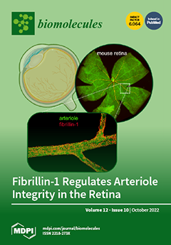
Cover Story (view full-size image): Fibrillin-1 is an extracellular matrix protein that provides critical functions in connective tissues. Mutations of this protein give rise to Marfan syndrome, a genetic disease characterized by severe cardiovascular, ocular, and skeletal symptoms. Typically, fibrillin-1 mutations cause the progressive enlargement of the ascending aorta, leading to aneurysms and dissections. Marfan patients also suffer from retinal detachment. However, if and how fibrillin-1 deficiency affects the microvasculature have never been investigated. We show that fibrillin-1 plays a critical role in the homeostasis of retinal arterioles. Fibrillin-1 deficiency impairs basement membrane and smooth muscle cell coverage, resulting in arteriolar leakage and enlargement. Losartan treatment, which reduces aortic root dilation in mouse models of Marfan syndrome, rescues the arteriolar defects. View this paper
  • Issues are regarded as officially published after their release is announced to the table of contents alert mailing list.
  • You may sign up for e-mail alerts to receive table of contents of newly released issues.
  • PDF is the official format for papers published in both, html and pdf forms. To view the papers in pdf format, click on the "PDF Full-text" link, and use the free Adobe Reader to open them.
Order results
Result details
Section
Select all
Export citation of selected articles as:
12 pages, 808 KiB  
Article
Cholesterol Content of Very-Low-Density Lipoproteins Is Associated with 1-Year Mortality in Acute Heart Failure Patients
by Vesna Degoricija, Iva Klobučar, Ines Potočnjak, Sanda Dokoza Terešak, Luka Vidović, Gudrun Pregartner, Andrea Berghold, Hansjörg Habisch, Tobias Madl and Saša Frank
Biomolecules 2022, 12(10), 1542; https://doi.org/10.3390/biom12101542 - 21 Oct 2022
Cited by 2 | Viewed by 2258
Abstract
Considering the relationship between the extent of metabolic derangement and the disease severity in heart failure, we hypothesized that the lipid content of very-low-density lipoprotein (VLDL) may have prognostic value for 1 year mortality in acute heart failure (AHF). Baseline serum levels of [...] Read more.
Considering the relationship between the extent of metabolic derangement and the disease severity in heart failure, we hypothesized that the lipid content of very-low-density lipoprotein (VLDL) may have prognostic value for 1 year mortality in acute heart failure (AHF). Baseline serum levels of VLDL cholesterol (VLDL-C), VLDL triglycerides (VLDL-TG), VLDL phospholipids (VLDL-PL), and VLDL apolipoprotein B (VLDL-apoB) were measured using NMR spectroscopy. We calculated the ratios of the respective VLDL lipids and VLDL apoB (VLDL-C/VLDL-apoB, VLDL-TG/VLDL-apoB, and VLDL-PL/VLDL-apoB), as estimators of the cholesterol, triglyceride, and phospholipid content of VLDL particles and tested their association with mortality. Out of 315 AHF patients, 118 (37.5%) patients died within 1 year after hospitalization for AHF. Univariable Cox regression analyses revealed a significant inverse association of VLDL-C/VLDL-apoB (hazard ratio (HR) 0.43, 95% confidence interval (CI) 0.29–0.64, p < 0.001), VLDL-TG/VLDL-apoB (HR 0.79, 95% CI 0.71–0.88, p < 0.001), and VLDL-PL/VLDL-apoB (HR 0.37, 95% CI 0.25–0.56, p < 0.001) with 1 year mortality. Of the tested parameters, only VLDL-C/VLDL-apoB remained significant after adjustment for age and sex, as well as other clinical and laboratory parameters that showed a significant association with 1 year mortality in the univariable analyses. We conclude that cholesterol content of circulating VLDL (VLDL-C/VLDL-apoB) might be of prognostic value in AHF. Full article
(This article belongs to the Special Issue Molecular Biomarkers In Cardiology 2022–2023)
Show Figures

Figure 1

14 pages, 3161 KiB  
Article
Type I Interferon Receptor Subunit 1 Deletion Attenuates Experimental Abdominal Aortic Aneurysm Formation
by Takahiro Shoji, Jia Guo, Yingbin Ge, Yankui Li, Gang Li, Toru Ikezoe, Wei Wang, Xiaoya Zheng, Sihai Zhao, Naoki Fujimura, Jianhua Huang, Baohui Xu and Ronald L. Dalman
Biomolecules 2022, 12(10), 1541; https://doi.org/10.3390/biom12101541 - 21 Oct 2022
Cited by 7 | Viewed by 2475
Abstract
Objective: Type I interferon receptor signaling contributes to several autoimmune and vascular diseases such as lupus, atherosclerosis and stroke. The purpose of this study was to assess the influence of type I interferon receptor deficiency on the formation and progression of experimental abdominal [...] Read more.
Objective: Type I interferon receptor signaling contributes to several autoimmune and vascular diseases such as lupus, atherosclerosis and stroke. The purpose of this study was to assess the influence of type I interferon receptor deficiency on the formation and progression of experimental abdominal aortic aneurysms (AAAs). Methods: AAAs were induced in type I interferon receptor subunit 1 (IFNAR1)-deficient and wild type control male mice via intra-infrarenal aortic infusion of porcine pancreatic elastase. Immunostaining for IFNAR1 was evaluated in experimental and clinical aneurysmal abdominal aortae. The initiation and progression of experimental AAAs were assessed via ultrasound imaging prior to (day 0) and days 3, 7 and 14 following elastase infusion. Aneurysmal histopathology was analyzed at sacrifice. Results: Increased aortic medial and adventitial IFNAR1 expression was present in both clinical AAAs harvested at surgery and experimental AAAs. Following AAA induction, wild type mice experienced progressive, time-dependent infrarenal aortic enlargement. This progression was substantially attenuated in IFNAR1-deficient mice. On histological analyses, medial elastin degradation, smooth muscle cell depletion, leukocyte accumulation and neoangiogenesis were markedly diminished in IFNAR1-deficient mice in comparison to wild type mice. Conclusion: IFNAR1 deficiency limited experimental AAA progression in response to intra-aortic elastase infusion. Combined with clinical observations, these results suggest an important role for IFNAR1 activity in AAA pathogenesis. Full article
Show Figures

Figure 1

17 pages, 2102 KiB  
Article
New Betulin Derivatives with Nitrogen Heterocyclic Moiety—Synthesis and Anticancer Activity In Vitro
by Ewa Bębenek, Elwira Chrobak, Zuzanna Rzepka and Dorota Wrześniok
Biomolecules 2022, 12(10), 1540; https://doi.org/10.3390/biom12101540 - 21 Oct 2022
Cited by 7 | Viewed by 1949
Abstract
As part of the search for new medicinal substances with potential application in oncology, the synthesis of new compounds combining the betulin molecule and the indole system was carried out. The structure of the ester derivatives obtained in the Steglich reaction was confirmed [...] Read more.
As part of the search for new medicinal substances with potential application in oncology, the synthesis of new compounds combining the betulin molecule and the indole system was carried out. The structure of the ester derivatives obtained in the Steglich reaction was confirmed by spectroscopic methods (1H and 13C NMR, HR-MS). The obtained new 3-indolyl betulin derivatives were evaluated for anticancer activity against several human cancer cell lines (melanomas, breast cancers, colorectal adenocarcinomas, lung cancer) as well as normal human fibroblasts. The significant reduction in MCF-7 cells viability for 28-hydroxy-(lup-20(29)-ene)-3-yl 2-(1H-indol-3-yl)acetate was observed at a concentration of 10 µg/mL (17 µM). In addition, cytometric analysis showed that this compound strongly reduces the proliferation rate of breast cancer cells. For this, the derivative showing the promising cytotoxic effect on MCF-7 breast cancer cells, the pharmacokinetic profile prediction was performed using in silico methods. Based on the results obtained in the study, it can be concluded that indole-functionalized triterpene EB367 is a promising starting point for further research in the field of breast cancer therapy or the synthesis of new derivatives. Full article
Show Figures

Graphical abstract

13 pages, 16819 KiB  
Article
Automatic Cell Type Annotation Using Marker Genes for Single-Cell RNA Sequencing Data
by Yu Chen and Shuqin Zhang
Biomolecules 2022, 12(10), 1539; https://doi.org/10.3390/biom12101539 - 21 Oct 2022
Cited by 4 | Viewed by 3323
Abstract
Recent advancement in single-cell RNA sequencing (scRNA-seq) technology is gaining more and more attention. Cell type annotation plays an essential role in scRNA-seq data analysis. Several computational methods have been proposed for automatic annotation. Traditional cell type annotation is to first cluster the [...] Read more.
Recent advancement in single-cell RNA sequencing (scRNA-seq) technology is gaining more and more attention. Cell type annotation plays an essential role in scRNA-seq data analysis. Several computational methods have been proposed for automatic annotation. Traditional cell type annotation is to first cluster the cells using unsupervised learning methods based on the gene expression profiles, then to label the clusters using the aggregated cluster-level expression profiles and the marker genes’ information. Such procedure relies heavily on the clustering results. As the purity of clusters cannot be guaranteed, false detection of cluster features may lead to wrong annotations. In this paper, we improve this procedure and propose an Automatic Cell type Annotation Method (ACAM). ACAM delineates a clear framework to conduct automatic cell annotation through representative cluster identification, representative cluster annotation using marker genes, and the remaining cells’ classification. Experiments on seven real datasets show the better performance of ACAM compared to six well-known cell type annotation methods. Full article
Show Figures

Figure 1

19 pages, 3087 KiB  
Article
Heat Shock Protein Upregulation Supplemental to Complex mRNA Alterations in Autoimmune Glaucoma
by Sabrina Reinehr, Armin Safaei, Pia Grotegut, Annika Guntermann, Teresa Tsai, Stephan A. Hahn, Steffen Kösters, Carsten Theiss, Katrin Marcus, H. Burkhard Dick, Caroline May and Stephanie C. Joachim
Biomolecules 2022, 12(10), 1538; https://doi.org/10.3390/biom12101538 - 21 Oct 2022
Cited by 4 | Viewed by 2474
Abstract
Glaucomatous optic neuropathy is a common cause for blindness. An elevated intraocular pressure is the main risk factor, but also a contribution of the immune system seems likely. In the experimental autoimmune glaucoma model used here, systemic immunization with an optic nerve homogenate [...] Read more.
Glaucomatous optic neuropathy is a common cause for blindness. An elevated intraocular pressure is the main risk factor, but also a contribution of the immune system seems likely. In the experimental autoimmune glaucoma model used here, systemic immunization with an optic nerve homogenate antigen (ONA) leads to retinal ganglion cell (RGC) and optic nerve degeneration. We processed retinae for quantitative real-time PCR and immunohistology 28 days after immunization. Furthermore, we performed mRNA profiling in this model for the first time. We detected a significant RGC loss in the ONA retinae. This was accompanied by an upregulation of mRNA expression of genes belonging to the heat shock protein family. Furthermore, mRNA expression levels of the genes of the immune system, such as C1qa, C1qb, Il18, and Nfkb1, were upregulated in ONA animals. After laser microdissection, inner retinal layers were used for mRNA microarrays. Nine of these probes were significantly upregulated in ONA animals (p < 0.05), including Hba-a1 and Cxcl10, while fifteen probes were significantly downregulated in ONA animals (p < 0.05), such as Gdf15 and Wwox. Taken together, these findings provide further insights into the pivotal role of the immune response in glaucomatous optic neuropathy and could help to identify novel diagnostic or therapeutic strategies. Full article
(This article belongs to the Special Issue Molecular and Cellular Mechanisms of Optic Neuropathies)
Show Figures

Figure 1

10 pages, 10631 KiB  
Article
Gerstmann–Sträussler–Scheinker Disease with F198S Mutation Induces Independent Tau and Prion Protein Pathologies in Bank Voles
by Rosalia Bruno, Laura Pirisinu, Geraldina Riccardi, Claudia D’Agostino, Elena De Cecco, Giuseppe Legname, Franco Cardone, Pierluigi Gambetti, Romolo Nonno, Umberto Agrimi and Michele Angelo Di Bari
Biomolecules 2022, 12(10), 1537; https://doi.org/10.3390/biom12101537 - 21 Oct 2022
Cited by 1 | Viewed by 2035
Abstract
Gerstmann–Sträussler–Scheinker disease (GSS) is a rare genetic prion disease. A large GSS kindred linked to the serine-for-phenylalanine substitution at codon 198 of the prion protein gene (GSS-F198S) is characterized by conspicuous accumulation of prion protein (PrP)-amyloid deposits and neurofibrillary tangles. Recently, we demonstrated [...] Read more.
Gerstmann–Sträussler–Scheinker disease (GSS) is a rare genetic prion disease. A large GSS kindred linked to the serine-for-phenylalanine substitution at codon 198 of the prion protein gene (GSS-F198S) is characterized by conspicuous accumulation of prion protein (PrP)-amyloid deposits and neurofibrillary tangles. Recently, we demonstrated the transmissibility of GSS-F198S prions to bank vole carrying isoleucine at 109 PrP codon (BvI). Here we investigated: (i) the transmissibility of GSS-F198S prions to voles carrying methionine at codon 109 (BvM); (ii) the induction of hyperphosphorylated Tau (pTau) in two vole lines, and (iii) compared the phenotype of GSS-F198S-induced pTau with pTau induced in BvM following intracerebral inoculation of a familial Alzheimer’s disease case carrying Presenilin 1 mutation (fAD-PS1). We did not detect prion transmission to BvM, despite the high susceptibility of BvI previously observed. Immunohistochemistry established the presence of induced pTau depositions in vole brains that were not affected by prions. Furthermore, the phenotype of pTau deposits in vole brains was similar in GSS-F198S and fAD-PS1. Overall, results suggest that, regardless of the cause of pTau deposition and its relationship with PrPSc in GSS-F198S human-affected brains, the two components possess their own seeding properties, and that pTau deposition is similarly induced by GSS-F198S and fAD-PS1. Full article
(This article belongs to the Special Issue New Insights into Prion and Prion-Like Diseases)
Show Figures

Figure 1

3 pages, 184 KiB  
Editorial
Aortic Aneurysm and Dissection: Heterogeneity and Molecular Mechanisms
by Hong S. Lu, Hisashi Sawada and Congqing Wu
Biomolecules 2022, 12(10), 1536; https://doi.org/10.3390/biom12101536 - 21 Oct 2022
Cited by 1 | Viewed by 1602
Abstract
Aortic aneurysms and dissections (AAD) are devastating aortic diseases with high risks for aortic rupture, leading to uncontrolled bleeding and death [...] Full article
16 pages, 2435 KiB  
Article
Effects of Psychotropic Medication on Somatic Sterol Biosynthesis of Adult Mice
by Marta Balog, Allison C Anderson, Marija Heffer, Zeljka Korade and Karoly Mirnics
Biomolecules 2022, 12(10), 1535; https://doi.org/10.3390/biom12101535 - 21 Oct 2022
Cited by 4 | Viewed by 1990
Abstract
Polypharmacy is commonly used to treat psychiatric disorders. These combinations often include drugs with sterol biosynthesis inhibiting side effects, including the antipsychotic aripiprazole (ARI), and antidepressant trazodone (TRZ). As the effects of psychotropic medications are poorly understood across the various tissue types to [...] Read more.
Polypharmacy is commonly used to treat psychiatric disorders. These combinations often include drugs with sterol biosynthesis inhibiting side effects, including the antipsychotic aripiprazole (ARI), and antidepressant trazodone (TRZ). As the effects of psychotropic medications are poorly understood across the various tissue types to date, we investigated the effects of ARI, TRZ, and ARI + TRZ polypharmacy on the post-lanosterol biosynthesis in three cell lines (Neuro2a, HepG2, and human dermal fibroblasts) and seven peripheral tissues of an adult mouse model. We found that both ARI and TRZ strongly interfere with the function of 7-dehydrocholesterol reductase enzyme (DHCR7) and lead to robust elevation in 7-dehydrocholesterol levels (7-DHC) and reduction in desmosterol (DES) across all cell lines and somatic tissues. ARI + TRZ co-administration resulted in summative or synergistic effects across the utilized in vitro and in vivo models. These findings suggest that at least some of the side effects of ARI and TRZ are not receptor mediated but arise from inhibiting DHCR7 enzyme activity. We propose that interference with sterol biosynthesis, particularly in the case of simultaneous utilization of medications with such side effects, can potentially interfere with functioning or development of multiple organ systems, warranting further investigation. Full article
(This article belongs to the Collection Feature Papers in Biomacromolecules: Lipids)
Show Figures

Figure 1

15 pages, 1822 KiB  
Article
Modelling Hyperglycaemia in an Epithelial Membrane Model: Biophysical Characterisation
by Ana Reis, Joana P. F. Teixeira, Ana M. G. Silva, Mariana Ferreira, Paula Gameiro and Victor de Freitas
Biomolecules 2022, 12(10), 1534; https://doi.org/10.3390/biom12101534 - 21 Oct 2022
Viewed by 1950
Abstract
Biomimetic models are valuable platforms to improve our knowledge on the molecular mechanisms governing membrane-driven processes in (patho)physiological conditions, including membrane permeability, transport, and fusion. However, current membrane models are over simplistic and do not include the membrane’s lipid remodelling in response to [...] Read more.
Biomimetic models are valuable platforms to improve our knowledge on the molecular mechanisms governing membrane-driven processes in (patho)physiological conditions, including membrane permeability, transport, and fusion. However, current membrane models are over simplistic and do not include the membrane’s lipid remodelling in response to extracellular stimuli. Our study describes the synthesis of glycated dimyristoyl-phosphatidylethanolamine (DMPE-glyc), which was structurally characterised by mass spectrometry (ESI-MS) and quantified by NMR spectroscopy to be further incorporated in a complex phospholipid (PL) membrane model enriched in cholesterol (Chol) and (glyco)sphingolipids (GSL) designed to mimic epithelial membranes (PL/Chol/GSL) under hyperglycaemia conditions. Characterisation of synthesised DMPE-glyc adducts by tandem mass spectrometry (ESI-MS/MS) show that synthetic DMPE-glyc adducts correspond to Amadori products and quantification by 1H NMR spectroscopy show that the yield of glycation reaction was 8%. The biophysical characterisation of the epithelial membrane model shows that excess glucose alters the thermotropic behaviour and fluidity of epithelial membrane models likely to impact permeability of solutes. The epithelial membrane models developed to mimic normo- and hyperglycaemic scenarios are the basis to investigate (poly)phenol-lipid and drug–membrane interactions crucial in nutrition, pharmaceutics, structural biochemistry, and medicinal chemistry. Full article
(This article belongs to the Special Issue Bioactive Lipids: Sources, Synthesis, and Biological Roles)
Show Figures

Figure 1

14 pages, 1775 KiB  
Article
Molecular Classification of Genes Associated with Hypoxic Lipid Metabolism in Pancreatic Cancer
by Yaning Li, Xinyue Liang, Gang Che, Yutong Chen, Lisi Luo, Kecheng Liu, Rongzhi Xie and Linjuan Zeng
Biomolecules 2022, 12(10), 1533; https://doi.org/10.3390/biom12101533 - 21 Oct 2022
Cited by 6 | Viewed by 2753
Abstract
Abnormal lipid metabolism often occurs under hypoxic microenvironment, which is an important energy supplement for cancer cell proliferation and metastasis. We aimed to explore the lipid metabolism characteristics and gene expression features of pancreatic ductal adenocarcinoma (PDAC) related to hypoxia and identify biomarkers [...] Read more.
Abnormal lipid metabolism often occurs under hypoxic microenvironment, which is an important energy supplement for cancer cell proliferation and metastasis. We aimed to explore the lipid metabolism characteristics and gene expression features of pancreatic ductal adenocarcinoma (PDAC) related to hypoxia and identify biomarkers for molecular classification based on hypoxic lipid metabolism that are evaluable for PDAC prognosis and therapy. The multiple datasets were analyzed integratively, including corresponding clinical information of samples. PDAC possesses a distinct metabolic profile and oxygen level compared with normal pancreatic tissues, according to the bioinformatics methods. In addition, a study on untargeted metabolomics using Ultra Performance Liquid Chromatography Tandem Mass Spectrometry(UPLC-MS) revealed lipid metabolites differences affected by oxygen. Analysis of PDAC gene expression profiling in The Cancer Genome Atlas (TCGA) revealed that the sphingolipid process correlates closely with HIF1α. According to the characters of HIF-1 and sphingolipid, samples can be clustered into three subgroups using non-negative matrix factorization clustering. In cluster2, patients had an increased survival time. Relatively high MUC16 mutation arises in cluster2 and may positively influence the cancer survival rates. This study explored the expression pattern of lipid metabolism under hypoxia microenvironment in PDAC. On the basis of metabolic signatures, we identified the prognosis subtypes linking lipid metabolism to hypoxia. The classifications may be conducive to developing personalized treatment programs targeting metabolic profiles. Full article
(This article belongs to the Section Bioinformatics and Systems Biology)
Show Figures

Figure 1

13 pages, 3535 KiB  
Article
Mitochondrial Dysfunction in a High Intraocular Pressure-Induced Retinal Ischemia Minipig Model
by Michael Pasák, Marie Vanišová, Lucie Tichotová, Jana Křížová, Taras Ardan, Yaroslav Nemesh, Jana Čížková, Anastasiia Kolesnikova, Ruslan Nyshchuk, Natasha Josifovska, Lyubomyr Lytvynchuk, Miriam Kolko, Jan Motlík, Goran Petrovski and Hana Hansíková
Biomolecules 2022, 12(10), 1532; https://doi.org/10.3390/biom12101532 - 21 Oct 2022
Cited by 4 | Viewed by 2101
Abstract
Purpose: Retinal ischemia (RI) and progressive neuronal death are sight-threatening conditions. Mitochondrial (mt) dysfunction and fusion/fission processes have been suggested to play a role in the pathophysiology of RI. This study focuses on changes in the mt parameters of the neuroretina, retinal pigment [...] Read more.
Purpose: Retinal ischemia (RI) and progressive neuronal death are sight-threatening conditions. Mitochondrial (mt) dysfunction and fusion/fission processes have been suggested to play a role in the pathophysiology of RI. This study focuses on changes in the mt parameters of the neuroretina, retinal pigment epithelium (RPE) and choroid in a porcine high intraocular pressure (IOP)-induced RI minipig model. Methods: In one eye, an acute IOP elevation was induced in minipigs and compared to the other control eye. Activity and amount of respiratory chain complexes (RCC) were analyzed by spectrophotometry and Western blot, respectively. The coenzyme Q10 (CoQ10) content was measured using HPLC, and the ultrastructure of the mt was studied via transmission electron microscopy. The expression of selected mt-pathway genes was determined by RT-PCR. Results: At a functional level, increased RCC I activity and decreased total CoQ10 content were found in RPE cells. At a protein level, CORE2, a subunit of RCC III, and DRP1, was significantly decreased in the neuroretina. Drp1 and Opa1, protein-encoding genes responsible for mt quality control, were decreased in most of the samples from the RPE and neuroretina. Conclusions: The eyes of the minipig can be considered a potential RI model to study mt dysfunction in this disease. Strategies targeting mt protection may provide a promising way to delay the acute damage and onset of RI. Full article
(This article belongs to the Special Issue New Discoveries in Retinal Cell Degeneration and Retinal Diseases)
Show Figures

Figure 1

11 pages, 3221 KiB  
Article
Molecular Regulation of Heme Oxygenase-1 Expression by E2F Transcription Factor 2 in Lung Fibroblast Cells: Relevance to Idiopathic Pulmonary Fibrosis
by Qinmao Ye, Sarah J. Taleb, Heather Wang, Narasimham L. Parinandi, Daniel J. Kass, Mauricio Rojas, Cankun Wang, Qin Ma, Jing Zhao and Yutong Zhao
Biomolecules 2022, 12(10), 1531; https://doi.org/10.3390/biom12101531 - 21 Oct 2022
Cited by 4 | Viewed by 2345
Abstract
Idiopathic pulmonary fibrosis (IPF) is a fatal chronic lung disease. Heme oxygenase-1 (HMOX1/HO-1) is an enzyme that catalyzes the degradation of heme. The role of HO-1 in the pathogenesis of IPF has been studied; however, the molecular regulation of HO-1 and its role [...] Read more.
Idiopathic pulmonary fibrosis (IPF) is a fatal chronic lung disease. Heme oxygenase-1 (HMOX1/HO-1) is an enzyme that catalyzes the degradation of heme. The role of HO-1 in the pathogenesis of IPF has been studied; however, the molecular regulation of HO-1 and its role in IPF are still unclear. In this study, we found that HO-1 protein levels significantly increased in lung myofibroblasts in IPF patients and in lungs in a murine model of bleomycin-induced lung fibrosis. In addition, we observed that administration of a E2F transcription factor inhibitor elevated HO-1 mRNA and protein levels in lung fibroblasts. Downregulation of E2F2 by siRNA transfection increased HO-1 mRNA and protein levels, while overexpression of E2F2 reduced HO-1 levels. However, overexpression of E2F2 did not alter hemin-induced HO-1 protein levels. Furthermore, modulation of HO-1 levels regulated TGF-β1-induced myofibroblast differentiation without altering the phosphorylation of Smad2/3 in lung fibroblast cells. Moreover, the phosphorylation of protein kinase B (Akt) was significantly upregulated in HO-1-depleted lung fibroblast cells. In summary, this study demonstrated that E2F2 regulates the baseline expression of HO-1, but has no effect on modulating HO-1 expression by hemin. Finally, elevated HO-1 expression contributes to the TGF-β1-induced lung myofibroblast differentiation through the activation of the serine/threonine kinase AKT pathway. Overall, our findings suggest that targeting E2F2/HO-1 might be a new therapeutic strategy to treat fibrotic diseases such as IPF. Full article
(This article belongs to the Special Issue Molecular Mechanisms in Lung Injury, Repair, and Remodeling)
Show Figures

Figure 1

3 pages, 191 KiB  
Editorial
Advances in Molecular Biomarkers in Cardiology
by Pietro Scicchitano and Matteo Cameli
Biomolecules 2022, 12(10), 1530; https://doi.org/10.3390/biom12101530 - 21 Oct 2022
Cited by 1 | Viewed by 1531
Abstract
Cardiovascular diseases (CVD) represent the leading cause of death in the world despite innovations in therapies and advances in the general management of patients [...] Full article
(This article belongs to the Special Issue Molecular Biomarkers In Cardiology 2021)
14 pages, 2259 KiB  
Article
EP-Pred: A Machine Learning Tool for Bioprospecting Promiscuous Ester Hydrolases
by Ruite Xiang, Laura Fernandez-Lopez, Ana Robles-Martín, Manuel Ferrer and Victor Guallar
Biomolecules 2022, 12(10), 1529; https://doi.org/10.3390/biom12101529 - 21 Oct 2022
Cited by 4 | Viewed by 2449
Abstract
When bioprospecting for novel industrial enzymes, substrate promiscuity is a desirable property that increases the reusability of the enzyme. Among industrial enzymes, ester hydrolases have great relevance for which the demand has not ceased to increase. However, the search for new substrate promiscuous [...] Read more.
When bioprospecting for novel industrial enzymes, substrate promiscuity is a desirable property that increases the reusability of the enzyme. Among industrial enzymes, ester hydrolases have great relevance for which the demand has not ceased to increase. However, the search for new substrate promiscuous ester hydrolases is not trivial since the mechanism behind this property is greatly influenced by the active site’s structural and physicochemical characteristics. These characteristics must be computed from the 3D structure, which is rarely available and expensive to measure, hence the need for a method that can predict promiscuity from sequence alone. Here we report such a method called EP-pred, an ensemble binary classifier, that combines three machine learning algorithms: SVM, KNN, and a Linear model. EP-pred has been evaluated against the Lipase Engineering Database together with a hidden Markov approach leading to a final set of ten sequences predicted to encode promiscuous esterases. Experimental results confirmed the validity of our method since all ten proteins were found to exhibit a broad substrate ambiguity. Full article
Show Figures

Figure 1

16 pages, 29754 KiB  
Article
Acyl-Nω-methylserotonins and Branched-chain Acylserotonins in Lemon and Other Citrus Seeds—New Lipids with Antioxidant Properties and Potential Pharmacological Applications
by Jerzy Kruk, Agnieszka Trela-Makowej and Renata Szymańska
Biomolecules 2022, 12(10), 1528; https://doi.org/10.3390/biom12101528 - 20 Oct 2022
Cited by 1 | Viewed by 1756
Abstract
We have found 15 previously unknown compounds in seeds of lemon and other citrus species, such as tangerine, grapefruit and pomelo. The structure of these compounds was characterized by HR–MS spectrometry, fluorescence spectroscopy and chemical synthesis. These compounds were predominantly long-chain (C20–C25), saturated [...] Read more.
We have found 15 previously unknown compounds in seeds of lemon and other citrus species, such as tangerine, grapefruit and pomelo. The structure of these compounds was characterized by HR–MS spectrometry, fluorescence spectroscopy and chemical synthesis. These compounds were predominantly long-chain (C20–C25), saturated acyl-Nω-methylserotonins with the main contribution of C22 and C24 homologues, usually accounting for about 40% and 30% of all acylserotonins, respectively. The other, previously undescribed, minor compounds were branched-chain acylserotonins, as well as normal-chain acylserotonins, recently found in baobab seed oil. Within the seed, acylserotonins were found nearly exclusively in the inner seed coat, where probably their biosynthesis proceeds. On the other hand, lemon seedlings contained only trace amounts of these compounds that were not found in adult leaves. The compounds identified in the present studies were shown to have antioxidant properties in vitro, using 2,2-diphenyl-1-picrylhydrazyl (DPPH) assay. In the investigated reaction in hexane, Me-C22 and Me-C24-serotonins were less active than n-C22 and n-C24-serotonins and δ-tocopherol, while branched-chain acylserotonins (iso-C21 and -C25) showed higher antioxidant activity than all the normal-chain compounds. On the other hand, all these compounds showed a similar but considerably lower antioxidant activity in acetonitrile than in hexane. Full article
(This article belongs to the Special Issue Bioactive Lipids: Sources, Synthesis, and Biological Roles)
Show Figures

Figure 1

16 pages, 3782 KiB  
Article
The Epigenetic Dimension of Protein Structure Is an Intrinsic Weakness of the AlphaFold Program
by Fodil Azzaz, Nouara Yahi, Henri Chahinian and Jacques Fantini
Biomolecules 2022, 12(10), 1527; https://doi.org/10.3390/biom12101527 - 20 Oct 2022
Cited by 26 | Viewed by 6827
Abstract
One of the most important lessons we have learned from sequencing the human genome is that not all proteins have a 3D structure. In fact, a large part of the human proteome is made up of intrinsically disordered proteins (IDPs) which can adopt [...] Read more.
One of the most important lessons we have learned from sequencing the human genome is that not all proteins have a 3D structure. In fact, a large part of the human proteome is made up of intrinsically disordered proteins (IDPs) which can adopt multiple structures, and therefore, multiple functions, depending on the ligands with which they interact. Under these conditions, one can wonder about the value of algorithms developed for predicting the structure of proteins, in particular AlphaFold, an AI which claims to have solved the problem of protein structure. In a recent study, we highlighted a particular weakness of AlphaFold for membrane proteins. Based on this observation, we have proposed a paradigm, referred to as “Epigenetic Dimension of Protein Structure” (EDPS), which takes into account all environmental parameters that control the structure of a protein beyond the amino acid sequence (hence “epigenetic”). In this new study, we compare the reliability of the AlphaFold and Robetta algorithms’ predictions for a new set of membrane proteins involved in human pathologies. We found that Robetta was generally more accurate than AlphaFold for ascribing a membrane-compatible topology. Raft lipids (e.g., gangliosides), which control the structural dynamics of membrane protein structure through chaperone effects, were identified as major actors of the EDPS paradigm. We conclude that the epigenetic dimension of a protein structure is an intrinsic weakness of AI-based protein structure prediction, especially AlphaFold, which warrants further development. Full article
(This article belongs to the Special Issue Protein Structure Prediction with AlphaFold)
Show Figures

Figure 1

20 pages, 9039 KiB  
Article
TNP Analogues Inhibit the Virulence Promoting IP3-4 Kinase Arg1 in the Fungal Pathogen Cryptococcus neoformans
by Desmarini Desmarini, Daniel Truong, Lorna Wilkinson-White, Chandrika Desphande, Mario Torrado, Joel P. Mackay, Jacqueline M. Matthews, Tania C. Sorrell, Sophie Lev, Philip E. Thompson and Julianne Teresa Djordjevic
Biomolecules 2022, 12(10), 1526; https://doi.org/10.3390/biom12101526 - 20 Oct 2022
Cited by 1 | Viewed by 2503
Abstract
New antifungals with unique modes of action are urgently needed to treat the increasing global burden of invasive fungal infections. The fungal inositol polyphosphate kinase (IPK) pathway, comprised of IPKs that convert IP3 to IP8, provides a promising new target [...] Read more.
New antifungals with unique modes of action are urgently needed to treat the increasing global burden of invasive fungal infections. The fungal inositol polyphosphate kinase (IPK) pathway, comprised of IPKs that convert IP3 to IP8, provides a promising new target due to its impact on multiple, critical cellular functions and, unlike in mammalian cells, its lack of redundancy. Nearly all IPKs in the fungal pathway are essential for virulence, with IP3-4 kinase (IP3-4K) the most critical. The dibenzylaminopurine compound, N2-(m-trifluorobenzylamino)-N6-(p-nitrobenzylamino)purine (TNP), is a commercially available inhibitor of mammalian IPKs. The ability of TNP to be adapted as an inhibitor of fungal IP3-4K has not been investigated. We purified IP3-4K from the human pathogens, Cryptococcus neoformans and Candida albicans, and optimised enzyme and surface plasmon resonance (SPR) assays to determine the half inhibitory concentration (IC50) and binding affinity (KD), respectively, of TNP and 38 analogues. A novel chemical route was developed to efficiently prepare TNP analogues. TNP and its analogues demonstrated inhibition of recombinant IP3-4K from C. neoformans (CnArg1) at low µM IC50s, but not IP3-4K from C. albicans (CaIpk2) and many analogues exhibited selectivity for CnArg1 over the human equivalent, HsIPMK. Our results provide a foundation for improving potency and selectivity of the TNP series for fungal IP3-4K. Full article
Show Figures

Figure 1

12 pages, 758 KiB  
Review
Cyclosporine A Delivery Platform for Veterinary Ophthalmology—A New Concept for Advanced Ophthalmology
by Martyna Padjasek, Badr Qasem, Anna Cisło-Pakuluk and Krzysztof Marycz
Biomolecules 2022, 12(10), 1525; https://doi.org/10.3390/biom12101525 - 20 Oct 2022
Cited by 7 | Viewed by 3809
Abstract
Cyclosporine A (CsA) is a selective and reversible immunosuppressant agent that is widely used as a medication for a wide spectrum of diseases in humans such as graft versus host disease, non-infectious uveitis, rheumatoid arthritis, psoriasis, and atopic dermatitis. Furthermore, the CsA is [...] Read more.
Cyclosporine A (CsA) is a selective and reversible immunosuppressant agent that is widely used as a medication for a wide spectrum of diseases in humans such as graft versus host disease, non-infectious uveitis, rheumatoid arthritis, psoriasis, and atopic dermatitis. Furthermore, the CsA is used to treat keratoconjunctivitis sicca, chronic superficial keratitis, immune-mediated keratitis and equine recurrent uveitis in animals. The selective activity of Cyclosporine A (CsA) was demonstrated to be an immunomodulation characteristic of T-lymphocyte proliferation and inhibits cytokine gene expression. Moreover, the lipophilic characteristics with poor bioavailability and low solubility in water, besides the side effects, force the need to develop new formulations and devices that will provide adequate penetration into the anterior and posterior segments of the eye. This review aims to summarize the effectiveness and safety of cyclosporine A delivery platforms in veterinary ophthalmology. Full article
(This article belongs to the Special Issue Cyclodextrin-Based Drug Release and Drug Delivery Systems)
Show Figures

Figure 1

25 pages, 9005 KiB  
Article
SMN Is Physiologically Downregulated at Wild-Type Motor Nerve Terminals but Aggregates Together with Neurofilaments in SMA Mouse Models
by Julio Franco-Espin, Alaó Gatius, José Ángel Armengol, Saravanan Arumugam, Mehri Moradi, Michael Sendtner, Jordi Calderó and Lucia Tabares
Biomolecules 2022, 12(10), 1524; https://doi.org/10.3390/biom12101524 - 20 Oct 2022
Cited by 5 | Viewed by 2973
Abstract
Survival motor neuron (SMN) is an essential and ubiquitously expressed protein that participates in several aspects of RNA metabolism. SMN deficiency causes a devastating motor neuron disease called spinal muscular atrophy (SMA). SMN forms the core of a protein complex localized at the [...] Read more.
Survival motor neuron (SMN) is an essential and ubiquitously expressed protein that participates in several aspects of RNA metabolism. SMN deficiency causes a devastating motor neuron disease called spinal muscular atrophy (SMA). SMN forms the core of a protein complex localized at the cytoplasm and nuclear gems and that catalyzes spliceosomal snRNP particle synthesis. In cultured motor neurons, SMN is also present in dendrites and axons, and forms part of the ribonucleoprotein transport granules implicated in mRNA trafficking and local translation. Nevertheless, the distribution, regulation, and role of SMN at the axons and presynaptic motor terminals in vivo are still unclear. By using conventional confocal microscopy and STED super-resolution nanoscopy, we found that SMN appears in the form of granules distributed along motor axons at nerve terminals. Our fluorescence in situ hybridization and electron microscopy studies also confirmed the presence of β-actin mRNA, ribosomes, and polysomes in the presynaptic motor terminal, key elements of the protein synthesis machinery involved in local translation in this compartment. SMN granules co-localize with the microtubule-associated protein 1B (MAP1B) and neurofilaments, suggesting that the cytoskeleton participates in transporting and positioning the granules. We also found that, while SMN granules are physiologically downregulated at the presynaptic element during the period of postnatal maturation in wild-type (non-transgenic) mice, they accumulate in areas of neurofilament aggregation in SMA mice, suggesting that the high expression of SMN at the NMJ, together with the cytoskeletal defects, contribute to impairing the bi-directional traffic of proteins and organelles between the axon and the presynaptic terminal. Full article
Show Figures

Figure 1

12 pages, 3317 KiB  
Review
Synthesis of Protein-Oligonucleotide Conjugates
by Emma E. Watson and Nicolas Winssinger
Biomolecules 2022, 12(10), 1523; https://doi.org/10.3390/biom12101523 - 20 Oct 2022
Cited by 5 | Viewed by 3907
Abstract
Nucleic acids and proteins form two of the key classes of functional biomolecules. Through the ability to access specific protein-oligonucleotide conjugates, a broader range of functional molecules becomes accessible which leverages both the programmability and recognition potential of nucleic acids and the structural, [...] Read more.
Nucleic acids and proteins form two of the key classes of functional biomolecules. Through the ability to access specific protein-oligonucleotide conjugates, a broader range of functional molecules becomes accessible which leverages both the programmability and recognition potential of nucleic acids and the structural, chemical and functional diversity of proteins. Herein, we summarize the available conjugation strategies to access such chimeric molecules and highlight some key case study examples within the field to showcase the power and utility of such technology. Full article
(This article belongs to the Collection Feature Papers in Biomacromolecules: Nucleic Acids)
Show Figures

Figure 1

19 pages, 701 KiB  
Review
Methods for Radiolabelling Nanoparticles: SPECT Use (Part 1)
by Michela Varani, Valeria Bentivoglio, Chiara Lauri, Danilo Ranieri and Alberto Signore
Biomolecules 2022, 12(10), 1522; https://doi.org/10.3390/biom12101522 - 20 Oct 2022
Cited by 8 | Viewed by 3107
Abstract
The use of nanoparticles (NPs) is rapidly increasing in nuclear medicine (NM) for diagnostic and therapeutic purposes. Their wide use is due to their chemical–physical characteristics and possibility to deliver several molecules. NPs can be synthetised by organic and/or inorganic materials and they [...] Read more.
The use of nanoparticles (NPs) is rapidly increasing in nuclear medicine (NM) for diagnostic and therapeutic purposes. Their wide use is due to their chemical–physical characteristics and possibility to deliver several molecules. NPs can be synthetised by organic and/or inorganic materials and they can have different size, shape, chemical composition, and charge. These factors influence their biodistribution, clearance, and targeting ability in vivo. NPs can be designed to encapsulate inside the core or bind to the surface several molecules, including radionuclides, for different clinical applications. Either diagnostic or therapeutic radioactive NPs can be synthetised, making a so-called theragnostic tool. To date, there are several methods for radiolabelling NPs that vary depending on both the physical and chemical properties of the NPs and on the isotope used. In this review, we analysed and compared different methods for radiolabelling NPs for single-photon emission computed tomography (SPECT) use. Full article
(This article belongs to the Section Biological and Bio- Materials)
Show Figures

Figure 1

13 pages, 5677 KiB  
Article
Zebrafish Model of Severe Combined Immunodeficiency (SCID) Due to JAK3 Mutation
by Faiza Basheer, Effie Lee, Clifford Liongue and Alister C. Ward
Biomolecules 2022, 12(10), 1521; https://doi.org/10.3390/biom12101521 - 20 Oct 2022
Cited by 6 | Viewed by 2730
Abstract
JAK3 is principally activated by members of the interleukin-2 receptor family and plays an essential role in lymphoid development, with inactivating JAK3 mutations causing autosomal-recessive severe combined immunodeficiency (SCID). This study aimed to generate an equivalent zebrafish model of SCID and to characterize [...] Read more.
JAK3 is principally activated by members of the interleukin-2 receptor family and plays an essential role in lymphoid development, with inactivating JAK3 mutations causing autosomal-recessive severe combined immunodeficiency (SCID). This study aimed to generate an equivalent zebrafish model of SCID and to characterize the model across the life-course. Genome editing of zebrafish jak3 created mutants similar to those observed in human SCID. Homozygous jak3 mutants showed reduced embryonic T lymphopoiesis that continued through the larval stage and into adulthood, with B cell maturation and adult NK cells also reduced and neutrophils impacted. Mutant fish were susceptible to lymphoid leukemia. This model has many of the hallmarks of human SCID resulting from inactivating JAK3 mutations and will be useful for a variety of pre-clinical applications. Full article
(This article belongs to the Topic Kinases in Cancer and Other Diseases)
Show Figures

Figure 1

20 pages, 1745 KiB  
Review
Copper-Dependent Kinases and Their Role in Cancer Inception, Progression and Metastasis
by Alessandra Vitaliti, Anastasia De Luca and Luisa Rossi
Biomolecules 2022, 12(10), 1520; https://doi.org/10.3390/biom12101520 - 20 Oct 2022
Cited by 13 | Viewed by 3236
Abstract
In recent years, copper function has been expanded beyond its consolidated role as a cofactor of enzyme catalysis. Recent papers have demonstrated a new dynamic role for copper in the regulation of cell signaling pathways through direct interaction with protein kinases, modulating their [...] Read more.
In recent years, copper function has been expanded beyond its consolidated role as a cofactor of enzyme catalysis. Recent papers have demonstrated a new dynamic role for copper in the regulation of cell signaling pathways through direct interaction with protein kinases, modulating their activity. The activation of these pathways is exacerbated in cancer cells to sustain the different steps of tumor growth and dissemination. This review will focus on a novel proposed role for the transition metal copper as a regulator of cell signaling pathways through direct interaction with known protein kinases, which exhibit binding domains for this metal. Activation of these pathways in cancer cells supports both tumor growth and dissemination. In addition to the description of the results recently reported in the literature on the subject, relevance will be given to the possibility of controlling the cellular levels of copper and its homeostatic regulators. Overall, these findings may be of central relevance in order to propose copper and its homeostatic regulators as possible targets for novel therapies, which may act synergistically to those already existing to control cancer growth and dissemination. Full article
(This article belongs to the Special Issue Kinases Signaling in Cancers)
Show Figures

Figure 1

11 pages, 291 KiB  
Article
Grape Pulp Fiber as Possible Fining Agents for Red Wine
by Andrea Osete-Alcaraz, Lucía Osete-Alcaraz, Ana Eugenia Ortega-Regules, Ana Belen Bautista-Ortín and Encarna Gómez-Plaza
Biomolecules 2022, 12(10), 1519; https://doi.org/10.3390/biom12101519 - 20 Oct 2022
Cited by 3 | Viewed by 2049
Abstract
One of the biggest problems with the use of traditional fining agents is that some of them present allergenic characteristics or are not suitable for vegan consumers due to their animal origin. An alternative to these traditional proteinaceous agents could be cell walls [...] Read more.
One of the biggest problems with the use of traditional fining agents is that some of them present allergenic characteristics or are not suitable for vegan consumers due to their animal origin. An alternative to these traditional proteinaceous agents could be cell walls from grape pulp. This material could be used to modify the final phenolic concentration of a wine due to its affinity for phenolic compounds. In this study, the ability of freeze-dried grape pulp fiber, rich in pulp cell walls, to act as a fining agent was analyzed in wines from three different varieties: Cabernet Sauvignon, Syrah, and Monastrell. After the use of this material, the wine chromatic characteristics and total tannin concentration were analyzed by spectrophotometric and chromatographic techniques. In addition, the wines were contaminated with ochratoxin A and histamine to check whether this material could also be a tool for removing these wine contaminants. The pulp fiber presented a high capacity to retain phenolic compounds, especially tannins; however, there were differences depending on the studied wine. The largest reduction in tannin concentration after fining was observed when this material was used in Cabernet Sauvignon wines (23%), whereas for Monastrell wines the reduction was lower (18.3%) and even lower for Syrah wines (14.3%). This fining agent also reduced the anthocyanin concentration of the three red wines, although to a lesser extent than the reduction observed for tannins. A really interesting result was that the addition of this fining agent reduced the concentration of ochratoxin A by 50% in all the studied wines. Full article
Show Figures

Graphical abstract

16 pages, 1567 KiB  
Review
A Promising Candidate in Tendon Healing Events—PDGF-BB
by Yixuan Chen, Li Jiang, Kexin Lyu, Jingwei Lu, Longhai Long, Xiaoqiang Wang, Tianzhu Liu and Sen Li
Biomolecules 2022, 12(10), 1518; https://doi.org/10.3390/biom12101518 - 20 Oct 2022
Cited by 12 | Viewed by 3059
Abstract
Tendon injuries are one of the most common musculoskeletal disorders for which patients seek medical aid, reducing not only the quality of life of the patient but also imposing a significant economic burden on society. The administration of growth factors at the wound [...] Read more.
Tendon injuries are one of the most common musculoskeletal disorders for which patients seek medical aid, reducing not only the quality of life of the patient but also imposing a significant economic burden on society. The administration of growth factors at the wound site is a feasible solution for enhancing tendon healing. Platelet-derived growth factor-BB (PDGF-BB) has a well-defined safety profile compared to other growth factors and has been approved by the Food and Drug Administration (FDA). The purpose of this review is to summarize the role of PDGF-BB in tendon healing through a comprehensive review of the published literature. Experimental studies suggest that PDGF-BB has a positive effect on tendon healing by enhancing inflammatory responses, speeding up angiogenesis, stimulating tendon cell proliferation, increasing collagen synthesis and increasing the biomechanics of the repaired tendon. PDGF-BB is regarded as a promising candidate in tendon healing. However, in order to realize its full potential, we still need to carefully consider and study key issues such as dose and application time in the future, so as to explore further applications of PDGF-BB in the tendon healing process. Full article
(This article belongs to the Special Issue Recent Advances in Skeletal Development and Diseases)
Show Figures

Figure 1

20 pages, 540 KiB  
Review
Methods for Radiolabelling Nanoparticles: PET Use (Part 2)
by Valeria Bentivoglio, Michela Varani, Chiara Lauri, Danilo Ranieri and Alberto Signore
Biomolecules 2022, 12(10), 1517; https://doi.org/10.3390/biom12101517 - 20 Oct 2022
Cited by 4 | Viewed by 2351
Abstract
The use of radiolabelled nanoparticles (NPs) is a promising nuclear medicine tool for diagnostic and therapeutic purposes. Thanks to the heterogeneity of their material (organic or inorganic) and their unique physical and chemical characteristics, they are highly versatile for their use in several [...] Read more.
The use of radiolabelled nanoparticles (NPs) is a promising nuclear medicine tool for diagnostic and therapeutic purposes. Thanks to the heterogeneity of their material (organic or inorganic) and their unique physical and chemical characteristics, they are highly versatile for their use in several medical applications. In particular, they have shown interesting results as radiolabelled probes for positron emission tomography (PET) imaging. The high variability of NP types and the possibility to use several isotopes in the radiolabelling process implies different radiolabelling methods that have been applied over the previous years. In this review, we compare and summarize the different methods for NP radiolabelling with the most frequently used PET isotopes. Full article
(This article belongs to the Section Biological and Bio- Materials)
Show Figures

Figure 1

18 pages, 2869 KiB  
Article
Ligand-Promoted Surface Solubilization of TiO2 Nanoparticles by the Enterobactin Siderophore in Biological Medium
by Jérôme Laisney, Mireille Chevallet, Caroline Fauquant, Camille Sageot, Yohann Moreau, Daniela Predoi, Nathalie Herlin-Boime, Colette Lebrun and Isabelle Michaud-Soret
Biomolecules 2022, 12(10), 1516; https://doi.org/10.3390/biom12101516 - 19 Oct 2022
Cited by 3 | Viewed by 2182
Abstract
Titanium dioxide nanoparticles (TiO2-NPs) are increasingly used in consumer products for their particular properties. Even though TiO2 is considered chemically stable and insoluble, studying their behavior in biological environments is of great importance to figure their potential dissolution and transformation. [...] Read more.
Titanium dioxide nanoparticles (TiO2-NPs) are increasingly used in consumer products for their particular properties. Even though TiO2 is considered chemically stable and insoluble, studying their behavior in biological environments is of great importance to figure their potential dissolution and transformation. The interaction between TiO2-NPs with different sizes and crystallographic forms (anatase and rutile) and the strong chelating enterobactin (ent) siderophore was investigated to look at a possible dissolution. For the first time, direct evidence of anatase TiO2-NP surface dissolution or solubilization (i.e., the removal of Ti atoms located at the surface) in a biological medium by this siderophore was shown and the progressive formation of a hexacoordinated titanium–enterobactin (Ti–ent) complex observed. This complex was characterized by UV–visible and Fourier transform infrared (FTIR) spectroscopy (both supported by Density Functional Theory calculations) as well as electrospray ionization mass spectrometry (ESI-MS) and X-ray photoelectron spectroscopy (XPS). A maximum of ca. 6.3% of Ti surface atoms were found to be solubilized after 24 h of incubation, releasing Ti–ent complexes in the micromolar range that could then be taken up by bacteria in an iron-depleted medium. From a health and environmental point of view, the effects associated to the solubilization of the E171 TiO2 food additive in the presence of enterobactin and the entrance of the Ti–enterobactin complex in bacteria were questioned. Full article
(This article belongs to the Special Issue Biomolecule-Metal Ion Interaction)
Show Figures

Figure 1

12 pages, 3995 KiB  
Article
How Streptococcus mutans Affects the Surface Topography and Electrochemical Behavior of Nanostructured Bulk Ti
by Agata Sotniczuk, Agnieszka Jastrzębska, Adrian Chlanda, Agnieszka Kwiatek and Halina Garbacz
Biomolecules 2022, 12(10), 1515; https://doi.org/10.3390/biom12101515 - 19 Oct 2022
Cited by 1 | Viewed by 1759
Abstract
The metabolization of carbohydrates by Streptococcus mutans leads to the formation of lactic acid in the oral cavity, which can consequently accelerate the degradation of dental implants fabricated from commercially available microcrystalline Ti. Microstructure influences surface topography and hence interaction between bacteria cells [...] Read more.
The metabolization of carbohydrates by Streptococcus mutans leads to the formation of lactic acid in the oral cavity, which can consequently accelerate the degradation of dental implants fabricated from commercially available microcrystalline Ti. Microstructure influences surface topography and hence interaction between bacteria cells and Ti surfaces. This work offers the first description of the effect of S. mutans on the surface topography and properties of nanostructured bulk Ti, which is a promising candidate for modern narrow dental implants owing to its superior mechanical strength. It was found that S. mutans incubation resulted in the slight, unexpected decrease of surface nanoroughness, which was previously developed owing to privileged oxidation in areas of closely spaced boundaries. However, despite the changes in nanoscale surface topography, bacteria incubation did not reduce the high level of protection afforded by the oxide layer formed on the nanostructured Ti surface. The results highlight the need–hitherto ignored–to consider Ti microstructure when analyzing its behavior in the presence of carbohydrate-metabolizing bacteria. Full article
(This article belongs to the Special Issue Interface of Aging and Biomaterials)
Show Figures

Figure 1

22 pages, 2285 KiB  
Review
Amygdalin: A Review on Its Characteristics, Antioxidant Potential, Gastrointestinal Microbiota Intervention, Anticancer Therapeutic and Mechanisms, Toxicity, and Encapsulation
by Hassan Barakat, Thamer Aljutaily, Mona S. Almujaydil, Reham M. Algheshairy, Raghad M. Alhomaid, Abdulkarim S. Almutairi, Saleh I. Alshimali and Ahmed A. H. Abdellatif
Biomolecules 2022, 12(10), 1514; https://doi.org/10.3390/biom12101514 - 19 Oct 2022
Cited by 28 | Viewed by 7108
Abstract
Bioactive amygdalin, found in high concentrations in bitter almonds, has been recognized as a symbol of the cyanogenic glycoside chemical organic substance, which was initially developed as a pharmaceutical for treating cancer after being hydrolyzed to hydrogen cyanide (HCN). Regrettably, research has shown [...] Read more.
Bioactive amygdalin, found in high concentrations in bitter almonds, has been recognized as a symbol of the cyanogenic glycoside chemical organic substance, which was initially developed as a pharmaceutical for treating cancer after being hydrolyzed to hydrogen cyanide (HCN). Regrettably, research has shown that HCN can also damage normal cells, rendering it non-toxic to the human body. Extreme controversy surrounds both in vivo and in vitro studies, making its use risky. This review provides an extensive update on characteristics, antioxidant potential, gastrointestinal microbiota intervention, anticancer therapeutic, mechanisms, toxicity, and encapsulation of amygdalin. Antioxidant, anti-tumor, anti-fibrotic, antiatherosclerosis, anti-inflammatory, immunomodulatory, and analgesic characteristics, and the ability to improve digestive and reproductive systems, neurodegeneration, and cardiac hypertrophy are just some of the benefits of amygdalin. Studies verified the HCN-produced amygdalin to be harmful orally, but only at very high doses. Although intravenous treatment was less effective than the oral method, the oral route has a dose range of 0.6 to 1 g daily. Amygdalin’s toxicity depends heavily on the variety of bacteria in the digestive tract. Unfortunately, there is currently no foolproof method for determining the microbial consortium and providing a safe oral dosage for every patient. Amygdalin encapsulation in alginate-chitosan nanoparticles (ACNPs) is a relatively new area of research. Amygdalin has an enhanced cytotoxic effect on malignant cells, and ACNPs can be employed as an active drug-delivery system to release this compound in a regulated, sustained manner without causing any harm to healthy cells or tissues. In conclusion, a large area of research for a substance that might be the next step in cancer therapy is opened up due to unverified and conflicting data. Full article
Show Figures

Figure 1

23 pages, 2305 KiB  
Review
Antidepressant Drug Sertraline against Human Cancer Cells
by Diana Duarte and Nuno Vale
Biomolecules 2022, 12(10), 1513; https://doi.org/10.3390/biom12101513 - 19 Oct 2022
Cited by 15 | Viewed by 6184
Abstract
The use of FDA-approved drugs for new indications represents a faster and more economical way to find novel therapeutic agents for cancer therapy, compared to the development of new drugs. Repurposing drugs is advantageous in a pharmacological context since these drugs already have [...] Read more.
The use of FDA-approved drugs for new indications represents a faster and more economical way to find novel therapeutic agents for cancer therapy, compared to the development of new drugs. Repurposing drugs is advantageous in a pharmacological context since these drugs already have extensive data related to their pharmacokinetics, facilitating their approval process for different diseases. Several studies have reported the promising anticancer effects of sertraline, both alone and combined, in different types of cancer cell lines. Here, we performed a literature review on the anticancer potential of sertraline against different human cancer cells, more specifically in lung, colorectal, breast, hepatocellular, leukemia, brain, skin, oral, ovarian, and prostate cancer. Taken together, these findings suggest that sertraline decreases cell viability, proliferation, migration, and invasion, induces apoptosis, and causes cell cycle arrest in different types of cancer cells, besides being an established P-glycoprotein modulator. It was also found that this drug is able to modulate autophagy, cause DNA fragmentation, and induce radical oxygen species (ROS) formation. Moreover, it was found this drug targets important cellular pathways involved in tumorigeneses such as the TNF-MAP4K4-JNK pathway, the antiapoptotic pathway PI3K/Akt/mTOR, and the AMPK/mTOR axis. This drug also interferes with the TCTP/P53 feedback loop and with the cytosolic free Ca2+ levels. Together, these results suggest that sertraline may be a promising compound for further evaluation in novel cancer therapies. Full article
(This article belongs to the Special Issue New Advances in Drug Repurposing for Oncology)
Show Figures

Figure 1

Previous Issue
Next Issue
Back to TopTop