Communication

Application of Peroxidase-Mimic Mn2BPMP Boosted by ADP to Enzyme Cascade Assay for Glucose and Cholesterol

Department of Chemistry, Gwangju Institute of Science and Technology (GIST), Gwangju 61005, Korea
*
Author to whom correspondence should be addressed.
These authors contributed equally to this work.
Chemosensors 2022, 10(2), 89; https://doi.org/10.3390/chemosensors10020089
Submission received: 22 December 2021 / Revised: 24 January 2022 / Accepted: 18 February 2022 / Published: 21 February 2022
(This article belongs to the Special Issue Applications of Probe Sensing in Medicine)

Abstract

:
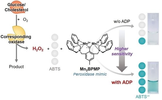
The Mn2BPMP complex has an intrinsic peroxidase-like activity in the pH range of 5 to 8, especially a maximum activity at pH 7, while most peroxidase mimics operate at an acidic pH (mainly pH 4). Its peroxidase-like activity is high among small-molecule-based peroxidase mimics with a high reproducibility. In addition, we recently revealed that adenosine mono/diphosphate (AMP and ADP) significantly boosted the peroxidase-like activity of Mn2BPMP. These advantages imply that Mn2BPMP is suitable for biosensing as a substitute for horseradish peroxidase (HRP). Herein, we established a colorimetric one-pot assay system using the enzyme cascade reaction between analyte oxidase and ADP-boosted Mn2BPMP. The simple addition of ADP to the Mn2BPMP-based assay system caused a greater increase in absorbance for the same concentration of H2O2, which resulted in a higher sensitivity. It was applied to one-pot detection of glucose and cholesterol at 25 °C and pH 7.0 for a few minutes.

Graphical Abstract

1. Introduction

Horseradish peroxidase (HRP) is a natural metalloenzyme that catalyzes the oxidation of various optical organic substrates in the presence of hydrogen peroxide (H2O2). That is, peroxidase activity causes quantitative optical signal changes such as color and fluorescence in proportion to the concentration of H2O2. Due to its high specificity and efficiency, HRP has been widely utilized in biosensors for the detection of various biomolecules such as glucose and antigens through enzyme cascade assays [1,2,3], immunoassays [4,5,6], and aptasensors [7,8,9]. However, HRP has intrinsic drawbacks, such as vulnerability of the catalytic activity to environmental conditions and the high cost of preparation, purification, and storage processes. These disadvantages limit its further practical applications.
Various peroxidase mimics have emerged as an alternative to HRP, such as magnetic nanoparticles [10,11], gold nanoparticles [12,13], metal-organic frameworks [14,15,16], graphene oxides [17,18], and small molecules [19,20]. However, most of the reported peroxidase mimics still suffer from the requirement of high temperature and acidic conditions for their peroxidase-like activities, and from poor reproducibility due to the batch-to-batch variation of nanomaterials [21]. Furthermore, most of them demand relatively long response times and multistep processes for biosensing. Therefore, the development of a peroxidase mimic that overcomes these disadvantages is highly desirable for the practical and simple detection of various biomolecules.
Recently, our group reported that Mn2BPMP (two Mn2+ ions-coordinated BPMP; 2,6-bis[(bis(2-pyridylmethyl)amino)-methyl]-4-methylphenolate) possesses a peroxidase-like activity [22]. This was demonstrated using a colorimetric assay with 2,2’-azino-bis(3-ethylbenzothiazoline-6-sulfonic acid) (ABTS) as the chromogenic peroxidase substrate and H2O2. Notably, Mn2BPMP works well at a neutral pH (pH 7.0) and responds quickly to ABTS and H2O2 within a minute. Moreover, the batch-to-batch variation is irrelevant in the use of Mn2BPMP. Based on the peroxidase-like activity of Mn2BPMP, H2O2 can be quantitatively detected by the Mn2BPMP and ABTS system. This suggests that Mn2BPMP can be further utilized for the indirect detection of various biomolecules engaged in reactions generating H2O2. Recently, it was revealed that adenosine mono/diphosphate (AMP and ADP) could boost the peroxidase-like activity of Mn2BPMP, lowering the Michaelis−Menten constant (KM) toward H2O2 and ABTS [23]. A high peroxidase-like activity is necessary for peroxidase mimics for the sensitive detection of biomolecules in the application. In addition, the Kong group reported that adenosine di/triphosphate (ADP and ATP) not only enhanced the peroxidase-like activity of peroxidase mimic, G-quadruplex-hemin DNAzyme, but also stabilized the bluish-green ABTS•+, which is unstable and decays to a colorless compound [24,25]. Therefore, we recruited them for the rapid and sensitive detection of biomolecules based on enzyme cascade reactions.
Herein, we constructed an Mn2BPMP/ADP/ABTS/oxidase system for the one-pot colorimetric assay of biomolecules by coupling two cascade reactions: oxidation of the target analyte by related analyte oxidase (AOx) and oxidation of ABTS by generated H2O2 and ADP-boosted Mn2BPMP (Scheme 1). As the oxidized ABTS radical has a bluish-green color, the color of the assay solution became darker in proportion to the concentration of the analyte. The enhanced peroxidase-like activity of Mn2BPMP by ADP caused a greater change in absorbance for the same concentration of analyte than in the assay system without ADP. In addition, ADP, a stabilizer of ABTS•+, maintained a darker bluish-green color for the assay solution than without ADP, making it possible to distinguish the analyte concentration more clearly with the naked eye. This system was successfully applied to the one-pot detection of glucose, which is related to human diseases such as diabetes mellitus and hyperlipidemia, and has great importance in clinical diagnosis [26]. In addition, it was extended to the detection of cholesterol, with great potential for further applications.

2. Materials and Methods

2.1. Materials and Instrumentation

2,6-Bis(hydroxymethyl)-p-cresol, manganese acetate tetrahydrate (Mn(OAc)2·4H2O), sodium perchlorate (NaClO4), cholesterol bioreagent, cholesterol oxidase from microorganisms, D-(-)-fructose, D-(+)-glucose, glucose oxidase from Aspergillus niger, D-(+)-maltose monohydrate, adenosine 5′-triphosphate disodium salt hydrate (ATP), adenosine 5′-monophosphate monohydrate (AMP), and Triton X-100 were purchased from Sigma-Aldrich, Seoul, Korea. Adenosine 5′-diphosphate (ADP) disodium salt hydrate, 2,2′-dipicolylamine, hydrogen peroxide, and D-(+)-sucrose were purchased from Tokyo Chemical Industry, Seoul, Korea. 2,2′-Azino-bis(3-ethylbenzothiazoline-6-sulfonic acid) ammonium salt (ABTS) was purchased from Alfa Aesar, Seoul, Korea. Thionyl chloride and triethylamine were purchased from Daejung Chemical Industry, Gyeongbuk, Korea. Sodium acetate trihydrate (NaOAc·3H2O) was purchased from Junsei Chemical Industry, Seoul, Korea. D-(+)-Galactose was purchased from Janssen Pharmaceuticals, Seoul, Korea. Lactose monohydrate was purchased from Samchun Chemicals, Seoul, Korea. All of the chemicals were used without further purification.
The optical density spectrum was recorded on a BioTek Cytation 3 Cell Imaging Multi-Mode Reader (BioTek, Winooski, VT, USA) using a 96 well plate. Absorbance spectra were recorded on an Agilent Cary 8454 UV−VIS spectrophotometer (Agilent, Santa Clara, CA, USA) using a 1 cm path length quartz cell. 1H-NMR and 13C-NMR spectra were recorded on a JEOL (400 MHz) NMR spectrometer (JEOL, Tokyo, Japan). Melting points were measured using a BUCHI melting point M-565 apparatus (Büchi, Flawil, Switzerland). The mass spectrum was recorded on an Agilent 6520 accurate-mass quadrupole time-of-flight mass spectrometer (MS) (Agilent, Santa Clara, CA, USA) with an electrospray ionization (ESI) source.

2.2. Effect of Adenosine Phosphates on the Peroxidase-like Activity of Mn2BPMP

Various adenosine phosphates (ATP, ADP, and AMP; 2 μM) were added to a buffer solution (Tris, pH 7.0, 20 mM, H2O to acetonitrile v/v = 19:1) containing Mn2BPMP (1 μM) and ABTS (1 mM). Then, H2O2 (5 mM) was added. The optical density spectra for these samples were recorded at 10-s intervals for 5 min at 420 nm and 25.0 °C.

2.3. Effect of ADP on the Peroxidase-like Activity of Mn2BPMP

Various concentrations of ADP were added to a buffer solution (Tris, pH 7.0, 20 mM, H2O to acetonitrile v/v = 19:1) containing Mn2BPMP (1 μM) and ABTS (1 mM). Then, H2O2 (5 mM) was added. The UV−VIS spectra for these samples were recorded at 10-s intervals for 3 min at 420 nm and 25.0 °C.

2.4. Colorimetric Detection of H2O2 in the Presence or Absence of ADP

Various concentrations of H2O2 were added to a buffer solution (Tris, pH 7.0, 20 mM, H2O to acetonitrile v/v = 19:1) containing Mn2BPMP (1 μM) and ABTS (1 mM) in the presence or absence of ADP (2 μM). The UV−VIS spectra for these samples were recorded at 10-s intervals for 3 min at 420 nm and 25.0 °C.

2.5. Colorimetric Detection of Glucose and Selectivity/Interference Experiments in the Presence or Absence of ADP

Various concentrations of glucose were added to a buffer solution (Tris, pH 7.0, 20 mM, H2O to acetonitrile v/v = 19:1) containing Mn2BPMP (2 μM), ABTS (1 mM), and glucose oxidase (GOx) (1 U/mL) in the presence or absence of ADP (4 μM). The UV−VIS spectra for these samples were recorded at 30-s intervals for 15 min at 420 nm and 25.0 °C.
For the selectivity and interference experiments, various glucose analogs (7 mM), such as fructose, galactose, lactose, sucrose, and maltose, were added to a buffer solution (Tris, pH 7.0, 20 mM, H2O to acetonitrile v/v = 19:1) containing Mn2BPMP (2 μM), ABTS (1 mM), ADP (4 μM), and GOx (1 U/mL) in the presence or absence of glucose (700 μM). After 7 min, the UV−VIS spectra for these samples were recorded at 420 nm and 25.0 °C.

2.6. Colorimetric Detection of Glucose in Human Serum

Human serum was prepared by ultrafiltration and the glucose concentration of the human serum was measured using a glucose meter. The 10-fold diluted serum samples, which were spiked with various concentrations of glucose (5, 10, and 25 mM), were added to a buffer solution (Tris, pH 7.0, 20 mM, H2O to acetonitrile v/v = 19:1) containing Mn2BPMP (2 μM), ABTS (1 mM), glucose oxidase (GOx) (1 U/mL), and ADP (4 μM). The UV−VIS spectra for these samples were recorded at 7 min at 420 nm and 25.0 °C.

2.7. Colorimetric Detection of Cholesterol in the Presence or Absence of ADP

To prepare a 10 mM stock solution of cholesterol, 38.6 mg of cholesterol was first dissolved in a mixture of 1 mL of Triton X-100 and 0.5 mL of isopropyl alcohol in a warm water bath at 60 °C. Then, 8.5 mL of 200 mM of Tris pH 7.0 buffer solution was added to give a stock solution of cholesterol. It was diluted using 200 mM of Tris pH 7.0 buffer solution for working solutions.
Various concentrations of cholesterol were added to a buffer solution (Tris, pH 7.0, 20 mM, H2O to acetonitrile v/v = 19:1) containing Mn2BPMP (2 μM), ABTS (1 mM), and cholesterol oxidase (ChOx) (1 U/mL) in the presence or absence of ADP (4 μM). The UV−VIS spectra for these samples were recorded at 30-s intervals for 5 min at 420 nm and 25.0 °C.

3. Results and Discussion

3.1. Effect of ADP on the Peroxidase-like Activity of Mn2BPMP

Mn2BPMP was synthesized as the pale-yellow crystals in three steps: chlorination of 2,6-bis(hydroxymethyl)-p-cresol with thionyl chloride, nucleophilic substitution with 2,2′-dipicolylamine to produce H-BPMP, and H-BPMP chelation with two Mn2+ ions. The synthesized H-BPMP and Mn2BPMP were confirmed by 1H and 13C NMR spectra and mass spectrum, respectively (Figures S1–S4). Mn2BPMP is a small molecule-based peroxidase mimic with an intrinsic peroxidase-like activity at pH 5–8, especially at pH 7 [22]. Mn2BPMP oxidizes colorless ABTS to bluish-green ABTS•+ using H2O2, and the concentration of H2O2 can be quantified through the color change. In this study, it was first confirmed that H2O2 of sub 100 μM could be detected within 1 min using only 1μM of Mn2BPMP as a control (Figure S8).
The effect of adenosine phosphates (ATP, ADP, and AMP) as the additive booster on the peroxidase-like activity of Mn2BPMP was confirmed to enhance the sensitivity for the detection of H2O2. Adenosine phosphates corresponding to two equivalents of Mn2BPMP were added to the Mn2BPMP/ABTS/H2O2 system in a pH 7.0 Tris buffer solution. In the case of ATP, the increase in absorbance versus time occurred more slowly than the control assay solution without adenosine phosphates, whereas in the case of ADP and AMP, the absorbance of the assay solution increased more rapidly with time than the control (Figure 1a). The assay solution containing ADP not only increased the absorbance approximately six times compared to the control at 1 min, but also showed a significantly dark bluish-green color compared to the other assay solutions (Figure 1b). This pronounced color difference was presumably due to the two beneficial functions of ADP: (1) enhancement of peroxidase-like activity of Mn2BPMP and (2) stabilization of ABTS•+. In particular, compared to other conditions, the assay solution containing the ADP showed a remarkably rapid increase in absorbance versus time at the beginning of catalytic reaction, indicating that Mn2BPMP used H2O2 with the help of ADP to rapidly oxidize ABTS. This result suggests that ADP plays a crucial role in greatly enhancing the peroxidase-like activity of Mn2BPMP. Unstable ABTS•+ usually is decayed or over-oxidized to form a colorless compound over time, which gradually loses the bluish-green color of the assay solution. For this reason, bluish-green ABTS•+ with a molar extinction coefficient of 36,000 M−1 cm−1 at 420 nm are present in a very small proportion compared to other stable ABTS analogues in the ABTS-based assay systems [24,25,27]. Interestingly, in the Mn2BPMP/ABTS/H2O2 system, it is speculated that ADP stabilized ABTS•+, allowing for a greater proportion of ABTS•+ to be present in the assay solution.
In this study, ADP was recruited as the additive booster of the peroxidase-like activity of Mn2BPMP and the stabilizer of ABTS•+ in order to improve the detection sensitivity for H2O2, resulting in a high sensitivity in the enzyme cascade assay system for the detection of various analytes. The optimization concentration of ADP as the additive booster was confirmed in the Mn2BPMP/ABTS/H2O2 system. As shown in Figure 2 and Figure S5, the absorbance at 420 nm dramatically increased with the concentration of ADP and reached a plateau at 2 μM within 1 min. Hence, subsequent experiments were conducted by adding ADP corresponding to twice the concentration of Mn2BPMP in Mn2BPMP-based colorimetric assay systems.
Colorimetric detection of H2O2 was conducted in the presence or absence of 2 μM of ADP to compare the effect of ADP. As shown in Figure 3 and Figures S6 and S7, the absorption signal was notably increased when ADP was present, and it was also observed with the naked eye. For H2O2, the linear range was 100 to 1000 μM, and the limit of detection (LOD) (S/N = 3) was 9.7 μM with R2 = 0.997 in the presence of ADP, and the linear range was 100 to 1000 μM and the LOD (S/N = 3) was 85 μM with R2 = 0.983 in the absence of ADP (Figure S8). Notably, a difference in the color of the assay solution was evident even at a low concentration of H2O2 in the presence of ADP. In addition, the LOD was decreased by about 9-fold when ADP was added, indicating that ADP could enhance the sensitivity for H2O2 in the Mn2BPMP/ABTS/H2O2 system. As this enhanced sensitivity enabled the detection of lower concentrations of analytes in a shorter time, the ADP-boosted Mn2BPMP/ABTS/H2O2 system would be more suitable for application in enzyme cascade assays.

3.2. Colorimetric Detection of Glucose Using the ADP/Mn2BPMP/ABTS/GOx System

Next, based on the ADP-boosted peroxidase-like activity of Mn2BPMP, colorimetric detection of glucose was conducted under an ADP/Mn2BPMP/ABTS/GOx system. One equivalent of glucose was oxidized by GOx generating one equivalent of H2O2, as shown in Equation (1):
β - D - glucose   +   O 2   GOx   D - glucono - 1 , 5 - lactone + H 2 O 2
Therefore, a quantitative analysis of H2O2 enables an indirect quantitative analysis of glucose. In detail, glucose was oxidized by GOx, which generated H2O2 as a byproduct. Subsequently, with the help of the generated H2O2, the enhanced peroxidase-like activity of Mn2BPMP by ADP catalyzed the oxidation of ABTS to ABTS•+ to give a bluish-green color in the solution. As higher concentrations of ABTS•+ gave a deeper bluish-green color, a quantitative analysis of glucose was enabled via measuring the absorbance by UV−VIS spectroscopy and naked eye observation. Scheme 1 illustrates the principle of the biosensing of the Mn2BPMP/ABTS/GOx system based on these cascade reactions.
Various concentrations of glucose were added to the Mn2BPMP/ABTS/GOx system in a Tris buffer (pH 7.0) solution in the presence or absence of ADP. The absorbance at 420 nm increased with an increasing concentration of glucose (Figure 4a and Figures S9 and S10). For glucose, the linear range was 100 to 700 μM and the LOD (S/N = 3) was 16 μM with R2 = 0.996 in the presence of ADP, and the linear range was 100 to 500 μM and the LOD (S/N = 3) was 59 μM in the absence of ADP with R2 = 0.982 (Figure S11). Due to the highly enhanced peroxidase-like activity of Mn2BPMP by ADP, not only was the color change clearly observed by the naked eye (Figure 4b), but it also lowered the LOD for glucose. In previous literature, most enzyme cascade-based glucose assay systems using nanomaterial-based peroxidase mimics such as ferromagnetic nanoparticles and gold nanoparticles were a method of pre-incubating glucose and glucose oxidase, lowering the pH, and detecting H2O2 generated by using peroxidase mimics with a low working pH, as shown in Table S1 [10,11]. Due to this, there are disadvantages, in that it is inconvenient to go through a two-step process and it takes tens of minutes. On the other hand, our system showed that it was possible to quantitatively detect a glucose concentration in 7 min in one pot. Moreover, our system is cost-effective and stable compared with the use of HRP, owing to the characteristics of a small-molecule-based peroxidase mimic.
To confirm that the ADP/Mn2BPMP/ABTS/GOx system has a high selectivity toward glucose and there was no interference by glucose analogues, a 10-fold higher concentration of fructose (Fru), galactose (Gal), lactose (Lac), maltose (Mal), and sucrose (Suc) was added to the system, individually, in the absence or presence of glucose. Then, the absorbance at 420 nm was measured after 7 min by UV−VIS spectroscopy. As shown in Figure 5a, no significant signals in absorbance at 420 nm were observed without glucose and there was no interference effect by the glucose analogues with glucose. It was also observed with the naked eye, as shown in Figure 5b. These results demonstrate that the proposed system can efficiently and selectively detect glucose, even with the naked eye. More importantly, real-time detection of glucose is possible, as this system does not require any multistep processes or long response time. Furthermore, the applicability of the ADP/Mn2BPMP/ABTS/GOx system for the detection of glucose in human serum was verified. Human serum was prepared by ultrafiltration to remove yellowness, and the glucose concentration of the filtered human serum was measured to be 5.35 ± 0.02 mM using the glucose meter. The serum samples spiked with known glucose concentrations (5, 10, and 25 mM) were added to the ADP/Mn2BPMP/ABTS/GOx system, and the glucose concentration was detected by measuring the absorbance. Recovery of the glucose concentrations in the serum samples ranged from 82.79% to 100.16%, with the relative standard deviation (RSD) ranging from 5.26% to 9.12% (Table S2). It was shown that glucose can be quantitatively detected at concentrations much higher than normal in serum samples, suggesting the possibility that the ADP/Mn2BPMP/ABTS/GOx can be utilized in the diagnosis of diseases related to high glucose levels.

3.3. Colorimetric Detection of Cholesterol Using the ADP/Mn2BPMP/ABTS/ChOx System

To confirm whether the ADP/Mn2BPMP/ABTS/oxidase system could be extended to the general platform for the colorimetric detection of other biomolecules, the detection of cholesterol was also conducted by simply substituting ChOx for GOx. As cholesterol, the precursor of steroid hormones and bile acids, is associated with many human diseases, such as arteriosclerosis, hypertension, hypothyroidism, and hypocholesterolemia [28,29], fast and reliable detection of cholesterol is highly important in clinical diagnosis. One equivalent of cholesterol is oxidized by ChOx generating one equivalent of H2O2, as shown in Equation (2):
cholesterol   +   O 2   ChOx   cholest - 4 - en - 3 - one + H 2 O 2
Therefore, a quantitative analysis of cholesterol could be conducted in a similar way to the detection of glucose.
Various concentrations of cholesterol were added to the Mn2BPMP/ABTS/ChOx system in the Tris buffer (pH 7.0) solution in the presence or absence of ADP. The absorbance at 420 nm increased with an increasing concentration of cholesterol and the color change could be noticed with the naked eye in the presence of ADP (Figure 6 and Figures S12 and S13). For cholesterol, the linear range was 20 to 150 μM and the LOD was 4.8 μM with R2 = 0.994 in the presence of ADP, and the linear range was 20 to 100 μM and the LOD was 12 μM with R2 = 0.986 in the absence of ADP (Figure S14). The proposed ADP/Mn2BPMP/ABTS/oxidase system could be applied for the detection of cholesterol as well. We envision that our system can be further utilized as a general platform in colorimetric biosensors for the real-time detection of various biomolecules accompanied by the generation of H2O2.

4. Conclusions

In this study, we demonstrated that the ADP/Mn2BPMP/ABTS/GOx system can be efficiently applied to the colorimetric detection of glucose based on the highly enhanced peroxidase-like activity of Mn2BPMP by ADP with the ability to stabilize ABTS•+. In this system, the linear range was 100–700 μM and LOD was 16 μM for glucose. In particular, because of the highly increased signals due to the involvement of ADP, glucose can be easily detected even with the naked eye, which is considerably advantageous in the detection of biomolecules. As this system does not require a long response time or any multistep processes, the real-time detection of glucose was successfully demonstrated within a few minutes in a simple way. As an extension, we also demonstrated that the colorimetric detection of cholesterol was enabled based on our system simply replacing GOx with ChOx. Therefore, we anticipate that the proposed ADP/Mn2BPMP/ABTS/oxidase systems can be further developed into a general platform for the real-time detection of various biomolecules. Furthermore, as a peroxidase mimic, Mn2BPMP may be a practical alternative to the natural enzyme HRP in a variety of fields, such as biosensors.

Supplementary Materials

The following supporting information can be downloaded at: https://www.mdpi.com/article/10.3390/chemosensors10020089/s1. Figures S1–S3: 1H and 13C NMR spectra of the synthesized compounds, Figure S4: Mass spectrum of Mn2BPMP, Figure S5: Optimization of ADP concentration in the Mn2BPMP/ABTS/H2O2 system, Figures S6–S8: H2O2 titration using the Mn2BPMP/ABTS/H2O2 system in the presence or absence of ADP, Figures S9–S11: Glucose titration using the Mn2BPMP/ABTS/GOx system in the presence or absence of ADP, Figures S12–S14: Cholesterol titration using the Mn2BPMP/ABTS/ChOx system in the presence or absence of ADP.

Author Contributions

Conceptualization, M.S.H.; funding acquisition, M.S.H.; investigation, N.L. and S.Y.; methodology, N.L. and S.Y.; project administration, M.S.H.; resources, N.L. and Y.L.; supervision, M.S.H.; validation, S.Y.; visualization, N.L. and S.Y.; writing—original draft, N.L. and S.Y.; writing—review and editing, S.Y. and M.S.H. All authors have read and agreed to the published version of the manuscript.

Funding

This research was supported by the National Research Foundation of Korea (NRF) grant funded by the Korea government (MSIT), grant number NRF-2020R1A2B5B01002392.

Conflicts of Interest

The authors declare no conflict of interest.

References

  1. Delvaux, M.; Walcarius, A.; Demoustier-Champagne, S. Bienzyme HRP-GOx-modified gold nanoelectrodes for the sensitive amperometric detection of glucose at low overpotentials. Biosens. Bioelectron. 2005, 20, 1587–1594. [Google Scholar] [CrossRef] [PubMed]
  2. Martinez, A.W.; Phillips, S.T.; Butte, M.J.; Whitesides, G.M. Patterned Paper as a Platform for Inexpensive, Low-Volume, Portable Bioassays. Angew. Chem.-Int. Ed. 2007, 119, 1340–1342. [Google Scholar] [CrossRef]
  3. Han, M.; Liu, S.; Bao, J.; Dai, Z. Pd nanoparticle assemblies-as the substitute of HRP, in their biosensing applications for H2O2 and glucose. Biosens. Bioelectron. 2012, 31, 151–156. [Google Scholar] [CrossRef] [PubMed]
  4. Whitehead, T.P.; Thorpe, G.H.G.; Carter, T.J.N.; Groucutt, C.; Kricka, L.J. Enhanced luminescence procedure for sensitive determination of peroxidase-labelled conjugates in immunoassay. Nature 1983, 305, 158–159. [Google Scholar] [CrossRef]
  5. Zhang, S.; Yang, J.; Lin, J. 3,3’-diaminobenzidine (DAB)-H2O2-HRP voltammetric enzyme-linked immunoassay for the detection of carcionembryonic antigen. Bioelectrochemistry 2008, 72, 47–52. [Google Scholar] [CrossRef] [PubMed]
  6. Tang, L.; Zeng, G.-M.; Shen, G.-L.; Li, Y.-P.; Zhang, Y.; Huang, D.-L. Rapid Detection of Picloram in Agricultural Field Samples Using a Disposable Immunomembrane-Based Electrochemical Sensor. Environ. Sci. Technol. 2008, 42, 1207–1212. [Google Scholar] [CrossRef] [PubMed]
  7. Mir, M.; Vreeke, M.; Katakis, I. Different strategies to develop an electrochemical thrombin aptasensor. Electrochem. Commun. 2006, 8, 505–511. [Google Scholar] [CrossRef]
  8. Zhang, D.W.; Sun, C.J.; Zhang, F.T.; Xu, L.; Zhou, Y.L.; Zhang, X.X. An electrochemical aptasensor based on enzyme linked aptamer assay. Biosens. Bioelectron. 2012, 31, 363–368. [Google Scholar] [CrossRef]
  9. Bai, L.; Chai, Y.; Yuan, R.; Yuan, Y.; Xie, S.; Jiang, L. Amperometric aptasensor for thrombin detection using enzyme-mediated direct electrochemistry and DNA-based signal amplification strategy. Biosens. Bioelectron. 2013, 50, 325–330. [Google Scholar] [CrossRef]
  10. Gao, L.; Zhuang, J.; Nie, L.; Zhang, J.; Zhang, Y.; Gu, N.; Wang, T.; Feng, J.; Yang, D.; Perrett, S.; et al. Intrinsic peroxidase-like activity of ferromagnetic nanoparticles. Nat. Nanotechnol. 2007, 2, 577–583. [Google Scholar] [CrossRef]
  11. Su, L.; Qin, W.; Zhang, H.; Rahman, Z.U.; Ren, C.; Ma, S.; Chen, X. The peroxidase/catalase-like activities of MFe2O4 (M = Mg, Ni, Cu) MNPs and their application in colorimetric biosensing of glucose. Biosens. Bioelectron. 2015, 63, 384–391. [Google Scholar] [CrossRef] [PubMed]
  12. Wang, S.; Chen, W.; Liu, A.L.; Hong, L.; Deng, H.H.; Lin, X.H. Comparison of the peroxidase-like activity of unmodified, amino-modified, and citrate-capped gold nanoparticles. ChemPhysChem 2012, 13, 1199–1204. [Google Scholar] [CrossRef] [PubMed]
  13. Ni, P.; Dai, H.; Wang, Y.; Sun, Y.; Shi, Y.; Hu, J.; Li, Z. Visual detection of melamine based on the peroxidase-like activity enhancement of bare gold nanoparticles. Biosens. Bioelectron. 2014, 60, 286–291. [Google Scholar] [CrossRef]
  14. Zhang, J.W.; Zhang, H.T.; Du, Z.Y.; Wang, X.; Yu, S.H.; Jiang, H.L. Water-stable metal-organic frameworks with intrinsic peroxidase-like catalytic activity as a colorimetric biosensing platform. Chem. Commun. 2014, 50, 1092–1094. [Google Scholar] [CrossRef] [PubMed]
  15. Ai, L.; Li, L.; Zhang, C.; Fu, J.; Jiang, J. MIL-53(Fe): A metal-organic framework with intrinsic peroxidase-like catalytic activity for colorimetric biosensing. Chem. Eur. J. 2013, 19, 1515–14108. [Google Scholar] [CrossRef] [PubMed]
  16. Tan, H.; Li, Q.; Zhou, Z.; Ma, C.; Song, Y.; Xu, F.; Wang, L. A sensitive fluorescent assay for thiamine based on metal-organic frameworks with intrinsic peroxidase-like activity. Anal. Chim. Acta 2015, 856, 90–95. [Google Scholar] [CrossRef]
  17. Dong, Y.L.; Zhang, H.G.; Rahman, Z.U.; Su, L.; Chen, X.J.; Hu, J.; Chen, X.G. Graphene oxide-Fe3O4 magnetic nanocomposites with peroxidase-like activity for colorimetric detection of glucose. Nanoscale 2012, 4, 3969–3976. [Google Scholar] [CrossRef]
  18. Tao, Y.; Lin, Y.; Huang, Z.; Ren, J.; Qu, X. Incorporating graphene oxide and gold nanoclusters: A synergistic catalyst with surprisingly high peroxidase-like activity over a broad pH range and its application for cancer cell detection. Adv. Mater. 2013, 25, 2594–2599. [Google Scholar] [CrossRef]
  19. Li, R.; Zhen, M.; Guan, M.; Chen, D.; Zhang, G.; Ge, J.; Gong, P.; Wang, C.; Shu, C. A novel glucose colorimetric sensor based on intrinsic peroxidase-like activity of C60-carboxyfullerenes. Biosens. Bioelectron. 2013, 47, 502–507. [Google Scholar] [CrossRef]
  20. Wang, Q.; Ma, K.; Yu, Z.; Ding, J.; Hu, Q.; Liu, Q.; Sun, H.; Wen, D.; Liu, Q.; Kong, J. The peroxidase-like catalytic activity of ferrocene and its application in the biomimetic synthesis of microsphere polyaniline. New J. Chem. 2018, 42, 13536–13540. [Google Scholar] [CrossRef]
  21. Liu, L.; Shi, Y.; Yang, Y.; Li, M.; Long, Y.; Huang, Y.; Zheng, H. Fluorescein as an artificial enzyme to mimic peroxidase. Chem. Commun. 2016, 52, 13912–13915. [Google Scholar] [CrossRef] [PubMed]
  22. Lee, Y.; Yoo, S.; Kang, S.; Hong, S.; Han, M.S. An [Mn2(bpmp)]3+ complex as an artificial peroxidase and its applications in colorimetric pyrophosphate sensing and cascade-type pyrophosphatase assay. Analyst 2018, 143, 1780–1785. [Google Scholar] [CrossRef]
  23. Kang, S.; Park, B.Y.; Lee, S.; Lee, N.; Han, M.S. Colorimetric discrimination of nucleoside phosphates based on catalytic signal amplification strategy and its application to related enzyme assays. Analyst 2021, 146, 463–470. [Google Scholar] [CrossRef] [PubMed]
  24. Kong, D.-M.; Xu, J.; Shen, H.-X. Positive Effects of ATP on G-Quadruplex-Hemin DNAzyme-Mediated Reactions. Anal. Chem. 2010, 82, 6148–6153. [Google Scholar] [CrossRef] [PubMed]
  25. Jia, S.M.; Liu, X.F.; Kong, D.M.; Shen, H.X. A simple, post-additional antioxidant capacity assay using adenosine triphosphate-stabilized 2,2’-azinobis(3-ethylbenzothiazoline)-6-sulfonic acid (ABTS) radical cation in a G-quadruplex DNAzyme catalyzed ABTS-H2O2 system. Biosens. Bioelectron. 2012, 35, 407–412. [Google Scholar] [CrossRef]
  26. Heller, A.; Feldman, B. Electrochemical Glucose Sensors and Their Applications in Diabetes Management. Chem. Rev. 2008, 108, 2482–2505. [Google Scholar] [CrossRef] [Green Version]
  27. Bourbonnais, R.; Leech, D.; Paice, M.G. Electrochemical analysis of the interactions of laccase mediators with lignin model compounds. Biochim. Biophys. Acta-Gen. Subj. 1998, 1379, 381–390. [Google Scholar] [CrossRef]
  28. Motonaka, J.; Faulkner, L.R. Determination of cholesterol and cholesterol ester with novel enzyme microsensors. Anal. Chem. 1993, 65, 3258–3261. [Google Scholar] [CrossRef]
  29. Martin, S.P.; Lamb, D.J.; Lynch, J.M.; Reddy, S.M. Enzyme-based determination of cholesterol using the quartz crystal acoustic wave sensor. Anal. Chim. Acta 2003, 487, 91–100. [Google Scholar] [CrossRef]
Scheme 1. Schematic illustration of the Mn2BPMP/ADP/ABTS/AOx system.
Chemosensors 10 00089 sch001
Figure 1. (a) Changes in the optical density and (b) photograph of the Mn2BPMP/ABTS/H2O2 system in the presence of various adenosine phosphates (ATP, ADP, and AMP). Adenosine phosphates = 2 µM; Mn2BPMP = 1 µM; ABTS = 1 mM; H2O2 = 5 mM in a buffer solution (Tris, 20 mM, pH 7.0).
Chemosensors 10 00089 g001
Figure 2. Changes in the absorbance of the Mn2BPMP/ABTS/H2O2 system in the presence of various concentrations of ADP (from 0 to 3 µM) after 3 min. Inset: The plot of the absorbance at 420 nm versus the concentration of ADP (from 0 to 3 µM) after 3 min. Mn2BPMP = 1 µM; ABTS = 1 mM; H2O2 = 5 mM in a buffer solution (Tris, 20 mM, pH 7.0).
Chemosensors 10 00089 g002
Figure 3. (a) The plot of the absorbance at 420 nm and (b) the photograph versus the concentration of H2O2 (0 to 5 mM) for the Mn2BPMP/ABTS system in the presence or absence of ADP (2 µM) after 1 min. Mn2BPMP = 1 µM; ABTS = 1 mM in a buffer solution (Tris, 20 mM, pH 7.0).
Chemosensors 10 00089 g003
Figure 4. (a) The plot of the absorbance at 420 nm and (b) photograph versus the concentration of glucose (0 to 2 mM) for the Mn2BPMP/ABTS/GOx system in the presence or absence of ADP (4 µM) after 1 min. Mn2BPMP = 2 µM; ABTS = 1 mM; GOx = 1 U/mL in a buffer solution (Tris, 20 mM, pH 7.0).
Chemosensors 10 00089 g004
Figure 5. (a) The absorbance at 420 nm and (b) photograph of the Mn2BPMP/ADP/ABTS/GOx system with various glucose analogs (7 mM) in the presence or absence of glucose (700 µM) after 7 min. Mn2BPMP = 2 µM; ADP = 4 µM; ABTS = 1 mM; GOx = 1 U/mL in a buffer solution (Tris, 20 mM, pH 7.0).
Chemosensors 10 00089 g005
Figure 6. (a) The plot of the absorbance at 420 nm and (b) photograph versus the concentration of cholesterol (0 to 300 µM) for the Mn2BPMP/ABTS/ChOx system in the presence or absence of ADP (4 µM) after 1 min. Mn2BPMP = 2 µM; ABTS = 1 mM; ChOx = 1 U/mL in a buffer solution (Tris, 20 mM, pH 7.0).
Chemosensors 10 00089 g006
Publisher’s Note: MDPI stays neutral with regard to jurisdictional claims in published maps and institutional affiliations.