Article

Fatty Acids from Paracentrotus lividus Sea Urchin Shells Obtained via Rapid Solid Liquid Dynamic Extraction (RSLDE)

1
Department of Chemical Sciences, University of Naples “Federico II”, 80126 Naples, Italy
2
Stazione Zoologica Anton Dohrn, 80121 Naples, Italy
3
Department of Molecular Medicine and Medical Biotechnology, University of Naples “Federico II”, 80131 Naples, Italy
*
Authors to whom correspondence should be addressed.
Separations 2019, 6(4), 50; https://doi.org/10.3390/separations6040050
Submission received: 17 September 2019 / Revised: 11 October 2019 / Accepted: 18 October 2019 / Published: 22 October 2019
(This article belongs to the Special Issue Extraction and Isolation of Natural Products)

Abstract

:
Sea urchins (Echinodermata, Echinoidea) are good a source of bioactive compounds belonging to different classes of natural substances. The edible Mediterranean sea urchin Paracentrotus lividus is a renowned animal model for study in different fields of biology, but it is intensively harvested for high commercial value due to the delicacy of its gonads. Most studies have focused on the composition and the nutritional value of P. lividus gonads (the edible part), but little interest has been taken in the other body parts, such as the shells and spines, which are generally considered waste material. The purpose of this study was to obtain an extract from sea urchin shells, with a green methodology of extraction, and to characterize the lipophilic components for potential applications. The shells of P. lividus were extracted via a very well performing technology based on rapid solid liquid dynamic extraction (RSLDE) implemented via an automated device (Naviglio Extractor®). The obtained extract shows the presence of fatty acids and their esters (methyl, ethyl and 1-glycerol esters). Gas chromatography-mass spectrometry (GC-MS) measurements were used to determine fatty acid abundance in the chromatographic fractions of the extract. Arachidonic acid (ARA), 5,8,11,14,17-eicosapentanoic acid (EPA), and 11-eicosenoic acids and their esters are the most abundant components. The presence of many polyunsaturated fatty acids (PUFA) in the extract, even in low percentages allows a future application in nutrition or medical use.

Graphical Abstract

1. Introduction

Food wastes are produced by many sources, ranging from agricultural operations to household consumption. In particular breeding, seafood, and dairy processing are the main source of animal-derived wastes [1,2,3].
Food industry wastes derive from the processing of raw materials into foodstuffs for human consumption, which consists in the extraction or separation of the nutritional part from the remains, having scarce nutritional value or inedible components [4]. Industrial processing of vegetable and animal materials produces tons of by-products and the non-utilization of those materials not only leads to the loss of potential profits, but the bio-waste generated raised serious concerns about the cost of disposal of these products considering the environmental impact. Therefore, converting food wastes to higher value products not only reduces the environmental impact but also provides potential revenue for industry in various fields, such as nutraceutical, cosmeceutical, and agrifood [5,6]. The recovered biomolecules and by-products can be applied in nutraceutical and pharmaceutical fields. In fact, waste material can be valorized through the extraction of high-value components, which can be re-used as additives, adjuvants, and pharmacologically functional ingredients. The choice of the extractive technique depends on the type of compounds and matrix. In recent years the aim of the scientific community has been the optimization of the extraction conditions using conventional or advanced techniques and the treatment of food waste [7].
The huge amount of marine food waste has generated significant activity on its alternative utilizations to turn these products into some economic and commercial value. Marine bio-waste includes unwanted materials derived from the fish processing industry, such as skin, bones, and shells. The global market demand for edible sea urchins is estimated at several tons annually [8]. The Mediterranean species Paracentrotus lividus is strongly harvested for processing and consumption of the gonads, which are considered a luxury sea food. Consequently, a significant portion of the raw material, essentially shells and spines, is produced as waste.
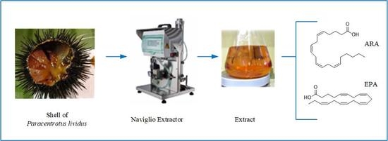
The aim of this study is to obtain extracts of P. lividus shells using the rapid solid liquid dynamic extraction (RSLDE) implemented via Naviglio Extractor®, as this technique has already successfully applied in the pharmaceutical, cosmetic, herbal, food and beverage fields [9,10,11,12,13,14,15]. Here we present our results on the qualitative and quantitative analysis via gas chromatography-mass spectrometry (GC-MS) of lipophilic components of the crude extract. Obtained results could be generalized to other species and may help to understand the use of sea urchin shell by-product in nutraceutical and cosmeceutical fields.

2. Materials and Methods

2.1. General Experimental Procedures

Nuclear magnetic resonance (NMR) spectra were recorded at 400 MHz in CDCl3 on a Bruker spectrometer (AscendTM400) (Bremen, Germany) and the same solvent was used as an internal standard. All reagents and solvents were analytical grade and purchased from Carlo Erba (Milan, Italy), Sigma-Aldrich (Saint Louis, MO, USA), and Fluka (Buchs, Switzerland).
An Agilent 6850 GC equipped with an HP-5MS capillary column (5% phenyl methyl polysiloxane stationary phase) and the Agilent 5973 Inert MS detector (Milan, Italy) (used in the scan mode) was used for GC-MS analysis, with ionization achieved by electron impact (EI) at 70 eV and 200 °C. Helium was employed as the carrier gas (flow rate of 1 mL min−1). The operating conditions were set with the injector temperature at 250 °C and during the run a temperature ramp raised the column temperature from 50 °C to 240 °C: starting with 50 °C for 2 min, 10 °C min−1 until reaching 180 °C, 180 °C for 5 min, 5 °C min−1 until reaching 240 °C, and 240 °C for 25 min. For identification purposes, the mass spectrum of each peak was recorded in the total ion current mode of the mass spectrometer, within a m/z range of 45 to 550 at a frequency of 3.9 Hz.
Solutions of standard compounds were prepared in n-hexane at a concentration range of 50–200 mg L−1 and analyzed by GC-MS. Methyl esters of nonanoic and pentadecanoic acids (Sigma-Aldrich, Saint Louis, MO, USA) were used as internal standards, a stock solution at concentration of 1 g L−1 was prepared and stored at −20 °C.
Analytical thin layer chromatography (TLC) was performed on silica gel plates (Kieselgel 60, F254, 0.25 mm) purchased from Merck (Darmstadt, Germany). The spots were visualized by exposure to UV radiation (253 nm), or by spraying with 10% H2SO4 in MeOH followed by heating at 110 °C for 10 min. Column chromatography (CC) was performed using silica gel (Kieselgel 60, 0.063–0.200 mm) purchased from Merck (Darmstadt, Germany).

2.2. Sea Urchins Collection

Specimens of sea Paracentrotus lividus were hand collected by scuba-diving from rocky shores of the Gulf of Naples, along the southern Tyrrhenian coasts of Italy. Animals were placed in a cooler and carried to the laboratory under moist conditions within 2 h. Sea urchins were held in suspended baskets in a semi-closed recirculating system as described in [16].
Sea urchin shells were collected from individuals processed for harvesting of gonads and gametes. Shells with spines were emptied of body residues and washed in sea water. Samples which were not immediately processed for extraction were stored frozen at −20 °C.

2.3. Shell Extraction and Purification Process

Before extraction, sea urchin shells obtained as described above were air dried for 24 h. Eighty grams of dried shells (P. lividus) were gently broken into big pieces and subsequently extracted. The rapid solid liquid dynamic extraction (RSLDE) was carried out using Naviglio Extractor® (Atlas Filtri Engineering S.r.l., Padova, Italy). This is an apparatus that allows a rapid solid–liquid extraction maintaining the liquid in contact with the solid in a programmable pressurization cycle between atmospheric pressure and 8–9 bar [17]. The Naviglio Extractor Mod. 500 cc was used in these experiments. An ethanol/water mixture (50:50, v/v) was used as the solvent for an extraction time of 70 h. Ultrapure water was obtained from a Milli-Q system (Millipore, Burlington, MA, USA). The hydro-alcoholic extract (500 mL) was evaporated under reduced pressure to remove the organic solvent. The aqueous phase (250 mL) was acidified to pH 2 with 2 M HCl and extracted three times with the same volume of ethyl acetate (EtOAc). The EtOAc phases were combined, dried with Na2SO4, and evaporated under reduced pressure to give a brown-red oil extract (180.0 mg). The extract was fractioned by CC, eluted with CHCl3/i-PrOH (95:5; v/v), originating six homogeneous fractions (frs) namely: A, 21.1 mg; B, 19.8 mg; C, 21.8 mg; D, 8.0 mg; E, 12.5 mg; and F, 11.8 mg (Figure 1).

2.4. GC-MS analysis of Chromatographic Fractions

2.4.1. Sample Preparation

The collected NMR data led us to analyze the CC fractions (frs A, C, and D) via GC-MS and according to the compounds potentially present in them, samples were treated in different ways. Hence, preliminary NMR data (see Results section) suggest the presence of methyl and ethyl ester of fatty acids in fr A (FAME, FAEE), free fatty acids in fr C (FA), and monoglycerides in fr D (MG). Due to the presence of methyl and ethyl esters of fatty acids, fr A was detected via GC-MS without preliminary treatment.
In order to detect free fatty acids in fr C via GC-MS, fatty acids were trimethylsilylated with N,O-bis(trimethylsilyl)trifluoroacetamide (BSTFA) according to a previously reported method [18].
Fr D was analyzed via GC-MS after a transesterification process due to the presence of typical signals of monoglycerides in the NMR spectrum of this fraction. Three milligrams of the sample was dissolved in 1 mL of n-hexane and then 200 µL of KOH 2 M in MeOH was added to make the reaction happen. After thoroughly mixing the solution, a short period of rest was given to allow the separation into two phases, where the upper phase contains methyl esters of fatty acids [19].

2.4.2. Qualitative and Quantitative analysis of Fatty Acids

Fatty acids and their esters were identified by their Kovats non-isothermal retention index (KI) and by comparing their mass spectra with those present in databases by employing the National Institute of Standards and Technology (NIST) Mass Spectral Search Program v.2.0g which, among others, could explore the NIST 14 Mass Spectral library (2014) and the Golm Metabolome Database. To overcome problems caused by matrix components and compounds coelution, GC-MS data were analyzed by the deconvolution program Automated Mass Spectral Deconvolution and Identification System (AMDIS) distributed by NIST [20,21].
The quantification of individual silylated (TMS), methylated (ME), and ethylated (EE) fatty acids was obtained by linear regression of the chromatographic peak areas and known concentrations of certified standards (ranging from 50 to 200 mg L−1). The response of each compound was normalized to the response of an internal standard (9:0 ME or 15:0 ME), which was added to the sample at the constant concentration of 100 mg L−1. The concentrations of individual FAs in each sample were converted to percentage contributions of the total FAs [22,23].

3. Results

The chromatographic purification process of crude extract gave different fractions that were identified as mixtures of methylated and ethylated fatty acids, fatty acids, and monoglycerides by 1H NMR. In particular, the protonic spectrum of fraction (fr) A showed the typical signals of a mixtuere of methyl and ethyl esters of sutured, monounsaturated, and polyunsaturated fatty acids (SFA, MUFA, and PUFA) [24]. The following are the chemical shifts, the multiplicity, and main signals: δ 5.46–5.33 (m) (–CH=CH–); 4.18–4.12 (dq, J = 2.3, 16 Hz) (CH3CH2O–); 3.70 (s) (CH3O–); 2.87–2.80 (m) (=CH–CH2–CH=); 2.37–2.29 (m) (–O–CO–CH2); 2.16–1.70 (m) (CH2–CH=CH–), 1.67–1.62 (m) (–O–CO–CH2–CH2–); 1.02–0.98 (t, J = 7.3 Hz) (CH3CH2–); and 0.93–0.88 (m) (CH3CH2–).
Fr C was identified as a mixture of fatty acids. In fact, the main difference from the fr A spectrum was the absence of signals relating to methoxy and ethoxy groups. Fr D was identified as a mixture of monoglycerides. In fact, its proton spectrum, in addition to the signals related to fatty acid residues, showed the typical signals of a residue of glycerol esterified in C-1 as reported below: δ 4.26–4.16 (ddd, J = 5, 6, 12 Hz, ROCH2–CHOH–CH2OH); 3.99–3.93 (m, ROCH2–CHOH–CH2OH); 3.74–3.60 (ddd, J = 4, 5, 12 Hz, ROCH2–CHOH–CH2OH).

Fatty Acids Composition

In all samples, FAs and their esters were identified and quantified via GC-MS. Figure 1 shows the total ion chromatograms (TICs) obtained from the fractions treated in different ways according to NMR data.
GC-MS analysis of fraction A (Figure 2A), without treatments, confirms the NMR results and revealed the presence of methyl esters and ethyl esters of fatty acids as listed in Table 1. 20:4n-6 EE (24.86%) and 20:5n-3 ME (26.51%) are the most abundant esters of fatty acids revealed in this fraction. TIC of fraction C (Figure 2B), acquired after trimethylsilylation, shows some peaks produced by the derivatization process and just two significant peaks which were identified as 20:4n-6 TMS (62.44%) and 20:5n-3 TMS (38.56%) (Table 1).
Fraction D was acquired after a transesterification process. Although it is feasible that GC-MS measurements for monoglycerides, especially with low molecular weight, are possible without derivatization procedures [25], in order to facilitate the detection and increase sensitivity, the residue was subjected to a common transesterification reaction by methanol (methanolysis) and monoglycerides were converted in their methyl esters. TIC of this fraction (Figure 2C) shows the presence of many 1-monoglycerol esters of fatty acids already identified as ME and the most abundant are 20:4n-6 ME (30.21%), 20:5n-3 ME (32.34%), and 20:1n-9 ME (14.42%) (Table 1).
Using the RSLDE process, 180 mg of extract were obtained from 80 g of shells, which corresponds to a yield of 0.225%.
In fr A, the concentrations of eicosapentanoic acid (EPA) (26.5%) in the injected solution ( C E P A ) (1 µL) and in the crude extract ( C E P A * ) were respectively:
C E P A = 153.1 m g L ; C E P A * = 9.9 m g g e x t r a c t .
From the data of the percent concentrations in Table 1, the concentration of any component, x, may be calculated using formulas:
C x = C E P A % x % E P A ; C x * = C E P A * g f r g e x t r a c t .
Analogously for frs C and D, concentrations can be calculated by taking into account that the concentrations of arachidonic acid (ARA) (61.4%) were respectively C A R A = 142.1 m g L and C A R A * = 24.6 m g g e x t r a c t and the concentrations for EPA (32.3%) were C A R A = 138.2 m g L and C A R A * = 1.8 m g g e x t r a c t .
The lipid component in the extract represented 28.27% and the percentage of any class of fatty acids and their esters are summarized in Table 2.
After developing a strategy and considering the fatty acid yield, the economic profitability should be estimated in order to generate a model to maximize the potential profits from the recovered material. The RSLDE application to the shells of sea urchins has an advantage in this respect, to transform waste material into an economic resource for industry and to reduce the environmental impact derived from the disposal. Apart from the advantages, the possibility of exploiting a by-product should take into account the costs related to the proposed strategy. In this respect, in Table 3 there is an overview of cost for the activities in laboratory of the shell extraction. If an optimization result is required, more detailed data (i.e., cost of chemicals) should be considered.
Although this strategy has been developed in a laboratory, the RSLDE could be used in large-scale industrial processes to produce shell extract, making the process cheap, and more efficient in terms of time, as well as ecologically benign.

4. Discussion

In Mediterranean countries there is a long and historical tradition of harvesting and eating sea urchins. In particular, Italy represents the largest market for Paracentrotus lividus in Europe, because the annual consumption is about 2000 tons [8]. The edible portion of the sea urchins constitutes approximately 10% of the total weight [26]. Hence, in Italy 1800 tons of sea urchin shells are unwanted material. It would be very important and of interest for a broad audience to reuse these shells as an example of circular economy.
In this study, analysis of P. lividus sea urchin shells was performed, and the fatty acid composition was reported for the first time.
Echinoderms, and particularly edible sea urchins, have been widely regarded for the proximate analysis and fatty acid contents of their tissues [27,28,29]. Investigations were performed on the gonadal tissue of diverse species of sea urchins, due to being of interest as seafood for human consumption [30,31,32,33,34,35,36]. The importance of polyunsaturated fatty acids (PUFAs) for human health and nutrition has stimulated chemists, biochemists and biotechnologists to focus their attention on lipids and fatty acids from marine organisms. In fact, marine FAs were extensively reviewed for their occurrence, roles, and for analytical strategies used for their screening [37,38].
The extracts analyzed in the present work were obtained by rapid solid liquid dynamic extraction (RSLDE), a convenient extractive technique which is easy to implement on the base of automated devices and does not require special expertise [12,13,14]. Contrary to previous studies conducted on red (Strongylocentrotus franciscanus) and green (S. droebachiensis) sea urchins using different hard strategies to obtain extracts [39,40], RSLDE allowed us to investigate the composition of shells without using a high volume of acidic solvents. In fact, RSLDE was preferred for the extraction and detection of shell components because the solvents, such as water and ethanol, do not alter the natural composition of the shells, which might be affected by hard treatments. The application of RSLDE to the shells of sea urchin, commonly considered to be material with low or no commercial value, permits the extraction of fatty acids of much higher commercial value to be reused in different fields of application, from aquafeed and agri-food industries to cosmeceuticals and nutraceuticals.
The shells RSLDE yield is 0.225% and, according to the Italian sea urchins’ consumption [8], the present strategy is capable of providing annually 4.1 tons of shell extract which contain approximately 1.2 tons of fatty acids and their esters.
The main fatty acid component found in shell extract of P. lividus was 5,8,11,14,17-eicosapentaenoic acid (EPA) and its esters followed by arachidonic acid (ARA) and its esters in all studied fractions and also many monounsaturated and polyunsaturated fatty acids, in lower percentages.
EPA performs many essential functions in biological membranes and acts as a precursor of a variety of lipid regulators in cell metabolism. These pieces of evidence have led to the flourishing of notable interest in the development of commercial processes to produce EPA. Oils rich in EPA have been marketed for centuries. In addition to their cardiovascular benefits, these oils are generally required for health advantages associated with the nervous system and eyes, as well as general inflammation and multiple inflammatory affections [41]. ARA is becoming increasingly considered as a needed nutritional supplement, especially important for newborns. Due to its importance in development, especially of the central nervous system and retina, the Food and Agricultural Organization (FAO)/World Health Organization (WHO) recommended ARA supplements in infant formula [42]. Interestingly, the 1-monoglycerol ester of arachidonic acid was found in a high percentage in the studied extract. 1-arachidonoylglycerol has antiproliferative effects, as reported by Fowler et al. who studied the activity of this 1-monoglycerol ester in comparison with the endocannabinoid 2-arachidonoylglycerol [43].

5. Conclusions

A circular economy is a system where the value of products is maintained for as long as possible, waste and the use of resources are minimized and these are kept in the economy when a product has reached the end of its life cycle, in order to reuse it several times and create additional value. In this vein, certain research sectors have been focusing on the recovery of food waste for some years now, to solve the problem of waste disposal, transforming the latter into raw materials. Therefore, in this study, the green methodology of extraction, RSLDE, was used for the recovery and enhancement of the shells of sea urchins (P. lividus). Subsequently, the obtained extract was characterized from the molecular point of view, in order to find applications for various sectors, such as animal feed, ingredients, nutraceuticals, and cosmetics.

Author Contributions

P.C., A.T., D.N. and A.A. conceived and organized the manuscript; P.C. and A.T. provided the samples; M.M.S. and M.C. performed the experimental part; M.M.S., F.S., M.G. and A.A. wrote, edited and reviewed the manuscript.

Funding

M.G, D.N. and A.A. acknowledge the financial support of the Finanziamento delle Attività Base della Ricerca (FFABR) 2017 of the Ministero dell’Educazione e della Ricerca Scientifica (MIUR, Italy).

Conflicts of Interest

The authors declare no conflict of interest.

References

  1. European Commission. Preparatory Study on Food Waste across EU 27 (2014) Technical Report-2010-054. Available online: http://ec.europa.eu/environment/eussd/pdf/bio_foodwaste_report.pdf (accessed on 16 September 2019).
  2. Girotto, F.; Alibardi, L.; Cossu, R. Food waste generation and industrial uses: A review. Waste Manag. 2015, 45, 32–41. [Google Scholar] [CrossRef]
  3. Parfitt, J.; Barthel, M.; Macnaughton, S. Food waste within food supply chains: Quantification and potential for change to 2050. Philos. Trans. R. Soc. Lond. B Biol. Sci. 2010, 365, 3065–3081. [Google Scholar] [CrossRef] [PubMed]
  4. Huie, C.W. A review of modern sample-preparation techniques for the extraction and analysis of medicinal plants. Anal. Bioanal. Chem. 2002, 373, 23–30. [Google Scholar] [CrossRef] [PubMed]
  5. Martínez-Alvarez, O.; Chamorro, S.; Brenes, A. Protein hydrolysates from animal processing by-products as a source of bioactive molecules with interest in animal feeding: A review. Food Res. Int. 2015, 73, 204–212. [Google Scholar] [CrossRef] [Green Version]
  6. Henchion, M.; McCarthy, M.; O’Callaghan, J. Transforming Beef By-products into Valuable ingredients: Which spell/recipe to Use? Front. Nutr. 2016, 3, 53. [Google Scholar] [CrossRef]
  7. Xiong, X.; Iris, K.M.; Tsang, D.C.; Bolan, N.S.; Ok, Y.S.; Igalavithana, A.; Kirkham, M.B.; Kim, K.-H.; Vikrant, K. Value-added Chemicals from Food Supply Chain Wastes: State-of-the-art Review and Future Prospects. Chem. Eng. J. 2019, 375, 121983. [Google Scholar] [CrossRef]
  8. Stefánsson, G.; Kristinsson, H.; Ziemer, N.; Hannon, C.; James, P. Markets for sea urchins: A review of global supply and markets. Int. Matis Rep. Skýrsla Matís 2017, 10–17. [Google Scholar] [CrossRef]
  9. Cozzolino, I.; Vitulano, M.; Conte, E.; D’Onofrio, F.; Aletta, L.; Ferrara, L.; Andolfi, A.; Naviglio, D.; Gallo, M. Extraction and curcuminoids activity from the roots of Curcuma longa by RSLDE using the Naviglio extractor. Eur. Sci. J. 2016, 12, 119–127. [Google Scholar]
  10. Caprioli, G.; Iannarelli, R.; Sagratini, G.; Vittori, S.; Zorzetto, C.; Sánchez-Mateo, C.C.; Rabanal, R.M.; Quassinti, L.; Bramucci, M.; Vitali, L.A.; et al. Phenolic acids, antioxidant and antiproliferative activities of Naviglio® extracts from Schizogyne sericea (Asteraceae). Nat. Prod. Res. 2017, 31, 515–522. [Google Scholar] [CrossRef]
  11. Gigliarelli, G.; Pagiotti, R.; Persia, D.; Marcotullio, M.C. Optimisation of a Naviglio-assisted extraction followed by determination of piperine content in Piper longum extracts. Nat. Prod. Res. 2017, 31, 214–217. [Google Scholar] [CrossRef]
  12. Naviglio, D.; Formato, A.; Vitulano, M.; Cozzolino, I.; Ferrara, L.; Zanoelo, E.F.; Gallo, M. Comparison between the kinetics of conventional maceration and a cyclic pressurization extraction process for the production of lemon liqueur using a numerical model. J. Food Process Eng. 2017, 40, e12350. [Google Scholar] [CrossRef]
  13. Gallo, M.; Formato, A.; Ianniello, D.; Andolfi, A.; Conte, E.; Ciaravolo, M.; Varchetta, V.; Naviglio, D. Supercritical fluid extraction of pyrethrins from pyrethrum flowers (Chrysanthemum cinerariifolium) compared to traditional maceration and cyclic pressurization extraction. J. Supercrit. Fluids 2017, 119, 104–112. [Google Scholar] [CrossRef]
  14. Gallo, M.; Vitulano, M.; Andolfi, A.; DellaGreca, M.; Naviglio, D. Rapid solid-liquid dynamic extraction (RSLDE), a new rapid and greener method to extract two steviol glycosides (stevioside and rebaudioside a) from stevia leaves. Plant Foods Hum. Nutr. 2017, 72, 141–148. [Google Scholar] [CrossRef] [PubMed]
  15. Posadino, A.; Biosa, G.; Zayed, H.; Abou-Saleh, H.; Cossu, A.; Nasrallah, G.; Giordo, R.; Pagnozzi, D.; Porcu, M.C.; Pretti, L.; et al. Protective effect of cyclically pressurized solid–liquid extraction polyphenols from Cagnulari grape pomace on oxidative endothelial cell death. Molecules 2018, 23, 2105. [Google Scholar] [CrossRef]
  16. Cirino, P.; Ciaravolo, M.; Paglialonga, A.; Toscano, A. Long-term maintenance of the sea urchin Paracentrotus lividus in culture. Aquac. Rep. 2017, 7, 27–33. [Google Scholar] [CrossRef]
  17. Naviglio, D. Naviglio’s principle and presentation of an innovative solid-liquid extraction technology: Extractor Naviglio. Anal. Lett. 2003, 36, 1647–1659. [Google Scholar] [CrossRef]
  18. Guida, M.; Salvatore, M.M.; Salvatore, F. A strategy for GC/MS quantification of polar compounds via their silylated surrogates: Silylation and quantification of biological amino acids. J. Anal. Bioanal. Tech. 2015, 6, 1. [Google Scholar]
  19. Naviglio, D.; DellaGreca, M.; Ruffo, F.; Andolfi, A.; Gallo, M. Rapid analysis procedures for triglycerides and fatty acids as pentyl and phenethyl esters for the detection of butter adulteration using chromatographic techniques. J. Food Qual. 2017, 2017, 9698107. [Google Scholar] [CrossRef]
  20. AMDIS NET. Available online: http://www.amdis.net/ (accessed on 12 September 2019).
  21. Kopka, J.; Schauer, N.; Krueger, S.; Birkemeyer, C.; Usadel, B.; Bergmüller, E.; Dormann, P.; Weckwerth, Y.G.S.; Fernie, A.R.; Steinhauser, D.; et al. GMD@ CSB. DB: The Golm metabolome database. Bioinformatics 2004, 21, 1635–1638. [Google Scholar] [CrossRef]
  22. Sparkman, O.D.; Penton, Z.E.; Kitson, F.G. Gas Chromatography and Mass Spectrometry: A Practical Guide, 2nd ed.; Elsevier: Burlington, MA, USA, 2011; ISBN 978-0-12-373628-4. [Google Scholar]
  23. Salvatore, M.; Giambra, S.; Naviglio, D.; DellaGreca, M.; Salvatore, F.; Burruano, S.; Andolfi, A. Fatty Acids Produced by Neofusicoccum vitifusiforme and N. parvum, Fungi Associated with Grapevine Botryosphaeria Dieback. Agriculture 2018, 8, 189. [Google Scholar] [CrossRef]
  24. Nieva-Echevarría, B.; Encarnación Goicoechea, M.; Manzanos, J.; Guillén, M.D. A method based on 1H NMR spectral data useful to evaluate the hydrolysis level in complex lipid mixtures. Food Res. Int. 2014, 66, 379–387. [Google Scholar] [CrossRef]
  25. Salvatore, M.M.; DellaGreca, M.; Nicoletti, R.; Salvatore, F.; Vinale, F.; Naviglio, D.; Andolfi, A. Talarodiolide, a New 12-Membered Macrodiolide, and GC/MS Investigation of Culture Filtrate and Mycelial Extracts of Talaromyces pinophilus. Molecules 2018, 23, 950. [Google Scholar] [CrossRef] [PubMed]
  26. Salem, Y.B.; Amri, S.; Hammi, K.M.; Abdelhamid, A.; Le Cerf, D.; Bouraoui, A.; Majdoub, H. Physico-chemical characterization and pharmacological activities of sulfated polysaccharide from sea urchin, Paracentrotus lividus. Int. J. Biol. Macromol. 2017, 97, 8–15. [Google Scholar] [CrossRef] [PubMed]
  27. Pereira, R.B.; Taveira, M.; Valentão, P.; Sousa, C.; Andrade, P.B. Fatty acids from edible sea hares: Anti-inflammatory capacity in LPS-stimulated RAW 264.7 cells involves iNOS modulation. RSC Adv. 2015, 5, 8981–8987. [Google Scholar] [CrossRef]
  28. Zárate, E.V.; de Vivar, M.E.D.; Avaro, M.G.; Epherra, L.; Sewell, M.A. Sex and reproductive cycle affect lipid and fatty acid profiles of gonads of Arbacia dufresnii (Echinodermata: Echinoidea). Mar. Ecol. Prog. Ser. 2016, 551, 185–199. [Google Scholar] [CrossRef]
  29. Corsolini, S.; Borghesi, N. A comparative assessment of fatty acids in Antarctic organisms from the Ross Sea: Occurrence and distribution. Chemosphere 2017, 174, 747–753. [Google Scholar] [CrossRef]
  30. Bragadeeswaran, S.; Sri Kumaran, N.; Prasath Sankar, P.; Prabahar, R. Bioactive potential of sea urchin Temnopleurus toreumaticus from Devanampattinam, Southeast coast of India. J. Pharm. Altern. Med. 2013, 2, 9–17. [Google Scholar]
  31. Lennikov, A.; Kitaichi, N.; Noda, K.; Mizuuchi, K.; Ando, R.; Dong, Z.; Fukuhara, J.; Kinoshita, S.; Namba, K.; Ohno, S.; et al. Amelioration of endotoxin-induced uveitis treated with the sea urchin pigment echinochrome in rats. Mol. Vis. 2014, 20, 171–177. [Google Scholar]
  32. Archana, A.; Babu, K.R. Nutrient composition and antioxidant activity of gonads of sea urchin Stomopneustes variolaris. Food Chem. 2016, 197, 597–602. [Google Scholar] [CrossRef]
  33. Siliani, S.; Melis, R.; Loi, B.; Guala, I.; Baroli, M.; Sanna, R.; Uzzau, S.; Roggio, T.; Addis, M.F.; Anedda, R. Influence of seasonal and environmental patterns on the lipid content and fatty acid profiles in gonads of the edible sea urchin Paracentrotus lividus from Sardinia. Mar. Environ. Res. 2016, 113, 124–133. [Google Scholar] [CrossRef]
  34. Sanna, R.; Siliani, S.; Melis, R.; Loi, B.; Baroli, M.; Roggio, T.; Baroli, M.; Roggio, T.; Uzzau, S.; Anedda, R. The role of fatty acids and triglycerides in the gonads of Paracentrotus lividus from Sardinia: Growth, reproduction and cold acclimatization. Mar. Environ. Res. 2017, 130, 113–121. [Google Scholar] [CrossRef] [PubMed]
  35. Prato, E.; Chiantore, M.; Kelly, M.S.; Hughes, A.D.; James, P.; Ferranti, M.P.; Biandolino, F.; Parlapiano, I.; Sicuro, B.; Fanelli, G. Effect of formulated diets on the proximate composition and fatty acid profiles of sea urchin Paracentrotus lividus gonad. Aquac. Int. 2018, 26, 185–202. [Google Scholar] [CrossRef]
  36. Zuo, R.; Li, M.; Ding, J.; Chang, Y. Higher Dietary Arachidonic Acid Levels Improved the Growth Performance, Gonad Development, Nutritional Value, and Antioxidant Enzyme Activities of Adult Sea Urchin (Strongylocentrotus intermedius). J. Ocean Univ. China 2018, 17, 932–940. [Google Scholar] [CrossRef]
  37. Parzanini, C.; Parrish, C.C.; Hamel, J.F.; Mercier, A. Functional diversity and nutritional content in a deep-sea faunal assemblage through total lipid, lipid class, and fatty acid analyses. PLoS ONE 2018, 13, e0207395. [Google Scholar] [CrossRef]
  38. Salvatore, M.M.; Nicoletti, R.; Salvatore, F.; Naviglio, D.; Andolfi, A. GC–MS approaches for the screening of metabolites produced by marine-derived Aspergillus. Mar. Chem. 2018, 206, 19–33. [Google Scholar] [CrossRef]
  39. Amarowicz, R.; Synowiecki, J.; Shahidi, F. Chemical composition of shells from red (Strongylocentrotus franciscanus) and green (Strongylocentrotus droebachiensis) sea urchin. Food Chem. 2012, 133, 822–826. [Google Scholar] [CrossRef]
  40. Shikov, A.N.; Ossipov, V.I.; Martiskainen, O.; Pozharitskaya, O.N.; Ivanova, S.A.; Makarov, V.G. The offline combination of thin-layer chromatography and high-performance liquid chromatography with diode array detection and micrOTOF-Q mass spectrometry for the separation and identification of spinochromes from sea urchin (Strongylocentrotus droebachiensis) shells. J. Chromatogr. A 2011, 1218, 9111–9114. [Google Scholar]
  41. Rice, H.B.; Ismail, A. Fish Oils in Human Nutrition: History and Current Status. In Fish and Fish Oil in Health and Disease Prevention; Elsevier: Amsterdam, The Netherlands, 2016; pp. 75–84. ISBN 978-0-12-802844-5. [Google Scholar]
  42. Tallima, H.; El Ridi, R. Arachidonic acid: Physiological roles and potential health benefits—A Review. J. Adv. Res. 2017, 11, 33–41. [Google Scholar] [CrossRef]
  43. Fowler, C.J.; Jonsson, K.O.; Andersson, A.; Juntunen, J.; Järvinen, T.; Vandevoorde, S.; Lambert, D.M.; Jerman, C.J.; Smart, D. Inhibition of C6 glioma cell proliferation by anandamide, 1-arachidonoylglycerol, and by a water soluble phosphate ester of anandamide: Variability in response and involvement of arachidonic acid. Biochem. Pharmacol. 2003, 66, 757–767. [Google Scholar] [CrossRef]
Figure 1. Schematic representation of extraction and purification processes of Paracentrotus lividus shells. Nuclear magnetic resonance (NMR), thin layer chromatography (TLC), gas chromatography-mass spectrometry (GC-MS).
Separations 06 00050 g001
Figure 2. Total ion chromatograms (TICs) acquired from chromatographic fractions of crude extract obtained from P. lividus shells: fraction A (A); fraction C (B), and fraction D (C).
Separations 06 00050 g002
Table 1. Fatty acid profile of chromatographic fractions (frs) from the crude extract of Paracentrotus lividus shells.
Code *NameKIAbundance (%)
Fr A (Methyl and Ethyl Esters of Fatty Acids)
14:0 MEMyristic acid ME17226.7
14:0 EEMyristic acid EE17916.1
16:1n-7 MEPalmitoleic acid ME19033.4
16:0 MEPalmitic acid ME19266.0
16:0 EEPalmitic acid EE19922.2
18:2n-6 MELinoleic acid ME20921.4
18:1n-9 MEOleic acid ME21030.7
18:0 MEStearic acid ME21252.1
18:0 EEStearic acid EE21910.8
20:4n-6 EEArachidonic acid EE226224.9
20:5n-3 ME5,8,11,14,17-Eicosapentanoic acid (EPA) ME226726.5
20:1n-11 ME11-Eicosenoic acid ME230012.4
20:4n-6 MEArachidonic acid ME23216.8
Fr C (free fatty acids)
20:4n-6 TMSArachidonic acid TMS237661.4
20:5n-3 TMS5,8,11,14,17-Eicosapentanoic acid (EPA) TMS240038.6
Fr D (1-monoglycerol esters)
16:1n-7 MEPalmitoleic acid ME19035.0
16:0 MEPalmitic acid ME19267.8
18:2n-6 MELinoleic acid ME20921.1
18:1n-3 ME15-Octadecenoic acid ME20120.4
18:0 MEStearic acid ME21258.3
20:4n-6 MEArachidonic acid ME226730.2
20:5n-3 ME5,8,11,14,17-Eicosapentanoic acid (EPA) ME227132.3
20:2n-6 ME11,14-Eicosadienoic acid ME22880.5
20:1n-9 ME11-Eicosenoic acid ME230014.4
* ME = methylated; EE = ethylated; TMS = trimethylsilylated.
Table 2. Summary of the yield (%) of any class of fatty acids in the extract.
ComponentYield (%)
Fatty acids ME7.7
Fatty acids EE4
Free fatty acids12.1
1-Monoglycerides4.5
Total28.3
Table 3. Overview of cost for the rapid solid liquid dynamic extraction (RSLDE) application to the shells of sea urchins.
CostLowMediumHigh
Shell preparation
Chemicals
Naviglio Extractor
Energy
Labor *
* Naviglio extractor is an automated device and does not require special expertise.