Next Issue
Volume 12, July
Previous Issue
Volume 12, May


Dent. J., Volume 12, Issue 6 (June 2024) – 43 articles

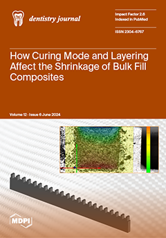
Cover Story (view full-size image): Bulk fill composites allow applications in layers of 4–5 mm. Different layering and polymerization techniques can be used depending on the polymerization shrinkage of the materials. The introduction of different light curing modes, the intensity and orientation of light during light curing affect the shrinkage of the material. This study aimed to identify the most favorable curing mode and layering strategy for bulk fill dental composites to reduce shrinkage stress and cuspal deflection. Measurement of cuspal tip deflection is one of the methods used to test internal stresses resulting from polymerization shrinkage. Simulated aluminum cavities were used to test different curing modes and two types of layering. The light curing mode showed a greater effect on material shrinkage than the layering technique. View this paper
  • Issues are regarded as officially published after their release is announced to the table of contents alert mailing list.
  • You may sign up for e-mail alerts to receive table of contents of newly released issues.
  • PDF is the official format for papers published in both, html and pdf forms. To view the papers in pdf format, click on the "PDF Full-text" link, and use the free Adobe Reader to open them.
Order results
Result details
Section
Select all
Export citation of selected articles as:
22 pages, 22805 KiB  
Article
Trabecular Bone Component Assessment under Orthodontic Loads and Movements during Periodontal Breakdown—A Finite Elements Analysis
by Radu-Andrei Moga, Cristian Doru Olteanu and Ada Gabriela Delean
Dent. J. 2024, 12(6), 190; https://doi.org/10.3390/dj12060190 - 20 Jun 2024
Viewed by 898
Abstract
This numerical analysis, by employing Tresca and Von Mises failure criteria, assessed the biomechanical behavior of a trabecular bone component subjected to 0.6, 1.2, and 2.4 N orthodontic forces under five movements (intrusion, extrusion, tipping, rotation, and translation) and during a gradual horizontal [...] Read more.
This numerical analysis, by employing Tresca and Von Mises failure criteria, assessed the biomechanical behavior of a trabecular bone component subjected to 0.6, 1.2, and 2.4 N orthodontic forces under five movements (intrusion, extrusion, tipping, rotation, and translation) and during a gradual horizontal periodontal breakdown (0–8 mm). Additionally, they assessed the changes produced by bone loss, and the ischemic and resorptive risks. The analysis employed eighty-one models of nine patients in 405 simulations. Both failure criteria showed similar qualitative results, with Tresca being quantitatively higher by 1.09–1.21. No qualitative differences were seen between the three orthodontic loads. Quantitatively, a doubling (1.2 N) and quadrupling (2.4 N) were visible when compared to 0.6 N. Rotation and translation followed by tipping are the most stressful, especially for a reduced periodontium, prone to higher ischemic and resorptive risks. In an intact periodontium, 1.2 N can be safely applied but only in a reduced periodontium for extrusion and intrusion. More than 0.6 N is prone to increasing ischemic and resorptive risks for the other three movements. In an intact periodontium, stress spreads in the entire trabecular structure. In a reduced periodontium, stress concentrates (after a 4 mm loss—marker for the stress change distribution) and increases around the cervical third of the remaining alveolar socket. Full article
(This article belongs to the Special Issue New Trends in Digital Dentistry)
Show Figures

Figure 1

18 pages, 3797 KiB  
Article
Evaluation of Ilex guayusa and Piper marginatum Extract Cytotoxicity on Human Dental Pulp Mesenchymal Stem Cells
by Luis G. Sequeda-Castañeda, Luisa F. Suárez-Carvajal, Mayra A. Téllez-Corral, Sandra J. Gutiérrez-Prieto and Henry A. Méndez-Pinzón
Dent. J. 2024, 12(6), 189; https://doi.org/10.3390/dj12060189 - 20 Jun 2024
Viewed by 1465
Abstract
Background: Amelogenesis imperfecta is a hereditary disorder affecting dental enamel. Among its phenotypes, hypocalcified AI is characterized by mineral deficiency, leading to tissue wear and, consequently, dental sensitivity. Excessive fluoride intake (through drinking water, fluoride supplements, toothpaste, or by ingesting products such as [...] Read more.
Background: Amelogenesis imperfecta is a hereditary disorder affecting dental enamel. Among its phenotypes, hypocalcified AI is characterized by mineral deficiency, leading to tissue wear and, consequently, dental sensitivity. Excessive fluoride intake (through drinking water, fluoride supplements, toothpaste, or by ingesting products such as pesticides or insecticides) can lead to a condition known as dental fluorosis, which manifests as stains and teeth discoloration affecting their structure. Our recent studies have shown that extracts from Colombian native plants, Ilex guayusa and Piper marginatum, deposit mineral ions such as phosphate and orthophosphate into the dental enamel structure; however, it is unknown whether these extracts produce toxic effects on the dental pulp. Objective: To assess cytotoxicity effects on human dental pulp stem cells (hDPSCs) exposed to extracts isolated from I. guayusa and P. marginatum and, hence, their safety for clinical use. Methods: Raman spectroscopy, fluorescence microscopy, and flow cytometry techniques were employed. For Raman spectroscopy, hDPSCs were seeded onto nanobiochips designed to provide surface-enhanced Raman spectroscopy (SERS effect), which enhances their Raman signal by several orders of magnitude. After eight days in culture, I. guayusa and P. marginatum extracts at different concentrations (10, 50, and 100 ppm) were added. Raman measurements were performed at 0, 12, and 24 h following extract application. Fluorescence microscopy was conducted using an OLIMPUS fv1000 microscope, a live–dead assay was performed using a kit employing a BD FACS Canto TM II flow cytometer, and data analysis was determined using a FlowJo program. Results: The Raman spectroscopy results showed spectra consistent with viable cells. These findings were corroborated using fluorescence microscopy and flow cytometry techniques, confirming high cellular viability. Conclusions: The analyzed extracts exhibited low cytotoxicity, suggesting that they could be safely applied on enamel for remineralization purposes. The use of nanobiochips for SERS effect improved the cell viability assessment. Full article
Show Figures

Figure 1

10 pages, 1443 KiB  
Article
Effect of Different Primers on the Shear Bond Strength of Orthodontic Brackets Bonded to Reinforced Polyetheretherketone (PEEK) Substrate
by Ahmed Akram EL-Awady, Khaled Samy ElHabbak, Hussein Ramadan Mohamed, Ahmed Elsayed Elwan, Karim Sherif Adly, Moamen Ahmed Abdalla, Ehab Mohamed Kamal and Ahmed Leithy Alameldin
Dent. J. 2024, 12(6), 188; https://doi.org/10.3390/dj12060188 - 20 Jun 2024
Viewed by 877
Abstract
This in vitro study assessed the effect of different primers on the shear bond strength (SBS) and adhesive remnant index (ARI) of orthodontic brackets bonded to reinforced polyetheretherketone (PEEK) substrate. A total of 40 specimens were randomly distributed to two groups based on [...] Read more.
This in vitro study assessed the effect of different primers on the shear bond strength (SBS) and adhesive remnant index (ARI) of orthodontic brackets bonded to reinforced polyetheretherketone (PEEK) substrate. A total of 40 specimens were randomly distributed to two groups based on the primer used for orthodontic bonding: group 1 (control)—Transbond XT adhesive with Visio.link primer and group 2 (test)—orthodontic adhesive (Transbond XT) with traditional orthodontic primer. After bonding, specimens were thermocycled followed by SBS testing and ARI scoring of debonded specimens. Data were analyzed using the unpaired independent t-test and the Chi-square test. Group 1 specimens showed significantly higher SBS values (21.38 ± 1.48 MPa) compared to group 2 specimens (18.63 ± 1.29 MPa) (p < 0.0001). Adhesive remnant index scores showed no significant variations in bond failure modes and distributions between groups. The SBS obtained by the tested primers exceeded the clinically recommended value. Consequently, there is a comparable clinical application for both tested primers in orthodontic bonding, especially the traditional orthodontic primer, where the availability of Visio.link in clinical practice is not ensured. Full article
Show Figures

Graphical abstract

10 pages, 790 KiB  
Article
Factors Influencing Peri-Extraction Anxiety: A Cross-Sectional Study
by Wojciech Niemczyk, Agnieszka Balicz, Karolina Lau, Tadeusz Morawiec and Janusz Kasperczyk
Dent. J. 2024, 12(6), 187; https://doi.org/10.3390/dj12060187 - 20 Jun 2024
Cited by 1 | Viewed by 1303
Abstract
Background: Fear and anxiety are common psychological responses to unpleasant stimuli, with dental fear being the fourth most prevalent type of fear or phobia. However, not all dental procedures cause the same level of anxiety, with dental surgery and tooth extraction being among [...] Read more.
Background: Fear and anxiety are common psychological responses to unpleasant stimuli, with dental fear being the fourth most prevalent type of fear or phobia. However, not all dental procedures cause the same level of anxiety, with dental surgery and tooth extraction being among the top five most frightening procedures in the field. Tooth extractions are also the most common surgical procedure in dental surgery. It is important to manage anxiety in the dental office by identifying the main factors. Methods: The study was conducted on a group of 250 patients. The survey technique and the Beliefs about Pain Control Questionnaire (BPCQ) were used in the study. Pain and stress intensity on a Visual Analog Scale (VAS) were measured in each patient before and after tooth extraction procedures. Results: Young women and people from small towns had the highest levels of anxiety. The factor causing the highest level of anxiety was fear of complications. Patients undergoing extraction of retained teeth were characterized by higher levels of anxiety. Conclusions: Perioperative stress is strongly dependent on numerous factors. For patient comfort, it may be crucial for dentists to have knowledge about these factors and the ability to utilize them to reduce stress before and after tooth extractions. Full article
(This article belongs to the Special Issue Preventive Dentistry and Dental Public Health)
Show Figures

Figure 1

14 pages, 1676 KiB  
Article
Molar Incisor Hypomineralization: Optimizing Treatment Protocols for Hypersensitivity: A Randomized Clinical Trial
by Elena Bardellini, Francesca Amadori, Laura Rosselli, Maria Luisa Garo, Alessandra Majorana and Giulio Conti
Dent. J. 2024, 12(6), 186; https://doi.org/10.3390/dj12060186 - 19 Jun 2024
Cited by 1 | Viewed by 1342
Abstract
Dentin hypersensitivity (DH) is a common challenge in pediatric patients with molar incisor hypomineralization (MIH), stemming from enamel porosity or exposed dentin after enamel breakdown. This three-arm randomized controlled clinical trial aims to evaluate the effectiveness of three different desensitizing treatment protocols. The [...] Read more.
Dentin hypersensitivity (DH) is a common challenge in pediatric patients with molar incisor hypomineralization (MIH), stemming from enamel porosity or exposed dentin after enamel breakdown. This three-arm randomized controlled clinical trial aims to evaluate the effectiveness of three different desensitizing treatment protocols. The study was conducted on 39 children, aged 6–14 years old, with MIH and DH. Group A received casein phosphopeptide plus amorphous calcium phosphate fluoride (CPP-ACPF) mousse and sham light therapy, Group B received placebo mousse and photo-bio-modulation therapy (PMBT), and Group C received both CPP-ACPF mousse and PMBT. DH evaluation using a visual analogue scale was performed at multiple time points. Both CPP-ACPF mousse and PMBT individually demonstrated desensitizing effects on dental elements affected by MIH. While PMBT had a greater immediate effect, the combination of the two therapies proved most effective in reducing DH. The VAS scores were statistically lower in group C compared to groups A and B, both after the first session (p = 0.0001) and after 28 days (p = 0.0005). This study suggests promising avenues for managing DH in MIH patients, highlighting the potential of combined therapies, specifically CPP-ACPF mousse and PMBT, for enhanced clinical outcomes. Full article
(This article belongs to the Special Issue Oral Health Care in Paediatric Dentistry Volume 2)
Show Figures

Figure 1

23 pages, 2613 KiB  
Systematic Review
Survival Rates of Short Dental Implants (≤6 mm) Used as an Alternative to Longer (>6 mm) Implants for the Rehabilitation of Posterior Partial Edentulism: A Systematic Review of RCTs
by Rodopi Emfietzoglou and Xanthippi Dereka
Dent. J. 2024, 12(6), 185; https://doi.org/10.3390/dj12060185 - 17 Jun 2024
Cited by 1 | Viewed by 1351
Abstract
Short dental implants have been proposed as an alternative treatment option to bone regeneration procedures for the rehabilitation of resorbed alveolar ridges. The aim of this paper was to systematically review randomized controlled trials (RCTs) comparing short implants (≤6 mm) and longer implants [...] Read more.
Short dental implants have been proposed as an alternative treatment option to bone regeneration procedures for the rehabilitation of resorbed alveolar ridges. The aim of this paper was to systematically review randomized controlled trials (RCTs) comparing short implants (≤6 mm) and longer implants (>6 mm) in atrophic alveolar ridges in terms of implant survival rates, peri-implant marginal bone loss (MBL), prevalence of peri-implantitis and technical complications. A thorough electronic search was performed until September 2023. RCTs with follow-up of at least 1-year post-loading comparing short implants with rough surfaces to longer implants in the posterior jaws of systemically and periodontally healthy, partially edentulous adults were considered. Studies with incomplete information on the number of patients, follow-up or definition of “short implants” were excluded. The revised Cochrane risk-of-bias tool for randomized trials was used for Risk of bias assessment. Fixed-effects meta-analysis of the selected studies was applied to compare the outcome variables. Random-effect meta-analysis was performed, on the basis of within-study comparisons. In total, 16 articles were selected for meta-analysis and incorporated 408 short implants and 475 longer implants inserted in 317 and 388 patients, respectively. The survival rates of longer implants in pristine or augmented bone were significantly increased compared to short implants (95%CI: 2–5%, p < 0.001). Standard-length implants displayed increased, although non-statistically significant MBL (95%CI: −0.17–0.04, p > 0.05), and prevalence of peri-implantitis (95%CI: 0–5%, p > 0.05). No statistically significant differences were observed between short and long implants in terms of technical complications (implant-level 95%CI: −4–6%, p > 0.05). Short implants represent a promising alternative treatment option for the rehabilitation of posterior jaws to avoid additional bone augmentation procedures. Nonetheless, they should be selected cautiously due to a potentially limited survival rate compared to longer implants. A major limitation of this study is the variability in the included studies regarding sample size, patient profile, type of bone, loading protocol, definition of peri-implantitis, among others. This study received no external funding. The study protocol was registered in PROSPERO (CRD42023485514). Full article
(This article belongs to the Special Issue Oral Implantology and Rehabilitation)
Show Figures

Figure 1

10 pages, 2467 KiB  
Article
Evaluation of a New Simplified Approach for Upper Superficial Impacted Third Molar Extraction—A Retrospective Split-Mouth Study
by Alberto Materni, Karol Alí Apaza Alccayhuaman, Alberto Maltagliati, Daniele Botticelli and Stefano Benedicenti
Dent. J. 2024, 12(6), 184; https://doi.org/10.3390/dj12060184 - 17 Jun 2024
Viewed by 1485
Abstract
Background: A new access technique was developed to reduce postoperative adverse events after the extraction of impacted maxillary wisdom teeth. Hence, this study aimed to assess the occurrence of adverse events after the extraction of maxillary impacted wisdom teeth using a traditional access [...] Read more.
Background: A new access technique was developed to reduce postoperative adverse events after the extraction of impacted maxillary wisdom teeth. Hence, this study aimed to assess the occurrence of adverse events after the extraction of maxillary impacted wisdom teeth using a traditional access (TA) or a new technique (NT). Methods: Two different surgical incision designs were used for bilateral wisdom tooth extractions in 30 patients. The traditional incision was performed distal to the second molar in the center of the tuberosity, followed by a buccal releasing incision. After the tooth extraction, the wound was secured by sutures. The new technique consists of an oblique incision from the distal palatal aspect of the tuberosity towards the buccal aspect of the second molar. After the tooth extraction, cyanoacrylate glue was used on the wound. Results: Lower pain was reported by patients at the site treated with the new technique (p < 0.01). Edema, postsurgical bleeding, and hematoma were similar in both groups. The surgical time was shorter for the new technique (p < 0.01). Conclusions: The new technique applied for the extraction of impacted maxillary wisdom teeth reduced postsurgical pain and the duration of surgery. Full article
Show Figures

Figure 1

14 pages, 1948 KiB  
Systematic Review
Association of Connective Tissue Grafts in Immediate Implants: Systematic Review and Meta-Analysis
by Marta Torra-Moneny, Elisabet Mauri-Obradors, Sonia Egido-Moreno, Joan Valls-Roca-Umbert, Antonio Marí-Roig and José López-López
Dent. J. 2024, 12(6), 183; https://doi.org/10.3390/dj12060183 - 17 Jun 2024
Cited by 1 | Viewed by 1648
Abstract
Background: The increase in soft tissue (ST) around implants can benefit peri-implant health and aesthetic results. The objective was to compare the gingival and esthetic health benefits of immediate implant placement (IIP) with simultaneous or delayed connective tissue graft (CTG) compared to IIP [...] Read more.
Background: The increase in soft tissue (ST) around implants can benefit peri-implant health and aesthetic results. The objective was to compare the gingival and esthetic health benefits of immediate implant placement (IIP) with simultaneous or delayed connective tissue graft (CTG) compared to IIP without CTG. Methods: A systematic review was carried out by two reviewers in Medline-Pubmed, Scopus, and Cochrane. The Preferred Reporting Items for Systematic Reviews and Meta-Analyses (PRISMA) were considered. Randomized Clinical Trials (RCTs) that were published between April 2017 and February 2024 were used. Studies that analyzed the performance of a simultaneous or deferred CTG after the placement of an implant in the aesthetic zone, with or without immediate provisionalization, without previous regeneration, with a follow-up of 6 months, and that were performed in humans were included. Results: Quantitative analysis was performed using data provided by the RCTs. The five RCTs that were selected analyzed a total “n” of 245 subjects who met the inclusion criteria and focused on the subject of the study. In the quantitative analysis, four RCTs were included. The studies evaluated buccal gingiva levels when placing the IIP with and without CTG, obtaining a mean buccal gingiva level difference of 0.09 mm (95% CI: −0.54 to 0.72, p = 0.05), statistically not significant, but with a favorable trend. Conclusions: The use of CTG associated with the II can maintain the gum level but not increase the volume. CTG is favorable for achieving successful esthetic results when immediate placement of an implant with a provisional prosthesis is planned. Full article
(This article belongs to the Special Issue Oral Implantology and Rehabilitation)
Show Figures

Graphical abstract

8 pages, 974 KiB  
Article
Impact of Different Glidepath Techniques on the Overall Performance of WaveOne Gold in an Artificial S-Shape Canal
by Vlad Mircea Lup, Olivia Andreea Marcu, Carlo Gaeta and Gabriela Ciavoi
Dent. J. 2024, 12(6), 182; https://doi.org/10.3390/dj12060182 - 13 Jun 2024
Viewed by 1303
Abstract
Objectives: The article’s aim is to test if rotary or reciprocating glide path influences the overall performance of WaveOne Gold in S-shaped canals. Methods: Sixty endo training blocks with an S-shape curvature were divided into three groups based on the glide path method [...] Read more.
Objectives: The article’s aim is to test if rotary or reciprocating glide path influences the overall performance of WaveOne Gold in S-shaped canals. Methods: Sixty endo training blocks with an S-shape curvature were divided into three groups based on the glide path method used: no glide path; glide path preparation with ProGlider; glide path preparation with WaveOne Gold Glider. All blocks were then shaped with WaveOne Gold Primary. The time for shaping, the incidence in reaching working length and the number of pecking motions were recorded. ANOVA with Turkey’s test was used, and the p-value was set to 0.05. Results: WaveOne Gold Primary reached working length faster in the control group when comparing total working times. No significant differences in the ability of the WaveOne Gold Primary to reach working length in all groups (p > 0.05). The mean number of pecking motions was higher in the control group compared to other groups. Conclusions: No significant differences in the time needed to achieve a glide path between Proglider and WaveOne Gold Glider. WaveOne Gold Primary can shape a double curved canal faster if a glide path is present but takes less time to reach length if it is the only file used. No difference in the ability to reach working length. Full article
(This article belongs to the Special Issue Modern Endodontics)
Show Figures

Figure 1

11 pages, 463 KiB  
Article
Additive Manufacturing Titanium Dental Implants Placed in Sinuses Grafted with 70HA:30-TCP: A One-Year Retrospective Study for Evaluation of Survival Rate
by Ilton José Mafra, Dimorvan Bordin, Rafael S. Siroma, Vittorio Moraschini, Leonardo P. Faverani, João Gabriel Souza, Carlos Fernando Mourão and Jamil Awad Shibli
Dent. J. 2024, 12(6), 181; https://doi.org/10.3390/dj12060181 - 13 Jun 2024
Cited by 1 | Viewed by 1045
Abstract
The present short-term retrospective study evaluated the implant survival rate and peri-implant bone loss around additive-manufactured titanium implants placed in sinuses grafted with Plenum Osshp (Plenum Bioengenharia, Jundia, SP, Brazil) (70HA:30β-TCP) material. A total of 39 implants were inserted after 23 sinus floor [...] Read more.
The present short-term retrospective study evaluated the implant survival rate and peri-implant bone loss around additive-manufactured titanium implants placed in sinuses grafted with Plenum Osshp (Plenum Bioengenharia, Jundia, SP, Brazil) (70HA:30β-TCP) material. A total of 39 implants were inserted after 23 sinus floor elevation procedures in 16 consecutive patients. Prosthetic rehabilitation included fixed partial prostheses (three units), single crowns (eleven units), and fixed full arches (three units). Clinical and radiographic parameters of implant-supported restorations were evaluated after at least one year of occlusal loading. The implant–crown success criteria included the absence of pain, suppuration, and clinical mobility, an average distance between the implant shoulder and the first visible bone contact (DIB) < 1.0 mm from the initial surgery, and the absence of prosthetic complications at the implant–abutment interface. The overall cumulative implant survival rate was 97.43%. No prosthetic complications at the implant–abutment interface were reported. After one year, the mean DIB was 0.23 mm ± 0.14. Within the limits of this retrospective study, it can be concluded that 70 HA:30 β-TCP allowed stable and reliable bone support to maintain healthy conditions around titanium dental implants produced by additive manufacturing. Full article
(This article belongs to the Special Issue New Perspectives in Periodontology and Implant Dentistry)
Show Figures

Figure 1

12 pages, 4578 KiB  
Article
Efficacy of the Sausage Technique in Rebuilding the Crestal Buccal Bone Thickness: A Retrospective Analysis
by Stefano Pieroni, Benedetta Miceli, Luca Giboli, Leonardo Romano, Lorenzo Azzi and Davide Farronato
Dent. J. 2024, 12(6), 180; https://doi.org/10.3390/dj12060180 - 12 Jun 2024
Viewed by 1352
Abstract
The goal was to evaluate the efficacy of the sausage technique in reconstructing the crestal buccal bone thickness, focusing on the distribution shape of the regenerated volume. Ten implants were placed in five patients with Cawood–Howell class IV defects. A cone beam computed [...] Read more.
The goal was to evaluate the efficacy of the sausage technique in reconstructing the crestal buccal bone thickness, focusing on the distribution shape of the regenerated volume. Ten implants were placed in five patients with Cawood–Howell class IV defects. A cone beam computed tomography (CBCT) was executed at T0 (before surgery). Guided bone regeneration (GBR) with the sausage technique utilized a resorbable collagen membrane, made of a 50% autologous bone and a 50% anorganic bovine bone matrix (ABBM) mixture. After 6 months, a CBCT (T1) was performed before implant placement. Using CBCT software, a plane parallel to the implant axis intersected perpendicular planes every 1.5 mm from the crest level. T0 and T1 CBCT sections were analyzed, yielding 140 measurements. Statistical analysis via SPSS revealed a significant increase in thickness (average 2.82 ± 1.79 mm). Maximum gains occurred at 4.5 mm from the coronal crest line (3.8 ± 1.51 mm). The GBR sausage technique was effective with minimal post-operative complications, yielding the biggest gain at the mid-ridge sagittal area. Within the analysis limitations, it can be assumed that the sausage technique is effective for horizontal GBR in the maxilla, but a lesser volume might be achieved at the crestal level because it seems to follow a bowed regeneration shape. Full article
Show Figures

Figure 1

11 pages, 272 KiB  
Article
Frequency of Oral Lesions, Olfactory, and Gustatory Disorders and Xerostomia in Patients with COVID-19
by Natália Lopes Castilho, Daniella R. Barbosa Martelli, Renato Assis Machado, Zêus Araujo Cunha, Claudiojanes dos Reis, Priscila Regina Queiroz, Dayane de Sá Silva, Eduardo Araujo Oliveira, Ricardo D. Coletta and Hercílio Martelli-Júnior
Dent. J. 2024, 12(6), 179; https://doi.org/10.3390/dj12060179 - 11 Jun 2024
Viewed by 1383
Abstract
COVID-19, a respiratory illness with a global impact on millions, has recently been linked to manifestations affecting various bodily systems, including the oral cavity. Studies highlight oral issues, like ulcers, blisters, and white patches, alongside olfactory and gustatory dysfunction, influencing an individual’s quality [...] Read more.
COVID-19, a respiratory illness with a global impact on millions, has recently been linked to manifestations affecting various bodily systems, including the oral cavity. Studies highlight oral issues, like ulcers, blisters, and white patches, alongside olfactory and gustatory dysfunction, influencing an individual’s quality of life. In this context, our study aimed to assess the frequency of oral lesions, olfactory and gustatory disorders, and xerostomia resulting from COVID-19. An observational study was conducted with 414 patients to evaluate the frequency of oral symptoms resulting from COVID-19. Patients were diagnosed with mild symptoms and evaluated through clinical examination of the oral cavity and a questionnaire to assess functional alterations. The findings showed that 139 out of 414 patients presented clinical manifestations, with oral lesions being the most prevalent (19.1%), followed by gustatory disorders (18.1%), xerostomia (14.2%), and olfactory dysfunction (14%). The most prevalent oral lesions were ulcerations (n = 51), candidiasis (n = 8), and erythema or red plaques (n = 7). Unfortunately, 50 (12.1%) patients died during this study. Therefore, oral lesions, olfactory and gustatory dysfunctions, and xerostomia are common symptoms associated with COVID-19. Full article
Show Figures

Graphical abstract

13 pages, 2930 KiB  
Article
Analysis of the Pulpal Blood Flow Microdynamics during Prosthetic Tooth Preparation Using Diamond Burs with Different Degrees of Wear
by Edmond Ciora, Mariana Miron, Diana Lungeanu, Andreea Igna and Anca Jivanescu
Dent. J. 2024, 12(6), 178; https://doi.org/10.3390/dj12060178 - 7 Jun 2024
Cited by 1 | Viewed by 1214
Abstract
Pulpal modifications taking place during prosthetic tooth preparation using worn-out burs may represent a risk for the vitality of the dental pulp. The aim of this in vivo study was to evaluate whether the wear of diamond burs has an influence on the [...] Read more.
Pulpal modifications taking place during prosthetic tooth preparation using worn-out burs may represent a risk for the vitality of the dental pulp. The aim of this in vivo study was to evaluate whether the wear of diamond burs has an influence on the vascular microdynamics at the level of the dental pulp, during vertical preparation for zirconia crowns. The study was performed with a split-mouth design and included 32 vital permanent monoradicular teeth (20 maxillary and 12 mandibular), from six subjects, aged between 20 and 50 years. The teeth were randomly assigned to two study groups of 16 teeth each. For prosthetic preparation, new burs were used in the first group, and burs at their 5th use were used in the second group. Four consecutive determinations of the pulpal blood flow by Laser Doppler flowmetry (LDF—laser Doppler MoorLab instrument VMS-LDF2, Moor Instruments Ltd., Axminster, UK) were taken for each tooth included in the study: before the preparation (control values), immediately, at 24 h, and at 7 days after the prosthetic preparation. A four-way ANOVA statistical analysis was applied to analyze the effect of four considered factors (bur wear degree, time of measurement, tooth number, and tooth location) on the pulpal blood flow (PBF). A significant increase in pulpal blood flow compared to the baseline was recorded immediately after preparation (p < 0.01), at 24 h (p < 0.01), and at 7 days (p < 0.05) in both groups, but more pronounced in the case of burs at the 5th use. The blood flow was significantly higher in upper jaw teeth, irrespective of the measurement time. In conclusion, the use of worn-out diamond burs produces lasting modifications in the pulpal blood flow of teeth that undergo prosthetic crown preparation. ISRCTN registry: ISRCTN49594720. Full article
Show Figures

Figure 1

7 pages, 641 KiB  
Brief Report
The Use of Potassium Iodide in Pediatric Dentistry Does Not Change the Retention of Glass Ionomer Cement on a Dentin Treated with Silver Fluoride: In Vitro Results
by Louise Gotas, Thibault Canceill and Sabine Joniot
Dent. J. 2024, 12(6), 177; https://doi.org/10.3390/dj12060177 - 7 Jun 2024
Viewed by 1472
Abstract
In pediatric and operative dentistry, caries treatment benefits from a therapeutic option based on the use of silver fluoride (AgF) associated with potassium iodide (KI) to avoid dark colorations on dental tissues. The objective of this in vitro study is to evaluate the [...] Read more.
In pediatric and operative dentistry, caries treatment benefits from a therapeutic option based on the use of silver fluoride (AgF) associated with potassium iodide (KI) to avoid dark colorations on dental tissues. The objective of this in vitro study is to evaluate the retention of glass ionomer cement (GIC) on a dentin when treated with AgF and KI. Twenty-two healthy human permanent molars and eight human primary teeth, all free of any decay, were cut to obtain occlusal flat dentinal surfaces and were then treated with AgF for a duration of 60 s. For half of the teeth, a drop of KI was applied for a duration of 30 s. All samples were covered with a plot of GIC and their resistance to shear bond strength was measured. The fracture resistance in both permanent and primary teeth does not show any statistically significant differences whether KI was applied or not. For permanent teeth, the resistance is slightly higher in the group treated with KI than in the group treated with AgF alone. To our knowledge, these data are the first to describe the possible application of KI both on primary and permanent teeth. In any case, further studies are needed to investigate the bond strength between dentin and GIC on a wider range of samples. Full article
(This article belongs to the Special Issue Current Advances in Pediatric Odontology)
Show Figures

Figure 1

16 pages, 822 KiB  
Review
Iron Deficiency Anemia and Its Impact on Oral Health—A Literature Review
by Kabilan Velliyagounder, Krupa Chavan and Kenneth Markowitz
Dent. J. 2024, 12(6), 176; https://doi.org/10.3390/dj12060176 - 7 Jun 2024
Viewed by 3443
Abstract
Oral disease interventions primarily focus on behavioral changes like dietary improvements and ensuring better oral hygiene. However, recognizing the influence of biological factors, including genetics and early-life nutrition, is crucial. Iron deficiency (ID) and its advanced form, iron deficiency anemia (IDA), affect nearly [...] Read more.
Oral disease interventions primarily focus on behavioral changes like dietary improvements and ensuring better oral hygiene. However, recognizing the influence of biological factors, including genetics and early-life nutrition, is crucial. Iron deficiency (ID) and its advanced form, iron deficiency anemia (IDA), affect nearly two billion people globally, especially children and pregnant women. We conducted a comprehensive search using Medline via EndNote and Web of Science, employing keywords related to iron deficiency anemia (IDA), and we identified 36 studies deemed relevant for inclusion in this literature review. IDA prevalence is notably high among pregnant women and young children. Both IDA and early-childhood caries (ECC) disproportionately affect impoverished populations, highlighting the socioeconomic dimension of this issue. IDA presents with various oral mucosal changes and is closely linked to candidiasis. Additionally, IDA can hinder tooth development and weaken the immune response. Multiple population surveys have revealed a significant association between ECC and IDA. While some studies have explored the IDA–periodontal disease link, the current evidence is relatively limited in its robustness. In conclusion, more comprehensive longitudinal studies are essential to deepen our understanding of the IDA–oral disease connection. Investigating the underlying biological mechanisms is critical to developing effective interventions, particularly for vulnerable populations affected by IDA. Full article
(This article belongs to the Section Oral Hygiene, Periodontology and Peri-implant Diseases)
Show Figures

Figure 1

12 pages, 2923 KiB  
Article
Injectable Platelet-Rich Fibrin and Advanced Platelet-Rich Fibrin Demonstrate Enhanced Anti-Biofilm Effect Compared to Enamel Matrix Derivatives on Decontaminated Titanium Surfaces
by Jothi Varghese, Liza L. Ramenzoni, Padmaja A. Shenoy, Patrick R. Schmidlin, Shubhankar Mehrotra and Vinayak Kamath
Dent. J. 2024, 12(6), 175; https://doi.org/10.3390/dj12060175 - 6 Jun 2024
Viewed by 1087
Abstract
Background: The search for effective antimicrobial agents to mitigate peri-implant infections remains a crucial aspect of implant dentistry. This study aimed to evaluate and compare the antimicrobial efficacy of i-PRF, A-PRF+, and enamel matrix derivative (EMD) on decontaminated rough and smooth titanium (Ti) [...] Read more.
Background: The search for effective antimicrobial agents to mitigate peri-implant infections remains a crucial aspect of implant dentistry. This study aimed to evaluate and compare the antimicrobial efficacy of i-PRF, A-PRF+, and enamel matrix derivative (EMD) on decontaminated rough and smooth titanium (Ti) discs. Materials and Methods: Rough and smooth Ti discs were coated with multispecies biofilm and thoroughly debrided using a chitosan-bristled brush. Subsequently, i-PRF, A-PRF+, and EMD were applied. Untreated discs served as control. Residual adherent bacteria present on the treated Ti discs were visualized by SEM and quantified using culture technique, and colony-forming units (CFUs) were measured after 48 h and 7 days. Results: i-PRF demonstrated better antimicrobial effectiveness on both smooth and rough implant surfaces as compared to A-PRF+ and EMD (p < 0.001). In all the experimental groups, smooth Ti discs displayed a greater reduction in microbes compared to rough Ti discs when treated with the biologics. The major reduction in CFU values was determined after seven days. Conclusions: i-PRF as a regenerative material may also be suitable for decontaminating implant surfaces, which could influence tissue healing and regenerative outcomes positively. Full article
Show Figures

Figure 1

10 pages, 561 KiB  
Article
Evaluation of the Association of Chewing Function and Oral Health-Related Quality of Life in a Population of Individuals Aged ≥ 45 Years and Residing in Communities in Switzerland: A Cross-Sectional Study
by Christian Tennert, Roberta Borg-Bartolo, Maria Prasinou, Maurus Kurt Jaeggi, Martin Schimmel, Andrea Roccuzzo and Guglielmo Campus
Dent. J. 2024, 12(6), 174; https://doi.org/10.3390/dj12060174 - 6 Jun 2024
Viewed by 1029
Abstract
Purpose: To analyse the association of masticatory performance and oral health-related quality of life in a representative population of individuals residing in communities in Switzerland aged ≥ 45 years. Materials and Methods: In total, 100 subjects completed two dedicated and validated questionnaires on [...] Read more.
Purpose: To analyse the association of masticatory performance and oral health-related quality of life in a representative population of individuals residing in communities in Switzerland aged ≥ 45 years. Materials and Methods: In total, 100 subjects completed two dedicated and validated questionnaires on their demographic data and the Geriatric Oral Health Assessment Index. A mixing ability test was performed for assessing masticatory performance. The qualitative analysis of the test was performed by categorizing the images into five categories, while the quantitative analysis was performed via a validated custom-made software. Results: Sixty-six samples could be analysed. Participants younger than 65 years of age showed significantly less frequent chewing deficiencies (17%) compared to those 65 years and older (50%, p < 0.01). However, retired participants had chewing deficiencies significantly more frequently (8%) compared to workers (51%, p < 0.01). A statistically significant positive association of having chewing deficiency was found between employment status (p < 0.01) and the presence of restorations (p = 0.04), while GOHAI did not show any statistically significant association. Overall, the enrolled subjects displayed moderate chewing function. Masticatory performance was positively associated with the number of present restorations. Conclusions: The enrolled subjects residing in communities in Switzerland aged ≥ 45 years displayed moderate chewing function. Their masticatory performance was positively associated with the number of present restorations but not associated with oral health related quality of life (GOHAI). Full article
Show Figures

Graphical abstract

14 pages, 841 KiB  
Article
Hospitalisations Due to Dental Infection: A Retrospective Clinical Audit from an Australian Public Hospital
by Mafaz Ullah, Muhammad Irshad, Albert Yaacoub, Eric Carter and Stephen Cox
Dent. J. 2024, 12(6), 173; https://doi.org/10.3390/dj12060173 - 6 Jun 2024
Cited by 1 | Viewed by 1521
Abstract
The aim of this clinical audit is to evaluate the characteristics of dental infections requiring hospitalisations, which may help improve preventative and management policies. This study retrospectively evaluated the records of patients admitted to the Nepean hospital, Kingswood, New South Wales, Australia, due [...] Read more.
The aim of this clinical audit is to evaluate the characteristics of dental infections requiring hospitalisations, which may help improve preventative and management policies. This study retrospectively evaluated the records of patients admitted to the Nepean hospital, Kingswood, New South Wales, Australia, due to dental infections between 2018 and 2019. A total of 102 patients, mostly in their thirties with equal gender distribution, were admitted with dental infections, presenting with pain (100%), swelling (99%), trismus (40.2%), dysphagia (27.4%), fever (21%) [>37 °C], tachycardia (24.8%) and tachypnoea (9.3%). Most patients (68%) presented on weekends, outside regular working hours, and public holidays. A total of 52.5% of patients had taken prior antibiotics. Dental caries, smoking, mental health issues, and illicit drug use were featured strongly. The majority of patients (56.4%) underwent treatment under local anaesthesia. The total length of hospital stay was 271 days (mean 2.7, SD 1.6). Augmentin was the most prescribed antibiotic. Complications were reported in 8.8% of the patients, primarily due to airway compromise. Dental infections leading to hospitalisations continue to be a burden on the healthcare system. A notable finding was that the presentations were primarily on weekends, outside regular working hours, and public holidays, and the majority required dental interventions under local anaesthesia. The provision of on-call emergency dental services may reduce potentially preventable hospitalisations and the length of hospital stay. Full article
Show Figures

Figure 1

12 pages, 11069 KiB  
Article
A Minimally Invasive Surgical Procedure to Harvest Palate Periosteum as a Source of Mesenchymal Stromal/Stem Cells for Bone Tissue Engineering
by André Antonio Pelegrine, David Gonzalo Montero López, Antonio Carlos Aloise, João Pedro Grandini Zeferino, Carolina Guassi Mannina, Raul Canal, Daniel Navarro da Rocha, Tamara Cristina Lopes de Castro, Elizabeth Ferreira Martinez, Lexie Shannon Holliday, Roberto Dalto Fanganiello and José Ricardo Muniz Ferreira
Dent. J. 2024, 12(6), 172; https://doi.org/10.3390/dj12060172 - 5 Jun 2024
Viewed by 909
Abstract
The aim of this study is to validate a minimally invasive surgical procedure to harvest palate periosteum as a source of tissue for mesenchymal stromal/stem cells. We performed a standardized procedure to harvest the palate periosteum in ten subjects, which consisted of a [...] Read more.
The aim of this study is to validate a minimally invasive surgical procedure to harvest palate periosteum as a source of tissue for mesenchymal stromal/stem cells. We performed a standardized procedure to harvest the palate periosteum in ten subjects, which consisted of a 3 mm disposable punch and a Molt periosteal elevator to harvest a small full-thickness fragment of soft tissue at the hard palate area, between the upper bicuspids, 3 to 4 mm apical to the cement enamel junction. The one-third inner portion was fragmented, and following standard cell culture procedures, the adherent cells were cultured for three passages, after obtaining 70–90% confluence. Cell morphology analysis, flow cytometry analysis, and viability and osteogenic differentiation assays were performed. In all 10 cases, uneventful healing was observed, with no need for analgesic intake. The evaluation of cell morphology showed elongated spindle-shaped cells distributed in woven patterns. A high viability range was verified as well as an immunophenotype compatible with mesenchymal stem cell lineage. The differentiation assay showed the potential of the cells to differentiate into the osteogenic lineage. These results demonstrate that the minimally invasive proposed surgical technique is capable of supplying enough periosteum source tissue for stem cell culture and bone tissue engineering. Full article
Show Figures

Figure 1

10 pages, 2113 KiB  
Article
New Perspectives in Overcoming Bulk-Fill Composite Polymerization Shrinkage: The Impact of Curing Mode and Layering
by Zsuzsanna Bardocz-Veres, Mátyás Levente Miklós, Edina-Kata Biró, Éva Andrea Kántor, József Kántor, Csaba Dudás and Bernadette Kerekes-Máthé
Dent. J. 2024, 12(6), 171; https://doi.org/10.3390/dj12060171 - 5 Jun 2024
Viewed by 1176
Abstract
Background: The purpose of this study was to investigate the effect of different light curing modes on the polymerization shrinkage of a bulk-fill composite and to evaluate the impact of two layering techniques on the cuspal deflection. Methods: Nine different light curing modes [...] Read more.
Background: The purpose of this study was to investigate the effect of different light curing modes on the polymerization shrinkage of a bulk-fill composite and to evaluate the impact of two layering techniques on the cuspal deflection. Methods: Nine different light curing modes were tested on bulk-fill composite samples in aluminum MOD cavities. Intensity, duration, and illumination distance were the factors that changed during the different curing modes. The digital image correlation method was used to visually represent the displacement of carbon particles on the materials’ surface caused by shrinkage along both the horizontal and vertical axes. For simulating cuspal deflection, a separate protocol was used, with a bulk and horizontal layering technique. Results: The results showed that the largest horizontal displacements were present in the soft start group (6.00 ± 0.82 µm) and in the X-tra power group (5.67 ± 1.21 µm). The smallest horizontal displacements were detected in normal curing modes (4.00 ± 1.58 µm; 4.00 ± 2.68 µm). The largest vertical displacements, at the bottom layer, were present in the normal curing mode group with a 20 s curing time (5.22 ± 1.56 µm), while the smallest vertical displacements were shown in the X-tra power group (2.89 ± 0.60 µm). The observed particle displacements showing the shrinkage of the composite were correlated with the curing mode. The bulk-fill group showed less cuspal deflection than the horizontal layering group did, but the difference was not statistically significant (p = 0.575). Conclusions: Within the limitations of this in vitro study, it can be concluded that lower intensities of curing lights (1200 mW/cm2) may perform better from the point of view of material shrinkage than high and extreme light intensities do. The pulse delay mode might be recommended in the case of bulk-fill materials. The number of layers did not significantly affect the cuspal deflection in the case of the studied composite. Full article
(This article belongs to the Special Issue Endodontics and Restorative Sciences)
Show Figures

Figure 1

13 pages, 6131 KiB  
Case Report
Periodontal Regeneration of Vital Poor Prognosis Teeth with Attachment Loss Involving the Root Apex: Two Cases with up to 5 Years Follow-Up
by Ethan Ng and John Rong Hao Tay
Dent. J. 2024, 12(6), 170; https://doi.org/10.3390/dj12060170 - 5 Jun 2024
Viewed by 1099
Abstract
Teeth with attachment loss involving the root apex are severely compromised and have a poor periodontal prognosis. In cases where periodontal regeneration is possible, current guidelines suggest that endodontic treatment is performed first. However, root canal treatment increases the overall treatment time and [...] Read more.
Teeth with attachment loss involving the root apex are severely compromised and have a poor periodontal prognosis. In cases where periodontal regeneration is possible, current guidelines suggest that endodontic treatment is performed first. However, root canal treatment increases the overall treatment time and costs, has risks of endodontic complications, and could predispose teeth to mechanical failure. In this case report, two patients diagnosed with periodontitis stage III/IV grade C, no history of smoking or diabetes, and attachment loss involving the root apex of a tooth, were treated with guided tissue regeneration. These two cases are unique because successful periodontal regeneration was carried out without endodontic treatment, and the vitality of these teeth was maintained longitudinally. This report presents the management that led to this clinical outcome, and important guidelines for case selection are identified. Within the limitations of this study, vital teeth with radiographic bone loss involving the apex may be treated successfully with periodontal regeneration and remain vital at least in the short- to medium-term. Full article
(This article belongs to the Special Issue Advances in Periodontal and Peri-Implant Tissues Health Management)
Show Figures

Figure 1

15 pages, 569 KiB  
Article
Knowledge, Attitudes, and Practices towards Silver Diamine Fluoride among Dentists in Vietnam
by Hollis Haotian Chai, Quang Khai Dao, Trong Hung Hoang, Sherry Shiqian Gao, Edward Chin Man Lo and Chun Hung Chu
Dent. J. 2024, 12(6), 169; https://doi.org/10.3390/dj12060169 - 5 Jun 2024
Viewed by 1123
Abstract
Silver diamine fluoride (SDF) is a topical solution used for managing dental caries. The mixed-methods study consists of a quantitative study (questionnaire survey) and a qualitative study (in-depth interview) to explore the knowledge, attitudes, and practices towards SDF among dentists in Vietnam. A [...] Read more.
Silver diamine fluoride (SDF) is a topical solution used for managing dental caries. The mixed-methods study consists of a quantitative study (questionnaire survey) and a qualitative study (in-depth interview) to explore the knowledge, attitudes, and practices towards SDF among dentists in Vietnam. A descriptive approach and a thematic approach were performed to analyze data, respectively. The questionnaire survey invited 436 licensed dentists registered for the national annual dental meeting and 226 dentists participated (response rate: 52%). Among them, 174 (77%, 174/226) dentists knew SDF, and 69 (40%, 69/174) dentists used SDF for caries management. Most of them considered SDF therapy as non-invasive (84%, 146/174) and simple (80%, 140/174). However, most of them expressed concern that SDF could discolor the tooth (74%, 128/174). Their most preferred teeth for SDF therapy were primary posterior teeth (92%, 160/174). The in-depth interview consulted 16 dentists to reach data saturation. They learned about SDF from outside curriculum resources as an effective anti-caries agent. They understood the advantages (simple, non-invasive, timesaving) and disadvantages (tooth discoloring, ammonia odor) of SDF. They used SDF to arrest caries in uncooperative children in the clinic and people living in rural areas in outreach services. Most dentists in Vietnam are supportive of SDF therapy, and they know its advantages and disadvantages for caries management. The results addressed the aim of the study to investigate Vietnamese dentists’ perspectives towards SDF. Full article
Show Figures

Figure 1

12 pages, 1213 KiB  
Article
Microbiological Analysis of Plaque and Its Composition in Three Patient Groups under Different Orthodontic Treatments
by Niccolò Cenzato, Chiara Occhipinti, Elena D’amici, Paolo Savadori, Benedetta Baldini and Cinzia Maspero
Dent. J. 2024, 12(6), 168; https://doi.org/10.3390/dj12060168 - 4 Jun 2024
Cited by 2 | Viewed by 1143
Abstract
Background: This article analyzes differences in microbiological parameters and periodontal health conditions among three patient groups: those undergoing conventional orthodontic treatment with fixed appliances, patients undergoing orthodontic treatment with clear aligners, and a control group receiving no treatment. Materials and Methods: In this [...] Read more.
Background: This article analyzes differences in microbiological parameters and periodontal health conditions among three patient groups: those undergoing conventional orthodontic treatment with fixed appliances, patients undergoing orthodontic treatment with clear aligners, and a control group receiving no treatment. Materials and Methods: In this study, 60 patients were enrolled. The microbiological analysis employed a qualitative and semi-quantitative methodology of bacterial morphotype analysis. Results: The analyses revealed a significant difference in favor of clear oral and periodontal health aligners. This could be attributed to better bacterial biofilm removal and reduced mechanical stress on the periodontal ligament, factors facilitated by the ease of clear aligner removal. Significant differences (p-value < 0.05) were observed for the Full-Mouth Plaque Score, Full-Mouth Bleeding Score, Plaque Index, and periodontal health assessment measurements. Conclusions: Although overall hygiene appears to be improved in patients in the aligners group compared to those treated with conventional orthodontic appliances, there are no statistically significant results regarding plaque composition. Microbiological aspects will be further addressed using more specific techniques in the follow-up of this research. Full article
Show Figures

Figure 1

15 pages, 3312 KiB  
Article
Concordance Analysis of Lower Third Molar Surgery Classifications: A Comparative Study
by Selene Barone, Francesco Bennardo, Marianna Salviati, Vincenzo Cosentino, Riccardo Finamore, Vincenzo Greco, Antonio Madonna, Anna Procopio, Alessandro Antonelli and Amerigo Giudice
Dent. J. 2024, 12(6), 167; https://doi.org/10.3390/dj12060167 - 3 Jun 2024
Viewed by 597
Abstract
The high frequency and complexity of mandibular third molar (M3M) surgery have led several authors to the development of classification systems for better evaluation and management in oral surgery. This study compared the classifications of Juodzabalys and Daugela et al. (JD), Sammartino et [...] Read more.
The high frequency and complexity of mandibular third molar (M3M) surgery have led several authors to the development of classification systems for better evaluation and management in oral surgery. This study compared the classifications of Juodzabalys and Daugela et al. (JD), Sammartino et al., Chang et al., Jhamb et al., Maglione et al., and Nemsi et al. to understand the concordance between the scores of M3M surgery. Two types of analysis were conducted: the relationship between the M3M and the inferior alveolar nerve (IAN), and the overall difficulty score based on the tooth’s angulation and its spatial position with the adjacent structure. The analysis of the classifications on the relationship between M3M and IAN resulted in a concordance of 26.1%. In the pairwise comparisons, the classifications of Nemsi et al. and Jhamb et al. showed the highest concordance of 59.5%. Analyzing the total scores difficulty, the JD et al., Chang et al., and Sammartino et al. classifications demonstrated a concordance level of 25.5%. A pairwise assessment revealed a higher concordance degree between the classifications of Sammartino et al. and Chang et al. (57.4%). The results highlight the limits in establishing a comprehensive and objective classification for the surgical difficulty of M3M, possibly attributed to variations in the methodology for computing total scores. An objective, automated, and non-operator-dependent classification method for assessing the surgical difficulty of M3M is still needed. Full article
Show Figures

Figure 1

8 pages, 7115 KiB  
Case Report
Management of Calcified Canals with a New Type of Endodontic Static Guide: A Case Report
by Roberto Fornara, Massimo Pisano, Giovanni Salvati, Giulia Malvicini, Alfredo Iandolo and Carlo Gaeta
Dent. J. 2024, 12(6), 166; https://doi.org/10.3390/dj12060166 - 3 Jun 2024
Cited by 1 | Viewed by 4194
Abstract
(1) Background: Pulp canal obliteration (PCO) is a common condition characterized by an abnormal dentinal apposition within the canal wall, which often rises as a consequence of dental trauma. In recent years, “guided endodontics” has offered a reliable and safer procedure for endodontic [...] Read more.
(1) Background: Pulp canal obliteration (PCO) is a common condition characterized by an abnormal dentinal apposition within the canal wall, which often rises as a consequence of dental trauma. In recent years, “guided endodontics” has offered a reliable and safer procedure for endodontic access in case of PCO. The present case report aims to introduce a new endodontic guided system with a sleeveless, open-frame titanium guide designed to reduce costs compared to traditional templates. (2) Methods: The patient is a 19-year-old female who was referred to a private clinic to treat a calcified and symptomatic tooth (3.3) with apical periodontitis. Following the first unsuccessful treatment attempt using the operating microscope, a three-dimensional cone beam computed tomography (CBCT) examination and an optical impression were performed in agreement with the patient. The Digital Imaging and Communications in Medicine (DICOM) and Standard Tessellation Language (STL) files were sent to the lab for the template packaging, and the patient was scheduled for a second appointment. The endodontic guide was accurately positioned, and shaping and filling were concluded with success. The canal was sealed with the single-cone technique and bioceramic sealer. (3) Results: The patient reported no significant post-operative symptoms. Notably, the treatment achieved total symptom resolution, as evidenced by radiographic assessments conducted 3 and 24 months post-treatment, confirming the procedure’s success. (4) Conclusions: This innovative sleeveless, open-frame static titanium guide presents a promising advancement in guided endodontics for PCO treatment. The advantages include preserving healthy dental tissue, reduced chairside operating time, and cost savings compared to conventional templates. This approach holds promise for enhancing the quality and efficiency of endodontic procedures in cases of PCO. While the static guide holds promise, larger prospective studies are necessary to validate its efficacy, safety, and broader applicability in routine endodontic procedures. Full article
Show Figures

Graphical abstract

10 pages, 370 KiB  
Article
The Influence of Anxiety on Postoperative Quality of Life Regarding Implant Treatments: An Epidemiological Study
by Carmen de la Calle Cañadas, Natalia Martínez-Rodríguez, Juan Santos-Marino, José María Martínez-González and Cristina Barona-Dorado
Dent. J. 2024, 12(6), 165; https://doi.org/10.3390/dj12060165 - 3 Jun 2024
Cited by 1 | Viewed by 945
Abstract
Dental treatment anxiety is highly prevalent worldwide. This is particularly important in the field of implantology since, in daily clinical practice, it translates into an increase in the difficulty of treatments, extending surgical times and having repercussions in the postoperative period. The aim [...] Read more.
Dental treatment anxiety is highly prevalent worldwide. This is particularly important in the field of implantology since, in daily clinical practice, it translates into an increase in the difficulty of treatments, extending surgical times and having repercussions in the postoperative period. The aim of this multicentre, cross-sectional, epidemiological study was to determine the influence of anxiety levels in the postoperative period of an implant treatment on patients treated at two dental departments in Extremadura (Spain). To analyse anxiety levels, the modified Corah’s Dental Anxiety Scale questionnaire was administered before surgical treatment. To analyse the postoperative period, another questionnaire was provided 7 days after surgery. The study was conducted on a total of 102 patients (55 men and 47 women), with a mean age of 47.99 years. The results indicated that patients with a high and severe degree of anxiety had a poorer quality of life in general. Patients with a higher degree of anxiety perceived greater swelling at 24, 48, 72 h and one week after surgery. Full article
Show Figures

Graphical abstract

18 pages, 2876 KiB  
Review
Intraoral Applications of Lasers in the Prosthetic Rehabilitation with Fixed Partial Dentures—A Narrative Review
by Magdalena Kwaśna, Paulina Cłapińska, Zuzanna Piosik, Kamila Barysz, Iga Dubiec, Adam Bęben and Iwona Ordyniec-Kwaśnica
Dent. J. 2024, 12(6), 164; https://doi.org/10.3390/dj12060164 - 31 May 2024
Cited by 2 | Viewed by 1194
Abstract
Laser, an acronym for Light Amplification by Stimulated Emission of Radiation, is a powerful tool with diverse applications in modern dentistry. It emits monochromatic, coherent light resulting from photon-induced chain reactions. Available dental lasers include diode, argon, Er,Cr:YSGG, Er:YAG, Nd:YAG, and CO2 [...] Read more.
Laser, an acronym for Light Amplification by Stimulated Emission of Radiation, is a powerful tool with diverse applications in modern dentistry. It emits monochromatic, coherent light resulting from photon-induced chain reactions. Available dental lasers include diode, argon, Er,Cr:YSGG, Er:YAG, Nd:YAG, and CO2. The unique property of these lasers, allowing them to be effectively used on both soft and hard tissues based on the operational parameters, positions them as particularly suited for a wide range of dental procedures. Compared to traditional methods, lasers offer advantages such as improved hemostasis and quicker wound healing. Such benefits stress the shift towards laser technology in dental treatment. In the realm of dental prosthodontics, which focuses on esthetics, functionality, and the physiological aspects of dental prostheses, lasers provide promising outcomes. Among the prosthetic options, fixed partial dentures stand out for their ability to mimic natural teeth, offering both esthetic and functional features, leading to satisfactory long-term outcomes if managed properly. This review paper delves into the specific application of laser technology in the context of prosthetic rehabilitation involving fixed partial dentures. By investigating intraoral laser procedures, it contributes to understanding laser’s role in improving patients’ satisfaction and clinical efficiency in this field. Full article
Show Figures

Figure 1

17 pages, 3654 KiB  
Systematic Review
Interventions to Reduce Intra-Operative and Post-Operative Pain Associated with Routine Dental Procedures in Children: A Systematic Review and Meta-Analysis
by Mohammed A. Alzubaidi, Bernadette K. Drummond, Jianhua Wu, Adam Jones and Vishal R. Aggarwal
Dent. J. 2024, 12(6), 163; https://doi.org/10.3390/dj12060163 - 28 May 2024
Cited by 1 | Viewed by 1149
Abstract
Objective: implementing appropriate pharmacological and non-pharmacological interventions to alleviate pain related to routine dental procedures in paediatric patients could enhance children’s ability to manage dental care. The aim of this review was to investigate the effectiveness of and provide recommendations for interventions that [...] Read more.
Objective: implementing appropriate pharmacological and non-pharmacological interventions to alleviate pain related to routine dental procedures in paediatric patients could enhance children’s ability to manage dental care. The aim of this review was to investigate the effectiveness of and provide recommendations for interventions that can be used to reduce intra-operative and post-operative pain associated with routine paediatric dental procedures. Methods: A systematic review of randomised controlled clinical trials (RCT) was conducted. Multiple electronic databases were systematically searched. The Cochrane risk-of-bias tool for RCTs was used to evaluate the quality of the included studies. A meta-analysis was performed to determine the effectiveness of the interventions using the Cohen’s d standardised mean differences (SMD) and 95% confidence intervals (CIs) for continuous outcomes. The GRADE tool was used to assess the certainty of evidence to make recommendations. Results: The review included forty-five RCTs comprising 3093 children. Thirty-seven RCTs were included in the meta-analysis, which showed the effectiveness of behavioural interventions (SMD = −0.50, 95% CI −0.83 to −0.18), mechanoreceptor and thermal receptor stimulation (SMD = −1.38, 95% CI −2.02 to −0.73) for intra-operative pain, and pre-emptive oral analgesics (SMD = −0.77, 95% CI −1.21 to −0.33) for reducing post-operative pain in children receiving routine dental care. Conclusion: The GRADE results for these interventions were strong recommendation (IB) for their use, based on moderate evidence and their benefits far outweighing the harm, and they can be delivered readily with minimal training to reduce the pain experience of paediatric patients. Full article
Show Figures

Figure 1

10 pages, 1570 KiB  
Article
Traditional Hands-On Puzzle Method versus Fully Digital Approach in Teaching Tooth Morphology: A Comparative Study of Educational Outcomes
by Amer Sehic and Qalbi Khan
Dent. J. 2024, 12(6), 162; https://doi.org/10.3390/dj12060162 - 27 May 2024
Cited by 1 | Viewed by 839
Abstract
Objective: Tooth morphology education is a critical component of dental curricula, providing a foundational understanding of the intricate structural anatomy of teeth. This study evaluates the learning outcomes in relation to tooth morphology of two student cohorts from different academic terms, comparing the [...] Read more.
Objective: Tooth morphology education is a critical component of dental curricula, providing a foundational understanding of the intricate structural anatomy of teeth. This study evaluates the learning outcomes in relation to tooth morphology of two student cohorts from different academic terms, comparing the traditional ‘tooth puzzle’ method to an alternative fully digital approach. Materials and Methods: Two groups of Master of Dentistry students were retrospectively analyzed. The control group (55 students) was taught via the ‘tooth puzzle’ method in 2021, while the experimental group (55 students) underwent a fully digital course in 2020 due to COVID-19 restrictions. Both groups completed a digital examination involving the identification of 40 teeth, presented as images and videos. Results: In the control group of 55 students, nearly half (49.1%) achieved faultless results, while 65.5% had at most two faults, and 74.5% had no more than four faults. The group had a total of 163 faults, averaging 3.0 per student, with only one student (1.8%) failing the test. In stark contrast, the experimental group had no students without faults, 9.1% had four or fewer faults, and a significant 61.8% made 10 or more faults, with 29.1% failing their first test attempt by exceeding 12 faults. Overall, the experimental group registered 582 faults, averaging 10.6 per student. Conclusions: The ‘tooth puzzle’ method, with its interactive and tactile elements, proved more effective in teaching tooth morphology than the digital-only approach. The increased number of faults and failed tests in the experimental group suggest that while digital tools offer meaningful support in learning tooth morphology, their main advantage is seen when coupled with traditional hands-on techniques, not unassisted and independently. Full article
Show Figures

Figure 1

11 pages, 419 KiB  
Article
Skeletal Anterior Open Bite Attenuates the Chewing-Related Increase in Brain Blood Flow
by Chihiro Kariya, Hiroyuki Kanzaki, Masao Kumazawa, Saaya Sahara, Kana Yoshida, Yuri Inagawa, Fumitaka Kobayashi and Hiroshi Tomonari
Dent. J. 2024, 12(6), 161; https://doi.org/10.3390/dj12060161 - 27 May 2024
Cited by 1 | Viewed by 1160
Abstract
The masticatory function of patients with skeletal anterior open bite (OPEN) is reported to be impaired compared with that of patients with normal occlusion (NORM). In this study, we compared brain blood flow (BBF) in patients with OPEN and NORM and investigated the [...] Read more.
The masticatory function of patients with skeletal anterior open bite (OPEN) is reported to be impaired compared with that of patients with normal occlusion (NORM). In this study, we compared brain blood flow (BBF) in patients with OPEN and NORM and investigated the factors related to BBF during mastication in patients with OPEN. The study included 17 individuals with NORM and 33 patients with OPEN. The following data were collected: number of occlusal contacts, jaw movement variables during mastication, and BBF measured with functional near-infrared spectroscopy during chewing. The number of occlusal contacts, maximum closing and opening speeds, closing angle, and vertical amplitude were smaller in the OPEN than in the NORM group. Interestingly, BBF increased less in the OPEN group. Correlation analysis revealed that several parameters, including number of occlusal contacts and closing angle, were correlated with changes in BBF during mastication. These results suggest that not only occlusion but also jaw movement variables and factors related to masticatory muscles contribute to the chewing-related increase in BBF. In conclusion, BBF increases less during mastication in patients with OPEN than in those with NORM. In addition, the higher increase in BBF is correlated with jaw movement. Together, we discovered that OPEN exhibits significant adverse effects not only on masticatory function but also on brain function. Full article
(This article belongs to the Special Issue Orthodontics and New Technologies)
Show Figures

Graphical abstract

Previous Issue
Next Issue
Back to TopTop