Next Issue
Volume 7, June
Previous Issue
Volume 6, December


Reports, Volume 7, Issue 1 (March 2024) – 21 articles

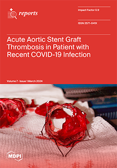
Cover Story (view full-size image): Intraoperative pictures of the tenacious adhesion of the residual clot to the fabric.
This higher density of clot, a typical aspect of COVID-19 thrombus, can explain the tenacious adhesion to the fabric of the stent graft and the difficulties in completely removing the clot with fibrinolytic infusion and mechanical thrombectomy. View this paper
  • Issues are regarded as officially published after their release is announced to the table of contents alert mailing list.
  • You may sign up for e-mail alerts to receive table of contents of newly released issues.
  • PDF is the official format for papers published in both, html and pdf forms. To view the papers in pdf format, click on the "PDF Full-text" link, and use the free Adobe Reader to open them.
Order results
Result details
Section
Select all
Export citation of selected articles as:
12 pages, 2339 KiB  
Case Report
Life-Threatening Obstetrical Emergency: Spontaneous Uterine Rupture Associated with Placenta Percreta in the First Trimester of Pregnancy—Case Report and Literature Review
by Mihaela Amza, Mihai-George Loghin, Didel-Ionuț Vâlcea, Nicolae Gică, Ileana-Maria Conea, Gabriel-Petre Gorecki, Alexandra Mirică, Romina-Marina Sima and Liana Pleș
Reports 2024, 7(1), 21; https://doi.org/10.3390/reports7010021 - 18 Mar 2024
Viewed by 2006
Abstract
Background: The greatest risk for the occurrence of the placenta accreta spectrum (PAS) is represented by uterine scars, which most frequently result after cesarean sections. Uterine rupture is a rare condition and appears mainly in the third trimester of pregnancy. The association between [...] Read more.
Background: The greatest risk for the occurrence of the placenta accreta spectrum (PAS) is represented by uterine scars, which most frequently result after cesarean sections. Uterine rupture is a rare condition and appears mainly in the third trimester of pregnancy. The association between these two conditions is extremely rare in the first trimester of pregnancy. Methods: We performed a systematic review of abnormal placental adhesions and spontaneous uterine ruptures in the first trimester of pregnancy. We also reported a case of spontaneous uterine rupture in a 12-week pregnancy that presented with massive hemoperitoneum and hemorrhagic shock. Results: A 33-year-old patient with two previous cesarean sections, at the twelfth week of pregnancy at the time to this visit to the emergency room, presented with syncope and intense pelvic–abdominal pain. A clinical examination and ultrasound scan established the diagnosis of hemoperitoneum and hemorrhagic shock. Surgical exploration was performed, uterine rupture was identified, and hemostasis hysterectomy was necessary. The histopathological results showed placenta percreta. There have been eight reported cases of spontaneous uterine rupture in the first trimester of pregnancy associated with PAS. In these cases, it was found that 62.5% of the patients had undergone at least one cesarean section in the past; in 75% of the cases, hysterectomy was performed; and, in 87.5% of the cases, the presence of placenta percreta was confirmed. Conclusions: A high rate of cesarean sections determines the increase in the incidence of placenta accreta spectrum disorders. The possible life-threatening complications caused by this pathology can be observed in early pregnancies. Full article
Show Figures

Figure 1

4 pages, 567 KiB  
Interesting Images
Late Inguinal Swelling: Don’t Judge a Book by Its Cover! An Unusual Case of Lymphocele
by Francesco Natale and Giovanni Cimmino
Reports 2024, 7(1), 20; https://doi.org/10.3390/reports7010020 - 11 Mar 2024
Viewed by 1385
Abstract
A 58-year-old man with a history of diabetes type I and chronic coronary syndrome who underwent coronary artery bypass grafting (CABG) 7 years before was admitted to a cardiology unit for unplanned cardiac catheterization because of dyspnea and chest pain at rest. Femoral [...] Read more.
A 58-year-old man with a history of diabetes type I and chronic coronary syndrome who underwent coronary artery bypass grafting (CABG) 7 years before was admitted to a cardiology unit for unplanned cardiac catheterization because of dyspnea and chest pain at rest. Femoral access was chosen because of the previous CABG and a vascular closure device (VCD) was used at the end of the procedure. Because of femoral artery rupture during VCD implantation, surgical vascular repair was performed. About 45 days later, the patient experienced a growing inguinal swelling at the site of vascular access in the absence of fever and clinical features of inflammation. The swelling became painful over time. Despite the most probable hypothesis of a hematoma, pseudoaneurysm, and inguinal abscess, a final diagnosis of lymphocele was made. Full article
Show Figures

Figure 1

9 pages, 2022 KiB  
Case Report
Anterior Uveitis Secondary to Avelumab and Pembrolizumab in a Patient with Metastatic Renal Cell Carcinoma—A Case Report
by Wei-Lun Chu, Kai-Chun Cheng, Pei-Kang Liu, Hung-Chi Lai, Kuo-Jen Chen and Yo-Chen Chang
Reports 2024, 7(1), 19; https://doi.org/10.3390/reports7010019 - 6 Mar 2024
Viewed by 1696
Abstract
We present an unusual case of uveitis secondary to avelumab and pembrolizumab in a 39-year-old Taiwanese male with stage IV clear cell renal cell carcinoma (ccRCC) and lung metastasis, who initially received pembrolizumab as his primary treatment. However, the patient experienced skin and [...] Read more.
We present an unusual case of uveitis secondary to avelumab and pembrolizumab in a 39-year-old Taiwanese male with stage IV clear cell renal cell carcinoma (ccRCC) and lung metastasis, who initially received pembrolizumab as his primary treatment. However, the patient experienced skin and liver immune-related adverse events (irAEs) after the seventh dose of pembrolizumab, which prompted a switch to avelumab. The patient began to experience gradual blurring of vision after completing the fifth cycle of avelumab immunotherapy. Ophthalmic examinations revealed findings consistent with bilateral anterior uveitis. Despite an initial lack of significant improvement with steroid treatment, the patient’s vision and inflammation improved upon discontinuation of avelumab. Due to the occurrence of uveitis, avelumab was switched back to pembrolizumab. However, three months after initiating pembrolizumab, the patient developed foggy vision and bilateral anterior uveitis with cystoid macular edema (CME). The administration of topical, oral, and subconjunctival steroids resulted in an improvement in vision and the resolution of CME, without the need to discontinue pembrolizumab. Over the subsequent eighteen months, there has been no recurrence of uveitis, and there is no evidence of relapse or further metastasis in his ccRCC. Full article
Show Figures

Figure 1

10 pages, 1865 KiB  
Case Report
Comparison between SARS-CoV-2-Associated Acute Disseminated Encephalomyelitis and Acute Stroke: A Case Report
by Yu-Xuan Jiang, Ming-Hua Chen, Yen-Yue Lin, Yung-Hsi Kao, Ting-Wei Liao, Chih-Chien Chiu and Po-Jen Hsiao
Reports 2024, 7(1), 18; https://doi.org/10.3390/reports7010018 - 1 Mar 2024
Viewed by 1868
Abstract
The neurological manifestations of severe acute respiratory syndrome coronavirus 2 (SARS-CoV-2) infection are underrecognized. Ischemic stroke and thrombotic complications have been documented in patients with SARS-CoV-2 infection. Acute disseminated encephalomyelitis (ADEM) associated with coronavirus disease 2019 (COVID-19) is rare but can occur; the [...] Read more.
The neurological manifestations of severe acute respiratory syndrome coronavirus 2 (SARS-CoV-2) infection are underrecognized. Ischemic stroke and thrombotic complications have been documented in patients with SARS-CoV-2 infection. Acute disseminated encephalomyelitis (ADEM) associated with coronavirus disease 2019 (COVID-19) is rare but can occur; the incidence of COVID-19-associated ADEM is still not clear due to the lack of reporting of cases. ADEM may have atypical stroke-like manifestations, such as hemiparesis, hemiparesthesia and dysarthria. The treatment strategies for ADEM and acute stroke are different. Early identification and prompt management may prevent further potentially life-threatening complications. We report a patient with SARS-CoV-2 infection presenting with stroke-like manifestations. We also make a comparison between demyelinating diseases, COVID-19-associated ADEM and acute stroke. This case can prompt physicians to learn about the clinical manifestations of SARS-CoV-2-associated ADEM. Full article
Show Figures

Figure 1

9 pages, 2739 KiB  
Case Report
Aggressive Vaccine-Induced Immune Thrombocytopenia and Thrombosis in a Young Woman with a Past Mild SARS-CoV-2 Infection
by Filippo Luciani, Maria Cristina Caroleo, Alfredo Zanolini, Lucio Taranto, Pino Pasqua, Alfredo Petrone, Manuela Colosimo, Roberto Cannataro and Erika Cione
Reports 2024, 7(1), 17; https://doi.org/10.3390/reports7010017 - 23 Feb 2024
Viewed by 2162
Abstract
Vaccine- induced immune thrombocytopenia and thrombosis (VITT) is a rare adverse event occurring after immunization with adenoviral vector-based vaccines against SARS-CoV-2. This life-threatening condition is characterized by thrombocytopenia, systemic activation of coagulation, and anti-platelet factor 4 antibodies, often resulting in extensive venous thrombosis. [...] Read more.
Vaccine- induced immune thrombocytopenia and thrombosis (VITT) is a rare adverse event occurring after immunization with adenoviral vector-based vaccines against SARS-CoV-2. This life-threatening condition is characterized by thrombocytopenia, systemic activation of coagulation, and anti-platelet factor 4 antibodies, often resulting in extensive venous thrombosis. Arterial thrombosis is less common and mainly affects the aorta, peripheral arteries, heart, and brain. Several cases of ischemic stroke have been reported in VITT patients, frequently being associated with large vessel occlusion (LVO). Here, we present a case of aggressive VITT in a 46-year-old woman with a past mild SARS-CoV-2 infection, who was admitted with a left-middle cerebral artery (MCA) territory stroke and thrombocytopenia eight days after her first dose of the ChAdOx1 nCoV-19 vaccine. The patient developed a diffuse arterial thrombosis with concomitant thrombotic events in the intrahepatic portal branches. The patient’s clinical condition worsened rapidly due to a significant enlargement of the ischemic cerebral lesion in the left hemisphere, cerebral herniation, and incipient hydrocephalus requiring decompressive neurosurgery with an unfavorable outcome. Our observations may be indicative of a stroke variant in VITT and highlight the diverse clinical manifestations of the syndrome. Full article
Show Figures

Figure 1

8 pages, 2226 KiB  
Case Report
Cystic Duct Remnant–Duodenal Fistula following Laparoscopic Cholecystectomy: A Case Report and Literature Review
by Aleksandra Polikarpova, Ngee-Soon Lau and David Yeo
Reports 2024, 7(1), 16; https://doi.org/10.3390/reports7010016 - 23 Feb 2024
Viewed by 1831
Abstract
Laparoscopic cholecystectomy is the most common procedure performed for the management of symptomatic gallstone disease. This, however, can be complicated by the formation of fistulous communications between the biliary tree and the gastrointestinal tract. This abnormal communication allows for the flow of bile [...] Read more.
Laparoscopic cholecystectomy is the most common procedure performed for the management of symptomatic gallstone disease. This, however, can be complicated by the formation of fistulous communications between the biliary tree and the gastrointestinal tract. This abnormal communication allows for the flow of bile and bowel contents between two systems (biliary system and intestine), which can cause abdominal pain, nausea, vomiting, and biliary sepsis. We would like to present a rare case of fistulous communication between the cystic duct stump and duodenum and outline possible contributing factors. The literature review describes the most common interventions for the management of fistulas with emphasis on ERCP and stent preferences to eliminate transpapillary pressure gradient, which directly contributes to fistula closure. Full article
Show Figures

Figure 1

5 pages, 4221 KiB  
Interesting Images
Significant Spontaneous Pneumomediastinum and Extensive Subcutaneous Emphysema in a COVID-19 Patient
by Arianna Gaspari, Francesca Carrieri, Matteo Villani and Elisabetta Bertellini
Reports 2024, 7(1), 15; https://doi.org/10.3390/reports7010015 - 19 Feb 2024
Viewed by 1776
Abstract
A 64-year-old man, who had no pre-existing health conditions, was admitted to the intensive care unit due to progressive shortness of breath resulting from COVID-19. Initially, the patient responded to non-invasive mechanical ventilation, which improved his breathing function. However, after six days, his [...] Read more.
A 64-year-old man, who had no pre-existing health conditions, was admitted to the intensive care unit due to progressive shortness of breath resulting from COVID-19. Initially, the patient responded to non-invasive mechanical ventilation, which improved his breathing function. However, after six days, his respiratory function worsened significantly, requiring invasive ventilation. Out of nowhere, the person experienced spontaneous pneumomediastinum and extensive subcutaneous emphysema. The next day, a spontaneous pneumothorax occurred and was successfully drained later. It became evident that there was extensive subcutaneous emphysema also. The CT scan of the chest confirmed the presence of spontaneous pneumomediastinum, but it did not have any effect on the patient’s hemodynamics. The physicians performed a bronchoscopy and imaging with a contrast medium, which ruled out any lesions to the trachea or esophagus. No other issues related to the problem were identified during the examination. Unfortunately, microscopic bullae of interstitial emphysema, or micro air leaks, are visible when they are an adequate size on computed tomography. Recent literature and studies not available during the case report have shown that if the Macklin effect is detected on the baseline CT scan, it could predict the occurrence of pneumothorax or pneumomediastinum. Full article
Show Figures

Figure 1

11 pages, 32571 KiB  
Case Report
Clinico-Morphological Features and Immunohistochemical Profile of a Rare Case of Three Synchronous Primary Malignancies in the Female Genital Tract
by Mădălina Boșoteanu, Raluca Ioana Vodă, Gabriela Izabela Balţǎtescu, Mariana Aşchie, Luana-Andreea Nurla and Cristian Ionuţ Orǎşanu
Reports 2024, 7(1), 14; https://doi.org/10.3390/reports7010014 - 17 Feb 2024
Viewed by 1646
Abstract
(1) Background: Synchronous tumors are defined as tumors that occur at the same time, appearing within 2 months, according to the Surveillance Epidemiology and End Results Program, with a frequency of tertiary tumors of 0.5%. The purpose of this presentation is to report [...] Read more.
(1) Background: Synchronous tumors are defined as tumors that occur at the same time, appearing within 2 months, according to the Surveillance Epidemiology and End Results Program, with a frequency of tertiary tumors of 0.5%. The purpose of this presentation is to report a case of three synchronous tumors of the female genital tract, given the fact that it represents a challenge both therapeutically and in demonstrating that the malignant lesions are completely different from each other. (2) Methods: We report the case of a 45 year-old patient diagnosed with three synchronous tumors developed in the genital tract: clear-cell ovarian carcinoma, uterine endometrioid adenocarcinoma, and cervical adenosquamous carcinoma. (3) Results: Total hysterectomy with bilateral anexectomy was performed and accompanied by a biopsy of the greater omentum. The evolution of the patient was favorable during chemotherapy, but she died two weeks after the completion of this treatment, from a cause secondary to the adverse effects determined by it, namely, severe thrombopenia which caused a massive lower digestive hemorrhage. (4) Conclusions: This case demonstrates the maximum importance of the involvement of adjuvant diagnostic techniques, especially when it comes to a diagnostic challenge with direct implications in the subsequent therapy of the patient. Full article
Show Figures

Figure 1

10 pages, 4162 KiB  
Case Report
Hidradenitis Suppurativa in Association with Ulcerative Proctitis: Surgical Management in a Refractory Case to Topical and Systemic Treatment
by Ioana-Valentina Stoenică, Mihai Cristian Dumitrașcu, Aida Petca, Răzvan-Cosmin Petca and Florica Șandru
Reports 2024, 7(1), 13; https://doi.org/10.3390/reports7010013 - 17 Feb 2024
Viewed by 1934
Abstract
Hidradenitis suppurativa (HS), a challenging dermatological condition, can be described as a chronic, painful, follicular, occlusive disease that is characterized by painful nodules, abscesses, and sinus tracts generally located in the intertriginous skin areas. This disorder places a considerable burden on affected individuals [...] Read more.
Hidradenitis suppurativa (HS), a challenging dermatological condition, can be described as a chronic, painful, follicular, occlusive disease that is characterized by painful nodules, abscesses, and sinus tracts generally located in the intertriginous skin areas. This disorder places a considerable burden on affected individuals and often leads to significant impairment in quality of life for those grappling with its persistent and recurrent nature. We present the case of a 20-year-old male patient known to have ulcerative proctitis and diagnosed with hidradenitis suppurativa in 2020, for which he underwent various topical and systemic treatments, with periods of remission and recurrent flares without managing to obtain complete remission of HS. In our dermatology service, the patient underwent two surgical unroofing procedures combined with punch debridement, with the wounds being allowed to heal by secondary intention. This choice of treatment delivered great results with favorable outcomes, without recurrence of the lesions, proving to be an effective method for managing HS. By presenting this case, we underline the role of surgical procedures in managing HS, and our desire is to emphasize the importance of comprehensive understanding of this enigmatic and complex condition for more effective management strategies in patients with refractory disease. Full article
Show Figures

Figure 1

8 pages, 4881 KiB  
Case Report
A Newly Reported Late Complication of Endoscopic Fundoplication: A Case Report
by Walid Bukhari, Hager Aref, Mohammed Ghunaim and Ahmed Elaryan
Reports 2024, 7(1), 12; https://doi.org/10.3390/reports7010012 - 9 Feb 2024
Viewed by 1793
Abstract
Endoscopic methods are emerging as a possible adjunct to surgery in the management of gastroesophageal reflux disease. Initially, endoscopic treatment has failed because of inefficient suturing devices, and although over many years it proved to be safe, it still can result in serious [...] Read more.
Endoscopic methods are emerging as a possible adjunct to surgery in the management of gastroesophageal reflux disease. Initially, endoscopic treatment has failed because of inefficient suturing devices, and although over many years it proved to be safe, it still can result in serious adverse events. In this article, the authors present a rare case of a male patient who experienced severe reflux years following endoscopic fundoplication. Moreover, in this report, we discovered an interesting finding with the patient who was diagnosed and managed laparoscopically. Although considered minimally invasive, endoscopic fundoplication can have significant deleterious consequences, and early recognition of these complications is vital to limit associated morbidities. Full article
Show Figures

Figure 1

9 pages, 635 KiB  
Case Report
Suspected Hematuria: Adverse Effects of Rivaroxaban in Older Adult Treated for Atrial Fibrillation
by Aleksandra Rapaić, Ekaterina Milošević, Nemanja Todorović, Nataša Janjić, Mladena Lalić-Popović and Nataša Milošević
Reports 2024, 7(1), 11; https://doi.org/10.3390/reports7010011 - 8 Feb 2024
Viewed by 3137
Abstract
Background: The modern concept of pharmaceutical healthcare implies monitoring the pharmacotherapy outcomes and reporting adverse drug reactions. Objective: To present a suspected hematuria as the adverse rivaroxaban reaction in a patient with atrial fibrillation observed by pharmacists in a community pharmacy. Case presentation: [...] Read more.
Background: The modern concept of pharmaceutical healthcare implies monitoring the pharmacotherapy outcomes and reporting adverse drug reactions. Objective: To present a suspected hematuria as the adverse rivaroxaban reaction in a patient with atrial fibrillation observed by pharmacists in a community pharmacy. Case presentation: A 69-year-old female patient came to a pharmacy with a prescription for cranberry-based supplement. She was diagnosed with a mild urinary infection after experiencing blood in her urine for about two weeks. The pharmaceutical anamnesis revealed that the patient was treated with irbesartan and rivaroxaban. Rivaroxaban was applied for atrial fibrillation, and the patient was treated for nine months. The patient was treated with omeprazole gastro-resistant capsules for mild dyspepsia and stomach ache over a three-week period. The pharmacist counselled the patient to contact the clinician who introduced rivaroxaban, further suggesting substitution with different anticoagulant. Although the urine culture was negative, the physician introduced ciprofloxacin, which was followed by blood in the patient’s stool. Thus, gastroscopy, colonoscopy, and gynecological examination were advised. All findings were normal. Four days after rivaroxaban was substituted with acenocoumarol, no blood in the urine or stool was detected. Conclusions: Rivaroxaban can cause spot urine blood even when applied in therapeutic doses among older female patients when applied with omeprazole. Possible rivaroxaban interaction with omeprazole metabolites is suspected and should be carefully monitored. Full article
Show Figures

Figure 1

7 pages, 1505 KiB  
Case Report
Multiple Cold Abscesses of a Chest Wall with Ribs Destruction in a Hemodialysis Patient
by Chih-Chun Kuo, Po-Jen Hsiao, Tai-You Kuo and Wen-Fang Chiang
Reports 2024, 7(1), 9; https://doi.org/10.3390/reports7010009 - 1 Feb 2024
Viewed by 2571
Abstract
Cold abscess of the chest wall is an uncommon disease that is mainly caused by tuberculous infection. Staphylococcal cold abscesses of the chest wall are extremely rare and usually clinically occult, frequently leading to a delay in diagnosis and consequently severe infectious complications. [...] Read more.
Cold abscess of the chest wall is an uncommon disease that is mainly caused by tuberculous infection. Staphylococcal cold abscesses of the chest wall are extremely rare and usually clinically occult, frequently leading to a delay in diagnosis and consequently severe infectious complications. We report an 88-year-old woman with end-stage renal disease, who presented with an exit site infection of a cuffed tunneled dialysis catheter caused by methicillin-resistant Staphylococcus aureus (MRSA). Despite the removal of the catheter and administration of antibiotics, she developed refractory MRSA bacteremia. Computed tomography (CT) of the chest revealed septic thrombophlebitis with metastatic cold abscesses of the chest wall and ribs destruction. Although CT-guided drainage and vancomycin therapy eliminated MRSA bacteremia, the chest wall abscesses did not resolve. Patients fitted with a central venous dialysis catheter are at risk of septic thrombophlebitis with metastatic cold abscesses of the chest wall that are resistant to antibiotic therapy. Early identification is based on serial blood cultures and prompt CT scans. Surgical management coupled with antibiotic therapy can eradicate the source of infection and improve patients’ outcomes. Full article
Show Figures

Figure 1

19 pages, 726 KiB  
Review
Epidemiology and Screening of Developmental Dysplasia of the Hip in Europe: A Scoping Review
by Emmanuela Dionysia Laskaratou, Anna Eleftheriades, Ioannis Sperelakis, Nikolaos Trygonis, Periklis Panagopoulos, Theodoros H. Tosounidis and Rozalia Dimitriou
Reports 2024, 7(1), 10; https://doi.org/10.3390/reports7010010 - 1 Feb 2024
Cited by 1 | Viewed by 2832
Abstract
Developmental hip dysplasia or developmental dysplasia of the hip (DDH) includes a wide range of deformities of the hip, such as congenital dysplasia, subluxation, and dislocation. It is usually identified through neonatal screening during the first 6–8 weeks of life. The incidence of [...] Read more.
Developmental hip dysplasia or developmental dysplasia of the hip (DDH) includes a wide range of deformities of the hip, such as congenital dysplasia, subluxation, and dislocation. It is usually identified through neonatal screening during the first 6–8 weeks of life. The incidence of DDH ranges from 1–7% in neonates among some populations, but this may vary among different ethnicities and countries. A consensus about the ideal age for screening has not been reached to date. The aim of this study is to summarize the existing data regarding the incidence of congenital hip dysplasia and screening tests among European countries. The authors conducted a systematic search in PubMed/Medline and Scopus and collected original studies published in English, French or German. The incidence of DDH presents fluctuations, not only among European countries, but also within the same country. There is no unanimity regarding the screening methods of DDH; in some countries, universal ultrasound is proposed as the basic screening method for neonates for DDH; in other countries screening is performed only in high-risk cases. More robust data are needed to conclude which screening approach is associated with improved long-term outcomes. Full article
Show Figures

Graphical abstract

11 pages, 2688 KiB  
Case Report
Wharton’s Jelly Tissue Allograft for Connective Tissue Defects Surrounding Nerves in the Tarsal Tunnel: A Retrospective Case Series
by Ronald Bruton, Tracie L. Gilliland, John J. Shou, Crislyn G. Woods, Naomi E. Lambert and Tyler C. Barrett
Reports 2024, 7(1), 8; https://doi.org/10.3390/reports7010008 - 30 Jan 2024
Viewed by 2446
Abstract
Caused by age or trauma, collapsed connective tissue can cause nerve entrapment and damage within the tarsal tunnel. Tarsal tunnel syndrome is relatively underdiagnosed. This study presents an intervention targeting damaged tissues surrounding the nerves and replacing the structural cushioning with a Wharton’s [...] Read more.
Caused by age or trauma, collapsed connective tissue can cause nerve entrapment and damage within the tarsal tunnel. Tarsal tunnel syndrome is relatively underdiagnosed. This study presents an intervention targeting damaged tissues surrounding the nerves and replacing the structural cushioning with a Wharton’s jelly tissue allograft. The eight patients in our study, selected from four clinical sites, had tarsal tunnel-related defects. Patient outcomes were tracked on a 90-day calendar utilizing the Numeric Pain Rating Scale (NPRS) and the Western Ontario and McMaster University Arthritis Index (WOMAC). All patients had failed standard care practices for at least six weeks. Each patient received a Wharton’s jelly tissue allograft to sites around the affected tarsal tunnel. No patients experienced adverse reactions. The percent change results calculated from the initial application to the 90-day follow-up showed an improvement of 59.43% in NPRS and a 37.58% improvement in WOMAC. This study provides evidence that WJ allograft applications are safe, minimally invasive, and efficacious for patients who have failed standard care treatments for tissue defects associated with tarsal tunnel syndrome. The limitations of this study include its small cohort size and nonblinded nature. The results of this study warrant further research to confirm the efficacy, optimal dose, protocol, and durability of Wharton’s jelly. Full article
Show Figures

Figure 1

12 pages, 1966 KiB  
Case Report
Surgical Treatment of Multiple Bone Cysts Using a Platelet-Rich Fibrin and BoneAlbumin Composite Graft: A Case Report
by Martin Major, Márton Kivovics, Bence Tamás Szabó, Tamás Déri, Melinda Polyák, Noémi Piroska Jákob, Dániel Csete, Attila Mócsai, Zsolt Németh and György Szabó
Reports 2024, 7(1), 7; https://doi.org/10.3390/reports7010007 - 22 Jan 2024
Viewed by 2226
Abstract
Promising research results have been obtained on the tissue-regeneration properties of PRF (platelet-rich fibrin) in dentistry and maxillofacial surgery. PRF presumably promotes healing and accelerates ossification. In this case report, the patient had a history of Gorlin–Goltz syndrome, also called nevoid basal cell [...] Read more.
Promising research results have been obtained on the tissue-regeneration properties of PRF (platelet-rich fibrin) in dentistry and maxillofacial surgery. PRF presumably promotes healing and accelerates ossification. In this case report, the patient had a history of Gorlin–Goltz syndrome, also called nevoid basal cell carcinoma syndrome, an autosomal dominant neurocutaneous disease that was known for many years. As a consequence, cysts were detected in both the mandible and maxilla. We performed decompression on this 37-year-old patient, followed by a cystectomy on an extensive lesion in the right angle of the mandible. One cyst from each side of the body mandible and one from the maxilla were completely enucleated, as determined using an intraoral exploration. The resulting bone defect was filled with a composite graft composed of a mixture of A-PRF and a serum albumin-coated bone allograft (BoneAlbumin). The wound was then covered with a PRF membrane. The surgical sites were closed per primam. The postoperative period was uneventful. Biopsies were performed after three and six months of healing for histological micromorphometry analyses. Dental implants were placed at the sampling site. Three months after the implantation, the ossified implants were fitted with superstructures. To date, no complications have appeared with the bone augmentation. The authors interpret from the findings that the combination of A-PRF and BoneAlbumin can be validated as a prosperous bone substitute. It can be safely implanted after a 3-month ossification period. Full article
(This article belongs to the Special Issue Clinical Research on Oral Diseases)
Show Figures

Figure 1

14 pages, 741 KiB  
Article
Coronary Artery Disease Is Related to Methylation Disorders Caused by the c.1286A>C MTHFR Polymorphism and to Low Serum 5-MTHF and Folic Acid Concentrations—Preliminary Results
by Agnieszka Pietruszyńska-Reszetarska, Robert Pietruszyński, Ireneusz Majsterek, Tomasz Popławski, Maciej Skrzypek, Beata Kolesińska, Joanna Waśko, Joanna Kapusta, Cezary Watała and Robert Irzmański
Reports 2024, 7(1), 6; https://doi.org/10.3390/reports7010006 - 17 Jan 2024
Viewed by 2222
Abstract
Background: Single nucleotide polymorphisms in gene encoding is the key enzyme in the folates pathway, methyltetrahydrofolate reductase (MTHFR), which causes methylation disorders associated with coronary artery disease (CAD). We evaluated associations between methylation disorders caused by MTHFR gene polymorphisms and the blood folate [...] Read more.
Background: Single nucleotide polymorphisms in gene encoding is the key enzyme in the folates pathway, methyltetrahydrofolate reductase (MTHFR), which causes methylation disorders associated with coronary artery disease (CAD). We evaluated associations between methylation disorders caused by MTHFR gene polymorphisms and the blood folate concentrations (folic acid, 5-MTHF) in CAD patients. Methods: Study group: 34 patients with CAD confirmed by invasive coronary angiography (ICA). Controls: 14 patients without CAD symptoms or significant coronary artery stenosis, based on ICA or multislice computed tomography (MSCT) with coronary artery calcification (CAC) scoring. Real-time PCR genotyping was assessed using TaqMan™ probes. Folic acid and 5-MTHF concentrations in blood serum were determined using Liquid Chromatography-Mass Spectrometry (LC-MS). Results: The c.[1286A>C];[1286A>C] MTHFR polymorphism occurred significantly more often in (CAD+) patients compared to the (CAD) cohort and to the selected general European “CEU_GENO_PANEL” population sample. The concentration of 5-MTHF and folic acid in subgroups of CAD+ patients with methylation disorders categorized by genotypes and CAD presence (CAD+) was always lower in CAD+ subgroups compared to non-CAD individuals (CAD). Conclusions: Further studies on a larger scale are needed to implicate the homozygous c.1286A>C MTHFR variant as CAD genetic marker and the 5-MTHF as CAD biomarker. Identification of high CAD risk using genetic and phenotypic tests can contribute to personalized therapy using an active (methylated) form of folic acid (5-MTHF) in CAD patients with MTHFR polymorphisms. Full article
(This article belongs to the Special Issue Recent Advancement in Cardiovascular Diseases)
Show Figures

Figure 1

7 pages, 806 KiB  
Case Report
Serious and Progressive Neuropathy Presumably Post-Shingrix Vaccination
by Michael J. Wons, Avani Vaghela, Amna Khalid and Benjamin D. Brooks
Reports 2024, 7(1), 5; https://doi.org/10.3390/reports7010005 - 13 Jan 2024
Viewed by 31035
Abstract
We present a case of serious and progressive neuropathy shortly following the administration of the herpes zoster subunit (HZ/su) vaccine, otherwise known as Shingrix. The progressive neuropathy occurred a week following the vaccination and progressed until discharge 16 days post-admittance. The patient’s mild [...] Read more.
We present a case of serious and progressive neuropathy shortly following the administration of the herpes zoster subunit (HZ/su) vaccine, otherwise known as Shingrix. The progressive neuropathy occurred a week following the vaccination and progressed until discharge 16 days post-admittance. The patient’s mild symptoms persist. The development of neuropathy following HZ/su administration is exceedingly rare, with an attributable risk of three cases per million vaccines administered. A black box warning was issued for this indication, although diagnosis and treatment were not confirmed for this patient. Reporting cases like this is crucial for a comprehensive understanding of vaccine risks and to characterize the underlying etiology of these serious adverse events. Full article
Show Figures

Figure 1

8 pages, 6193 KiB  
Case Report
Acute Aortic Stent Graft Thrombosis in Patient with Recent COVID-19 Infection
by Antonio Marzano, Jihad Jabbour, Vincenzo Brizzi, Enrico Sbarigia and Simone Cuozzo
Reports 2024, 7(1), 4; https://doi.org/10.3390/reports7010004 - 12 Jan 2024
Viewed by 2415
Abstract
Although COVID-19 primarily affects the respiratory system, it can have various effects on other organs, including the cardiovascular system. COVID-19 can lead to a prothrombotic status, promoting blood clotting, which can potentially affect native vessels and implanted devices. The exact mechanisms through which [...] Read more.
Although COVID-19 primarily affects the respiratory system, it can have various effects on other organs, including the cardiovascular system. COVID-19 can lead to a prothrombotic status, promoting blood clotting, which can potentially affect native vessels and implanted devices. The exact mechanisms through which it leads to increased clotting are not yet fully understood but may involve inflammation, endothelial dysfunction, and a hyperactive immune response. In the present report, we describe a case of acute aortic stent graft thrombosis four days after the resolution of SARS-CoV-2 infection. The patient required emergent explantation of the stent graft after the failure of endovascular bailout procedures. Full article
(This article belongs to the Special Issue Novel Aspects of COVID-19 after a Four-Year Pandemic)
Show Figures

Figure 1

9 pages, 1210 KiB  
Case Report
Infantile Hemangioma: Risk Factors and Management in a Preterm Patient—A Case Report
by Florica Sandru, Aida Petca, Andreea-Maria Radu, Andrei Gheorghe Preda, Alina Turenschi, Andreea Teodora Constantin and Raluca-Gabriela Miulescu
Reports 2024, 7(1), 3; https://doi.org/10.3390/reports7010003 - 1 Jan 2024
Viewed by 2874
Abstract
Infantile hemangiomas (IHs), boasting a prevalence ranging from 4% to 10%, stand as the most commonly encountered benign tumors during the early stages of human life. We present the case of a 2-year-9-month-old child who was born preterm with very low birth weight [...] Read more.
Infantile hemangiomas (IHs), boasting a prevalence ranging from 4% to 10%, stand as the most commonly encountered benign tumors during the early stages of human life. We present the case of a 2-year-9-month-old child who was born preterm with very low birth weight (VLBW), 1010 g birth weight, at 27 weeks gestational age. During pregnancy, her mother had anemia and needed cervical cerclage. On her 10th day of life, the appearance of a frontal hemangioma could be observed. The hemangioma was situated at the hairline. At the age of one month, another hemangioma could be observed on her right arm. The hemangiomas were treated with propranolol oral suspension for 10 months and afterwards with local ointment for 2 months. This choice of treatment delivered great results, with no adverse reactions reported. In this case report, we underlined the risk factors for IH, possible complications, and available treatment options. Full article
Show Figures

Figure 1

7 pages, 1304 KiB  
Case Report
Fatal Hemoptysis Secondary to Severe Pulmonary Veins Stenosis and Fibrosing Mediastinitis following Radiofrequency Ablation for Atrial Fibrillation: A Case Report and Review of the Literature
by Vladut Mirel Burduloi, Flavia Catalina Corciova, Gabriela Dumachita Sargu, Raluca Ozana Chistol, Alexandra Cristina Rusu and Cristinel Ionel Stan
Reports 2024, 7(1), 2; https://doi.org/10.3390/reports7010002 - 26 Dec 2023
Viewed by 1731
Abstract
Fatal hemoptysis secondary to severe pulmonary veins stenosis and fibrosing mediastinitis is an exceptional late complication of radiofrequency ablation for atrial fibrillation. We report the case of a 53-year-old male with a history of atrial fibrillation treated by radiofrequency ablation and admitted in [...] Read more.
Fatal hemoptysis secondary to severe pulmonary veins stenosis and fibrosing mediastinitis is an exceptional late complication of radiofrequency ablation for atrial fibrillation. We report the case of a 53-year-old male with a history of atrial fibrillation treated by radiofrequency ablation and admitted in our center 6 months after the procedure because of aggravating dyspnea and fatigability. Transthoracic echocardiography showed moderate dilation of right heart cavities, severe pulmonary hypertension and a turbulent flow in superior pulmonary veins. The cardiologist suspected pulmonary vein(s) stenosis and so cardiac computed tomography (CT) angiography was performed, with findings of severe stenosis of the right superior, right inferior and left inferior pulmonary veins, near-occlusion of the left superior pulmonary vein and the vein draining the apical segment of the right lower lobe. The CT scan also revealed soft tissue attenuation of the mediastinum posterior to the left atrium suggesting fibrosing mediastinitis together with parenchymal findings consistent with pulmonary veno-oclusive disease and an area of hemorrhagic infarction. Fatal hemoptysis occurred 3 days later, before treatment was attempted. In conclusion, severe pulmonary vein stenosis and fibrosing mediastinitis are rare but devastating complications of radiofrequency ablation. Prevention and early diagnosis are the key elements as these entities are potentially life-threatening. Full article
Show Figures

Figure 1

13 pages, 4474 KiB  
Article
Post-Burn and Surgical Scar Reconstruction with Tissue Expanders: Review of the Literature and Our Local Experience
by Ziyad I. Alharbi, Leena H. Moshref, Rahaf E. Badr, Ola A. Zahran, Maan T. Almaghrabi and Sherif F. Khamis
Reports 2024, 7(1), 1; https://doi.org/10.3390/reports7010001 - 21 Dec 2023
Viewed by 2312
Abstract
Tissue expansion (TE) is a rather widely accepted technique in plastic surgery, with variable indications; among numerous other indications, it is commonly used in scar management. In our research, we focused on the significance of TE as an important armamentarium and valid solution [...] Read more.
Tissue expansion (TE) is a rather widely accepted technique in plastic surgery, with variable indications; among numerous other indications, it is commonly used in scar management. In our research, we focused on the significance of TE as an important armamentarium and valid solution for the treatment of large and old post-burn scars, with good cosmetic and functional outcomes. Our study is a retrospective analysis conducted at Dr. Soliman Fakeeh Hospital, Jeddah, during the period from 2020 to 2023. This retrospective analysis included 15 tissue expanders, which were inserted in eight patients with large post-burn scars. Each patient underwent two stages of scar repair using variable-size tissue expanders. Post-operative assessment for scar quality and patient satisfaction was conducted using the POSAS score system in addition to standard documentation. Our study sheds light on the satisfactory and accepted cosmetic and functional outcomes for skin tissue expanders for post-burn scars and deformities. Full article
Show Figures

Figure 1

Previous Issue
Next Issue
Back to TopTop