Article

Electrospun Fe3O4-PVDF Nanofiber Composite Mats for Cryogenic Magnetic Sensor Applications

1
Department of Mechanical Engineering, University of North Texas, Denton, TX 76207, USA
2
Department of Material Science and Engineering, University of North Texas, Denton, TX 76207, USA
*
Author to whom correspondence should be addressed.
Textiles 2021, 1(2), 227-238; https://doi.org/10.3390/textiles1020011
Submission received: 22 May 2021 / Revised: 17 June 2021 / Accepted: 20 July 2021 / Published: 27 July 2021
(This article belongs to the Special Issue Fibrous Materials (Textiles) for Functional Applications)

Abstract

:
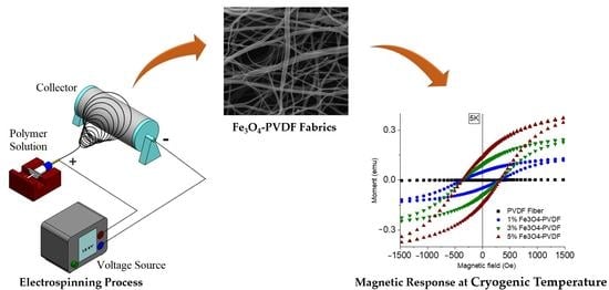
Magnetically responsive, mechanically stable and highly flexible iron (III) oxide-polyvinylidene fluoride (Fe3O4-PVDF) piezoelectric composite fiber mats were fabricated via one step electrospinning method for magnetic sensing at cryogenic temperature. The properties of Fe3O4-PVDF composite fiber mats were characterized using scanning electron microscopy (SEM), differential scanning calorimetry (DSC), X-ray diffraction (XRD), Fourier transform infrared (FT-IR) spectroscopy, d33 and magnetization test. The fiber diameter decreased as the concentration of Fe3O4 increased. The DSC results suggested a decrease in the crystallinity of the composite fiber mats after adding Fe3O4, and the XRD curves identified that the decrease in crystallinity took place in the β crystalline phases of the fibers. FT-IR results further confirmed the reduction of β phases of the composite fiber mats which dropped the piezoelectric response of the fiber mats by 38% for 5% Fe3O4-PVDF than PVDF fiber but still 400% higher than PVDF pellets. The magnetization test advocated a superparamagnetic state of the fiber at room temperature but a ferromagnetic behavior at a lower temperature. The coercivity values of the mats suggested a homogeneous dispersion of the Fe3O4 nanoparticles into the PVDF matrix. Young’s modulus (E) of the fibers remained the same before and after the magnetization test, indicating the mechanical stability of the fiber in the range of 5 K to 300 K. Its mechanical stability, superparamagnetic behavior at room temperature and ferromagnetic at low temperature could open up its application in spintronic devices at cryogenic temperature and cryogenic power electronic devices.

Graphical Abstract

1. Introduction

Magnetically responsive flexible fabrics are a growing need for shielding electromagnetic interference especially in microelectronic applications to reduce growing electromagnetic pollution, sheaths for portable electronics and stealth weapon systems in military applications [1,2]. These flexible fabrics are also useful for different biomedical applications like magnetic cell separation membranes [3], magnetic resonance imaging contrast agents [4], etc. Their application could also be extended to spintronics and power electronic devices at extremely low temperatures [5,6]. Iron (III) oxide (Fe3O4) nanoparticles are a valuable additive because of their electronic, magnetic, optical, and mechanical properties [7]. Composite nanofibers/thin films consisting of magnetic nanoparticles have been under intensive investigation in the last few years [7]. There remains a need to create flexible processable magnetically responsive and mechanically rugged films.
PVDF is a widely used piezoelectric semi-crystalline polymer with five different crystalline phases (α, β, δ, γ and ε) [8,9]. Its piezoelectric properties are directly related to its crystal phases [10]. Nonpolar α and polar β phase are the most common crystalline structures of PVDF where the β phase is responsible for the piezoelectric properties. Thereby, increasing β phase with eliminating α phase content in PVDF was a major area of focus for the last decade. Cold-drawing (stretching) [11], high-pressure quenching [12], and poling (applying a high electric field) of PVDF [13] were such processes to increase β phase. Electrospinning is another optional simple one-step method for fabricating PVDF nanofibers under a high electric field which converts the α phase into the β phase [14,15]. The high voltage that is involved in this process, boosts up the β phase of the fibers [16]. Moreover, electrospun fibers mats are highly flexible and mechanically strong compare to the solvent cast films [17].
Different filler materials have been introduced into the PVDF matrix for different functional properties such as Fe3O4, MnFe2O4 and MgFe2O4 for magnetic properties; graphene oxide, CNT for conductive properties, BaTiO3, PbTiO3, KNbO3 for piezoelectric properties [18]. Researchers prepared Fe3O4-PVDF nanocomposites films by solvent casting method. Porous systems have been produced using the phase inversion approach [19,20,21,22]. According to their reports, the magnetic property of Fe3O4-PVDF nanocomposites increased with an increase in Fe3O4 loading. However, their limitations were attributed to the poor dispersion of Fe3O4 nanoparticles and poor processability. The ruggedness of composites has also been limited at low temperature applications such as spintronics and power electronic devices at cryogenic conditions (<123 K) [5,6]. Thermal stresses paired with low temperature brittleness have been associated with this weakness. Iron oxide has proved valuable as an additive to polymers to increase low temperature retention of strength in aerospace applications [23]. For example, a modified diglycidyl ether of bisphenol-A (DGEBA) epoxy resin reinforced with carbon fiber showed excellent mechanical performance at cryogenic temperature after Fe3O4/GO nanofillers were added to it [24]. The filler material (Fe3O4/GO) helped the epoxy to retain its mechanical performance at cryogenic temperature.
The objective of this paper is to explore the viability of iron oxide-PVDF as electrospun textiles retaining mechanical and magnetic performance in cryogenic conditions. The effect of the addition of iron oxide on the polymer is explored through the investigation of the crystallinity and piezoelectricity of the composite fiber mats after the addition of Fe3O4 nanoparticles into the PVDF matrix. Magnetic response and mechanical properties were evaluated at room temperature and cryogenic temperature (5 K) to see the effect of cryogenic temperature on the performance of the fiber mats.

2. Materials and Methods

PVDF was obtained from Arkema (Kynar® 721, pellet form) with the following properties: density: 1.78 g/cm3, melt flow index (MFI): 10 g/10 min, tensile strength: 54 MPa, melting temperature: 168 °C. Fe3O4 was obtained from SkySpring Nanomaterials Inc. (Houston, TX, USA) with an average particle size of 20–30 nm. Acetone and N,N-dimethylformamide (DMF) were purchased from Sigma-Aldrich (St. Louis, MO, USA).

2.1. Preparation of Fe3O4-PVDF Fiber Mats

Firstly, 18 wt% (w/w) PVDF was dissolved in DMF/acetone (2:1) solution by stirring at 50 °C for 2 h using a magnetic stirrer in a 250 mL beaker at 200 rpm. After all the pellets dissolved, Fe3O4 nanoparticles (1 wt%, 3 wt% and 5 wt% (w/w) with respect to polymer) were added into the solution. Then, ultrasonication was performed with Sonic Vibracell II (25% pulse amplitude) for 5 min to disperse the Fe3O4 nanoparticles in the polymer solution. Next, 5 mL of this electrospinning solution was taken in a syringe and used for electrospinning. The electrospinning parameters were: voltage 15 kV, flow rate 0.05 mL/h, distance 15 cm, and the needle diameter of 0.038 in. At 400 rpm, a spinning roller with nonstick aluminum foil was capturing the fiber. The average thickness of the fabricated fiber mats was 0.1 mm.

2.2. Nanofiber Mats Characterization

Surface morphology of fiber mats was observed via FEI Nova Nano SEM 230 (Thermo Fisher Scientific, Hillsboro, OR, USA). All the samples were sputter coated to reduce the charge development on their surface. Using a Perkin Elmer DSC6 differential scanning calorimeter, the thermal properties of PVDF and Fe3O4-PVDF mats were investigated. The scanning temperature range was 30 °C to 210 °C at a rate of 10 °C per minute. Crystal structures were studied using a Rigaku Ultima III X-ray Diffractometer (Rigaku Americas Corporation, The Woodlands, TX, USA). A Scientific Nicolet 6700 Fourier Transform Infrared (FT-IR) Spectroscopy (Thermo Scientific, Waltham, MA, USA) with an attenuated total reflectance (ATR) detector was used to characterize the functional groups in PVDF and Fe3O4-PVDF mats. A d33 meter (APC International, Mackeyville, PA, USA) was used to measure the d33 value of the fiber. The fiber mats were rolled to make them thick enough for d33 testing. The d33 testing was done on 5 different spots of the fiber mats and average values were taken. Magnetic properties of the fiber mats were explored by a DynaCool-14 (Quantum Design, San Diego, CA, USA) Physical Properties Measurement System (PPMS) machine. Like in d33 testing, fiber mats were rolled and fitted into the coffee straws to do the magnetization testing. The weights of the samples used in the magnetization test were (0.1 ± 0.01) g.

3. Results

3.1. Scanning Electron Microscopy (SEM)

Scanning electron microscopy images of the fibers as a function of iron oxide concentration were analyzed using ImageJ software to calculate the diameter of electrospun fibers. The fiber diameter reduced gradually as the concentration of the Fe3O4 increased as shown in Figure 1 and Figure 2. The average fiber diameter reduced up to 26% for 5% Fe3O4-PVDF fiber. Similar results were obtained from electrospun PLA and PMMA fibers loaded with Fe3O4 nanomaterials [25]. At room temperature, Fe3O4 is a conductor material and adding the Fe3O4 particles into the PVDF solution increased the dielectric constant of the solution. A higher dielectric constant means higher polarity in the solution as well as a higher ability to stabilize the charges in the Taylor cone which leads to a decrease in nanofiber diameters [26]. O and Fe signals in the EDX chart of the fiber mats, shown in Figure 3, confirmed the presence of Fe3O4 in PVDF fiber. Au and Pd picks came from the sputter coated thin conductive film over the samples.

3.2. Differential Scanning Calorimetry (DSC)

The thermal behavior and crystallinity of the fiber mats were studied using the DSC. The results are presented in Figure 4a, where PVDF and all composite electrospun fiber mats showed a similar melting temperature (Tm) at ~160 °C, indicating no disruption in the PVDF thermal transitions. Furthermore, the melting enthalpy of the nanofiber mats was extracted from DSC curves and used to calculate the degree of crystallinity of the fiber mats based on Equation (1):
X c = H H 0 × 100
where Xc is the degree of crystallinity of the fiber mats; ∆H0 is the melting enthalpy of 100% crystalline PVDF (105 J/g), and ΔH is the melting enthalpy of Fe3O4-PVDF meshes. ΔH was corrected by multiplying it with the mass fraction of PVDF in the composite mats. The results, presented in Table 1, indicated a reduction in crystallinity and enthalpy as the magnetite content increases. For 5% Fe3O4-PVDF, the crystallinity of the fiber mats decreased by up to 55%. Previous experiments on Fe3O4-PVDF films yielded similar findings [14,27]. This may be linked to the partial inhibition effect of Fe3O4 addition on polymer crystal formation, similar to inorganic fillers like Al2O3, TiO2, c-LiAlO2, and Sm2O3 decreased the crystalline phase of PEO-based polymer electrolyte systems [28,29,30,31,32].

3.3. X-ray Diffraction (XRD)

The X-ray diffraction (XRD) patterns of PVDF and Fe3O4-PVDF nanocomposite fiber mats exhibited strong diffraction peak for all samples at = 20.5° corresponding to β(110) and β(200) planes, as shown Figure 4b. Weaker diffraction peaks at 36° assigned to β(201) planes were also present [33]. These results indicated that electrospun nanofiber membranes contain mainly β-phase crystal structure of PVDF. Due to the high electric voltage (15 kV) used during the electrospinning method, the random electric dipoles in the PVDF solution aligned, resulting in the forming of the β phase crystal structure. Thereby, the piezoelectric property increased. Also, with increasing loading of the Fe3O4 in the polymer matrix, the intensity of the peak at 20.5° decreased (7155, 5634, 4684 and 3401 a.u. for PVDF, 1%, 3% and 5% Fe3O4-PVDF respectively). So, the addition of the Fe3O4 into PVDF did not promote the crystallization of the PVDF in the β phase. This has been ascribed to inhibition of crystallization by the inorganic particles during the solidification process, which decreases the volume fraction of the crystalline phase in the fiber as discussed in the DSC section [34].

3.4. Fourier-Transform Infrared Spectroscopy (FTIR)

To confirm the XRD results, the authors compared the FTIR spectra of electrospun PVDF nanofiber and Fe3O4-PVDF nanofiber mats (Figure 5a). The PVDF pellet exhibited strong peaks at 615 cm−1 (CF2 bending and skeletal bending), 762 cm−1 (CF2 bending), 795 cm−1 (CF2 rocking), and 976 cm−1 (CH out-of-plane deformation) which were all recognized as α phase and relatively weak peaks at 840 cm−1 (CH2 rocking), 877 cm−1 (CF2 rocking), 1273 cm−1 (CF out-of-plane deformation) and 1402 cm−1 (CH2 scissoring) were considered as β phase [35,36,37]. Following electrospinning, the α peaks became weaker, while the β crystalline peaks grew stronger. Additionally, the percentage change in β phase can be determined using the following equation (Equation (2)) [35]:
F ( β ) = X β X α + X β × 100 = A β 1.26 A α + A β × 100
where, F ( β )   represents the β phase percentage of PVDF, A α   and   A β are their absorption bands at 763 and 840 cm−1.
F(β) of the PVDF fiber increased significantly by 68% than PVDF pellets due to the electrospinning method as shown in Table 2. With the addition of Fe3O4, the β phase decreased slightly compared to the β phase of pure PVDF fiber due to the partial inhibition effect as described in the DSC result discussion section and it kept decreasing as the loading of Fe3O4 increasing. The 5% Fe3O4-PVDF had the lowest F(β) compare to all other electrospun fiber mats but still that value is higher than PVDF pellet by 35%. So, the 5% Fe3O4-PVDF should have higher piezoelectric properties than the PVDF pellets which is discussed in d33 section.

3.5. Piezoelectric Coefficient (d33) Test

The piezoelectric coefficient (d33) of a material specifies the intensity of its piezoelectric effect. The greater the d33 value, the more effective the piezoelectric effect [38]. Figure 5b represents the piezoelectric coefficient d33 of the PVDF fiber mats as a function of Fe3O4 mass fraction. The d33 of PVDF mats was 32 pC/N and it dropped gradually to 20 pC/N as Fe3O4 increased to a 5% mass ratio. This result was in an agreement with FTIR data. The piezoelectric coefficient d33 is directly related to the β phase of the fiber mats. The β phase of the fiber decreased after adding Fe3O4 as recorded from FTIR data, which led to the decrease in the d33 value. It was also notable that the d33 of PVDF pellet was 5 pC/N as recorded in Table 2, whereas the lowest d33 value of electrospun fiber mat (5% Fe3O4-PVDF) was 20 pC/N; four times higher than PVDF pellets. So, adding Fe3O4 might reduce the piezoelectric property of the fiber mats but it has still four times higher piezoelectricity than the PVDF pellet.

3.6. Magnetization Test

M–H measurements were done at 300 and 5 K with the magnetic field varying from −2 T to 2 T (1 T = 10,000 Oe; 1 Oe = 79.577 Am−1) as presented in Figure 6. At 300 K (Figure 6a,b), the samples showed zero coercivity, indicating that the nanofiber mats were in the superparamagnetic state. However, samples showed ferromagnetic behavior at 5 K with a coercivity of ~370 Oe for all the samples as shown in Figure 6c,d. Superparamagnetic materials can fluctuate randomly by thermal fluctuation at high enough temperatures just as an atom spin in paramagnetic materials. When the temperature is low, this thermal energy decreases and the magnetic moments are blocked [39]. The temperature below this happens known as blocking temperature which is 290 K as shown in Figure 7. Below the blocking temperature, there is some net alignment of the particle spins, while above it, the spins are in random directions which caused the hysteresis loops to appear at 5 K and increased the magnetic saturation value as shown in Figure 6d and Table 3 [40]. Previous magnetization measurements also indicated that the Fe3O4 particles are superparamagnetic at room temperature and ferromagnetic at low temperatures [41]. So, the composites made of Fe3O4 particles show similar characteristics.
On the other hand, coercivity measures the resistance of a ferromagnetic material to become demagnetized. A similar coercivity for all the samples in our case indicated that the coercivity is independent of Fe3O4 nanoparticles loading, and a homogeneous distribution of nanoparticles in the polymer matrix [42]. PVDF fiber mats did not show any magnetic response at all on both the temperatures but the Fe3O4 nanoparticles loaded samples did. With the increase of Fe3O4 nanoparticles, the magnetic saturation value of the composites also increased (1.28, 3.29, 5.11 emu/g for 1%, 3% and 5% Fe3O4-PVDF respectively) which suggests that ferromagnetic behavior observed in the composites arose from the magnetic Fe3O4 nanoparticles.
The magnetic moment vs. temperature (M–T) graph as shown in Figure 7, the separation of zero-field-cooled (ZFC) and field-cooled (FC) curves occur at large temperatures (blocking temperature Tb~290 K) indicating thermal irreversibility of the samples below room temperature. In a superparamagnetic state around 5 K, when the samples were cooled at zero applied field, the magnetic moments were randomly oriented leading to the total magnetization to be around zero as shown in Figure 7. When the temperature started to increase, magnetic moments began to fluctuate, and the presence of an externally applied field aligned them that led to the increase in the magnetization. Similarly, in the case of the field-cooled sample, the magnetic moments were aligned at a higher applied field resulting in higher total magnetization at 5 K starting to decrease with heating. These observations are in agreement with previously reported studies on Fe3O4 nanoparticles incorporation in other materials, including ceramic matrices [43].

3.7. Dynamic Mechanical Analyzer (DMA)

To see the stability of the fiber mats, DMA was done for all the samples before and after the magnetization test. Since the magnetization test of fiber mats was done at cryogenic temperature (5 K), it was important to determine their mechanical stability after the test. Dynamic strain sweep was done for all the samples as shown in Figure 8. The Young’s modulus (E) calculated from the stress–strain curves are listed in Table 4. The retention of modulus following cryogenic temperature exposure in composite samples over the PVDF which shows a slight decrease indicates that Fe3O4 helped the PVDF matrix to retain its mechanical property after being exposed to cryogenic temperature.

4. Conclusions

Flexible and mechanically stable PVDF and Fe3O4-PVDF composite fiber mats were successfully prepared from one step electrospinning method. A decrease in the fiber diameter was revealed from SEM images as the mass fraction of Fe3O4 increased. DSC data suggested that the crystallinity of the composite fiber mats also decreased as a function of Fe3O4 concentration in the PVDF matrix. XRD curves identified the reduction in β crystalline phases of the fiber, causing the decrease detected in DSC results. Due to the reduction in the β crystalline phases, the piezoelectric response of the fiber mats dropped by 38% for the maximum Fe3O4 loading but that is still 4 times higher than PVDF pellets. The magnetization test advocated a superparamagnetic state of the fiber at room temperature but a ferromagnetic behavior at a lower temperature. The coercivity values of the mats suggested a homogeneous dispersion of the Fe3O4 nanoparticles into the PVDF matrix. Moreover, stable and reusable fibers mats were obtained from this process which was confirmed from DMA analysis. The overall results suggest the potential of using Fe3O4-PVDF fiber mats as a magnetically responsive textile in spintronics and power electronic devices at cryogenic conditions.

Author Contributions

Conceptualization, N.D.; Data curation, T.C. and D.B.; Formal analysis, T.C. and D.B.; Investigation, T.C.; Methodology, T.C.; Project administration, N.D.; Validation, T.C.; Writing—original draft, T.C.; Writing—review & editing, N.D. and D.B. All authors have read and agreed to the published version of the manuscript.

Funding

This research received no external funding.

Institutional Review Board Statement

Not applicable.

Informed Consent Statement

Not applicable.

Data Availability Statement

The data presented in this study are available within the article.

Acknowledgments

The authors would like to thank Torrance Walker of UNT for visualization work and to the Materials Research Facility (MRF) and Advanced Materials, Manufacturing Processing Institute (AMMPI) for characterization support.

Conflicts of Interest

The authors declare no conflict of interest.

References

  1. Zhan, Y.; Long, Z.; Wan, X.; Zhang, J.; He, S.; He, Y. 3D carbon fiber mats/nano-Fe3O4 hybrid material with high electromagnetic shielding performance. Appl. Surf. Sci. 2018, 444, 710–720. [Google Scholar] [CrossRef]
  2. Chiscan, O.; Dumitru, I.; Postolache, P.; Tura, V.; Stancu, A. Electrospun PVC/Fe3O4 composite nanofibers for microwave absorption applications. Mater. Lett. 2012, 68, 251–254. [Google Scholar] [CrossRef]
  3. Honda, H.; Kawabe, A.; Shinkai, M.; Kobayashi, T. Development of chitosan-conjugated magnetite for magnetic cell separation. J. Ferment. Bioeng. 1998, 86, 191–196. [Google Scholar] [CrossRef]
  4. Lutz, J.-F.; Stiller, S.; Hoth, A.; Kaufner, L.; Pison, U.; Cartier, R. One-Pot Synthesis of PEGylated Ultrasmall Iron-Oxide Na-noparticles and Their In Vivo Evaluation as Magnetic Resonance Imaging Contrast Agents. Biomacromolecules 2006, 7, 3132–3138. [Google Scholar] [CrossRef]
  5. Pelliccione, M.; Jenkins, A.; Ovartchaiyapong, P.; Reetz, C.; Emmanouilidou, E.; Ni, N.; Jayich, A.C.B. Scanned probe imaging of nanoscale magnetism at cryogenic temperatures with a single-spin quantum sensor. Nat. Nanotechnol. 2016, 11, 700–705. [Google Scholar] [CrossRef] [PubMed]
  6. Abderrahmane, A.; Ko, P.J.; Okada, H.; Sato, S.-I.; Ohshima, T.; Sandhu, A. Proton irradiation enhancement of low-field negative magnetoresistance sensitivity of AlGaN/GaN-based magnetic sensor at cryogenic temperature. IEEE Electron Device Lett. 2014, 35, 1130–1132. [Google Scholar] [CrossRef]
  7. Bajpai, O.; Setua, D.; Chattopadhyay, S. A brief overview on ferrite (Fe3O4) based polymeric nanocomposites: Recent developments and challenges. J. Res. Updat. Polym. Sci. 2014, 3, 184. [Google Scholar]
  8. He, F.; Fan, J.; Lau, S. Thermal, mechanical, and dielectric properties of graphite reinforced poly(vinylidene fluoride) composites. Polym. Test. 2008, 27, 964–970. [Google Scholar] [CrossRef]
  9. An, N.; Liu, H.; Ding, Y.; Zhang, M.; Tang, Y. Preparation and electroactive properties of a PVDF/nano-TiO2 composite film. Appl. Surf. Sci. 2011, 257, 3831–3835. [Google Scholar] [CrossRef]
  10. Samadi, A.; Hosseini, S.M.; Mohseni, M. Investigation of the electromagnetic microwaves absorption and piezoelectric properties of electrospun Fe3O4-GO/PVDF hybrid nanocomposites. Org. Electron. 2018, 59, 149–155. [Google Scholar] [CrossRef]
  11. Martins, P.; Lopes, A.C.; Lanceros-Mendez, S. Electroactive phases of poly(vinylidene fluoride): Determination, processing and applications. Prog. Polym. Sci. 2014, 39, 683–706. [Google Scholar] [CrossRef]
  12. Tasaka, S.; Miyata, S. Effects of crystal structure on piezoelectric and ferroelectric properties of copoly(vinylidenefluoride-tetrafluoroethylene). J. Appl. Phys. 1985, 57, 906–910. [Google Scholar] [CrossRef]
  13. Ting, Y.; Gunawan, H.; Sugondo, A.; Chiu, C.-W. A New Approach of Polyvinylidene Fluoride (PVDF) Poling Method for Higher Electric Response. Ferroelectrics 2013, 446, 28–38. [Google Scholar] [CrossRef]
  14. Chowdhury, T.; D’Souza, N.; Dahotre, N. Low-Cost Reliable Corrosion Sensors Using ZnO-PVDF Nanocomposite Textiles. Sensors 2021, 21, 4147. [Google Scholar] [CrossRef]
  15. Chowdhury, T.; D’Souza, N.; Ho, Y.H.; Dahotre, N.; Mahbub, I. Embedded Corrosion Sensing with ZnO-PVDF Sensor Textiles. Sensors 2020, 20, 3053. [Google Scholar] [CrossRef]
  16. Mansouri, S.; Sheikholeslami, T.F.; Behzadmehr, A. Investigation on the electrospun PVDF/NP-ZnO nanofibers for application in environmental energy harvesting. J. Mater. Res. Technol. 2019, 8, 1608–1615. [Google Scholar] [CrossRef]
  17. Ghosal, K.; Chandra, A.; Praveen, G.; Snigdha, S.; Roy, S.; Agatemor, C.; Thomas, S.; Provaznik, I. Electrospinning over Solvent Casting: Tuning of Mechanical Properties of Membranes. Sci. Rep. 2018, 8, 5058. [Google Scholar] [CrossRef] [PubMed] [Green Version]
  18. Jiang, P.; Lu, J.; Li, K.; Chen, X.; Dan, R. Research on hydrophobicity of electrospun Fe3O4/PVDF nanofiber membranes under different preparation conditions. Full Nanotub. Carbon Nanostruct. 2019, 28, 381–386. [Google Scholar] [CrossRef]
  19. Bhatt, A.S.; Bhat, D.K.; Santosh, M.S. Crystallinity, conductivity, and magnetic properties of PVDF-Fe3O4 composite films. J. Appl. Polym. Sci. 2010, 119, 968–972. [Google Scholar] [CrossRef]
  20. Jayakumar, O.D.; Mandal, B.P.; Majeed, J.; Lawes, G.; Naik, R.; Tyagi, A.K. Inorganic–organic multiferroic hybrid films of Fe3O4 and PVDF with significant magneto-dielectric coupling. J. Mater. Chem. C 2013, 1, 3710–3715. [Google Scholar] [CrossRef]
  21. Prabhakaran, T.; Hemalatha, J. Ferroelectric and magnetic studies on unpoled (Polyvinylidine fluoride)/Fe3O4 magnetoelectric nanocomposite structures. Mater. Chem. Phys. 2013, 137, 781–787. [Google Scholar] [CrossRef]
  22. Huang, Z.-Q.; Zheng, F.; Zhang, Z.; Xu, H.-T.; Zhou, K.-M. The performance of the PVDF-Fe3O4 ultrafiltration membrane and the effect of a parallel magnetic field used during the membrane formation. Desalination 2012, 292, 64–72. [Google Scholar] [CrossRef]
  23. Chen, D.; Li, J.; Yuan, Y.; Gao, C.; Cui, Y.; Li, S.; Liu, X.; Wang, H.; Peng, C.; Wu, Z. A Review of the Polymer for Cryogenic Application: Methods, Mechanisms and Perspectives. Polymers 2021, 13, 320. [Google Scholar] [CrossRef]
  24. He, Y.; Chen, Q.; Yang, S.; Lu, C.; Feng, M.; Jiang, Y.; Cao, G.; Zhang, J.; Liu, C. Micro-crack behavior of carbon fiber reinforced Fe3O4/graphene oxide modified epoxy composites for cryogenic application. Compos. Part A Appl. Sci. Manuf. 2018, 108, 12–22. [Google Scholar] [CrossRef]
  25. Zhang, C.C.; Li, X.; Yang, Y.; Wang, C. Polymethylmethacrylate/Fe3O4 composite nanofiber membranes with ultra-low dielectric permittivity. Appl. Phys. A 2009, 97, 281–285. [Google Scholar] [CrossRef]
  26. Sorayani-Bafqi, M.S.; Bagherzadeh, R.; Latifi, M. Fabrication of composite PVDF-ZnO nanofiber mats by electrospinning for energy scavenging application with enhanced efficiency. J. Polym. Res. 2015, 22, 130. [Google Scholar] [CrossRef]
  27. Xu, C.; Ouyang, C.; Jia, R.; Li, Y.; Wang, X. Magnetic and optical properties of poly(vinylidene difluoride)/Fe3O4 nanocomposite prepared by coprecipitation approach. J. Appl. Polym. Sci. 2009, 111, 1763–1768. [Google Scholar] [CrossRef]
  28. Croce, F.; Appetecchi, G.B.; Persi, L.; Scrosati, B. Nanocomposite polymer electrolytes for lithium batteries. Nat. Cell Biol. 1998, 394, 456–458. [Google Scholar] [CrossRef]
  29. Gang, W.; Roos, J.; Brinkmann, D.; Capuano, F.; Croce, F.; Scrosati, B. Comparison of NMR and conductivity in (PEP)8LiClO4+γ-LiAlO2. Solid State Ion. 1992, 53, 1102–1105. [Google Scholar] [CrossRef]
  30. Chu, P.P.; Reddy, M.J. Sm2O3 composite PEO solid polymer electrolyte. J. Power Sources 2003, 115, 288–294. [Google Scholar] [CrossRef]
  31. Weston, J.; Steele, B. Effects of inert fillers on the mechanical and electrochemical properties of lithium salt-poly(ethylene oxide) polymer electrolytes. Solid State Ion. 1982, 7, 75–79. [Google Scholar] [CrossRef]
  32. Wieczorek, W.; Such, K.; Wyciślik, H.; Płocharski, J. Modifications of crystalline structure of PEO polymer electrolytes with ceramic additives. Solid State Ion. 1989, 36, 255–257. [Google Scholar] [CrossRef]
  33. Sukitpaneenit, P.; Chung, T.-S. Molecular elucidation of morphology and mechanical properties of PVDF hollow fiber mem-branes from aspects of phase inversion, crystallization and rheology. J. Membr. Sci. 2009, 340, 192–205. [Google Scholar] [CrossRef]
  34. Leo, C.J.; Thakur, A.K.; Subba-Rao, G.V.; Chowdari, B.V.R. Effect of glass—Ceramic filler on properties of polyethylene oxide–LiCF3SO3 complex. J. Power Sources 2003, 115, 295–304. [Google Scholar] [CrossRef]
  35. Kim, M.; Wu, Y.; Kan, E.; Fan, J. Breathable and Flexible Piezoelectric ZnO@PVDF Fibrous Nanogenerator for Wearable Ap-plications. Polymers 2018, 10, 745. [Google Scholar] [CrossRef] [PubMed] [Green Version]
  36. Bormashenko, E.; Pogreb, R.; Stanevsky, O. Vibrational spectrum of PVDF and its interpretation. Polym. Test. 2004, 23, 791–796. [Google Scholar] [CrossRef]
  37. Salimi, A.; Yousefi, A.A. Analysis Method: FTIR studies of β-phase crystal formation in stretched PVDF films. Polym. Test. 2003, 22, 699–704. [Google Scholar] [CrossRef]
  38. Zhao, P.; Wang, S.; Kadlec, A. Piezoelectric and dielectric properties of nanoporous polyvinylidence fluoride (PVDF) films. Behav. Mech. Multifunct. Mater. Compos. 2016, 9800, 98000. [Google Scholar] [CrossRef]
  39. Goya, G.; Morales, M.D.P. Field Dependence of Blocking Temperature in Magnetite Nanoparticles. J. Metastab. Nanocryst. Mater. 2004, 20–21, 673–678. [Google Scholar] [CrossRef] [Green Version]
  40. Sara-Majetich, T.W.; Mefford, O.T. Magnetic Nanoparticles. MRS Bull. 2013, 38, 899–903. [Google Scholar] [CrossRef] [Green Version]
  41. Demir, A.; Topkaya, R.; Baykal, A. Green synthesis of superparamagnetic Fe3O4 nanoparticles with maltose: Its magnetic investigation. Polyhedron 2013, 65, 282–287. [Google Scholar] [CrossRef]
  42. Ramajo, L.A.; Cristóbal, A.A.; Botta, P.M.; Porto-López, J.M.; Reboredo, M.M.; Castro, M.S. Dielectric and magnetic response of Fe3O4/epoxy composites. Compos. Part A Appl. Sci. Manuf. 2009, 40, 388–393. [Google Scholar] [CrossRef]
  43. Berman, D.; Sha, Y.; Shevchenko, E. Effect of Polymer Removal on the Morphology and Phase of the Nanoparticles in All-Inorganic Heterostructures Synthesized via Two-Step Polymer Infiltration. Molecules 2021, 26, 679. [Google Scholar] [CrossRef] [PubMed]
Figure 1. SEM image of (a) PVDF (b) 1% Fe3O4-PVDF (c) 3% Fe3O4-PVDF and (d) 5% Fe3O4-PVDF nanofiber mats.
Textiles 01 00011 g001
Figure 2. Average fiber diameter for (a) PVDF, (b) 1% Fe3O4-PVDF, (c) 3% Fe3O4-PVDF and (d) 5% Fe3O4-PVDF nanofiber mats.
Textiles 01 00011 g002aTextiles 01 00011 g002b
Figure 3. Qualitative EDS analysis of (a) 5% Fe3O4-PVDF and (b) PVDF nanofiber mats.
Textiles 01 00011 g003
Figure 4. (a) DSC curves and (b) XRD curves for PVDF and Fe3O4-PVDF fiber mats.
Textiles 01 00011 g004
Figure 5. (a) FTIR spectra of PVDF and Fe3O4-PVDF fibers mats and (b) piezoelectric coefficient d33 of the fiber mats as a function of Fe3O4 mass fraction.
Textiles 01 00011 g005
Figure 6. Magnetic hysteresis curves of the samples as a function of the Fe3O4 nanoparticles concentration: (a) overall response of the samples at 300 K, (b) enlarged view of the hysteresis curve around zero field at 300 K, (c) overall response of the samples at 5 K, and (d) enlarged view of the hysteresis curve around zero field at 5 K.
Textiles 01 00011 g006
Figure 7. Zero-field-cooled (ZFC) and field-cooled (FC) at 1 T curves for the 5% Fe3O4-PVDF.
Textiles 01 00011 g007
Figure 8. DMA strain sweep curve for composite fiber mats (a) before magnetization test and (b) after magnetization test at 5 K.
Textiles 01 00011 g008
Table 1. Enthalpy and percentage crystallinity values obtained from DSC curves.
SampleTm (°C)H (J/g)Corrected ∆HXc (%)
PVDF Fiber160.1226.4026.4025.14
1% Fe3O4-PVDF Fiber160.4622.9122.6821.81
3% Fe3O4-PVDF Fiber160.3515.0714.6214.35
5% Fe3O4-PVDF Fiber160.1814.5213.7913.82
Table 2. Percentage of beta phase F(β) and piezoelectric coefficient (d33).
SampleF(β)d33 (pC/N)
PVDF Pellet48.45 ± 3.435 ± 3
PVDF Fiber81.16 ± 1.8132 ± 1.73
1% Fe3O4-PVDF73.39 ± 1.2428 ± 1
3% Fe3O4-PVDF71.69 ± 2.1626 ± 1
5% Fe3O4-PVDF65.26 ± 3.3920 ± 2.65
Table 3. Magnetic saturation values of composite fiber mats at 300 K and 5 K.
SamplePVDF1% Fe3O4-PVDF3% Fe3O4-PVDF5% Fe3O4-PVDF
emuemu/gemuemu/gemuemu/gemuemu/g
300 K000.151.280.283.290.445.11
5 K000.171.450.333.880.515.93
Table 4. Modulus of elasticity (E) before and after magnetization test.
SamplePVDF1% Fe3O4-PVDF3% Fe3O4-PVDF5% Fe3O4-PVDF
E Before magnetization test (MPa)0.11 ± 0.020.30 ± 0.030.62 ± 0.030.71 ± 0.01
E After magnetization test (MPa)0.09 ± 0.010.29 ± 0.020.62± 0.040.71 ± 0.03
Publisher’s Note: MDPI stays neutral with regard to jurisdictional claims in published maps and institutional affiliations.