Article

Photobehavior of an Acidochromic Dinitrophenyl-Hydrazinylidene Derivative: A Case of Total Internal Conversion

by
Letizia Mencaroni
1,
Alessio Cesaretti
1,*,
Giuseppe Consiglio
2,
Fausto Elisei
1,
Cosimo Gianluca Fortuna
2 and
Anna Spalletti
1
1
Department of Chemistry, Biology and Biotechnology and Center of Excellence on Innovative Nanostructured Materials (CEMIN), University of Perugia, Via Elce di Sotto n. 8, 06123 Perugia, Italy
2
Department of Chemical Sciences, University of Catania, Viale Andrea Doria 6, 95125 Catania, Italy
*
Author to whom correspondence should be addressed.
Photochem 2022, 2(4), 849-865; https://doi.org/10.3390/photochem2040054
Submission received: 31 August 2022 / Revised: 5 October 2022 / Accepted: 7 October 2022 / Published: 10 October 2022

Abstract

:
Research in photochemistry is always looking for novel compounds that can serve a role in applications ranging from medicine to environmental science. Push–pull compounds with protonable groups represent an interesting class of molecules in this sense, as they can prove to be sensitive to changes in both the acidity and polarity of the medium, becoming valuable as sensors and probes. Hence, in this work, a new dinitrophenyl-hydrazinylidene derivative with multiple protonable centers has been specifically designed and synthesized. The molecule showed an important acidochromism in the visible, with three differently-protonated species under acidic, neutral, and basic conditions, each characterized by a peculiar absorption spectrum. The photophysical characterization of this compound revealed an ultrafast excited-state deactivation, as described by femtosecond transient absorption experiments, and the hints of charge-transfer dynamics, as supported by the observed solvatochromism and quantum-mechanical calculations. These properties led to almost undetectable fluorescence that, together with negligible intersystem crossing and the absence of reactive pathways, points to the preference for a total non-radiative deactivation mechanism, i.e., internal conversion. This intriguing behavior stimulates interest in light of possible applications of the investigated acidochromic dye as a probe in photoacoustic imaging, which offers an alternative to classical fluorescence imaging.

Graphical Abstract

1. Introduction

Our long-term interest in the photobehavior of stilbenoid compounds has recently been addressed to the study of push-pull systems, bearing substituents consisting of different electron donor/acceptor groups [1,2,3,4,5,6,7,8], and with protonable centers, [9,10,11] owing to the sensitivity of their spectral and photophysical properties to the polarity and acidity of the medium. The latter behavior makes these compounds good candidates to be used in molecular optoelectronics [12] or as sensors and environment-sensitive probes [13,14,15,16].
Inter alia, the electron-withdrawing nitro group has been found particularly interesting [17,18,19,20,21], also because it would be an attractive substituent for electron-deficient conjugates and n-conducting materials [22,23,24,25]. On the other hand, compounds containing a hydrazone moiety, which can be seen as a donor group, [26], have been used in supramolecular chemistry as molecular switches [27], metallo-assemblies, and sensors [28].
The attachment of one or more nitro groups to the rings of an aromatic compound induces bathochromic spectral shifts, which is potentially an attractive means of shifting the optical absorption from the UV to the visible region of the spectrum and up to the near-infrared, very useful for applications in solar storage devices.
Usually, intersystem crossing (ISC) leading to triplet formation (from the first singlet excited state S1 to a quasi-isoenergetic upper triplet state and consequent fast internal conversion to T1) is the most prevalent and efficient decay pathway of excited nitro-derivatives [21,29,30,31]. The strength of the spin-orbit coupling determining the ISC quantum yield is related to the involved electronic states’ character, with ISC between excited states with different orbital nature being considerably more efficient than ISC between states with similar electronic configuration, as stated by the El Sayed’s rules [32,33].
Fast ISC and the consequent important fluorescence quenching cause the nitro-aromatics to have very short lifetimes (sub-ns/sub-ps) for the electronically excited singlet states. Such short lifetimes have made the use of nitro-aromatics impractical for photonic, optoelectronic, and particularly for efficient light-emitting applications and strengthened the search for fluorescent nitro compounds. The long-standing presumption about their non-fluorescent nature, although disregarded by several exceptions [18,21], led Gryko et al. to produce a review [34] that revisits the photochemical behavior of nitro derivatives outlining the guidelines for the synthesis of fluorescent systems.
However, very recently, non-emitting and photostable compounds have received particular attention for application purposes. Indeed, optical microscopy and imaging play an essential role in biological discovery, translational research, or clinical diagnosis [35,36,37,38], and fluorescent imaging is well-known and popularly used [39,40,41,42]. Nevertheless, due to photon scatter, the spatial resolution of optical methods falls drastically with increasing tissue depth, which limits the fluorescence visualization of fine biological details under the tissue surface [37].
In light of the above, the fine-tuning of optoacoustic imaging [43] offers an interesting alternative to conventional optical imaging methods, enabling high-resolution visualization in tissues with a penetration depth of several millimeters or centimeters [44]. Current advances in fast-tuning laser technology, image reconstruction, and spectral detection schemes have yielded real-time optoacoustic (photoacoustic) imaging with applications spanning from in vitro microscopy to human imaging [45].
This progress has generated an unprecedented need for photoacoustic imaging probes. Among others, small-molecule organic dyes with tailored physicochemical properties, such as significant water-solubility, high molar extinction coefficient, and great internal conversion quantum yield, have been studied for optoacoustic signal generation [45,46,47].
With a view to its potential as a photoacoustic probe, a dinitrophenyl-hydrazinylidene derivative has thus been scrutinized in this work for its photophysical behavior in both buffered organic solvent/water mixtures and solvents of different polarities. In fact, its molecular structure deserves special attention since the presence of two strong electron-withdrawing nitro-groups can foster excited-state charge-transfer dynamics, competing with fluorescence, thus favoring internal conversion, and its hydrazinylidene motif can lead to anti→syn and the reverse syn→anti isomerizations around the C=N bond, again resulting in a preferred non-radiative decay. Moreover, the investigated molecule shows some special appeal as a possible acid–base indicator in that it exhibits several protonable centers.

2. Materials and Methods

2.1. Synthesis

(E)-1-methyl-5-(2-(pyridin-4-yl)vinyl)-1H-pyrrole-2-carbaldehyde (0.202 g, 1.10 mmol) and (2,4-dinitrophenyl)hydrazine (0.198 g, 1.00 mmol) were dissolved in warm DMSO (1 mL) (Scheme 1). The resulting solution was stirred at room temperature for 48 h. Subsequently, diethyl ether (30 mL) was added under stirring and the resulting solid was filtered under vacuum and washed with fresh diethyl ether. After drying under vacuum, a brown solid was obtained (0.310 g, 79%). Yield: 0.310 g. 79%, brown powder. 1H NMR ([D6]DMSO) (Figure S1): δ = 4.00 (s, 3 H, NCH3), 6.77 (d, 3JH-H = 5.0 Hz, 1 H, pyrroleH), 6.83 (d, 3JH–H = 5.0 Hz, 1 H, pyrroleH), 7.11 (d, 3JH-H = 16.1 Hz, 1 H, CH=CH), 7.58 (m, 3 H, CH=CH + PyH), 7.92 (d, 3JH-H = 9.6 Hz, 1 H, ArH), 8.35 (d, 3JH-H = 9.6 Hz, 1 H, ArH), 8.52 (d, 3JH-H = 5.0 Hz, 2 H, PyH), 8.73 (s, 1 H, CH=N), 8.87 (s, 1 H, ArH) ppm; 13C NMR ([D6]DMSO) (Figure S2): δ = 36.1, 110.3, 114.8, 116.5, 123.0, 123.4, 129.1, 130.1, 130.3, 130.8, 130.9, 132.3, 138.8, 139.2, 144.5, 145.2, 149.6; MS (positive ESI): m/z = 393.2 [M + H]+. HRMS (positive ESI): calculated for C19H17N6O4+ 393.1306, found 393.1301.

2.2. Characterization

Elemental analyses were performed on a Carlo Erba 1106 elemental analyzer. ESI-MS spectra were recorded on a Thermo Fisher API 2000 mass spectrometer. High-resolution mass spectra were acquired on a Waters® SYNAPT® G2-S/Si mass spectrometer (Waters, Wilmslow, UK). Samples were infused at 5 μL min−1 and converted to gas-phase ions by electrospray ionization using the following parameters: source capillary voltage, 3.00 kV; source temperature, 80 °C; desolvation temperature, 300 °C. MS survey scanning conditions of 0.05 s were performed in resolution mode (20 K FWHM). Leucine enkephalin (m/z 556.2771 Da) was used as a lock mass compound and infused at 5 μL min−1. The reference sprayer was sampled periodically using the following settings: cone voltage, 3.00 V; scan time, 1 s; interval, 60 s; scan to average, 3; mass window, 0.5 Da. NMR experiments were performed at 27 °C on a Varian Unity S 500 (499.88 MHz for 1H) spectrometer. Tetramethylsilane (TMS) has been used as the internal reference for all NMR experiments.

2.3. Solvents

The compound Hy (shown in Scheme 1) was mainly investigated in aqueous solutions (w). The addition of some percentage (50%) of acetonitrile (AcCN) was necessary due to solubility issues. The different pHs for acid/base titrations were obtained by using Britton buffer solutions, pHs < 2 and > 12 were reached by adding HClO4 and NaOH, respectively. Toluene (Tol), anisole (An), dimethylsulfoxide (DMSO), tetrahydrofuran (THF), chloroform (CHCl3), and cyclohexane (CH) of spectroscopic grade purchased from Merck were also used. When AcCN was added to prepare aqueous-organic solvent mixtures, the actual pH of the final solution was corrected by applying a conversion parameter between pH scales as described by Gagliardi et al. [48].

2.4. Spectroscopic Stationary Techniques

Stationary absorption measurements were carried out by a PerkinElmer Lambda 800 spectrophotometer. A SPEX Fluorolog-2 F112AI spectrofluorimeter by HORIBA Scientific operated by FluorEssence was used for the fluorimetric measurements (absorbance < 0.1 at the excitation wavelength) employing tetracene (ϕF = 0.17 in air-equilibrated CH) [49] and 9,10-diphenylanthracene (ϕF = 0.90 in de-aerated CH) [50] as reference compounds. The eventual photoreaction was checked by irradiating with a high-pressure mercury lamp equipped with an interference filter at 436 nm and monitored spectrophotometrically.

2.5. Föster Cycle for Determining pKa*

Direct measurement of pKa* is usually performed by fluorimetric titration. In the case of the scarcely fluorescent system under investigation, an (approximate) estimation of pKa* was obtained by the indirect method proposed by Förster [51,52] and Weller [53,54,55]. pKa* can be estimated according to Equation (1) by knowing the pKa in the ground state and measuring the energy difference between the 0,0 transitions of the protonated and neutral forms (Δν).
The energy associated with the 0,0 transition of each species can be estimated from the intersection point between the normalized absorption and fluorescence spectra;
ΔpK = pKa* − pKa = 0.625 Δν(cm−1)/T

2.6. ns Flash Photolysis

Triplet properties were measured by laser flash photolysis (Edinburgh LP980) with a pump pulse centered at 355 nm (third harmonic of a Continuum Surelite II Nd:YAG laser, Spectra Physics) with nanosecond time-resolution (pulse width 7 ns and laser energy < 1 mJ pulse−1) coupled with a PMT for signal detection. The T-T extinction coefficients (εT) were obtained by energy transfer from benzophenone in AcCN (6500 M−1 cm−1 at 520 nm) [56].

2.7. fs Transient Absorption Measurements

The experimental setup for ultrafast transient absorption experiments has been previously reported [1,5,10,57]. The 400-nm-excitation pulses (ca. 60 fs) were generated by an amplified Ti:Sapphire laser system (Spectra Physics). The Helios transient absorption spectrometer (Ultrafast Systems) is characterized by a temporal resolution of about 150 fs and a spectral resolution of 1.5 nm. A small portion of the 800 nm light passes through an optical delay line (time window of 3200 ps) and is focused onto a sapphire crystal (2 mm thick) to generate white light in the 450–800 nm spectral range (probe pulse). All measurements were carried out under the magic angle in a 2 mm cell at an absorbance of about 0.5 at 400 nm (concentration ≈ 2 × 10⁻⁴ M). The solution was stirred during the experiments to avoid photoproduct interferences. Photodegradation was checked by recording the absorption spectra before and after the time-resolved measurement, where no significant change was observed.
Transient absorption data were analyzed using the Surface Xplorer PRO (Ultrafast Systems) and GloTarAn software. The former performs Singular Value Deconvolution of the 3D matrix to extract principal components (spectra and kinetics) and successively Global Analysis in order to obtain the lifetimes and Decay Associated Spectra (DAS) of the detected transients. GloTarAn was instead used to perform a Global Analysis model that assumes successive steps to describe the evolution of transients providing the Evolution Associated Spectra (EAS) [58,59].

2.8. Computational Details

Quantum–mechanical calculations were carried out using the Gaussian 16 package [60]. Density functional theory (DFT) based on the CAM-B3LYP method was used to optimize the geometry and to obtain the properties of the substrates in the ground state, while the lowest excited singlet states were characterized by time-dependent (TD) DFT (CAM-B3LYP) excited-state calculations [61,62,63]. In particular, the ground state of the differently-protonated forms of the investigated molecule was optimized by CAM-B3LYP/6-31G(d), while transition energies and probabilities of the excited singlet states were obtained by TD-DFT CAM-B3LYP/6-311G + (2d,p). AcCN solvation effects were included in the calculations by means of the conductor-like polarizable continuum model (CPCM) [64].
The spectrophotometric titrations were analyzed by a global fitting procedure using the React-LabTM Equilibria Program.

3. Results

3.1. Acidochromism

3.1.1. Spectral Properties

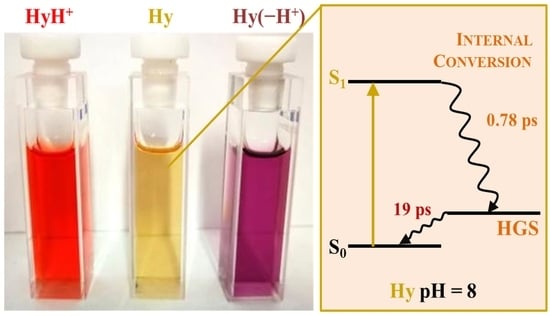
The absorption spectrum of the studied compound Hy was recorded in AcCN/w (50:50, v/v) mixture buffered at different pHs in the 1−13 pH range. A peculiar behavior was observed: the spectrum recorded in a neutral medium (red trace) showed red shifts either upon increasing or decreasing the acidity of the solvent (Figure 1) in the investigated pH range. In particular, the solution of the compound appeared yellow at pH 7.0, with its absorption spectrum peaked at 450 nm. The absorption maximum underwent red and hyperchromic shifts by lowering the pH, with the final absorption band (pH 1) centered at 495 nm and the solution acquiring an intense red color. The bathochromic shift was even more pronounced when increasing the basicity of the medium up to pH 12.7, where the absorption maximum was recorded at about 520 nm and the solution turned purple. However, an interesting thermal reaction was observed at pH < 1.

3.1.2. pKa Determination

The spectrophotometric titration followed at 495 nm, where the biggest spectral changes were observed, evinced two inflections due to the establishment of two acid–base equilibria in the ground state (Figure 2). The best fitting of the data at 495 nm provided the two pKa reported in the fourth column of Table 1. A global fitting procedure of the whole spectral data matrix using the React-LabTM Equilibria Program gave the molar absorption coefficients and the percentage concentration profiles, as a function of pH, of three different species (shown in Figure 3) and more reliable pKa values, reported in the fifth column of Table 1. The different species were identified with the help of a computational approach using the ACS/pKa DB program. Though approximated, these calculations, which use Hammet-type equations and electronic substituent constants to predict pKa values for ionizable groups, are a useful tool for understanding the sites involved in the protonation equilibria and their order (Scheme 2). The neutral molecule (Hy, red spectrum in Figure 3 with λmax = 451 nm) is the most stable species in the 5−10 pH range, while the predominant species at pH > 12 is the anion (Hy(−H+), black trace in Figure 3 with λmax = 520 nm) formed by losing the proton at the hydrazone moiety (see Scheme 2). The acid–base equilibrium observed in the acidic solution is due to the protonation of the pyridine ring (HyH+, green spectrum in Figure 3 with λmax = 496 nm). Moreover, the calculations predicted a further protonation of the molecule at negative H0 (see the formula at the right in Scheme 2) not experimentally detectable because of a fast thermal reaction taking hold at pH < 1. The three calculated pKa values are reported in the second to last column of Table 1. The first value (12.7 ± 0.5) is in fair agreement with the experimental measurements; conversely, the second (5.9 ± 0.3) was found to be higher than the one experimentally determined (3.5). An acid–base equilibrium has thus been experimentally observed among three species, whose concentrations vary on the basis of their pH range stability, as shown in the right graph of Figure 3.

3.1.3. Thermal Reaction

A fast thermal reaction accompanied by major spectral changes was observed at pH < 1 as shown by the temporal evolution of the spectrum at pH = 0.3 reported in Figure 4. A pseudo-first order reaction was evidenced by the linear trend of lnA against time at two different analysis wavelengths (400 and 495 nm, see Figure S3 and the inset of Figure 4). This thermal reaction was found to be accelerated by increasing the acidity of the medium. Eventually, albeit more slowly, it also took place at pH = 1.8 where the solution moved toward the thermal products if left in the dark for several days. The spectral evolutions obtained at pH = 0.3 and H0 = −1, followed by a diode array spectrophotometer for more reliable measurements as is the case with fast kinetics, and the single-wavelength fitting of lnA vs t are shown in Figures S4–S7. Table 2 collects all the kinetic results and related parameters.
The k values found at the two pH values with the same technique point to a thermal reaction of pseudo-first order five times faster at H0 = −1 than at pH = 0.3, which parallels the proton concentration, being [H+] ≅ 2.5 M and = 0.5 M, respectively. Therefore, a value of 2.7 × 10⁵ M−1 s−1 for the second-order kinetic constant k2 could be calculated.
It has to be noted that it was not possible to characterize the thermal product formed at pH ≤ 0.3 because the scarce solubility of the investigated compound Hy in AcCN/w mixtures prevented a sufficient quantity of the product to be stored for the necessary measurements. An attempt to follow the same thermal reaction in CHCl3, which allows greater Hy solubility, adding trifluoracetic acid to increase the acidity of the medium, proved unsuccessful (Figure S8). Indeed, no thermal reactions were triggered in CHCl3, and the spectral evolution with increasing [H+] probably points to a further protonation that leads to the bi-protonated species, predicted by calculations but not observed in water solutions because of the presence of the thermal reaction.
A plausible hypothesis (Scheme 3) about this fast thermal reaction was formulated on the basis of literature data [65] which reports, for hydrazone analogs at very low pH, protonation of the hydrazine unit followed by a nucleophilic attack of water on the carbon of C=N, which results in the fragmentation of the molecule into dinitrophenylhydrazine and the corresponding aldehyde. This speculation is in agreement with the fact that the reaction does not proceed in the absence of water, as is the case with CHCl3.
The definitive confirmation of this reaction path came from the comparison of the spectral evolution triggered by the thermal reaction with the absorption spectra of the hypothesized pure products. In fact, the absorption spectrum of 2,4-dinitrophenylhydrazine overlaps the band growing at 398 nm during the thermal spectral evolution. Conversely, the absorption band of the corresponding aldehyde falls in a higher energetic UV region of the spectrum (maximum at 320 nm and low molar extinction coefficient) and thus does not contribute significantly to the total absorption of the solution, causing the system to behave as a two-component system with a clear isosbestic point.

3.1.4. pK* Determination

Figure 1 shows that the absorption spectrum of the mono-protonated species HyH+ shifts towards longer wavelengths if compared with the neutral compound Hy, thus entailing an increase of basicity in the excited state according to the Förster cycle (Equation (2)):
pKa *   =   pKa   0.625   ×   Δ ν ¯ T
where pKa* is the pKa in the excited state and Δ ν ¯ is the difference between the 0,0 transition energies of the involved species. The latter was estimated by the calculated electronic transitions (see Tables S1–S3) since the experimental determination was not possible due to the lack of reliable fluorescence spectra for all the involved species. Moreover, a larger red shift was observed for the anion Hy(−H+) that points to a pKa* < pKa. The pKa* values computed through Equation (2) and reported in the last column of Table 1 indicate that the neutral Hy species that largely prevails in the ground state is expected to be not thermodynamically stable in the singlet excited state, with the excitation favoring the anion or the monoprotonated species depending on the pH range considered, according to the predicted pKa* values. The experimental observation of the anticipated excited-state acid–base re-equilibration was indeed prevented by the almost complete absence of fluorescence signals. However, it is unlikely that this process could take place because of the extremely short excited-state lifetimes assessed by ultrafast transient absorption spectroscopy, as described below.

3.1.5. Photobehaviour

The fluorescence quantum yield measured as a function of the pH medium is reported in Table 3. The investigated compound was found to be very poorly emitting in any case: a slight increase of ΦF was observed for the monoprotonated species HyH+, while the emission spectrum became totally undetectable for the anion Hy(−H+). A better ability to fluoresce (ΦF higher by two orders of magnitude) was instead found for the product of the thermal reaction observed at pH < 1.
The fluorescence excitation and emission spectra for the thermal product are reported in Figure S9, together with its absorption for comparison. The lack of overlap between the excitation and absorption spectra is not negligible and could point to the presence of some absorbing but not emitting species in the solution.

3.1.6. Transient Absorption (fs Domain)

The almost negligible fluorescence yields usually unveil very short lifetimes for the lowest excited singlet state, where the radiative pathway cannot compete with very fast non-radiative decay. The pump-probe transient absorption measurements, obtained by ultrafast techniques with an excitation laser light centered at 400 nm, shed some light on the decay dynamics of the excited states of the investigated compound Hy and its thermal product. In the case of the monoprotonated form, the signal was too low to collect reliable data. The obtained results are shown in Figure 5 and collected in Table 4.
In Figure 5, the upper panel (Panel A) contains the contour plot of the 3D experimental matrix; representative spectra at different delay times and representative kinetics are reported in the central panel (Panel B); the lower panel (Panel C) displays the Evolution-Associated Spectra (EAS) of the transient species resulting from Global Analysis with their relative lifetimes.
As a rule, with the only exception of the experiments conducted on the product of the thermal reaction, the measurements are dominated by positive signals of excited-state absorption (ESA) and the decay occurs very quickly, in line with the very low fluorescence quantum yields. The hint of negative signals recorded at λ < 500 nm can be associated with the ground-state bleaching (GSB) of the compound, while the negative band centered at 520 nm and observed in the case of the product is due to stimulated emission (SE), in agreement with the significant steady-state fluorescence only detected for this species.
The data analysis provided two or three transient species: the time of about 100 fs represents the inertial solvation (Solv.i) involving the vibrational and librational modes of the solvent around the excited molecules; the second transient observed in the case of the thermally-formed product (continuous green spectrum in Figure 5, right), considering its spectral form and lifetime, can be traced back to diffusive solvation (Solv.d); the main transient associated with the largest ΔA, whose lifetime ranges from fraction/few picoseconds in neutral or basic AcCN/w mixtures to ten of picoseconds in the case of the product, was instead assigned to S1 decay. In particular, the S1 lifetime of the product is larger than that of the other forms by more than one order of magnitude, in agreement with its higher fluorescence quantum yield.
In the peculiar case of neutral Hy at Ph 8, a further transient was found, characterized by an extremely small signal and a short lifetime of 19 ps. Its spectral profile features a negative signal in the region of GSB and an absorption band at its red edge, at higher energies relative to the ESA absorption of S1. These characteristics allow this minor transient to be assigned to a vibrationally Hot Ground State (HGS), which would be formed from the S1 decay to later return to the relaxed ground state with the typical time of vibrational relaxation [66].

3.2. Quantum-Mechanics Calculations

The investigated compound can exist in principle as an equilibrium among different conformers due to the free rotation around the quasi-single bonds between aromatic rings and double bonds [67,68]. A detailed conformational analysis is beyond the scope of this paper; nevertheless, very different photobehaviors are not expected for these structures. Quantum mechanical calculations were therefore performed only on the neutral, anionic, and monoprotonated conformer shown in Scheme S1.
The optimized ground state geometry of the three prototypic forms calculated by TD-DFT CAM-B3LYP/6-311G+(2d,p) in AcCN (Scheme S1) appears rather planar with the only exception of the nitro-group in the ortho position that deviates from the benzene plane by about 40 degrees. The quantum-mechanics calculations predicted a main bathochromic band together with other lower-intensity hypsochromic transitions for the neutral, monoprotonated, and anionic species (Tables S1–S3). They provided energies for the S0→Sn transitions in fair agreement with the experimental absorption spectra (Figure 3). The dominant configuration in the first transition is always the HOMO-LUMO. In the case of the neutral molecule Hy, there is a clear charge transfer towards the nitro-groups upon absorption, as shown in Scheme 4, Schemes S2 and S3. For the monoprotonated species HyH+, a greater charge delocalization in the excited state emerged due to the presence of the protonated pyridinium unit, which acts as a strong electron-acceptor, in addition to the dinitro-benzene placed on the other side of the system (Schemes S4 and S5). The anion Hy(−H+) features some charge movement as well, particularly towards the coplanar nitro group in the para position, better conjugated to the benzene ring (Schemes S6 and S7).

3.3. Solvatochromism

Spectral Properties and Photobehaviour

The normalized absorption spectrum of Hy in solvents of different polarities is reported in Figure 6. A modest positive solvatochromism was evidenced, with the spectrum showing a red shift of its maximum (from 445 to 475 nm) by increasing the solvent polarity. A shoulder around 390 nm, less influenced by the solvent, is also present.
As for the emission properties, low-intensity broad emission spectra were recorded for Hy in all the investigated solvents, and the representative example of Tol is shown in Figure S10. The fluorescence quantum yield remains very small independently of the medium, but it seems to further decrease in polar solvents, becoming undetectable in THF and DMSO (Table 5). The kF values indicate the fairly allowed nature of the emitting state in the non-polar investigated media, while the emission transition becomes less allowed in polar AcCN.
Ns-laser flash photolysis measurements pointed to a scarce triplet production both in polar and non-polar solvents despite the presence of two nitro groups that usually increase the ISC rate constant by El-Sayed rules. This behavior indicates that n,π* states are highly energetic and thus do not play an important role in the deactivation paths of the lower excited states. In fact, a small transient absorption was detected with a maximum at 560 and 510 nm in de-aerated DMSO and An, respectively, possibly assigned to the triplet state with very low ΦT × εT values ≅ 100 M−1 cm−1 (Figure S11).
The absorption spectrum of the investigated compound was found not to change under photoexcitation at room temperature; some very little photodegradation was only observed when leaving the mixture in the sunlight for several days. The photoisomerization around the C=C double bond is probably not efficient due to a high torsional energy barrier. As to the syn-anti isomerization around the C=N double bond, it is well known that the photochemically out-of-plane anti→syn rotation through a “perpendicular” form is often followed by a fast thermal syn→anti in-plane nitrogen inversion through a “linear” transition state. [27,69,70] Finally, the photoisomerization followed by reverse fast thermal isomerization results in not observing any photoreactions at high temperatures. In conclusion, the singlet state reached by light absorption seems to mainly decay through internal conversion, restoring its ground state.
Fs-transient absorption measurements were also carried out in solvents of different polarities (Figure 7, Figure S12 and Table 6). Independently of the medium considered, the signal was always dominated by a broad positive band assigned to ESA, which fast decays in a few picoseconds. The analysis always revealed two or three transients necessary to describe the decay of this excited state. As already discussed above, the ultrafast transient with a lifetime of the order of 100 fs represents the inertial solvation involving the vibrational and librational modes of the solvent. The lifetime of about1 ps, which is always peculiar to the most important component, is assigned to the S1 decay, and the longest transient, with a lifetime ranging from 5.3 ps in DMSO to 16 ps in Tol, features a very small positive signal which can be assigned to the absorption of a vibrationally HGS. However, when dealing with this transient, the contribution of a fast thermal reverse syn→anti nitrogen inversion cannot be ruled out.
The decay kinetics of S1 is slightly slowed down in less polar solvents in agreement with a partial charge transfer character for this state: in fact, polar solvents can assist charge separation favoring non-radiative de-excitation pathways. However, the extremely short lifetimes detected in all the investigated media imply a certain charge-transfer character for the S1 state, even in a non-polar solvent like Tol. This notable charge-transfer dynamic is indeed granted by the presence of the two strong electron-acceptor nitro groups, as also corroborated by the results of the quantum-mechanical calculations. This eventually leads to a relaxed charge-transfer S1 state, which prevents the population of inaccessible triplet states and thus decays almost entirely by internal conversion.

4. Discussion

In the search for novel photoacoustic imaging and/or acidochromic probes, the dinitrophenyl-hydrazinylidene derivative under investigation, Hy, needs careful consideration. This newly-synthesized molecule has been specifically designed to fulfill the required criteria of following non-radiative decays and featuring multiple protonable centers. In particular, the molecule was found not to have any net charge at neutral pH, while the pyridine ring could be easily protonated in an acidic environment, with a pKa ≈ 3.4; conversely, at pH > 11 the anion became the most stable form. Interestingly, all of the three differently-protonated species were characterized by peculiar absorption properties, with their colors changing from yellow at pH 7 to red and purple as a consequence of increasing acidity or basicity, respectively, thus appointing the compound as a good acidochromic probe in both low and high pH ranges. A second protonation, predicted by the calculations and involving the hydrazone, was not observed because of a fast acid-catalyzed reaction operative in water leading to the fragmentation of the molecule and the formation of dinitrophenylhydrazine as a product. The differently-protonated species and the thermally-formed product were studied for their photobehavior, and unimportant fluorescence was detected for the monoprotonated, neutral, and deprotonated forms, while a small but non-negligible emission was measured for the product. This is in agreement with the charge transfer process, predicted by quantum-mechanical calculations, from the hydrazone moiety to either the nitro groups in the neutral and anionic species or the protonated pyridine ring in HyH+. This push-pull character indeed stabilizes the excited state at the expense of emissive relaxation.
The photobehavior of the compound was also studied as a function of the polarity of the medium and a slight solvatochromic shift was observed in the absorption spectrum as a result of this intramolecular charge transfer process. A greater charge transfer could be expected to occur in the excited state, and in fact, femtosecond transient absorption experiments revealed ultrafast decays for the S1 state in all solvents, with the fastest dynamics on the sub-picosecond time-scale measured in highly-polar AcCN. This behavior allows a great stabilization to be foreseen for the relaxed charge-transfer S1 state, which, on the one hand, would take the involvement of triplet states off the table and, on the other hand, would dump fluorescence in favor of non-radiative decays. As a matter of fact, although intersystem crossing would be anticipated on the basis of the presence of nitro groups, almost no triplet population was revealed by ns-transient absorption measurements. Moreover, no signs of C=C photoisomerization emerged upon irradiation, and only some anti→syn and reverse syn→anti isomerizations around the C=N bond of the hydrazone moiety could be hypothesized. However, this two-step closed process would restore the initial ground state, not compromising the photostability of the compound but contributing to its non-radiative deactivation. In conclusion, the peculiar molecular structure of the compound is expressly detrimental for all deactivation pathways except for internal conversion, which becomes the main route followed by the excited molecule on its way back to the ground state. Hence, the compound under investigation represents an example of an acidochromic indicator that could also be potentially used as a non-fluorescent photoacoustic imaging probe.

Supplementary Materials

The following supporting information can be downloaded at: https://www.mdpi.com/article/10.3390/photochem2040054/s1, Figures S1 and S2: Characterization; Figures S3–S7: Thermal Reaction; Figures S8–S10: Spectral Properties; Figure S11: Transient absorption (ns domain); Figure S12: Transient absorption (fs domain); Schemes S1–S7: Quantum-mechanics calculations; Tables S1–S3: Quantum-mechanics calculations.

Author Contributions

Conceptualization, A.C., C.G.F. and A.S.; methodology, L.M., A.C., G.C., F.E. and A.S.; validation, L.M., A.C., G.C. and A.S.; formal analysis, L.M., A.C., G.C., F.E. and A.S.; investigation, L.M., A.C. and G.C.; resources, C.G.F., A.S. and F.E.; writing—original draft preparation, L.M., A.C. and A.S.; writing—review and editing, G.C., C.G.F. and F.E.; visualization, L.M., A.C. and A.S.; supervision, C.G.F. and A.S.; funding acquisition, F.E. All authors have read and agreed to the published version of the manuscript.

Funding

The authors acknowledge support from the University of Perugia for the FRB2019 grant.

Data Availability Statement

The data presented in this study are available in the article or Supplementary Material.

Acknowledgments

Francesco Cesarini is kindly thanked for his contribution to the initial steady state investigation.

Conflicts of Interest

The authors declare no conflict of interest.

References

  1. Carlotti, B.; Spalletti, A.; Šindler-Kulyk, M.; Elisei, F. Ultrafast Photoinduced Intramolecular Charge Transfer in Push–Pull Distyryl Furan and Benzofuran: Solvent and Molecular Structure Effect. Phys. Chem. Chem. Phys. 2011, 13, 4519–4528. [Google Scholar] [CrossRef]
  2. Carlotti, B.; Kikaš, I.; Škorić, I.; Spalletti, A.; Elisei, F. Photophysics of Push–Pull Distyrylfurans, Thiophenes and Pyridines by Fast and Ultrafast Techniques. ChemPhysChem 2013, 14, 970–981. [Google Scholar] [CrossRef]
  3. Carlotti, B.; Cesaretti, A.; Fortuna, C.G.; Spalletti, A.; Elisei, F. Experimental Evidence of Dual Emission in a Negatively Solvatochromic Push–Pull Pyridinium Derivative. Phys. Chem. Chem. Phys. 2015, 17, 1877–1882. [Google Scholar] [CrossRef]
  4. Carlotti, B.; Consiglio, G.; Elisei, F.; Fortuna, C.G.; Mazzucato, U.; Spalletti, A. Intramolecular Charge Transfer of Push–Pull Pyridinium Salts in the Singlet Manifold. J. Phys. Chem. A 2014, 118, 3580–3592. [Google Scholar] [CrossRef] [PubMed]
  5. Cesaretti, A.; Carlotti, B.; Consiglio, G.; Del Giacco, T.; Spalletti, A.; Elisei, F. Inclusion of Two Push–Pull N-Methylpyridinium Salts in Anionic Surfactant Solutions: A Comprehensive Photophysical Investigation. J. Phys. Chem. B 2015, 119, 6658–6667. [Google Scholar] [CrossRef]
  6. Cesaretti, A.; Carlotti, B.; Germani, R.; Spalletti, A.; Elisei, F. Inclusion of Push–Pull N -Methylpyridinium Salts within Surfactant Hydrogels: Is Their Excited State Intramolecular Charge Transfer Mediated by Twisting? Phys. Chem. Chem. Phys. 2015, 17, 17214–17220. [Google Scholar] [CrossRef] [PubMed]
  7. Carlotti, B.; Benassi, E.; Cesaretti, A.; Fortuna, C.G.; Spalletti, A.; Barone, V.; Elisei, F. An Ultrafast Spectroscopic and Quantum Mechanical Investigation of Multiple Emissions in Push–Pull Pyridinium Derivatives Bearing Different Electron Donors. Phys. Chem. Chem. Phys. 2015, 17, 20981–20989. [Google Scholar] [CrossRef]
  8. Mencaroni, L.; Bonaccorso, C.; Botti, V.; Carlotti, B.; Consiglio, G.; Elisei, F.; Fortuna, C.G.; Spalletti, A.; Cesaretti, A. Nonlinear Optical Properties of a New Panchromatic Series of Water-Soluble Bicationic Push-Pull Fluorophores. Dye. Pigment. 2021, 194, 109620. [Google Scholar] [CrossRef]
  9. Benassi, E.; Carlotti, B.; Fortuna, C.G.; Barone, V.; Elisei, F.; Spalletti, A. Acid–Base Strength and Acidochromism of Some Dimethylamino–Azinium Iodides. An Integrated Experimental and Theoretical Study. J. Phys. Chem. A 2015, 119, 323–333. [Google Scholar] [CrossRef] [PubMed]
  10. Cesaretti, A.; Bonaccorso, C.; Carboni, V.; Giubila, M.S.; Fortuna, C.G.; Elisei, F.; Spalletti, A. Four Styryl Phenanthroline Derivatives as Excellent Acidochromic Probes. Dye. Pigment. 2019, 162, 440–450. [Google Scholar] [CrossRef]
  11. Mencaroni, L.; Cesaretti, A.; Elisei, F.; Škorić, I.; Mlakić, M.; Spalletti, A. Acid–Base Strength and Acido(Fluoro)Chromism of Three Push–Pull Derivatives of 2,6-Distyrylpyridine. Photochem. Photobiol. Sci. 2022, 21, 935–947. [Google Scholar] [CrossRef] [PubMed]
  12. Kautny, P.; Kriegner, H.; Bader, D.; Dušek, M.; Reider, G.A.; Fröhlich, J.; Stöger, B. Ethyne-Linked Push–Pull Chromophores: Implications of Crystal Structure and Molecular Electronics on the Quadric Nonlinear Activity. Cryst. Growth Des. 2017, 17, 4124–4136. [Google Scholar] [CrossRef]
  13. Sezgin, E.; Sadowski, T.; Simons, K. Measuring Lipid Packing of Model and Cellular Membranes with Environment Sensitive Probes. Langmuir 2014, 30, 8160–8166. [Google Scholar] [CrossRef]
  14. Demchenko, A.P.; Mély, Y.; Duportail, G.; Klymchenko, A.S. Monitoring Biophysical Properties of Lipid Membranes by Environment-Sensitive Fluorescent Probes. Biophys. J. 2009, 96, 3461–3470. [Google Scholar] [CrossRef] [PubMed] [Green Version]
  15. Klymchenko, A.S.; Duportail, G.; Demchenko, A.P.; Mély, Y. Bimodal Distribution and Fluorescence Response of Environment-Sensitive Probes in Lipid Bilayers. Biophys. J. 2004, 86, 2929–2941. [Google Scholar] [CrossRef] [Green Version]
  16. Tiecco, M.; Di Guida, I.; Gentili, P.L.; Germani, R.; Bonaccorso, C.; Cesaretti, A. Probing the Structural Features and the Micro-Heterogeneity of Various Deep Eutectic Solvents and Their Water Dilutions by the Photophysical Behaviour of Two Fluorophores. J. Mol. Liq. 2021, 331, 115718. [Google Scholar] [CrossRef]
  17. Carlotti, B.; Elisei, F.; Spalletti, A. A Peculiar Dependence of Intersystem Crossing of P-Nitro-2,5-Distyrylfuran on the Dielectric Properties of the Solvent. Phys. Chem. Chem. Phys. 2011, 13, 20787–20793. [Google Scholar] [CrossRef] [PubMed]
  18. Carlotti, B.; Elisei, F.; Mazzucato, U.; Spalletti, A. Unusual High Fluorescence of Two Nitro-Distyrylbenzene-like Compounds Induced by CT Processes Affecting the Fluorescence/Intersystem-Crossing Competition. Phys. Chem. Chem. Phys. 2015, 17, 14740–14749. [Google Scholar] [CrossRef] [PubMed]
  19. Carlotti, B.; Cesaretti, A.; Cacioppa, G.; Elisei, F.; Odak, I.; Škorić, I.; Spalletti, A. Fluorosolvatochromism and Hyperpolarizability of One-Arm and Two-Arms Nitro-Compounds Bearing Heterocyclic Rings. J. Photochem. Photobiol. A Chem. 2019, 368, 190–199. [Google Scholar] [CrossRef]
  20. Cesaretti, A.; Bonaccorso, C.; Elisei, F.; Fortuna, C.G.; Mencaroni, L.; Spalletti, A. Photoinduced Intramolecular Charge Transfer and Hyperpolarizability Coefficient in Push-Pull Pyridinium Salts with Increasing Strength of the Acceptor Group. ChemPlusChem 2018, 83, 1021–1031. [Google Scholar] [CrossRef] [PubMed]
  21. Mencaroni, L.; Carlotti, B.; Cesaretti, A.; Elisei, F.; Grgičević, A.; Škorić, I.; Spalletti, A. Competition between Fluorescence and Triplet Production Ruled by Nitro Groups in One-Arm and Two-Arm Styrylbenzene Heteroanalogues. Photochem. Photobiol. Sci. 2020, 19, 1665–1676. [Google Scholar] [CrossRef]
  22. Wilson, T.M.; Tauber, M.J.; Wasielewski, M.R. Toward an N-Type Molecular Wire: Electron Hopping within Linearly Linked Perylenediimide Oligomers. J. Am. Chem. Soc. 2009, 131, 8952–8957. [Google Scholar] [CrossRef]
  23. Stolar, M.; Baumgartner, T. Organic N-Type Materials for Charge Transport and Charge Storage Applications. Phys. Chem. Chem. Phys. 2013, 15, 9007–9024. [Google Scholar] [CrossRef]
  24. Yue, Q.; Liu, W.; Zhu, X. N-Type Molecular Photovoltaic Materials: Design Strategies and Device Applications. J. Am. Chem. Soc. 2020, 142, 11613–11628. [Google Scholar] [CrossRef]
  25. Ashcraft, A.; Liu, K.; Mukhopadhyay, A.; Paulino, V.; Liu, C.; Bernard, B.; Husainy, D.; Phan, T.; Olivier, J.-H. A Molecular Strategy to Lock-in the Conformation of a Perylene Bisimide-Derived Supramolecular Polymer. Angew. Chem. 2020, 132, 7557–7563. [Google Scholar] [CrossRef]
  26. Abbotto, A.; Beverina, L.; Manfredi, N.; Pagani, G.A.; Archetti, G.; Kuball, H.-G.; Wittenburg, C.; Heck, J.; Holtmann, J. Second-Order Nonlinear Optical Activity of Dipolar Chromophores Based on Pyrrole-Hydrazono Donor Moieties. Chem.—Eur. J. 2009, 15, 6175–6185. [Google Scholar] [CrossRef]
  27. Lehn, J.-M. Conjecture: Imines as Unidirectional Photodriven Molecular Motors—Motional and Constitutional Dynamic Devices. Chem.—Eur. J. 2006, 12, 5910–5915. [Google Scholar] [CrossRef]
  28. Su, X.; Aprahamian, I. Hydrazone-Based Switches, Metallo-Assemblies and Sensors. Chem. Soc. Rev. 2014, 43, 1963–1981. [Google Scholar] [CrossRef] [Green Version]
  29. Chen, M.-C.; Chen, D.-G.; Chou, P.-T. Fluorescent Chromophores Containing the Nitro Group: Relatively Unexplored Emissive Properties. ChemPlusChem 2021, 86, 11–27. [Google Scholar] [CrossRef]
  30. Rodríguez-Córdoba, W.; Gutiérrez-Arzaluz, L.; Cortés-Guzmán, F.; Peon, J. Excited State Dynamics and Photochemistry of Nitroaromatic Compounds. Chem. Commun. 2021, 57, 12218–12235. [Google Scholar] [CrossRef]
  31. Görner, H. Photophysics and Photochemistry of Trans-4-Nitrostilbenes and Trans-2,4-Dinitrostilbenes: Effect of Intramolecular Charge Transfer. Ber. Der Bunsenges. Für Phys. Chem. 1998, 102, 726–737. [Google Scholar] [CrossRef]
  32. El-Sayed, M.A. Vanishing First- and Second-Order Intramolecular Heavy-Atom Effects on the (Π*→n) Phosphorescence in Carbonyls. J. Chem. Phys. 1964, 41, 2462–2467. [Google Scholar] [CrossRef]
  33. El-Sayed, M.A. Spin–Orbit Coupling and the Radiationless Processes in Nitrogen Heterocyclics. J. Chem. Phys. 1963, 38, 2834–2838. [Google Scholar] [CrossRef]
  34. Poronik, Y.M.; Sadowski, B.; Szychta, K.; Quina, F.H.; Vullev, V.I.; Gryko, D.T. Revisiting the Non-Fluorescence of Nitroaromatics: Presumption versus Reality. J. Mater. Chem. C 2022, 10, 2870–2904. [Google Scholar] [CrossRef]
  35. Amos, B. Lessons from the History of Light Microscopy. Nat. Cell Biol. 2000, 2, E151–E152. [Google Scholar] [CrossRef]
  36. James, M.L.; Gambhir, S.S. A Molecular Imaging Primer: Modalities, Imaging Agents, and Applications. Physiol. Rev. 2012, 92, 897–965. [Google Scholar] [CrossRef] [Green Version]
  37. Ntziachristos, V. Going Deeper than Microscopy: The Optical Imaging Frontier in Biology. Nat. Methods 2010, 7, 603–614. [Google Scholar] [CrossRef]
  38. Tsien, R.Y. Building and Breeding Molecules to Spy on Cells and Tumors. FEBS Lett. 2005, 579, 927–932. [Google Scholar] [CrossRef] [Green Version]
  39. Tsien, R.Y. Nobel Lecture: Constructing and Exploiting the Fluorescent Protein Paintbox. Integr. Biol. 2010, 2, 77–93. [Google Scholar] [CrossRef]
  40. Giepmans, B.N.G.; Adams, S.R.; Ellisman, M.H.; Tsien, R.Y. The Fluorescent Toolbox for Assessing Protein Location and Function. Science 2006, 312, 217–224. [Google Scholar] [CrossRef]
  41. Whitley, M.J.; Cardona, D.M.; Lazarides, A.L.; Spasojevic, I.; Ferrer, J.M.; Cahill, J.; Lee, C.-L.; Snuderl, M.; Blazer, D.G.; Hwang, E.S.; et al. A Mouse-Human Phase 1 Co-Clinical Trial of a Protease-Activated Fluorescent Probe for Imaging Cancer. Sci. Transl. Med. 2016, 8, 320ra4. [Google Scholar] [CrossRef] [PubMed] [Green Version]
  42. Cesaretti, A.; Mencaroni, L.; Bonaccorso, C.; Botti, V.; Calzoni, E.; Carlotti, B.; Fortuna, C.G.; Montegiove, N.; Spalletti, A.; Elisei, F. Amphiphilicity-Controlled Localization of Red Emitting Bicationic Fluorophores in Tumor Cells Acting as Bio-Probes and Anticancer Drugs. Molecules 2022, 27, 3713. [Google Scholar] [CrossRef]
  43. Rosencwaig, A.; Gersho, A. Theory of the Photoacoustic Effect with Solids. J. Appl. Phys. 1976, 47, 64–69. [Google Scholar] [CrossRef]
  44. Ntziachristos, V.; Razansky, D. Molecular Imaging by Means of Multispectral Optoacoustic Tomography (MSOT). Chem. Rev. 2010, 110, 2783–2794. [Google Scholar] [CrossRef] [PubMed]
  45. Gujrati, V.; Mishra, A.; Ntziachristos, V. Molecular Imaging Probes for Multi-Spectral Optoacoustic Tomography. Chem. Commun. 2017, 53, 4653–4672. [Google Scholar] [CrossRef]
  46. Nie, L.; Chen, X. Structural and Functional Photoacoustic Molecular Tomography Aided by Emerging Contrast Agents. Chem. Soc. Rev. 2014, 43, 7132–7170. [Google Scholar] [CrossRef]
  47. Luo, S.; Zhang, E.; Su, Y.; Cheng, T.; Shi, C. A Review of NIR Dyes in Cancer Targeting and Imaging. Biomaterials 2011, 32, 7127–7138. [Google Scholar] [CrossRef]
  48. Gagliardi, L.G.; Castells, C.B.; Ràfols, C.; Rosés, M.; Bosch, E. δ Conversion Parameter between PH Scales (and) in Acetonitrile/Water Mixtures at Various Compositions and Temperatures. Anal. Chem. 2007, 79, 3180–3187. [Google Scholar] [CrossRef]
  49. Birks, J.B. Photophysics of Aromatic Molecules; Wiley Monographs in Chemical Physics; Wiley-Interscience: London, UK, 1970; ISBN 978-0-471-07420-5. [Google Scholar]
  50. Bartocci, G.; Masetti, F.; Mazzucato, U.; Spalletti, A.; Baraldi, I.; Momicchioli, F. Photophysical and Theoretical Studies of Photoisomerism and Rotamerism of Trans-Styrylphenanthrenes. J. Phys. Chem. 1987, 91, 4733–4743. [Google Scholar] [CrossRef]
  51. Förster, T. Fluoreszenzspektrum und Wasserstoffionen-Konzentration. Naturwissenschaften 1949, 36, 186–187. [Google Scholar] [CrossRef]
  52. Förster, T. Die pH-Abhängigkeit der Fluoreszenz von Naphthalinderivaten. Z. Für Elektrochem. Und Angew. Phys. Chem. 1950, 54, 531–535. [Google Scholar] [CrossRef]
  53. Weller, A. Allgemeine Basenkatalyse bei der elektrolytischen Dissoziation angeregter Naphthole. Z. Für Elektrochem. Ber. Der Bunsenges. Für Phys. Chem. 1954, 58, 849–853. [Google Scholar] [CrossRef]
  54. Urban, W.; Weller, A. Fluoreszenzuntersuchungen zum Mechanismus protolytischer Reaktionen in nichtwäßrigen Medien I. Die Dissoziationsreaktion starker Säuren. Ber. Der Bunsenges. Für Phys. Chem. 1963, 67, 787–791. [Google Scholar] [CrossRef]
  55. Weller, A. Outer and Inner Mechanism of Reactions of Excited Molecules. Discuss. Faraday Soc. 1959, 27, 28–33. [Google Scholar] [CrossRef]
  56. Carmichael, I.; Hug, G.L. Triplet–Triplet Absorption Spectra of Organic Molecules in Condensed Phases. J. Phys. Chem. Ref. Data 1986, 15, 1–250. [Google Scholar] [CrossRef]
  57. Cesaretti, A.; Carlotti, B.; Elisei, F.; Fortuna, C.G.; Spalletti, A. Photoinduced ICT vs. Excited Rotamer Intercoversion in Two Quadrupolar Polyaromatic N -Methylpyridinium Cations. Phys. Chem. Chem. Phys. 2018, 20, 2851–2864. [Google Scholar] [CrossRef] [PubMed]
  58. Van Stokkum, I.H.M.; Larsen, D.S.; van Grondelle, R. Global and Target Analysis of Time-Resolved Spectra. Biochim. Biophys. Acta (BBA)—Bioenerg. 2004, 1657, 82–104. [Google Scholar] [CrossRef] [Green Version]
  59. Snellenburg, J.J.; Laptenok, S.; Seger, R.; Mullen, K.M.; van Stokkum, I.H.M. Glotaran: A Java-Based Graphical User Interface for the R Package TIMP. J. Stat. Softw. 2012, 49, 1–22. [Google Scholar] [CrossRef] [Green Version]
  60. Frisch, M.E.; Trucks, G.W.; Schlegel, H.B.; Scuseria, G.E.; Robb, M.A.; Cheeseman, J.R.; Scalmani, G.; Barone, V.; Petersson, G.A.; Nakatsuji, H. Gaussian 16; Gaussian inc.: Wallingford, CT, USA, 2016. [Google Scholar]
  61. Yanai, T.; Tew, D.P.; Handy, N.C. A New Hybrid Exchange–Correlation Functional Using the Coulomb-Attenuating Method (CAM-B3LYP). Chem. Phys. Lett. 2004, 393, 51–57. [Google Scholar] [CrossRef] [Green Version]
  62. Yanai, T.; Harrison, R.J.; Handy, N.C. Multiresolution Quantum Chemistry in Multiwavelet Bases: Time-Dependent Density Functional Theory with Asymptotically Corrected Potentials in Local Density and Generalized Gradient Approximations. Mol. Phys. 2005, 103, 413–424. [Google Scholar] [CrossRef]
  63. Chai, J.-D.; Head-Gordon, M. Long-Range Corrected Hybrid Density Functionals with Damped Atom–Atom Dispersion Corrections. Phys. Chem. Chem. Phys. 2008, 10, 6615–6620. [Google Scholar] [CrossRef] [PubMed] [Green Version]
  64. Barone, V.; Cossi, M. Quantum Calculation of Molecular Energies and Energy Gradients in Solution by a Conductor Solvent Model. J. Phys. Chem. A 1998, 102, 1995–2001. [Google Scholar] [CrossRef]
  65. Kalia, J.; Raines, R.T. Hydrolytic Stability of Hydrazones and Oximes. Angew. Chem. Int. Ed. 2008, 47, 7523–7526. [Google Scholar] [CrossRef] [Green Version]
  66. Duvanel, G.; Grilj, J.; Chaumeil, H.; Jacques, P.; Vauthey, E. Ultrafast Excited-State Dynamics of a Series of Zwitterionic Pyridinium Phenoxides with Increasing Sterical Hindering. Photochem. Photobiol. Sci. 2010, 9, 908–915. [Google Scholar] [CrossRef] [PubMed]
  67. Saltiel, J.; Sun, Y.P. Photochromism: Molecules and Systems; Elsevier: Amsterdam, The Netherlands, 1990. [Google Scholar]
  68. Mazzucato, U.; Momicchioli, F. Rotational Isomerism in Trans-1, 2-Diarylethylenes. Chem. Rev. 1991, 91, 1679–1719. [Google Scholar] [CrossRef]
  69. Fischer, E.; Frei, Y. Photoisomerization Equilibria Involving the C=N Double Bond. J. Chem. Phys. 1957, 27, 808–809. [Google Scholar] [CrossRef]
  70. Wettermark, G.; Weinstein, J.; Sousa, J.; Dogliotti, L. Kinetics of Cis-Trans Isomerization of Para-Substituted N-Benzylideneanilines. J. Phys. Chem. 1965, 69, 1584–1587. [Google Scholar] [CrossRef]
Scheme 1. Synthesis of 4-[(E)-2-(5-{(E)-[2-(2,4-dinitrophenyl)hydrazinylidene]methyl}-1methyl- pyrrol-2-yl)ethenyl]pyridine (Hy).
Photochem 02 00054 sch001
Figure 1. Spectral changes of the UV-Vis absorption of the investigated compound Hy as a function of pH.
Photochem 02 00054 g001
Figure 2. Spectrophotometric titration of the investigated compound Hy at 495 nm.
Photochem 02 00054 g002
Figure 3. Experimental molar absorption coefficient (a) and percentage concentration profiles as a function of pH (b) of the three different species of Hy in solution as obtained through the global fitting of the all absorption data matrix analyzed by the React-LabTM Equilibria program. Theoretical absorption spectra (bars) of Hy, HyH+, and Hy(−H+) in AcCN (CPCM) calculated by the CAM-B3LYP/6-311+G(2d,p)//CAM-B3LYP/6-311+G(2d,p) model are also reported as vertical bars for comparison (see below paragraph quantum mechanical calculations).
Photochem 02 00054 g003
Scheme 2. Protonation sites for Hy as derived by ACS/pKa DB calculations.
Photochem 02 00054 sch002
Figure 4. Temporal evolution of the absorption spectrum of the investigated compound Hy at pH = 0.3. The inset shows the linear plot of lnA against time at 495 nm.
Photochem 02 00054 g004
Scheme 3. Molecular structure of the bi-protonated species and the hypothesized thermal reaction products.
Photochem 02 00054 sch003
Figure 5. Pump-probe absorption spectroscopy (λexc = 400 nm) of the investigated compound Hy in AcCN/w at pH 8 (left) and the product in AcCN/w H0 = −1 (right): contour plot of the experimental data (upper panels A), representative time-resolved absorption spectra recorded at different delays (central panels B, inset: decay kinetics recorded at meaningful wavelengths), and Evolution Associated Spectra (EAS) calculated by Global Analysis (lower panels C).
Photochem 02 00054 g005
Scheme 4. Frontier molecular orbitals of Hy.
Photochem 02 00054 sch004
Figure 6. Normalized absorption spectra of the investigated compound Hy in solvents of different polarities.
Photochem 02 00054 g006
Figure 7. Pump-probe absorption spectroscopy of the investigated compound Hyexc = 400nm) in DMSO (left) and Tol (right): contour plot of the experimental data (upper panels A), representative time-resolved absorption spectra recorded at different delays (central panels B, inset: decay kinetics recorded at meaningful wavelengths), and Evolution Associated Spectra (EAS) calculated by Target Analysis (lower panels C).
Photochem 02 00054 g007
Table 1. Spectral properties and pKa of the ground and excited states of the differently-protonated species of Hy as obtained by both experimental and theoretical approaches (n.d. = not detected).
SpeciesProtonation Siteλmax bpKaexp apKaexp bpKath cpK* d
Anion
Hy(−H+)
N2-hydrazone52012.1 ± 0.111.461 ± 0.00812.7 ± 0.56.7
Neutral
Hy
4513.2 ± 0.13.45 ± 0.015.9 ± 0.36.4
Monoprotonated
HyH+
N-pyridine496
Biprotonated
HyH2++
N1-hydrazonen.dn.d.n.d.0.5 ± 0.7
a from a best-fit procedure at 495 nm; b from a global fitting of all the spectral data by the React-Lab Equilibria Program; c from ACS/pKa DB calculations; d from Förster cycle calculation at 293 K (see below).
Table 2. Kinetic constant (k) for the thermal reaction of Hy and related parameters as a function of the acidity of the medium followed at different wavelengths of analysis (λan).
pH/H0λan/nmk/104 s−1r2
0.34005.61 ± 0.190.983
4957.03 ± 0.070.999
0.3 a40013.6 ± 0.40.990
49513.9 ± 0.30.995
−1 a49564 ± 30.987
a followed by a diode array spectrophotometer.
Table 3. Fluorescence quantum yield (ΦF) of the investigated compound Hy as a function of pH.
SolventCompoundλabsmax (nm)λemmax (nm)ΦF
AcCN/w pH 2HyH+4956902 × 10−5
AcCN/w pH 8Hy450broad1 × 10−5
AcCN/w pH 13Hy(−H+)520broadnot detected
AcCN/w pH 0.3Thermal Product4005053 × 10−3
Table 4. Results obtained by Global Analysis of femtosecond time-resolved Transient Absorption experiments.
SolventCompoundτ (ps)λmax (nm)Assignment
AcCN/w pH = 13Hy(−H+)0.1
1.9
600 (+)<490 (−); 560 (+)Solv.i
S1
AcCN/w pH = 8Hy0.78
19
<495(−); 575 (+)
<505 (−); 550 (+)
S1
HGS
AcCN/w H0 = −1Thermal Product0.1
1.3
31
broad
510 (−); 660 (+)
520 (−); 650 (+)
Solv.i
Solv.d
S1
Table 5. Fluorescence quantum yield of Hy in different solvents.
SolventΦFkF (s−1) a
Tol1 × 10−41 × 108
An3 × 10−42 × 108
CHCl38 × 10−5
THFnot detected
AcCN8 × 10−61 × 107
DMSOnot detected
a calculated through the τF values measured by fs transient experiments (Table 6).
Table 6. Results obtained by Global Analysis of femtosecond time-resolved Transient Absorption experiments for Hy in different solvents.
Solventτ (ps)λmax (ps)Assignment
DMSO< 0.1
0.84
5.3
610 (+)
<500 (−); 605 (+)
<505 (−); 575 (+)
Solv.i
S1
HGS
AcCN0.77
12
<495(−); 580 (+)
<505 (−); 550 (+)
S1
HGS
THF0.1
1.4
8.8
630 (+)
<505 (−); 620 (+)
<505 (−); 555 (+)
Solv.i
S1
HGS
An0.11
1.7
12
610 (+)
<520 (−); 630 (+)
<530 (−); 580 (+)
Solv.i
S1
HGS
Tol0.17
1.5
16
620 (+)
<505 (−); 625 (+)
<490 (−); 580 (+)
Solv.i
S1
HGS
Publisher’s Note: MDPI stays neutral with regard to jurisdictional claims in published maps and institutional affiliations.