Feature Papers in Biomacromolecules: Lipids

A topical collection in Biomolecules (ISSN 2218-273X). This collection belongs to the section "Biomacromolecules: Lipids".

Viewed by 34557

Editor


E-Mail Website
Collection Editor
Department of of Medicinal Chemistry and Molecular Pharmacology, College of Pharmacy, Purdue University, West Lafayette, IN 47907, USA
Interests: biological membranes; sphingolipids signaling in cancers; host cell lipid metabolism; lipid-binding proteins

Topical Collection Information

Dear Colleagues, 

This Topical Collection, “Feature Papers in Biomacromolecules: Lipids”, will bring together high-quality research articles, review articles, and communications on all aspects of Lipids. It is dedicated to diverse recent advances in lipids research and comprises a selection of exclusive papers from the Editorial Board Members (EBMs) of the Biomacromolecules: Lipids Section as well as invited papers from relevant experts. We also welcome established experts in the field to make contributions to this Topical Collection. Please note that all invited papers will be published online once accepted. We aim to represent our Section as an attractive open access publishing platform for lipids research.

Prof. Dr. Robert V. Stahelin
Collection Editor

Manuscript Submission Information

Manuscripts should be submitted online at www.mdpi.com by registering and logging in to this website. Once you are registered, click here to go to the submission form. Manuscripts can be submitted until the deadline. All submissions that pass pre-check are peer-reviewed. Accepted papers will be published continuously in the journal (as soon as accepted) and will be listed together on the collection website. Research articles, review articles as well as short communications are invited. For planned papers, a title and short abstract (about 100 words) can be sent to the Editorial Office for announcement on this website.

Submitted manuscripts should not have been published previously, nor be under consideration for publication elsewhere (except conference proceedings papers). All manuscripts are thoroughly refereed through a single-blind peer-review process. A guide for authors and other relevant information for submission of manuscripts is available on the Instructions for Authors page. Biomolecules is an international peer-reviewed open access monthly journal published by MDPI.

Please visit the Instructions for Authors page before submitting a manuscript. The Article Processing Charge (APC) for publication in this open access journal is 2700 CHF (Swiss Francs). Submitted papers should be well formatted and use good English. Authors may use MDPI's English editing service prior to publication or during author revisions.

Published Papers (14 papers)

2024

Jump to: 2023, 2022, 2021

18 pages, 2068 KiB  
Article
Rapid Movement of Palmitoleic Acid from Phosphatidylcholine to Phosphatidylinositol in Activated Human Monocytes
by Miguel A. Bermúdez, Alvaro Garrido, Laura Pereira, Teresa Garrido, María A. Balboa and Jesús Balsinde
Biomolecules 2024, 14(6), 707; https://doi.org/10.3390/biom14060707 - 15 Jun 2024
Viewed by 921
Abstract
This work describes a novel route for phospholipid fatty acid remodeling involving the monounsaturated fatty acid palmitoleic acid. When administered to human monocytes, palmitoleic acid rapidly incorporates into membrane phospholipids, notably into phosphatidylcholine (PC). In resting cells, palmitoleic acid remains within the phospholipid [...] Read more.
This work describes a novel route for phospholipid fatty acid remodeling involving the monounsaturated fatty acid palmitoleic acid. When administered to human monocytes, palmitoleic acid rapidly incorporates into membrane phospholipids, notably into phosphatidylcholine (PC). In resting cells, palmitoleic acid remains within the phospholipid pools where it was initially incorporated, showing no further movement. However, stimulation of the human monocytes with either receptor-directed (opsonized zymosan) or soluble (calcium ionophore A23187) agonists results in the rapid transfer of palmitoleic acid moieties from PC to phosphatidylinositol (PI). This is due to the activation of a coenzyme A-dependent remodeling route involving two different phospholipase A2 enzymes that act on different substrates to generate free palmitoleic acid and lysoPI acceptors. The stimulated enrichment of specific PI molecular species with palmitoleic acid unveils a hitherto-unrecognized pathway for lipid turnover in human monocytes which may play a role in regulating lipid signaling during innate immune activation. Full article
Show Figures

Figure 1

2023

Jump to: 2024, 2022, 2021

15 pages, 1645 KiB  
Article
Imipramine Treatment Alters Sphingomyelin, Cholesterol, and Glycerophospholipid Metabolism in Isolated Macrophage Lysosomes
by Jacob M. Albright, Matthew J. Sydor, Jonathan Shannahan, Christina R. Ferreira and Andrij Holian
Biomolecules 2023, 13(12), 1732; https://doi.org/10.3390/biom13121732 - 1 Dec 2023
Cited by 3 | Viewed by 1825
Abstract
Lysosomes are degradative organelles that facilitate the removal and recycling of potentially cytotoxic materials and mediate a variety of other cellular processes, such as nutrient sensing, intracellular signaling, and lipid metabolism. Due to these central roles, lysosome dysfunction can lead to deleterious outcomes, [...] Read more.
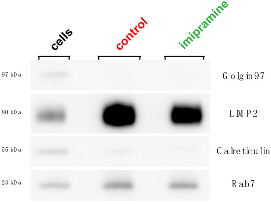
Lysosomes are degradative organelles that facilitate the removal and recycling of potentially cytotoxic materials and mediate a variety of other cellular processes, such as nutrient sensing, intracellular signaling, and lipid metabolism. Due to these central roles, lysosome dysfunction can lead to deleterious outcomes, including the accumulation of cytotoxic material, inflammation, and cell death. We previously reported that cationic amphiphilic drugs, such as imipramine, alter pH and lipid metabolism within macrophage lysosomes. Therefore, the ability for imipramine to induce changes to the lipid content of isolated macrophage lysosomes was investigated, focusing on sphingomyelin, cholesterol, and glycerophospholipid metabolism as these lipid classes have important roles in inflammation and disease. The lysosomes were isolated from control and imipramine-treated macrophages using density gradient ultracentrifugation, and mass spectrometry was used to measure the changes in their lipid composition. An unsupervised hierarchical cluster analysis revealed a clear differentiation between the imipramine-treated and control lysosomes. There was a significant overall increase in the abundance of specific lipids mostly composed of cholesterol esters, sphingomyelins, and phosphatidylcholines, while lysophosphatidylcholines and ceramides were overall decreased. These results support the conclusion that imipramine’s ability to change the lysosomal pH inhibits multiple pH-sensitive enzymes in macrophage lysosomes. Full article
Show Figures

Figure 1

13 pages, 800 KiB  
Review
Intracranial Aneurysms and Lipid Metabolism Disorders: From Molecular Mechanisms to Clinical Implications
by Tonglin Pan, Yuan Shi, Guo Yu, Abdureshid Mamtimin and Wei Zhu
Biomolecules 2023, 13(11), 1652; https://doi.org/10.3390/biom13111652 - 14 Nov 2023
Cited by 2 | Viewed by 2308
Abstract
Many vascular diseases are linked to lipid metabolism disorders, which cause lipid accumulation and peroxidation in the vascular wall. These processes lead to degenerative changes in the vessel, such as phenotypic transformation of smooth muscle cells and dysfunction and apoptosis of endothelial cells. [...] Read more.
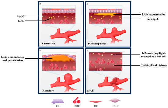
Many vascular diseases are linked to lipid metabolism disorders, which cause lipid accumulation and peroxidation in the vascular wall. These processes lead to degenerative changes in the vessel, such as phenotypic transformation of smooth muscle cells and dysfunction and apoptosis of endothelial cells. In intracranial aneurysms, the coexistence of lipid plaques is often observed, indicating localized lipid metabolism disorders. These disorders may impair the function of the vascular wall or result from it. We summarize the literature on the relationship between lipid metabolism disorders and intracranial aneurysms below. Full article
Show Figures

Figure 1

14 pages, 1351 KiB  
Article
Dynamics of Docosahexaenoic Acid Utilization by Mouse Peritoneal Macrophages
by Patricia Monge, Alma M. Astudillo, Laura Pereira, María A. Balboa and Jesús Balsinde
Biomolecules 2023, 13(11), 1635; https://doi.org/10.3390/biom13111635 - 10 Nov 2023
Cited by 1 | Viewed by 1519
Abstract
In this work, the incorporation of docosahexaenoic acid (DHA) in mouse resident peritoneal macrophages and its redistribution within the various phospholipid classes were investigated. Choline glycerophospholipids (PC) behaved as the major initial acceptors of DHA. Prolonged incubation with the fatty acid resulted in [...] Read more.
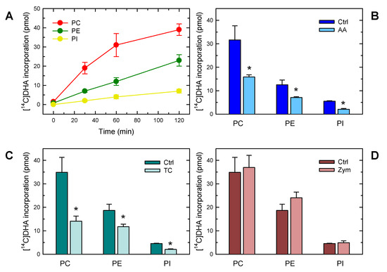
In this work, the incorporation of docosahexaenoic acid (DHA) in mouse resident peritoneal macrophages and its redistribution within the various phospholipid classes were investigated. Choline glycerophospholipids (PC) behaved as the major initial acceptors of DHA. Prolonged incubation with the fatty acid resulted in the transfer of DHA from PC to ethanolamine glycerophospholipids (PE), reflecting phospholipid remodeling. This process resulted in the cells containing similar amounts of DHA in PC and PE in the resting state. Mass spectrometry-based lipidomic analyses of phospholipid molecular species indicated a marked abundance of DHA in ether phospholipids. Stimulation of the macrophages with yeast-derived zymosan resulted in significant decreases in the levels of all DHA-containing PC and PI species; however, no PE or PS molecular species were found to decrease. In contrast, the levels of an unusual DHA-containing species, namely PI(20:4/22:6), which was barely present in resting cells, were found to markedly increase under zymosan stimulation. The levels of this phospholipid also significantly increased when the calcium-ionophore A23187 or platelet-activating factor were used instead of zymosan to stimulate the macrophages. The study of the route involved in the synthesis of PI(20:4/22:6) suggested that this species is produced through deacylation/reacylation reactions. These results define the increases in PI(20:4/22:6) as a novel lipid metabolic marker of mouse macrophage activation, and provide novel information to understand the regulation of phospholipid fatty acid turnover in activated macrophages. Full article
Show Figures

Figure 1

21 pages, 32336 KiB  
Article
Self-Assembly of Lipid Molecules under Shear Flows: A Dissipative Particle Dynamics Simulation Study
by Huan Zhang, Fan Pan and Shiben Li
Biomolecules 2023, 13(9), 1359; https://doi.org/10.3390/biom13091359 - 7 Sep 2023
Viewed by 1582
Abstract
The self-assembly of lipid molecules in aqueous solution under shear flows was investigated using the dissipative particle dynamics simulation method. Three cases were considered: zero shear flow, weak shear flow and strong shear flow. Various self-assembled structures, such as double layers, perforated double [...] Read more.
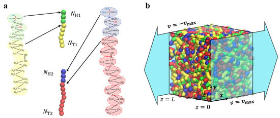
The self-assembly of lipid molecules in aqueous solution under shear flows was investigated using the dissipative particle dynamics simulation method. Three cases were considered: zero shear flow, weak shear flow and strong shear flow. Various self-assembled structures, such as double layers, perforated double layers, hierarchical discs, micelles, and vesicles, were observed. The self-assembly behavior was investigated in equilibrium by constructing phase diagrams based on chain lengths. Results showed the remarkable influence of chain length, shear flow and solution concentration on the self-assembly process. Furthermore, the self-assembly behavior of lipid molecules was analyzed using the system energy, particle number and shape factor during the dynamic processes, where the self-assembly pathways were observed and analyzed for the typical structures. The results enhance our understanding of biomacromolecule self-assembly in a solution and hold the potential for applications in biomedicine. Full article
Show Figures

Figure 1

15 pages, 3795 KiB  
Review
Infection, Inflammation, and Immunity in Sepsis
by Undurti N. Das
Biomolecules 2023, 13(9), 1332; https://doi.org/10.3390/biom13091332 - 31 Aug 2023
Cited by 18 | Viewed by 3517
Abstract
Sepsis is triggered by microbial infection, injury, or even major surgery. Both innate and adaptive immune systems are involved in its pathogenesis. Cytoplasmic presence of DNA or RNA of the invading organisms or damaged nuclear material (in the form of micronucleus in the [...] Read more.
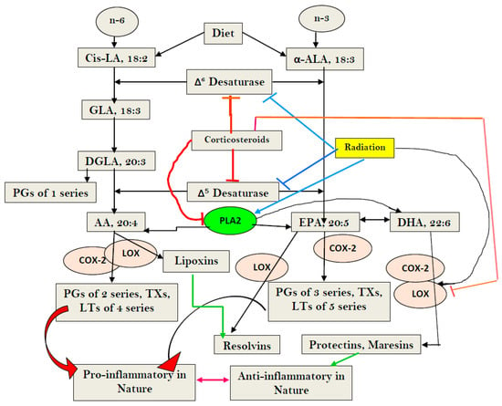
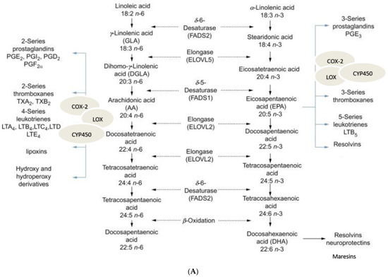
Sepsis is triggered by microbial infection, injury, or even major surgery. Both innate and adaptive immune systems are involved in its pathogenesis. Cytoplasmic presence of DNA or RNA of the invading organisms or damaged nuclear material (in the form of micronucleus in the cytoplasm) in the host cell need to be eliminated by various nucleases; failure to do so leads to the triggering of inflammation by the cellular cGAS-STING system, which induces the release of IL-6, TNF-α, and IFNs. These cytokines activate phospholipase A2 (PLA2), leading to the release of polyunsaturated fatty acids (PUFAs), gamma-linolenic acid (GLA), arachidonic acid (AA), eicosapentaenoic acid (EPA), and docosahexaenoic acid (DHA), which form precursors to various pro- and anti-inflammatory eicosanoids. On the other hand, corticosteroids inhibit PLA2 activity and, thus, suppress the release of GLA, AA, EPA, and DHA. PUFAs and their metabolites have a negative regulatory action on the cGAS-STING pathway and, thus, suppress the inflammatory process and initiate inflammation resolution. Pro-inflammatory cytokines and corticosteroids (corticosteroids > IL-6, TNF-α) suppress desaturases, which results in decreased formation of GLA, AA, and other PUFAs from the dietary essential fatty acids (EFAs). A deficiency of GLA, AA, EPA, and DHA results in decreased production of anti-inflammatory eicosanoids and failure to suppress the cGAS-STING system. This results in the continuation of the inflammatory process. Thus, altered concentrations of PUFAs and their metabolites, and failure to suppress the cGAS-STING system at an appropriate time, leads to the onset of sepsis. Similar abnormalities are also seen in radiation-induced inflammation. These results imply that timely administration of GLA, AA, EPA, and DHA, in combination with corticosteroids and anti-IL-6 and anti-TNF-α antibodies, may be of benefit in mitigating radiation-induced damage and sepsis. Full article
Show Figures

Figure 1

30 pages, 4934 KiB  
Article
Cholesterol and Sphingomyelin Polarize at the Leading Edge of Migrating Myoblasts and Involve Their Clustering in Submicrometric Domains
by Juliette Vanderroost, Noémie Avalosse, Danahe Mohammed, Delia Hoffmann, Patrick Henriet, Christophe E. Pierreux, David Alsteens and Donatienne Tyteca
Biomolecules 2023, 13(2), 319; https://doi.org/10.3390/biom13020319 - 7 Feb 2023
Cited by 3 | Viewed by 2286
Abstract
Myoblast migration is crucial for myogenesis and muscular tissue homeostasis. However, its spatiotemporal control remains elusive. Here, we explored the involvement of plasma membrane cholesterol and sphingolipids in this process. In resting C2C12 mouse myoblasts, those lipids clustered in sphingomyelin/cholesterol/GM1 ganglioside (SM/chol/GM1)- and [...] Read more.
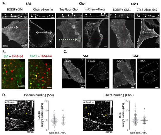
Myoblast migration is crucial for myogenesis and muscular tissue homeostasis. However, its spatiotemporal control remains elusive. Here, we explored the involvement of plasma membrane cholesterol and sphingolipids in this process. In resting C2C12 mouse myoblasts, those lipids clustered in sphingomyelin/cholesterol/GM1 ganglioside (SM/chol/GM1)- and cholesterol (chol)-enriched domains, which presented a lower stiffness than the bulk membrane. Upon migration, cholesterol and sphingomyelin polarized at the front, forming cholesterol (chol)- and sphingomyelin/cholesterol (SM/chol)-enriched domains, while GM1-enriched domains polarized at the rear. A comparison of domain proportion suggested that SM/chol- and GM1-enriched domains originated from the SM/chol/GM1-coenriched domains found at resting state. Modulation of domain proportion (through cholesterol depletion, combined or not with actin polymerization inhibition, or sphingolipid synthesis inhibition) revealed that the higher the chol- and SM/chol-enriched domains, the higher the myoblast migration. At the front, chol- and SM/chol-enriched domains were found in proximity with F-actin fibers and the lateral mobility of sphingomyelin in domains was specifically restricted in a cholesterol- and cytoskeleton-dependent manner while domain abrogation impaired F-actin and focal adhesion polarization. Altogether, we showed the polarization of cholesterol and sphingomyelin and their clustering in chol- and SM/chol-enriched domains with differential properties and roles, providing a mechanism for the spatial and functional control of myoblast migration. Full article
Show Figures

Figure 1

2022

Jump to: 2024, 2023, 2021

13 pages, 926 KiB  
Article
Differential Mobilization of the Phospholipid and Triacylglycerol Pools of Arachidonic Acid in Murine Macrophages
by Miguel A. Bermúdez, Julio M. Rubio, María A. Balboa and Jesús Balsinde
Biomolecules 2022, 12(12), 1851; https://doi.org/10.3390/biom12121851 - 11 Dec 2022
Cited by 2 | Viewed by 1864
Abstract
Innate immune cells such as monocytes and macrophages contain high levels of arachidonic acid (AA), part of which can be mobilized during cellular activation for the formation of a vast array of bioactive oxygenated metabolites. Monocytes and macrophages present in inflammatory foci typically [...] Read more.
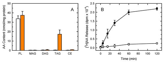
Innate immune cells such as monocytes and macrophages contain high levels of arachidonic acid (AA), part of which can be mobilized during cellular activation for the formation of a vast array of bioactive oxygenated metabolites. Monocytes and macrophages present in inflammatory foci typically incorporate large amounts of AA, not only in membrane phospholipids, but also in neutral lipids such as triacylglycerol. Thus, it was of interest to investigate the metabolic fate of these two AA pools in macrophages. Utilizing a variety of radiolabeling techniques to distinguish the phospholipid and triacylglycerol pools, we show in this paper that during an acute stimulation of the macrophages with yeast-derived zymosan, the membrane phospholipid AA pool acts as the major, if not the only, source of releasable AA. On the contrary, the AA pool in triacylglycerol appears to be used at a later stage, when the zymosan-stimulated response has declined, as a source to replenish the phospholipid pools that were consumed during the activation process. Thus, phospholipids and triacylglycerol play different in roles AA metabolism and dynamics during macrophage activation. Full article
Show Figures

Figure 1

16 pages, 2435 KiB  
Article
Effects of Psychotropic Medication on Somatic Sterol Biosynthesis of Adult Mice
by Marta Balog, Allison C Anderson, Marija Heffer, Zeljka Korade and Karoly Mirnics
Biomolecules 2022, 12(10), 1535; https://doi.org/10.3390/biom12101535 - 21 Oct 2022
Cited by 4 | Viewed by 1990
Abstract
Polypharmacy is commonly used to treat psychiatric disorders. These combinations often include drugs with sterol biosynthesis inhibiting side effects, including the antipsychotic aripiprazole (ARI), and antidepressant trazodone (TRZ). As the effects of psychotropic medications are poorly understood across the various tissue types to [...] Read more.
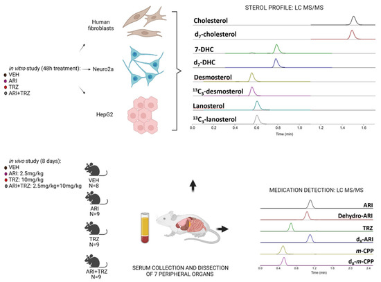
Polypharmacy is commonly used to treat psychiatric disorders. These combinations often include drugs with sterol biosynthesis inhibiting side effects, including the antipsychotic aripiprazole (ARI), and antidepressant trazodone (TRZ). As the effects of psychotropic medications are poorly understood across the various tissue types to date, we investigated the effects of ARI, TRZ, and ARI + TRZ polypharmacy on the post-lanosterol biosynthesis in three cell lines (Neuro2a, HepG2, and human dermal fibroblasts) and seven peripheral tissues of an adult mouse model. We found that both ARI and TRZ strongly interfere with the function of 7-dehydrocholesterol reductase enzyme (DHCR7) and lead to robust elevation in 7-dehydrocholesterol levels (7-DHC) and reduction in desmosterol (DES) across all cell lines and somatic tissues. ARI + TRZ co-administration resulted in summative or synergistic effects across the utilized in vitro and in vivo models. These findings suggest that at least some of the side effects of ARI and TRZ are not receptor mediated but arise from inhibiting DHCR7 enzyme activity. We propose that interference with sterol biosynthesis, particularly in the case of simultaneous utilization of medications with such side effects, can potentially interfere with functioning or development of multiple organ systems, warranting further investigation. Full article
Show Figures

Figure 1

19 pages, 5390 KiB  
Article
Phosphatidylinositol Monophosphates Regulate the Membrane Localization of HSPA1A, a Stress-Inducible 70-kDa Heat Shock Protein
by Larissa Smulders, Rachel Altman, Carolina Briseno, Alireza Saatchi, Leslie Wallace, Maha AlSebaye, Robert V. Stahelin and Nikolas Nikolaidis
Biomolecules 2022, 12(6), 856; https://doi.org/10.3390/biom12060856 - 20 Jun 2022
Cited by 5 | Viewed by 3340
Abstract
HSPA1A is a molecular chaperone that regulates the survival of stressed and cancer cells. In addition to its cytosolic pro-survival functions, HSPA1A also localizes and embeds in the plasma membrane (PM) of stressed and tumor cells. Membrane-associated HSPA1A exerts immunomodulatory functions and renders [...] Read more.
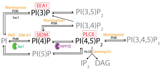
HSPA1A is a molecular chaperone that regulates the survival of stressed and cancer cells. In addition to its cytosolic pro-survival functions, HSPA1A also localizes and embeds in the plasma membrane (PM) of stressed and tumor cells. Membrane-associated HSPA1A exerts immunomodulatory functions and renders tumors resistant to standard therapies. Therefore, understanding and manipulating HSPA1A’s surface presentation is a promising therapeutic. However, HSPA1A’s pathway to the cell surface remains enigmatic because this protein lacks known membrane localization signals. Considering that HSPA1A binds to lipids, like phosphatidylserine (PS) and monophosphorylated phosphoinositides (PIPs), we hypothesized that this interaction regulates HSPA1A’s PM localization and anchorage. To test this hypothesis, we subjected human cell lines to heat shock, depleted specific lipid targets, and quantified HSPA1A’s PM localization using confocal microscopy and cell surface biotinylation. These experiments revealed that co-transfection of HSPA1A with lipid-biosensors masking PI(4)P and PI(3)P significantly reduced HSPA1A’s heat-induced surface presentation. Next, we manipulated the cellular lipid content using ionomycin, phenyl arsine oxide (PAO), GSK-A1, and wortmannin. These experiments revealed that HSPA1A’s PM localization was unaffected by ionomycin but was significantly reduced by PAO, GSK-A1, and wortmannin, corroborating the findings obtained by the co-transfection experiments. We verified these results by selectively depleting PI(4)P and PI(4,5)P2 using a rapamycin-induced phosphatase system. Our findings strongly support the notion that HSPA1A’s surface presentation is a multifaceted lipid-driven phenomenon controlled by the binding of the chaperone to specific endosomal and PM lipids. Full article
Show Figures

Figure 1

36 pages, 8608 KiB  
Article
Gamma-Linolenic Acid (GLA) Protects against Ionizing Radiation-Induced Damage: An In Vitro and In Vivo Study
by Poorani Rengachar, Anant Narayan Bhatt, Sailaja Polavarapu, Senthil Veeramani, Anand Krishnan, Monika Sadananda and Undurti N. Das
Biomolecules 2022, 12(6), 797; https://doi.org/10.3390/biom12060797 - 7 Jun 2022
Cited by 12 | Viewed by 4110
Abstract
Radiation is pro-inflammatory in nature in view of its ability to induce the generation of reactive oxygen species (ROS), cytokines, chemokines, and growth factors with associated inflammatory cells. Cells are efficient in repairing radiation-induced DNA damage; however, exactly how this happens is not [...] Read more.
Radiation is pro-inflammatory in nature in view of its ability to induce the generation of reactive oxygen species (ROS), cytokines, chemokines, and growth factors with associated inflammatory cells. Cells are efficient in repairing radiation-induced DNA damage; however, exactly how this happens is not clear. In the present study, GLA reduced DNA damage (as evidenced by micronuclei formation) and enhanced metabolic viability, which led to an increase in the number of surviving RAW 264.7 cells in vitro by reducing ROS generation, and restoring the activities of desaturases, COX-1, COX-2, and 5-LOX enzymes, TNF-α/TGF-β, NF-kB/IkB, and Bcl-2/Bax ratios, and iNOS, AIM-2, and caspases 1 and 3, to near normal. These in vitro beneficial actions were confirmed by in vivo studies, which revealed that the survival of female C57BL/6J mice exposed to lethal radiation (survival~20%) is significantly enhanced (to ~80%) by GLA treatment by restoring altered levels of duodenal HMGB1, IL-6, TNF-α, and IL-10 concentrations, as well as the expression of NF-kB, IkB, Bcl-2, Bax, delta-6-desaturase, COX-2, and 5-LOX genes, and pro- and anti-oxidant enzymes (SOD, catalase, glutathione), to near normal. These in vitro and in vivo studies suggest that GLA protects cells/tissues from lethal doses of radiation by producing appropriate changes in inflammation and its resolution in a timely fashion. Full article
Show Figures

Figure 1

25 pages, 1916 KiB  
Review
Utility of the Specialized Pro-Resolving Mediators as Diagnostic and Prognostic Biomarkers in Disease
by Jesmond Dalli, Esteban Alberto Gomez and Charlotte Camille Jouvene
Biomolecules 2022, 12(3), 353; https://doi.org/10.3390/biom12030353 - 23 Feb 2022
Cited by 6 | Viewed by 3847
Abstract
A precision medicine approach is widely acknowledged to yield more effective therapeutic strategies in the treatment of patients with chronic inflammatory conditions than the prescriptive paradigm currently utilized in the management and treatment of these patients. This is because such an approach will [...] Read more.
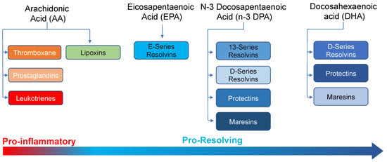
A precision medicine approach is widely acknowledged to yield more effective therapeutic strategies in the treatment of patients with chronic inflammatory conditions than the prescriptive paradigm currently utilized in the management and treatment of these patients. This is because such an approach will take into consideration relevant factors including the likelihood that a patient will respond to given therapeutics based on their disease phenotype. Unfortunately, the application of this precision medicine paradigm in the daily treatment of patients has been greatly hampered by the lack of robust biomarkers, in particular biomarkers for determining early treatment responsiveness. Lipid mediators are central in the regulation of host immune responses during both the initiation and resolution of inflammation. Amongst lipid mediators, the specialized pro-resolving mediators (SPM) govern immune cells to promote the resolution of inflammation. These autacoids are produced via the stereoselective conversion of essential fatty acids to yield molecules that are dynamically regulated during inflammation and exert potent immunoregulatory activities. Furthermore, there is an increasing appreciation for the role that these mediators play in conveying the biological actions of several anti-inflammatory therapeutics, including statins and aspirin. Identification and quantitation of these mediators has traditionally been achieved using hyphenated mass spectrometric techniques, primarily liquid-chromatography tandem mass spectrometry. Recent advances in the field of chromatography and mass spectrometry have increased both the robustness and the sensitivity of this approach and its potential deployment for routine clinical diagnostics. In the present review, we explore the evidence supporting a role for specific SPM as potential biomarkers for patient stratification in distinct disease settings together with methodologies employed in the identification and quantitation of these autacoids. Full article
Show Figures

Figure 1

13 pages, 4742 KiB  
Article
Single-Cellular Biological Effects of Cholesterol-Catabolic Bile Acid-Based Nano/Micro Capsules as Anti-Inflammatory Cell Protective Systems
by Armin Mooranian, Corina Mihaela Ionescu, Daniel Walker, Melissa Jones, Susbin Raj Wagle, Bozica Kovacevic, Jacqueline Chester, Thomas Foster, Edan Johnston, Jafri Kuthubutheen, Daniel Brown, Marcus D. Atlas, Momir Mikov and Hani Al-Salami
Biomolecules 2022, 12(1), 73; https://doi.org/10.3390/biom12010073 - 4 Jan 2022
Cited by 15 | Viewed by 2303
Abstract
Recent studies in our laboratories have shown promising effects of bile acids in ➀ drug encapsulation for oral targeted delivery (via capsule stabilization) particularly when encapsulated with Eudragit NM30D® and ➁ viable-cell encapsulation and delivery (via supporting cell viability and biological activities, [...] Read more.
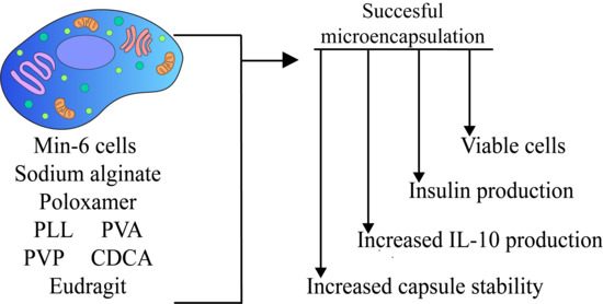
Recent studies in our laboratories have shown promising effects of bile acids in ➀ drug encapsulation for oral targeted delivery (via capsule stabilization) particularly when encapsulated with Eudragit NM30D® and ➁ viable-cell encapsulation and delivery (via supporting cell viability and biological activities, postencapsulation). Accordingly, this study aimed to investigate applications of bile acid-Eudragit NM30D® capsules in viable-cell encapsulation ready for delivery. Mouse-cloned pancreatic β-cell line was cultured and cells encapsulated using bile acid-Eudragit NM30D® capsules, and capsules’ images, viability, inflammation, and bioenergetics of encapsulated cells assessed. The capsules’ thermal and chemical stability assays were also assessed to ascertain an association between capsules’ stability and cellular biological activities. Bile acid-Eudragit NM30D® capsules showed improved cell viability (e.g., F1 < F2 & F8; p < 0.05), insulin, inflammatory profile, and bioenergetics as well as thermal and chemical stability, compared with control. These effects were formulation-dependent and suggest, overall, that changes in ratios of bile acids to Eudragit NM30D® can change the microenvironment of the capsules and subsequent cellular biological activities. Full article
Show Figures

Graphical abstract

2021

Jump to: 2024, 2023, 2022

12 pages, 1373 KiB  
Article
Rhamnolipids as a Tool for Eradication of Trichosporon cutaneum Biofilm
by Olga Maťátková, Irena Kolouchová, Kristýna Lokočová, Jana Michailidu, Petr Jaroš, Markéta Kulišová, Tomáš Řezanka and Jan Masák
Biomolecules 2021, 11(11), 1727; https://doi.org/10.3390/biom11111727 - 19 Nov 2021
Cited by 5 | Viewed by 1964
Abstract
Microbial biofilms formed by pathogenic and antibiotic-resistant microorganisms represent a serious threat for public health in medicine and many industrial branches. Biofilms are involved in many persistent and chronic infections, the biofouling of water and food contamination. Therefore, current research is involved in [...] Read more.
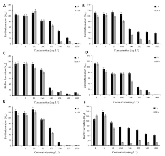
Microbial biofilms formed by pathogenic and antibiotic-resistant microorganisms represent a serious threat for public health in medicine and many industrial branches. Biofilms are involved in many persistent and chronic infections, the biofouling of water and food contamination. Therefore, current research is involved in the development of new treatment strategies. Biofilm is a complex system, and thus all aspects of the measurement and monitoring of its growth and eradication in various conditions, including static and dynamic flow, are issues of great importance. The antibiofilm character of rhamnolipid mixtures produced by four Pseudomonas aeruginosa strains was studied under different conditions. For this purpose, the biofilm of opportunistic pathogen Trichosporon cutaneum was used and treated under static conditions (microscope glass coverslip in a Petri dish) and under dynamic conditions (a single-channel flow cell). The results show that the biological activity of rhamnolipids depends both on their properties and on the conditions of the biofilm formation. Therefore, this aspect must be taken into account when planning the experimental or application design. Full article
Show Figures

Figure 1

Back to TopTop