Diagnosis and Treatment of Primary and Secondary Lung Cancers

A topical collection in Cancers (ISSN 2072-6694). This collection belongs to the section "Clinical Research of Cancer".

Viewed by 93436

Editor


E-Mail Website
Collection Editor
1. Department of Thoracic Surgery, IRCCS European Institute of Oncology, 20141 Milan, Italy
2. Department of Oncology and Hemato-Oncology, University of Milan, 20141 Milan, Italy
Interests: lung cancer surgery; thoracic oncology; early-stage lung cancer; locally advanced lung cancer
Special Issues, Collections and Topics in MDPI journals

Topical Collection Information

Dear Colleagues,

Both primary and secondary tumors of the chest are major issues for clinicians; in fact, on the one hand, primary lung cancer is one of the most frequently diagnosed cancers and is the leading cause of cancer-related death worldwide. On the other hand, the lung is the second most common site of metastases, and pulmonary metastasectomy is the most frequent surgical resection undertaken by thoracic surgeons. Significant developments have recently been shown in clinical settings, both in terms of diagnosis and treatment: for example, the early diagnosis of lung nodules by computed tomography screening, circulating miRNA detection, minimally invasive diagnostic and therapeutic approaches such as endobronchial ultrasound, video-assisted thoracic surgery and robot-assisted thoracic surgery, immunotherapy, TKI inhibitors, and stereotactic body radiation therapy (SBRT) represent only some of the most significant and recent goals.

Moreover, other thoracic malignancies such as chest wall tumors, mediastinal tumors, and mesothelioma, although rare, represent the most advanced frontier of modern oncology.

In this Topical Collection, we would like to focus on the most significant cornerstones in the diagnosis and therapy of both primary and secondary thoracic cancers.

It is our pleasure to lead such a comprehensive Topical Collection, and we look forward to receiving your valuable contributions.

Prof. Francesco Petrella
Collection Editor

Manuscript Submission Information

Manuscripts should be submitted online at www.mdpi.com by registering and logging in to this website. Once you are registered, click here to go to the submission form. Manuscripts can be submitted until the deadline. All submissions that pass pre-check are peer-reviewed. Accepted papers will be published continuously in the journal (as soon as accepted) and will be listed together on the collection website. Research articles, review articles as well as communications are invited. For planned papers, a title and short abstract (about 100 words) can be sent to the Editorial Office for announcement on this website.

Submitted manuscripts should not have been published previously, nor be under consideration for publication elsewhere (except conference proceedings papers). All manuscripts are thoroughly refereed through a single-blind peer-review process. A guide for authors and other relevant information for submission of manuscripts is available on the Instructions for Authors page. Cancers is an international peer-reviewed open access semimonthly journal published by MDPI.

Please visit the Instructions for Authors page before submitting a manuscript. The Article Processing Charge (APC) for publication in this open access journal is 2900 CHF (Swiss Francs). Submitted papers should be well formatted and use good English. Authors may use MDPI's English editing service prior to publication or during author revisions.

Keywords

  • lung cancer
  • lung metastases
  • diagnosis
  • lung resection
  • computed tomography
  • positron emission tomography
  • stereotactic body radiation therapy (SBRT)
  • immunotherapy
Published Papers (31 papers)

2024

Jump to: 2023, 2022, 2021

10 pages, 1747 KiB  
Article
Tumor Location Is an Independent Prognostic Factor in Completely Resected Pathological Stage I Non-Small Cell Lung Cancer: A Multicenter Retrospective Study
by Wei-Ke Kuo, Po-Ju Chen, Mei-Hsuan Wu, Hsin-Chung (Henry) Lee, Jiun-Kai Fan, Pang-Hung Hsu and Ching-Fu Weng
Cancers 2024, 16(9), 1710; https://doi.org/10.3390/cancers16091710 - 27 Apr 2024
Viewed by 1311
Abstract
Previous studies suggested that the location of the primary tumor in non-small cell lung cancer (NSCLC) is associated with clinical features and prognosis, but results are conflicting. The purpose of this study was to explore tumor location as an independent risk factor of [...] Read more.
Previous studies suggested that the location of the primary tumor in non-small cell lung cancer (NSCLC) is associated with clinical features and prognosis, but results are conflicting. The purpose of this study was to explore tumor location as an independent risk factor of survival for patients with completely resected pathological stage I NSCLC. This was a multicenter retrospective study conducted in Taiwan. Included patients were diagnosed with stage I NSCLC and had undergone primary tumor resection. Variables including tumor location, pathological stage, histological differentiation, and International Association for the Study of Lung Cancer (IASLC) grade were evaluated for predictive ability for disease-free survival (DFS) and overall survival (OS). A total of 208 patients were included, with 123 (59.1%) patients having a primary tumor in the upper and middle lobes. The median duration of follow-up for survivors was 60.5 months. Compared to patients with IASLC Grade 3 disease, patients with Grade 1 disease had significantly longer DFS. Tumor location and IASLC grade were independent predictors for OS in multivariate analysis. Specifically, patients with NSCLC in the lower lobe and patients who are histologically classified as IASLC Grade 3 may have poorer prognosis and require greater attention to improve outcomes. Full article
Show Figures

Figure 1

2023

Jump to: 2024, 2022, 2021

16 pages, 746 KiB  
Article
Immune-Related Adverse Events, Biomarkers of Systemic Inflammation, and Survival Outcomes in Patients Receiving Pembrolizumab for Non-Small-Cell Lung Cancer
by George Raynes, Mark Stares, Samantha Low, Dhania Haron, Hussain Sarwar, Dhruv Abhi, Colin Barrie, Barry Laird, Caledonian Cachexia Collaborative, Iain Phillips and Melanie MacKean
Cancers 2023, 15(23), 5502; https://doi.org/10.3390/cancers15235502 - 21 Nov 2023
Cited by 5 | Viewed by 1542
Abstract
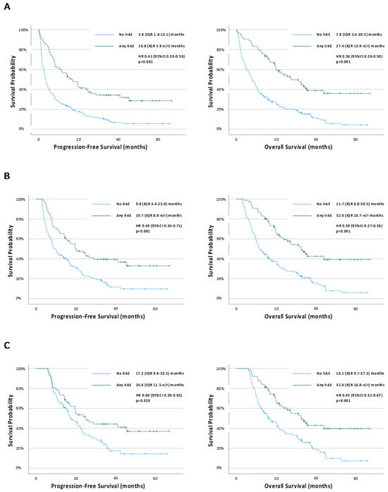
Background: Pembrolizumab monotherapy for non-small-cell lung cancer (NSCLC) expressing PD-L1 ≥ 50% doubles five-year survival rates compared to chemotherapy. However, immune-related adverse events (irAEs) can cause severe, long-term toxicity necessitating high-dose steroids and/or treatment cessation. Interestingly, patients experiencing irAEs demonstrate better survival outcomes. [...] Read more.
Background: Pembrolizumab monotherapy for non-small-cell lung cancer (NSCLC) expressing PD-L1 ≥ 50% doubles five-year survival rates compared to chemotherapy. However, immune-related adverse events (irAEs) can cause severe, long-term toxicity necessitating high-dose steroids and/or treatment cessation. Interestingly, patients experiencing irAEs demonstrate better survival outcomes. Biomarkers of systemic inflammation, including the Scottish Inflammatory Prognostic Score (SIPS), also predict survival in this patient group. This study examines the relationship between inflammatory status, irAEs, and survival outcomes in NSCLC. Methods: A retrospective analysis was conducted on patients with NSCLC expressing PD-L1 ≥ 50% receiving first-line pembrolizumab monotherapy at a large cancer centre in Scotland. Regression analyses were conducted to examine the relationship between SIPS, irAEs, and survival. Results: 83/262 eligible patients (32%) experienced an irAE. Dermatological, endocrine, gastrointestinal, and hepatic, but not pulmonary, irAEs were associated with prolonged PFS and OS (p <= 0.011). Mild irAEs were associated with better PFS and OS in all patients, including on time-dependent analyses (HR0.61 [95% CI 0.41–0.90], p = 0.014 and HR0.41 [95% CI 0.26–0.63], p < 0.001, respectively). SIPS predicted PFS (HR 1.60 [95% CI 1.34–1.90], p < 0.001) and OS (HR 1.69 [95% CI 1.41–2.02], p < 0.001). SIPS predicted the occurrence of any irAE in all patients (p = 0.011), but not on 24-week landmark analyses (p = 0.174). The occurrence of irAEs predicted favourable outcomes regardless of the baseline inflammatory status (p = 0.015). Conclusion: The occurrence of certain irAEs is associated with a survival benefit in patients with NSCLC expressing PD-L1 ≥ 50% receiving pembrolizumab. We find that the association between low levels of systemic inflammation and the risk of irAEs is confounded by their independent prognostic value. Full article
Show Figures

Figure 1

15 pages, 1293 KiB  
Article
MicroRNA Profiling of Red Blood Cells for Lung Cancer Diagnosis
by Xinyan Geng, Jie Ma, Pushpa Dhilipkannah and Feng Jiang
Cancers 2023, 15(22), 5312; https://doi.org/10.3390/cancers15225312 - 7 Nov 2023
Cited by 1 | Viewed by 1561
Abstract
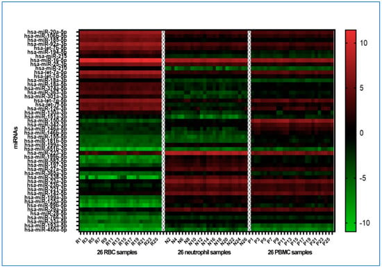
Background: Despite extensive endeavors to establish cell-free circulating biomarkers for lung cancer diagnosis, clinical adoption remains elusive. Noteworthy, emergent evidence suggests the pivotal roles of red blood cells (RBCs) and their derivatives in tumorigenesis, illuminating potential avenues for diagnostic advancements using blood cell-derived [...] Read more.
Background: Despite extensive endeavors to establish cell-free circulating biomarkers for lung cancer diagnosis, clinical adoption remains elusive. Noteworthy, emergent evidence suggests the pivotal roles of red blood cells (RBCs) and their derivatives in tumorigenesis, illuminating potential avenues for diagnostic advancements using blood cell-derived microRNAs (miRNAs). Methods: We executed microarray analyses on three principal blood cell types—RBCs, peripheral blood mononuclear cells (PBMCs), and neutrophils—encompassing 26 lung cancer patients and 26 healthy controls. Validation was performed using droplet digital PCR within an additional cohort comprising 42 lung cancer and 39 control cases. Results: Our investigation unearthed distinct miRNA profiles associated with lung cancer across all examined blood cell types. Intriguingly, RBC-miRNAs emerged as potential novel biomarkers for lung cancer, an observation yet to be documented. Importantly, integrating miRNAs from disparate blood cell types yielded a superior diagnostic accuracy for lung cancer over individual cell-type miRNAs. Subsequently, we formulated three diagnostic panels, adeptly discerning non-small cell lung cancer, adenocarcinoma, and squamous cell carcinoma, maintaining consistency across various disease stages. Conclusion: RBC-derived molecules introduce novel cancer biomarkers, and exploiting miRNA profiles across varied blood cell types unveils a promising frontier for lung cancer’s early detection and histological classification. Full article
Show Figures

Figure 1

14 pages, 937 KiB  
Review
Cellular Therapy for Lung Cancer: Focusing on Chimeric Antigen Receptor T (CAR T) Cells and Tumor-Infiltrating Lymphocyte (TIL) Therapy
by Vatsala Katiyar, Jason Chesney and Goetz Kloecker
Cancers 2023, 15(14), 3733; https://doi.org/10.3390/cancers15143733 - 23 Jul 2023
Cited by 11 | Viewed by 3077
Abstract
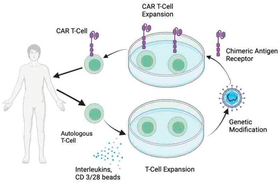
Lung cancer is a leading cause of morbidity and mortality in the United States and worldwide. The introduction of immune checkpoint inhibitors has led to a marked improvement in the outcomes of lung cancer patients. Despite these advances, there is a huge unmet [...] Read more.
Lung cancer is a leading cause of morbidity and mortality in the United States and worldwide. The introduction of immune checkpoint inhibitors has led to a marked improvement in the outcomes of lung cancer patients. Despite these advances, there is a huge unmet need for therapeutic options in patients who are not candidates for targeted or immunotherapy or those who progress after first-line treatment. With its high mutational burden, lung cancer appears to be an attractive target for novel personalized treatment approaches. In this review, we provide an overview of two adoptive cell therapy approaches–chimeric antigen receptors (CAR) T-cell therapy and Tumor-infiltrating lymphocytes (TILs) in lung cancer with an emphasis on current challenges and future perspectives. While both these therapies are still in the early phases of development in lung cancer and need more refinement, they harbor the potential to be effective treatment options for this group of patients with otherwise poor prognoses. Full article
Show Figures

Figure 1

12 pages, 1520 KiB  
Article
Longitudinal Assessment of Circulating Tumor Cells and Outcome in Small Cell Lung Cancer: A Sub-Study of RASTEN—A Randomized Trial with Low Molecular Weight Heparin
by Pär-Ola Bendahl, Mattias Belting and Emelie Gezelius
Cancers 2023, 15(12), 3176; https://doi.org/10.3390/cancers15123176 - 13 Jun 2023
Cited by 4 | Viewed by 1557
Abstract
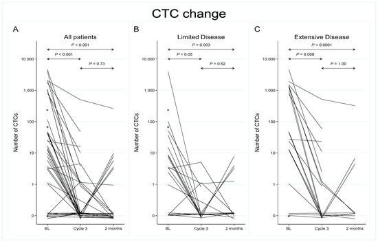
Circulating tumor cells (CTCs) may provide a liquid biopsy approach to disease monitoring in small cell lung cancer (SCLC), a particularly aggressive tumor subtype. Yet, the prognostic role of CTCs during and after treatment in relation to baseline remains ill-defined. Here, we assessed [...] Read more.
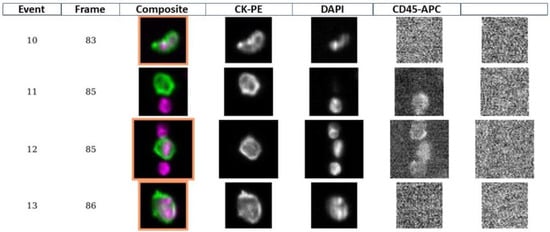
Circulating tumor cells (CTCs) may provide a liquid biopsy approach to disease monitoring in small cell lung cancer (SCLC), a particularly aggressive tumor subtype. Yet, the prognostic role of CTCs during and after treatment in relation to baseline remains ill-defined. Here, we assessed the value of longitudinal CTC analysis and the potential of low-molecular-weight heparin (LMWH) to reduce CTC abundance in SCLC patients from a randomized trial (RASTEN). Blood samples were collected at baseline, before chemotherapy Cycle 3, and at 2-month follow-up from 42 patients in total, and CTCs were quantified using the FDA-approved CellSearch system. We found a gradual decline in CTC count during and after treatment, independently of the addition of LMWH to standard therapy. Detectable CTCs at baseline correlated significantly to reduced survival compared to undetectable CTCs (unadjusted hazard ratio (HR) of 2.75 (95% CI 1.05–7.20; p = 0.040)). Furthermore, a persistent CTC count at 2-month follow-up was associated with a HR of 4.22 (95% CI 1.20–14.91; p = 0.025). Our findings indicate that persistently detectable CTCs during and after completion of therapy offer further prognostic information in addition to baseline CTC, suggesting a role for CTC in the individualized management of SCLC. Full article
Show Figures

Figure 1

18 pages, 1098 KiB  
Systematic Review
Lung Cancer as a Leading Cause among Paraneoplastic Non-Bacterial Thrombotic Endocarditis: A Meta-Analysis of Individual Patients’ Data
by Mohamed Rahouma, Sherif Khairallah, Anas Dabsha, Ismail A. M. H. Elkharbotly, Massimo Baudo, Amr Ismail, Omnia M. Korani, Mohamed Hossny, Arnaldo Dimagli, Leonard N. Girardi, Stephanie L. Mick and Mario Gaudino
Cancers 2023, 15(6), 1848; https://doi.org/10.3390/cancers15061848 - 20 Mar 2023
Cited by 11 | Viewed by 2530
Abstract
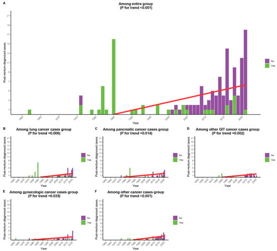
Hypercoagulability is strongly associated with cancer and may result in non-bacterial thrombotic endocarditis (NBTE). The aim of our meta-analysis was to explore the demographics and characteristics of this condition in cancer. Databases were systematically searched. The outcomes were to identify the annual trend [...] Read more.
Hypercoagulability is strongly associated with cancer and may result in non-bacterial thrombotic endocarditis (NBTE). The aim of our meta-analysis was to explore the demographics and characteristics of this condition in cancer. Databases were systematically searched. The outcomes were to identify the annual trend in premortem diagnosis among the entire cohort and different subgroups and to identify differences in characteristics and survival in the considered population. A total of 121 studies with 144 patients were included. The proportion of marantic endocarditis associated with lung cancer was 0.29 (95% CI, 0.21–0.37; p < 0.001), that associated with pancreatic cancer was 0.19 (95% CI, 0.13–0.27; p < 0.001), that associated with advanced cancer stage (metastasis) was 0.69 (95% CI, 0.61–0.76; p < 0.001), and that associated with adenocarcinoma was 0.65 (95% CI, 0.56–0.72; p < 0.001). Median and 6-month overall survival (OS) were 1.3 months and 32.3%, respectively, with 6-month OS of 20.8% vs. 37.0% in lung vs. other cancers, respectively (p = 0.06) and 42.9% vs. 31.1% among those who underwent intervention vs. those who did not (p = 0.07). Cases discovered in recent years had better survival (HR = 0.98 (95% CI, 0.96–0.99; p = 0.003). While cancer-associated NBTE is a rare entity, lung cancers were the most common tumor site and are frequently associated with more advanced and metastatic cancer stages. The prognosis is dismal, especially among lung cancers. Full article
Show Figures

Figure 1

11 pages, 1002 KiB  
Article
Real-World Impact of SABR on Stage I Non-Small-Cell Lung Cancer Outcomes at a Scottish Cancer Centre
by Mark Stares, Georgina Lewis, Maheva Vallet, Angus Killean, Giovanni Tramonti, Ailsa Patrizio, Melanie Mackean, Stephen Harrow, Colin Barrie, Kirsty MacLennan, Sorcha Campbell, Tamasin Evans, Aisha Tufail, Edinburgh Cancer Informatics Programme, Peter Hall and Iain Phillips
Cancers 2023, 15(5), 1431; https://doi.org/10.3390/cancers15051431 - 23 Feb 2023
Cited by 5 | Viewed by 2055
Abstract
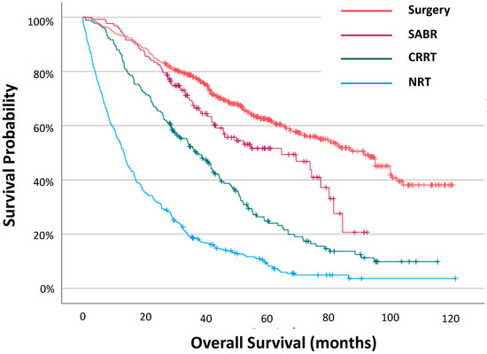
Introduction: Stereotactic ablative body radiotherapy (SABR) offers patients with stage I non-small-cell lung cancer (NSCLC) a safe, effective radical therapy option. The impact of introducing SABR at a Scottish regional cancer centre was studied. Methods: The Edinburgh Cancer Centre Lung Cancer Database was [...] Read more.
Introduction: Stereotactic ablative body radiotherapy (SABR) offers patients with stage I non-small-cell lung cancer (NSCLC) a safe, effective radical therapy option. The impact of introducing SABR at a Scottish regional cancer centre was studied. Methods: The Edinburgh Cancer Centre Lung Cancer Database was assessed. Treatment patterns and outcomes were compared across treatment groups (no radical therapy (NRT), conventional radical radiotherapy (CRRT), SABR and surgery) and across three time periods reflecting the availability of SABR (A, January 2012/2013 (pre-SABR); B, 2014/2016 (introduction of SABR); C, 2017/2019, (SABR established)). Results: 1143 patients with stage I NSCLC were identified. Treatment was NRT in 361 (32%), CRRT in 182 (16%), SABR in 132 (12%) and surgery in 468 (41%) patients. Age, performance status, and comorbidities correlated with treatment choice. The median survival increased from 32.5 months in time period A to 38.8 months in period B to 48.8 months in time period C. The greatest improvement in survival was seen in patients treated with surgery between time periods A and C (HR 0.69 (95% CI 0.56–0.86), p < 0.001). The proportion of patients receiving a radical therapy rose between time periods A and C in younger (age ≤ 65, 65–74 and 75–84 years), fitter (PS 0 and 1), and less comorbid patients (CCI 0 and 1–2), but fell in other patient groups. Conclusions: The introduction and establishment of SABR for stage I NSCLC has improved survival outcomes in Southeast Scotland. Increasing SABR utilisation appears to have enhanced the selection of surgical patients and increased the proportion of patients receiving a radical therapy. Full article
Show Figures

Figure 1

2022

Jump to: 2024, 2023, 2021

26 pages, 1256 KiB  
Review
Lung Cancer Immunotherapy: Beyond Common Immune Checkpoints Inhibitors
by Martina Catalano, Sonia Shabani, Jacopo Venturini, Carlotta Ottanelli, Luca Voltolini and Giandomenico Roviello
Cancers 2022, 14(24), 6145; https://doi.org/10.3390/cancers14246145 - 13 Dec 2022
Cited by 14 | Viewed by 6371
Abstract
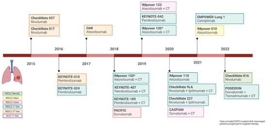
Immunotherapy is an ever-expanding field in lung cancer treatment research. Over the past two decades, there has been significant progress in identifying immunotherapy targets and creating specific therapeutic agents, leading to a major paradigm shift in lung cancer treatment. However, despite the great [...] Read more.
Immunotherapy is an ever-expanding field in lung cancer treatment research. Over the past two decades, there has been significant progress in identifying immunotherapy targets and creating specific therapeutic agents, leading to a major paradigm shift in lung cancer treatment. However, despite the great success achieved with programmed death protein 1/ligand 1 (PD-1/PD-L1) monoclonal antibodies and with anti-PD-1/PD-L1 plus anti-cytotoxic T-lymphocyte antigen 4 (CTLA-4), only a minority of lung cancer patients respond to treatment, and of these many subsequently experience disease progression. In addition, immune-related adverse events sometimes can be life-threatening, especially when anti-CTLA-4 and anti-PD-1 are used in combination. All of this prompted researchers to identify novel immune checkpoints targets to overcome these limitations. Lymphocyte activation gene-3 (LAG-3), T cell immunoglobulin (Ig) and Immunoreceptor Tyrosine-Based Inhibitory Motif (ITIM) domain (TIGIT), T cell immunoglobulin and mucin-domain containing-3 (TIM-3) are promising molecules now under investigation. This review aims to outline the current role of immunotherapy in lung cancer and to examine efficacy and future applications of the new immune regulating molecules. Full article
Show Figures

Figure 1

2 pages, 182 KiB  
Editorial
From Diagnosis to Treatment of Lung Cancer: An Update in “Cancers” in 2021
by Francesco Petrella
Cancers 2022, 14(22), 5639; https://doi.org/10.3390/cancers14225639 - 17 Nov 2022
Viewed by 1254
Abstract
After its successful launch in January 2021 by Cancers, the topic collection “Diagnosis and Treatment of Primary and Secondary Lung Cancers” experienced a productive first full year [...] Full article
16 pages, 757 KiB  
Review
RET Proto-Oncogene—Not Such an Obvious Starting Point in Cancer Therapy
by Tomasz Kucharczyk, Paweł Krawczyk, Dariusz M. Kowalski, Adam Płużański, Tomasz Kubiatowski and Ewa Kalinka
Cancers 2022, 14(21), 5298; https://doi.org/10.3390/cancers14215298 - 27 Oct 2022
Cited by 2 | Viewed by 3522
Abstract
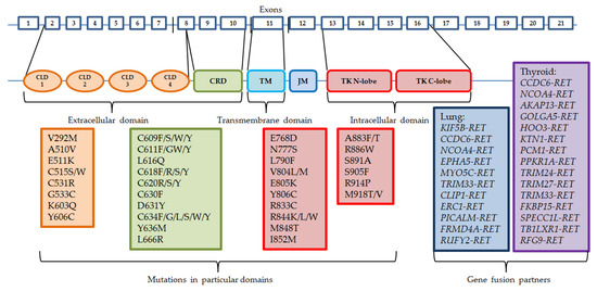
Mutations and fusions of RET (rearranged during transfection) gene are detected in a few common types of tumors including thyroid or non-small cells lung cancers. Multiple kinase inhibitors (MKIs) do not show spectacular effectiveness in patients with RET-altered tumors. Hence, recently, two [...] Read more.
Mutations and fusions of RET (rearranged during transfection) gene are detected in a few common types of tumors including thyroid or non-small cells lung cancers. Multiple kinase inhibitors (MKIs) do not show spectacular effectiveness in patients with RET-altered tumors. Hence, recently, two novel RET-specific inhibitors were registered in the US and in Europe. Selpercatinib and pralsetinib showed high efficacy in clinical trials, with fewer adverse effects, in comparison to previously used MKIs. However, the effectiveness of these new drugs may be reduced by the emergence of resistance mutations in RET gene and activation of different activating signaling pathways. This review presents the function of the normal RET receptor, types of molecular disturbances of the RET gene in patients with various cancers, methods of detecting these abnormalities, and the effectiveness of modern anticancer therapies (ranging from immunotherapies, through MKIs, to RET-specific inhibitors). Full article
Show Figures

Figure 1

11 pages, 808 KiB  
Article
Intraoperative Extra Corporeal Membrane Oxygenator for Lung Cancer Resections Does Not Impact Circulating Tumor Cells
by Francesco Petrella, Laura Zorzino, Samuele Frassoni, Vincenzo Bagnardi, Monica Casiraghi, Claudia Bardoni, Shehab Mohamed, Valeria Musso, Emanuele Simonini, Fabiana Rossi, Francesco Alamanni, Marco Venturino and Lorenzo Spaggiari
Cancers 2022, 14(20), 5004; https://doi.org/10.3390/cancers14205004 - 13 Oct 2022
Cited by 2 | Viewed by 1369
Abstract
Background: The diagnosis of active neoplastic disease was traditionally judged an absolute contraindication for extracorporeal membrane oxygenator (ECMO) because of the fear of tumor cells being scattered or seeded. The aim of this study is to compare the number of circulating tumor cells [...] Read more.
Background: The diagnosis of active neoplastic disease was traditionally judged an absolute contraindication for extracorporeal membrane oxygenator (ECMO) because of the fear of tumor cells being scattered or seeded. The aim of this study is to compare the number of circulating tumor cells (CTCs) before and after surgery in patients receiving lung cancer resection with and without intraoperative ECMO support. Methods: This is a prospective, non-randomized, two-arms observational study comparing the number of CTCs before and after surgery in patients receiving lung cancer resection with and without intraoperative ECMO support. The ECMO arm includes patients suffering from lung cancer undergoing pulmonary resection with planned intraoperative ECMO support. The non-ECMO arm includes patients suffering from non-early-stage lung cancer undergoing pulmonary resection without planned intraoperative ECMO support. Results: Twenty patients entered the study, eight in the ECMO arm and twelve in the non-ECMO arm. We did not observe any significant difference between the ECMO and non-ECMO groups in terms of postoperative complications (p = 1.00), ICU stay (p = 0.30), hospital stay (p = 0.23), circulating tumor cells’ increase or decrease after surgery (p = 0.24), and postoperative C-reactive protein and C-reactive protein increase (p = 0.80). The procedures in the non-ECMO arm were significantly longer than those in the ECMO arm (p = 0.043). Conclusions: Intraoperative ECMO for lung cancer resections did not impact CTC increase or decrease after the procedure. Full article
Show Figures

Figure 1

11 pages, 1045 KiB  
Review
Molecular and Immune Phenotypic Modifications during Metastatic Dissemination in Lung Carcinogenesis
by Drosos Tsavlis, Theodora Katopodi, Doxakis Anestakis, Savvas Petanidis, Charalampos Charalampidis, Evmorfia Chatzifotiou, Panagiotis Eskitzis, Paul Zarogoulidis and Konstantinos Porpodis
Cancers 2022, 14(15), 3626; https://doi.org/10.3390/cancers14153626 - 26 Jul 2022
Cited by 2 | Viewed by 2268
Abstract
The tumor microenvironment plays a key role in the progression of lung tumorigenesis, progression, and metastasis. Recent data reveal that disseminated tumor cells (DTCs) appear to play a key role in the development and progression of lung neoplasiaby driving immune system dysfunction and [...] Read more.
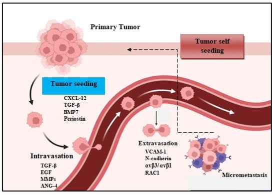
The tumor microenvironment plays a key role in the progression of lung tumorigenesis, progression, and metastasis. Recent data reveal that disseminated tumor cells (DTCs) appear to play a key role in the development and progression of lung neoplasiaby driving immune system dysfunction and established immunosuppression, which is vital for evading the host immune response. As a consequence, in this review we will discuss the role and function of DTCs in immune cell signaling routes which trigger drug resistance and immunosuppression. We will also discuss the metabolic biology of DTCs, their dormancy, and their plasticity, which are critical for metastasis and drive lung tumor progression. Furthermore, we will consider the crosstalk between DTCs and myeloid cells in tumor-related immunosuppression. Specifically, we will investigate the molecular immune-related mechanisms in the tumor microenvironment that lead to decreased drug sensitivity and tumor relapse, along with strategies for reversing drug resistance and targeting immunosuppressive tumor networks. Deciphering these molecular mechanisms is essential for preclinical and clinical investigations in order to enhance therapeutic efficacy. Furthermore, a better understanding of these immune cell signaling pathways that drive immune surveillance, immune-driven inflammation, and tumor-related immunosuppression is necessary for future personalized therapeutic approaches. Full article
Show Figures

Figure 1

13 pages, 303 KiB  
Review
New Implications of Patients’ Sex in Today’s Lung Cancer Management
by Jo Raskin, Annemiek Snoeckx, Annelies Janssens, Charlotte De Bondt, Reinier Wener, Mick van de Wiel, Jan P. van Meerbeeck and Evelien Smits
Cancers 2022, 14(14), 3399; https://doi.org/10.3390/cancers14143399 - 13 Jul 2022
Cited by 2 | Viewed by 1809
Abstract
This paper describes where and how sex matters in today’s management of lung cancer. We consecutively describe the differences between males and females in lung cancer demographics; sex-based differences in the immune system (including the poorer outcomes in women who are treated with [...] Read more.
This paper describes where and how sex matters in today’s management of lung cancer. We consecutively describe the differences between males and females in lung cancer demographics; sex-based differences in the immune system (including the poorer outcomes in women who are treated with immunotherapy but no chemotherapy); the presence of oncogenic drivers and the response to targeted therapies according to sex; the greater benefit women derive from lung cancer screening and why they get screened less; and finally, the barriers to smoking cessation that women experience. We conclude that sex is an important but often overlooked factor in modern-day thoracic oncology practice. Full article
15 pages, 7699 KiB  
Article
Durvalumab after Sequential High Dose Chemoradiotherapy versus Standard of Care (SoC) for Stage III NSCLC: A Bi-Centric Retrospective Comparison Focusing on Pulmonary Toxicity
by Romana Wass, Maximilian Hochmair, Bernhard Kaiser, Brane Grambozov, Petra Feurstein, Gertraud Weiß, Raphaela Moosbrugger, Felix Sedlmayer, Bernd Lamprecht, Michael Studnicka and Franz Zehentmayr
Cancers 2022, 14(13), 3226; https://doi.org/10.3390/cancers14133226 - 30 Jun 2022
Cited by 5 | Viewed by 2420
Abstract
Introduction: The standard of care (SoC) for unresectable stage III non-small-cell lung cancer (NSCLC) is durvalumab maintenance therapy after concurrent chemoradiation in patients with PD-L1 > 1%. However, the concurrent approach is only amenable for about one-third of patients due to co-morbidities. Although [...] Read more.
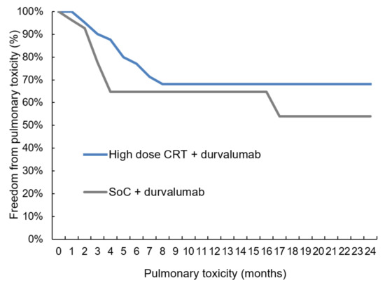
Introduction: The standard of care (SoC) for unresectable stage III non-small-cell lung cancer (NSCLC) is durvalumab maintenance therapy after concurrent chemoradiation in patients with PD-L1 > 1%. However, the concurrent approach is only amenable for about one-third of patients due to co-morbidities. Although sequential regimens are usually not regarded as curative, these schedules applied in a dose-escalated manner may be similarly radical as SoC. As combining high-dose radiation and durvalumab remains a question of debate this retrospective bi-center study aims to evaluate pulmonary toxicity after high-dose chemoradiotherapy beyond 70 Gy compared to SoC. Patients and Methods: Patients with NSCLC stage III received durvalumab after either sequential high-dose chemoradiation or concomitant SoC. Chemotherapy consisted of platinum combined with either pemetrexed, taxotere, vinorelbine, or gemcitabine. The primary endpoint was short-term pulmonary toxicity occurring within six months after the end of radiotherapy (RT). Results: A total of 78 patients were eligible for this analysis. 18F-FDG-PET-CT, cranial MRT, and histological/cytological verification were mandatory in the diagnostic work-up. The high-dose and SoC group included 42/78 (53.8%) and 36/78 (46.2%) patients, respectively, which were matched according to baseline clinical variables. While the interval between the end of RT and the start of durvalumab was equal in both groups (p = 0.841), more courses were administered in the high-dose cohort (p = 0.031). Pulmonary toxicity was similar in both groups (p = 0.599), whereas intrathoracic disease control was better in the high-dose group (local control p = 0.081, regional control p = 0.184). Conclusion: The data of this hypothesis-generating study suggest that sequential high-dose chemoradiation followed by durvalumab might be similar to SoC in terms of pulmonary toxicity and potentially more effective with respect to intra-thoracic disease control. Larger trials with a prospective design are warranted to validate these results. Full article
Show Figures

Figure 1

6 pages, 246 KiB  
Article
Role of Pre-Operative Brain Imaging in Patients with NSCLC Stage I: A Retrospective, Multicenter Analysis
by Luis Filipe Azenha, Pietro Bertoglio, Peter Kestenholz, Michel Gonzalez, Matyas Pal, Thorsten Krueger, Bassam Redwan, Volkan Koesek, Eyad Al Masri, Takuro Miyazaki, Farahnaz Sadegh Beigee, Benedetta Bedetti, Philipp Schnorr, Joachim Schmidt, Patrick Zardo, Laura Boschetti, Sven Oliver Schumann and Fabrizio Minervini
Cancers 2022, 14(10), 2419; https://doi.org/10.3390/cancers14102419 - 13 May 2022
Cited by 2 | Viewed by 2186
Abstract
Background: Lung cancer is the worldwide leading oncological cause of death in both genders combined and accounts for around 40–50% of brain metastases in general. In early-stage lung cancer, the incidence of brain metastases is around 3%. Since the early detection of asymptomatic [...] Read more.
Background: Lung cancer is the worldwide leading oncological cause of death in both genders combined and accounts for around 40–50% of brain metastases in general. In early-stage lung cancer, the incidence of brain metastases is around 3%. Since the early detection of asymptomatic cerebral metastases is of prognostic value, the aim of this study was to analyze the incidence of brain metastases in early-stage lung cancer and identify possible risk factors. Methods: We conducted a retrospective multicentric analysis of patients with Stage I (based on T and N stage only) Non-Small Cell Lung Cancer (NSCLC) who had received preoperative cerebral imaging in the form of contrast-enhanced CT or MRI. Patients with a history of NSCLC, synchronous malignancy, or neurological symptoms were excluded from the study. Analyzed variables were gender, age, tumor histology, cerebral imaging findings, smoking history, and tumor size. Results were expressed as mean with standard deviation or median with range. Results: In total, 577 patients were included in our study. Eight (1.4%) patients were found to have brain metastases in preoperative brain imaging. Tumor histology was adenocarcinoma in all eight cases. Patients were treated with radiotherapy (five), surgical resection (two), or both (one) prior to thoracic surgical treatment. Other than tumor histology, no statistically significant characteristics were found to be predictive of brain metastases. Conclusion: Given the low incidence of brain metastases in patients with clinical Stage I NSCLC, brain imaging in this cohort could be avoided. Full article
20 pages, 2877 KiB  
Article
Spread Through Air Spaces (STAS) Is an Independent Prognostic Factor in Resected Lung Squamous Cell Carcinoma
by Sami Dagher, Abdulrazzaq Sulaiman, Sophie Bayle-Bleuez, Claire Tissot, Valérie Grangeon-Vincent, David Laville, Pierre Fournel, Olivier Tiffet and Fabien Forest
Cancers 2022, 14(9), 2281; https://doi.org/10.3390/cancers14092281 - 3 May 2022
Cited by 6 | Viewed by 2125
Abstract
Objective: There is no histoprognostic grading for lung squamous cell carcinoma (LUSC). Different prognostic factors have been described in the recent literature and are not always studied in parallel. Our objective was to search for morphological histopathological prognostic factors in LUSC. Materials and [...] Read more.
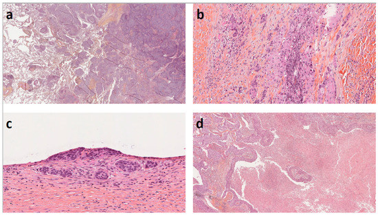
Objective: There is no histoprognostic grading for lung squamous cell carcinoma (LUSC). Different prognostic factors have been described in the recent literature and are not always studied in parallel. Our objective was to search for morphological histopathological prognostic factors in LUSC. Materials and Methods: In this single-center retrospective study of 241 patients, all patients with LUSC who underwent surgical excision over a 12-year period were included. The primary endpoint was 5-year overall survival. Results: STAS was present in 86 (35.7%) patients. The presence of Spread Through Air Spaces (STAS) was correlated with tumor location (p < 0.001), pathological stage (p = 0.039), tumor differentiation (p = 0.029), percentage of necrosis (p = 0.004), presence of vascular and/or lymphatic emboli, budding (p = 0.02), single cell invasion (p = 0.002) and tumor nest size (p = 0.005). The percentage of tumor necrosis was correlated with the overall survival at 5 years: 44.6% of patients were alive when the percentage of necrosis was ≥50%, whereas 68.5% were alive at 5 years when the necrosis was <30% (p < 0.001). When vasculolymphatic emboli were present, the percentage of survival at 5 years was 42.5% compared to 65.5% when they were absent (p = 0.002). The presence of isolated cell invasion was correlated with a lower 5-year survival rate: 51.1% in the case of presence, versus 66% in the case of absence (p = 0.02). In univariate analysis, performance status, pathological stage pT or pN, pleural invasion, histopathological subtype, percentage of tumor necrosis, vasculolymphatic invasion, single-cell invasion, budding and tumor nest size correlated with the percentage of survival at 5 years. On multivariate analysis, only STAS > 3 alveoli (HR, 2.74; 95% CI, 1.18–6.33) was related to overall survival. Conclusion: In conclusion, extensive STAS is an independent factor of poor prognosis in LUSC. STAS is correlated with the presence of other poor prognostic factors such as emboli and pleural invasion and would reflect greater tumor aggressiveness. Full article
Show Figures

Figure 1

9 pages, 251 KiB  
Article
Unplanned Return to the Operating Room after Elective Oncologic Thoracic Surgery: A Further Quality Indicator in Surgical Oncology
by Francesco Petrella, Monica Casiraghi, Davide Radice, Claudia Bardoni, Andrea Cara, Shehab Mohamed, Daniele Sances and Lorenzo Spaggiari
Cancers 2022, 14(9), 2064; https://doi.org/10.3390/cancers14092064 - 20 Apr 2022
Cited by 5 | Viewed by 2362
Abstract
Background: An unplanned return to the operating room (UROR) is defined as a readmission to the operating room because of a complication or an untoward outcome related to the initial surgery. The aim of the present report is to evaluate the role of [...] Read more.
Background: An unplanned return to the operating room (UROR) is defined as a readmission to the operating room because of a complication or an untoward outcome related to the initial surgery. The aim of the present report is to evaluate the role of URORs after elective oncologic thoracic surgery. Methods: In the study, 4012 consecutive patients were enrolled; among them, 71 patients (1.76%) had an unplanned return to the operating room. Age, sex, Charlson comorbidity index, induction treatments, type of the first operation, indication to readmission to the operating room and type of second operation, length of stay, complication after reoperation and outcomes were collected. Results: The mean age was 63.3 (SD: 13.0); there were 53 male patients (74.6%); the type of the first procedure was: lower lobectomy (11.3%), middle lobectomy (1.4%), upper lobectomy (22.5%), metastasectomy (5.6%), extrapleural pneumonectomy (4.2%), pneumonectomy (40.9%), pleural biopsy (5.6%) and other procedures (8.5%). Patients presenting complications after UROR had undergone a significantly longer first procedure (p < 0.02), had a longer length of stay (p < 0.001) and had higher post-operative mortality (p < 0.001). Conclusions: The patients experiencing UROR after elective oncologic thoracic surgery have significantly higher morbidity and mortality rates when compared to standard thoracic surgery. Bronchopleural fistula remains the most lethal complication in patients undergoing UROR. Full article
15 pages, 2222 KiB  
Article
Heterogeneity of Synchronous Lung Metastasis Calls for Risk Stratification and Prognostic Classification: Evidence from a Population-Based Database
by Shuncong Wang, Lei Chen, Yuanbo Feng, Johan V. Swinnen, Charles Jonscher, Chantal Van Ongeval and Yicheng Ni
Cancers 2022, 14(7), 1608; https://doi.org/10.3390/cancers14071608 - 22 Mar 2022
Cited by 3 | Viewed by 1951
Abstract
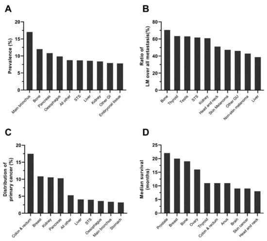
The epidemiology and associated potential heterogeneity of synchronous lung metastasis (sLM) have not been reported at a population-based level. Cancer patients with valid information about sLM status in the Surveillance, Epidemiology, and End Results database were enrolled. The prevalence of sLM, with a [...] Read more.
The epidemiology and associated potential heterogeneity of synchronous lung metastasis (sLM) have not been reported at a population-based level. Cancer patients with valid information about sLM status in the Surveillance, Epidemiology, and End Results database were enrolled. The prevalence of sLM, with a 95% confidential interval, and median survival of sLM, with interquartile range, were calculated and compared by Chi-square analyses and log-rank tests by primary cancer type and clinicopathological factors. Furthermore, the risk factors of sLM development were identified by multivariate logistic regression. Among 1,672,265 enrolled cases, 3.3% cases were identified with sLM, with a median survival of 7 months. Heterogeneity in prevalence and prognosis in sLM was observed among different primary cancers, with the highest prevalence in main bronchus cancer and best survival in testis cancer. Higher prevalence and poorer prognosis were observed in the older population, male population, African American, patients with lower socioeconomic status, and cases with advanced T stage, N stage, or more malignant pathological characteristics. Race, age, T stage, N stage, metastasis to other sites, insurance status and marital status were associated with sLM development (p < 0.001). The current study highlights the heterogeneity of the prevalence and prognosis in patients with sLM. Full article
Show Figures

Figure 1

18 pages, 398 KiB  
Review
Exploring the Impact of the Obesity Paradox on Lung Cancer and Other Malignancies
by Lindsay Joyce Nitsche, Sarbajit Mukherjee, Kareena Cheruvu, Cathleen Krabak, Rohit Rachala, Kalyan Ratnakaram, Priyanka Sharma, Maddy Singh and Sai Yendamuri
Cancers 2022, 14(6), 1440; https://doi.org/10.3390/cancers14061440 - 11 Mar 2022
Cited by 19 | Viewed by 4266
Abstract
There is a paradoxical relationship between obesity, as measured by BMI, and many types of cancer, including non-small-cell lung cancer. Obese non-small-cell lung cancer patients have been shown to fare better than their non-obese counterparts. To analyze the multifaceted effects of obesity on [...] Read more.
There is a paradoxical relationship between obesity, as measured by BMI, and many types of cancer, including non-small-cell lung cancer. Obese non-small-cell lung cancer patients have been shown to fare better than their non-obese counterparts. To analyze the multifaceted effects of obesity on oncologic outcomes, we reviewed the literature on the obesity paradox, methods to measure adiposity, the obesity-related derangements in immunology and metabolism, and the oncologic impact of confounding variables such as gender, smoking, and concomitant medications such as statins and metformin. We analyzed how these aspects may contribute to the obesity paradox and cancer outcomes with a focus on lung cancer. We concluded that the use of BMI to measure adiposity is limited and should be replaced by a method that can differentiate abdominal obesity. We also concluded that the concomitant metabolic and immunologic derangements caused by obesity contribute to the obesity paradox. Medications, gender, and smoking are additional variables that impact oncologic outcomes, and further research needs to be performed to solidify the mechanisms. Full article
17 pages, 1040 KiB  
Review
Application of Artificial Intelligence in Lung Cancer
by Hwa-Yen Chiu, Heng-Sheng Chao and Yuh-Min Chen
Cancers 2022, 14(6), 1370; https://doi.org/10.3390/cancers14061370 - 8 Mar 2022
Cited by 57 | Viewed by 11803
Abstract
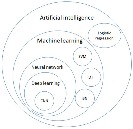
Lung cancer is the leading cause of malignancy-related mortality worldwide due to its heterogeneous features and diagnosis at a late stage. Artificial intelligence (AI) is good at handling a large volume of computational and repeated labor work and is suitable for assisting doctors [...] Read more.
Lung cancer is the leading cause of malignancy-related mortality worldwide due to its heterogeneous features and diagnosis at a late stage. Artificial intelligence (AI) is good at handling a large volume of computational and repeated labor work and is suitable for assisting doctors in analyzing image-dominant diseases like lung cancer. Scientists have shown long-standing efforts to apply AI in lung cancer screening via CXR and chest CT since the 1960s. Several grand challenges were held to find the best AI model. Currently, the FDA have approved several AI programs in CXR and chest CT reading, which enables AI systems to take part in lung cancer detection. Following the success of AI application in the radiology field, AI was applied to digitalized whole slide imaging (WSI) annotation. Integrating with more information, like demographics and clinical data, the AI systems could play a role in decision-making by classifying EGFR mutations and PD-L1 expression. AI systems also help clinicians to estimate the patient’s prognosis by predicting drug response, the tumor recurrence rate after surgery, radiotherapy response, and side effects. Though there are still some obstacles, deploying AI systems in the clinical workflow is vital for the foreseeable future. Full article
Show Figures

Figure 1

14 pages, 2006 KiB  
Article
Evaluation of Microscopic Tumour Extension in Localized Stage Non-Small-Cell Lung Cancer for Stereotactic Radiotherapy Planning
by Martin Schmitt, Lucie Aussenac, Joseph Seitlinger, Véronique Lindner, Georges Noël and Delphine Antoni
Cancers 2022, 14(5), 1282; https://doi.org/10.3390/cancers14051282 - 2 Mar 2022
Viewed by 2947
Abstract
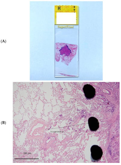
Background: Stereotactic radiotherapy for localised stage non-small-cell lung carcinoma (NSCLC) is an alternative indication for patients who are inoperable or refuse surgery. A study showed that the microscopic tumour extension (ME) of NSCLC varied according to the histological type, which allowed us to [...] Read more.
Background: Stereotactic radiotherapy for localised stage non-small-cell lung carcinoma (NSCLC) is an alternative indication for patients who are inoperable or refuse surgery. A study showed that the microscopic tumour extension (ME) of NSCLC varied according to the histological type, which allowed us to deduce adapted margins for the clinical target volume (CTV). However, to date, no study has been able to define the most relevant margins for patients with stage 1 tumours. Methods: We performed a retrospective analysis including patients with adenocarcinoma (ADC) or squamous cell carcinoma (SCC) of localised stage T1N0 or T2aN0 who underwent surgery. The ME was measured from this boundary. The profile of the type of tumour spread was also evaluated. Results: The margin required to cover the ME of a localised NSCLC with a 95% probability is 4.4 mm and 2.9 mm for SCC and ADC, respectively. A significant difference in the maximum distance of the ME between the tumour-infiltrating lymphocytes (TILs), 0–10% and 50–90% (p < 0.05), was noted for SCC. There was a significant difference in the maximum ME distance based on whether the patient had chronic obstructive pulmonary disease (COPD) (p = 0.011) for ADC. Multivariate analysis showed a statistically significant relationship between the maximum microextension distance and size with the shrinkage coefficient. Conclusion: This study definitively demonstrated that the ME depends on the pathology subtype of NSCLC. According to International Commission on Radiation Units and Measurements (ICRU) reports, 50, 62 and 83 CTV margins, proposed by these results, should be added to the GTV (Gross tumour volume). When stereotactic body radiation therapy is used, this approach should be considered in conjunction with the dataset and other margins to be applied. Full article
Show Figures

Figure 1

10 pages, 606 KiB  
Article
Factors Associated with Nodal Upstaging in Clinical T1a-bN0M0 Non-Small Cell Lung Cancers
by Tung-Ming Tsai, Chao-Yu Liu, Mong-Wei Lin, Hsao-Hsun Hsu and Jin-Shing Chen
Cancers 2022, 14(5), 1277; https://doi.org/10.3390/cancers14051277 - 1 Mar 2022
Cited by 11 | Viewed by 2330
Abstract
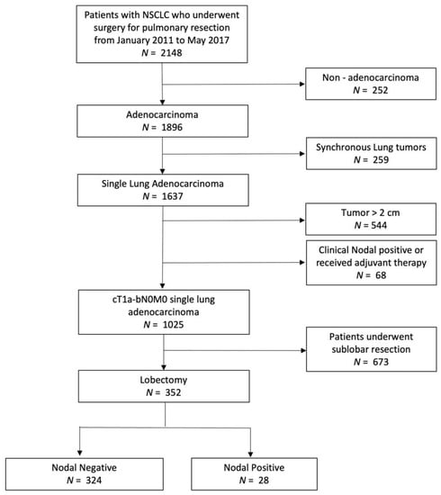
Nodal upstaging of lung adenocarcinoma occurs when unexpected pathological lymph node metastasis is found after surgical intervention, and may be associated with a worse prognosis. In this study, we aimed to determine the predictive factors of nodal upstaging in cT1a-bN0M0 primary lung adenocarcinoma. [...] Read more.
Nodal upstaging of lung adenocarcinoma occurs when unexpected pathological lymph node metastasis is found after surgical intervention, and may be associated with a worse prognosis. In this study, we aimed to determine the predictive factors of nodal upstaging in cT1a-bN0M0 primary lung adenocarcinoma. We retrospectively reviewed a prospective database (January 2011 to May 2017) at National Taiwan University Hospital and identified patients with cT1a-bN0M0 (solid part tumor diameter ≤ 2 cm) lung adenocarcinoma who underwent video-assisted thoracoscopic lobectomy. Logistic regression models and survival analysis were used to examine and compare the predictive factors of nodal upstaging. A total of 352 patients were included. Among them, 28 (7.8%) patients had nodal upstaging. Abnormal preoperative serum carcinoembryonic antigen (CEA) levels, solid part tumor diameter ≥ 1.3 cm, and consolidation–tumor (C/T) ratio ≥ 0.50 on chest computed tomography (CT) were significant predictive factors associated with nodal upstaging, and patients with nodal upstaging tended to have worse survival. Standard lobectomy is recommended for patients with these predictive factors. If neither of the predictive factors are positive, a less invasive procedure may be a reasonable alternative. Further studies are needed to verify these data. Full article
Show Figures

Figure 1

23 pages, 893 KiB  
Review
Review of Therapeutic Strategies for Anaplastic Lymphoma Kinase-Rearranged Non-Small Cell Lung Cancer
by Takafumi Fukui, Motoko Tachihara, Tatsuya Nagano and Kazuyuki Kobayashi
Cancers 2022, 14(5), 1184; https://doi.org/10.3390/cancers14051184 - 24 Feb 2022
Cited by 23 | Viewed by 4253
Abstract
Non-small cell lung cancer (NSCLC) with anaplastic lymphoma kinase rearrangement (ALK) was first reported in 2007. ALK-rearranged NSCLC accounts for about 3–8% of NSCLC. The first-line therapy for ALK-rearranged advanced NSCLC is tyrosine kinase inhibitors (TKI) targeting ALK. Following the development of crizotinib, [...] Read more.
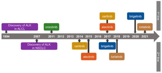
Non-small cell lung cancer (NSCLC) with anaplastic lymphoma kinase rearrangement (ALK) was first reported in 2007. ALK-rearranged NSCLC accounts for about 3–8% of NSCLC. The first-line therapy for ALK-rearranged advanced NSCLC is tyrosine kinase inhibitors (TKI) targeting ALK. Following the development of crizotinib, the first ALK-TKI, patient prognosis has been greatly improved. Currently, five TKIs are approved by the FDA. In addition, clinical trials of the novel TKI, ensartinib, and fourth-generation ALK-TKI for compound ALK mutation are ongoing. Treatment with angiogenesis inhibitors and immune checkpoint inhibitors is also being studied. However, as the disease progresses, cancers tend to develop resistance mechanisms. In addition to ALK mutations, other mechanisms, including the activation of bypass signaling pathways and histological transformation, cause resistance, and the identification of these mechanisms is important in selecting subsequent therapy. Studies on tissue and liquid biopsy have been reported and are expected to be useful tools for identifying resistance mechanisms. The purpose of this manuscript is to provide information on the recent clinical trials of ALK-TKIs, angiogenesis inhibitors, immune checkpoint inhibitors, and chemotherapy to describe tissue and liquid biopsy as a method to investigate the mechanisms of resistance against ALK-TKIs and suggest a proposed treatment algorithm. Full article
Show Figures

Figure 1

11 pages, 3882 KiB  
Article
Diagnostic Value of Bronchoscopy for Peripheral Metastatic Lung Tumors
by Yoshie Tsujimoto, Yuji Matsumoto, Midori Tanaka, Tatsuya Imabayashi, Keigo Uchimura and Takaaki Tsuchida
Cancers 2022, 14(2), 375; https://doi.org/10.3390/cancers14020375 - 13 Jan 2022
Cited by 4 | Viewed by 2169
Abstract
Although lungs are one of the most frequent sites of metastasis for malignant tumors, little has been reported about the value of bronchoscopy for lung metastases presenting with peripheral pulmonary lesions (PPLs). This retrospective cohort study investigated the diagnostic value of bronchoscopy for [...] Read more.
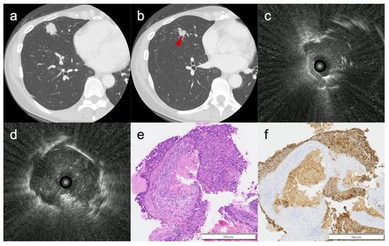
Although lungs are one of the most frequent sites of metastasis for malignant tumors, little has been reported about the value of bronchoscopy for lung metastases presenting with peripheral pulmonary lesions (PPLs). This retrospective cohort study investigated the diagnostic value of bronchoscopy for peripheral metastatic lung tumors. Consecutive patients who underwent diagnostic bronchoscopy with radial endobronchial ultrasound for PPLs and were finally diagnosed with metastatic lung tumors from April 2012 to March 2019 were included. We analyzed 235 PPLs, with a median size of 18.8 mm. The overall diagnostic yield was 76.6%. In a multivariable analysis, large lesion size (>20.0 mm vs. <20.0 mm: 87.6% vs. 67.7%, p = 0.043, OR = 2.26), inner location (inner 2/3 vs. outer 1/3: 84.8% vs. 69.1%, p = 0.004, OR = 2.79), and visibility on radiography (visible vs. invisible: 83.2% vs. 56.1%, p = 0.015, OR = 3.29) significantly affected the diagnostic yield. Although a positive bronchus sign tended to have a higher yield, no significant difference was observed (81.8% vs. 70.6%, p = 0.063). Only one case of lung abscess was observed, with no serious complications. In conclusion, bronchoscopy is a valuable technique for peripheral metastatic lung tumors, with good diagnostic accuracy and safety. Full article
Show Figures

Figure 1

12 pages, 292 KiB  
Review
Recent Advances in the Diagnosis and Management of Multiple Primary Lung Cancer
by Chi-Lu Chiang, Ping-Chung Tsai, Yi-Chen Yeh, Yuan-Hung Wu, Han-Shui Hsu and Yuh-Min Chen
Cancers 2022, 14(1), 242; https://doi.org/10.3390/cancers14010242 - 4 Jan 2022
Cited by 19 | Viewed by 4040
Abstract
With the wide application of computed tomography in lung cancer screening, the incidence of multiple primary lung cancer (MPLC) has been increasingly reported. Despite the established criteria, the differentiation between MPLC and intrapulmonary metastasis remains challenging. Although histologic features are helpful in some [...] Read more.
With the wide application of computed tomography in lung cancer screening, the incidence of multiple primary lung cancer (MPLC) has been increasingly reported. Despite the established criteria, the differentiation between MPLC and intrapulmonary metastasis remains challenging. Although histologic features are helpful in some circumstances, a molecular analysis is often needed. The application of next-generation sequencing could aid in distinguishing MPLCs from intrapulmonary metastasis, decreasing ambiguity. For MPLC management, surgery with lobectomy is the main operation method. Limited resection does not appear to negatively affect survival, and it is a reasonable alternative. Stereotactic ablative radiotherapy (SABR) has become a standard of care for patients refusing surgery or for those with medically inoperable early-stage lung cancer. However, the efficacy of SABR in MPLC management could only be found in retrospective series. Other local ablation techniques are an emerging alternative for the control of residual lesions. Furthermore, systemic therapies, such as targeted therapy for oncogene-addicted patients, and immunotherapy have shown promising results in MPLC management after resection. In this paper, the recent advances in the diagnosis and management of MPLC are reviewed. Full article

2021

Jump to: 2024, 2023, 2022

14 pages, 731 KiB  
Article
Sex-Based Clinical Outcome in Advanced NSCLC Patients Undergoing PD-1/PD-L1 Inhibitor Therapy—A Retrospective Bi-Centric Cohort Study
by David Lang, Anna Brauner, Florian Huemer, Gabriel Rinnerthaler, Andreas Horner, Romana Wass, Elmar Brehm, Bernhard Kaiser, Richard Greil and Bernd Lamprecht
Cancers 2022, 14(1), 93; https://doi.org/10.3390/cancers14010093 - 24 Dec 2021
Cited by 7 | Viewed by 2703
Abstract
Men with non-small cell lung cancer (NSCLC) have a more favorable response to immune-checkpoint inhibitor (ICI) monotherapy, while women especially benefit from ICI-chemotherapy (CHT) combinations. To elucidate such sex differences in clinical practice, we retrospectively analyzed two cohorts treated with either ICI monotherapy [...] Read more.
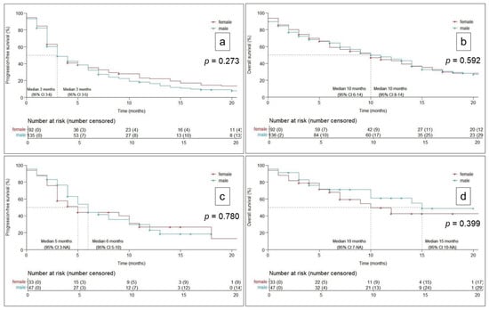
Men with non-small cell lung cancer (NSCLC) have a more favorable response to immune-checkpoint inhibitor (ICI) monotherapy, while women especially benefit from ICI-chemotherapy (CHT) combinations. To elucidate such sex differences in clinical practice, we retrospectively analyzed two cohorts treated with either ICI monotherapy (n = 228) or ICI-CHT combination treatment (n = 80) for advanced NSCLC. Kaplan–Meier analyses were used to calculate progression-free (PFS) and overall survival (OS), influencing variables were evaluated using Cox-regression analyses. No significant sex differences for PFS/OS could be detected in either cohort. Men receiving ICI monotherapy had a statistically significant independent impact on PFS by Eastern Cooperative Oncology Group performance status (ECOG) ≥2 (hazard ratio (HR) 1.90, 95% confidence interval (CI): 1.10–3.29, p = 0.021), higher C-reactive protein (CRP; HR 1.06, 95%CI: 1.00–1.11, p = 0.037) and negative programmed death-ligand 1 (PD-L1) status (HR 2.04, 95%CI: 1.32–3.15, p = 0.001), and on OS by CRP (HR 1.09, 95%CI: 1.03–1.14, p = 0.002). In men on ICI-CHT combinations, multivariate analyses (MVA) revealed squamous histology (HR 4.00, 95%CI: 1.41–11.2, p = 0.009) significant for PFS; and ECOG ≥ 2 (HR 5.58, 95%CI: 1.88–16.5, p = 0.002) and CRP (HR 1.19, 95%CI: 1.06–1.32, p = 0.002) for OS. Among women undergoing ICI monotherapy, no variable proved significant for PFS, while ECOG ≥ 2 had a significant interaction with OS (HR 1.90, 95%CI 1.04–3.46, p = 0.037). Women treated with ICI-CHT had significant MVA findings for CRP with both PFS (HR 1.09, 95%CI: 1.02–1.16, p = 0.007) and OS (HR 1.11, 95%CI: 1.03–1.19, p = 0.004). Although men and women responded similarly to both ICI mono- and ICI-CHT treatment, predictors of response differed by sex. Full article
Show Figures

Figure 1

11 pages, 1361 KiB  
Article
Endobronchial Ultrasonography with a Guide Sheath Transbronchial Biopsy for Diagnosing Peripheral Pulmonary Lesions within or near Fibrotic Lesions in Patients with Interstitial Lung Disease
by Takayasu Ito, Shotaro Okachi, Tomoki Kimura, Kensuke Kataoka, Yasuhiko Suzuki, Fumie Kinoshita, Keiko Wakahara, Naozumi Hashimoto and Yasuhiro Kondoh
Cancers 2021, 13(22), 5751; https://doi.org/10.3390/cancers13225751 - 17 Nov 2021
Cited by 6 | Viewed by 2061
Abstract
In patients with interstitial lung disease (ILD), the most frequent locations of lung cancer are within or near fibrotic lesions. However, the diagnostic yield for peripheral pulmonary lesions (PPLs) within or near fibrotic lesions using endobronchial ultrasonography with a guide sheath transbronchial biopsy [...] Read more.
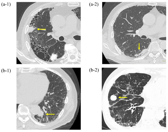
In patients with interstitial lung disease (ILD), the most frequent locations of lung cancer are within or near fibrotic lesions. However, the diagnostic yield for peripheral pulmonary lesions (PPLs) within or near fibrotic lesions using endobronchial ultrasonography with a guide sheath transbronchial biopsy (EBUS-GS TBB) may be unsatisfactory compared to that for PPLs distant from fibrotic lesions because of the difficulty in reaching the lesions. Our objectives were to evaluate the yield for PPLs using EBUS-GS TBB according to the proximity of PPLs to fibrotic lesions and to determine factors affecting the yield for PPLs. We retrospectively investigated 323 consecutive lesions using EBUS-GS TBB between 1 November 2014 and 31 December 2016. We identified PPLs with ILD in such lesions. PPLs with ILD were divided into PPLs within or near fibrotic lesions which met the criterion of PPLs, and of fibrotic lesions overlapping each other (PPLs-FL) and those distant from fibrotic lesions, which met the criterion of PPLs and the area of fibrotic lesion not overlapping each other (PPLs-NFL). Of the 323 lesions, 55 were included (31 PPLs-FL and 24 PPLs-NFL). The diagnostic yield for PPLs-FL was significantly lower than for PPLs-NFL (45.2% vs. 83.3%, p = 0.004). Multivariate analysis revealed that PPLs-NFL (odds ratio (OR) = 7.509) and a probe position within the lesion (OR = 4.172) were significant factors affecting diagnostic yield. Lesion’s positional relation to fibrotic lesions and the probe position were important factors affecting the successful diagnosis via EBUS-GS TBB in these patients. Full article
Show Figures

Figure 1

18 pages, 335 KiB  
Review
Management of Resectable Stage III-N2 Non-Small-Cell Lung Cancer (NSCLC) in the Age of Immunotherapy
by Xabier Mielgo-Rubio, Sara Montemuiño, Unai Jiménez, Javier Luna, Ana Cardeña, Laura Mezquita, Margarita Martín and Felipe Couñago
Cancers 2021, 13(19), 4811; https://doi.org/10.3390/cancers13194811 - 26 Sep 2021
Cited by 19 | Viewed by 4176
Abstract
Stage III non-small-cell lung cancer (NSCLC) with N2 lymph node involvement is a heterogeneous group with different potential therapeutic approaches. Patients with potentially resectable III-N2 NSCLC are those who are considered to be able to receive a multimodality treatment that includes tumour resection [...] Read more.
Stage III non-small-cell lung cancer (NSCLC) with N2 lymph node involvement is a heterogeneous group with different potential therapeutic approaches. Patients with potentially resectable III-N2 NSCLC are those who are considered to be able to receive a multimodality treatment that includes tumour resection after neoadjuvant therapy. Current treatment for these patients is based on neoadjuvant chemotherapy +/− radiotherapy followed by surgery and subsequent assessment for adjuvant chemotherapy and/or radiotherapy. In addition, some selected III-N2 patients could receive upfront surgery or pathologic N2 incidental involvement can be found a posteriori during analysis of the surgical specimen. The standard treatment for these patients is adjuvant chemotherapy and evaluation for complementary radiotherapy. Despite being a locally advanced stage, the cure rate for these patients continues to be low, with a broad improvement margin. The most immediate hope for improving survival data and curing these patients relies on integrating immunotherapy into perioperative treatment. Immunotherapy based on anti-PD1/PD-L1 immune checkpoint inhibitors is already a standard treatment in stage III unresectable and advanced NSCLC. Data from the first phase II studies in monotherapy neoadjuvant therapy and, in particular, in combination with chemotherapy, are highly promising, with impressive improved and complete pathological response rates. Despite the lack of confirmatory data from phase III trials and long-term survival data, and in spite of various unresolved questions, immunotherapy will soon be incorporated into the armamentarium for treating stage III-N2 NSCLC. In this article, we review all therapeutic approaches to stage III-N2 NSCLC, analysing both completed and ongoing studies that evaluate the addition of immunotherapy with or without chemotherapy and/or radiotherapy. Full article
14 pages, 284 KiB  
Review
Adjuvant Treatment with Tyrosine Kinase Inhibitors in Epidermal Growth Factor Receptor Mutated Non-Small-Cell Lung Carcinoma Patients, Past, Present and Future
by Walid Shalata, Binil Mathew Jacob and Abed Agbarya
Cancers 2021, 13(16), 4119; https://doi.org/10.3390/cancers13164119 - 16 Aug 2021
Cited by 11 | Viewed by 3012
Abstract
Lung cancer is the most common malignancy across the world. The new era in lung cancer treatments, especially this past decade, has yielded novel categories of targeted therapy for specific mutations and adjuvant therapy, both of which have led to improved survival rates. [...] Read more.
Lung cancer is the most common malignancy across the world. The new era in lung cancer treatments, especially this past decade, has yielded novel categories of targeted therapy for specific mutations and adjuvant therapy, both of which have led to improved survival rates. In the present study, we review the changes and development of treatments, with a special focus on adjuvant therapy using tyrosine kinase inhibitors (TKIs) administered to non-small-cell lung carcinoma patients who had a complete resection of the tumor harboring a mutated epidermal growth factor receptor. The clinical trials are dating from the past (chemotherapy trials), present (TKIs), and future (ongoing trials). Full article
9 pages, 290 KiB  
Article
Prognostic Value of the Hemoglobin/Red Cell Distribution Width Ratio in Resected Lung Adenocarcinoma
by Francesco Petrella, Monica Casiraghi, Davide Radice, Andrea Cara, Gabriele Maffeis, Elena Prisciandaro, Stefania Rizzo and Lorenzo Spaggiari
Cancers 2021, 13(4), 710; https://doi.org/10.3390/cancers13040710 - 9 Feb 2021
Cited by 20 | Viewed by 2347
Abstract
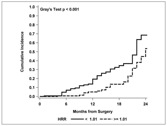
Background: The ratio of hemoglobin to red cell distribution width (HRR) has been described as an effective prognostic factor in several types of cancer. The aim of this study was to investigate the prognostic role of preoperative HRR in resected-lung-adenocarcinoma patients. Methods: We [...] Read more.
Background: The ratio of hemoglobin to red cell distribution width (HRR) has been described as an effective prognostic factor in several types of cancer. The aim of this study was to investigate the prognostic role of preoperative HRR in resected-lung-adenocarcinoma patients. Methods: We enrolled 342 consecutive patients. Age, sex, surgical resection, adjuvant treatments, pathological stage, preoperative hemoglobin, red cell distribution width, and their ratio were recorded for each patient. Results: Mean age was 66 years (SD: 9.0). There were 163 females (47.1%); 169 patients (49.4%) had tumors at stage I, 71 (20.8%) at stage II, and 102 (29.8%) at stage III. In total, 318 patients (93.0%) underwent lobectomy, and 24 (7.0%) pneumonectomy. Disease-free survival multivariable analysis disclosed an increased hazard ratio (HR) of relapse for preoperative HRR lower than 1.01 (HR = 2.20, 95%CI: (1.30–3.72), p = 0.004), as well as for N1 single-node (HR = 2.55, 95%CI: (1.33–4.90), p = 0.005) and multiple-level lymph node involvement compared to N0 for both N1 (HR = 9.16, 95%CI:(3.65–23.0), p < 0.001) and N2 (HR = 10.5, 95%CI:(3.44–32.2, p < 0.001). Conclusion: Pre-operative HRR is an effective prognostic factor of disease-free survival in resected-lung-adenocarcinoma patients, together with the level of pathologic node involvement. Full article
Show Figures

Figure 1

2 pages, 182 KiB  
Editorial
Diagnosis and Treatment of Primary and Secondary Lung Cancers
by Francesco Petrella
Cancers 2021, 13(3), 448; https://doi.org/10.3390/cancers13030448 - 25 Jan 2021
Cited by 1 | Viewed by 1889
Abstract
Primary and secondary lung cancers are the most common clinical conditions that thoracic surgeons have to deal with: primary lung cancer, in fact, is one of the most frequently diagnosed cancers and is the leading cause of cancer-related death worldwide [...] Full article
Back to TopTop