Cancer Nanomedicine

A topical collection in Cancers (ISSN 2072-6694). This collection belongs to the section "Cancer Therapy".

Viewed by 133213

Editor


E-Mail Website
Collection Editor
School of Pure and Applied Chemistry, University of Strathclyde, Glasgow G1 1XQ, UK
Interests: nanomedicine; cancer nanotechnology; targeted drug delivery; pancreatic cancer; theranostics
Special Issues, Collections and Topics in MDPI journals

Topical Collection Information

Dear Colleagues,

Cancer treatments are often hindered by the lack of drug specificity, poor physicochemical properties of active pharmaceutical ingredients, poor penetration ability and drug resistance. With the discovery and characterization of an increasing number of cancer types with little improvement of the ability to diagnose, treatment options or patient prognosis, more advanced technologies are urgently required.

Nanotechnology defines particulates within the 1x10⁻⁹ m range. Particulates within the nano-sized domain often exhibit unique properties compared to their larger size scale. These can be exploited in biomedicine for applications such as imaging, cell sorting, drug delivery and targeting.

Cancer nanomedicine is rapidly becoming one of the leading areas of promise for cancer therapy, with first-generation treatments already available to patients. This Collection invites articles and reviews which encompass the many areas under the nanomedicine umbrella, including diagnostics, drug delivery and advanced therapies.

Dr. Clare Hoskins
Collection Editor

Manuscript Submission Information

Manuscripts should be submitted online at www.mdpi.com by registering and logging in to this website. Once you are registered, click here to go to the submission form. Manuscripts can be submitted until the deadline. All submissions that pass pre-check are peer-reviewed. Accepted papers will be published continuously in the journal (as soon as accepted) and will be listed together on the collection website. Research articles, review articles as well as communications are invited. For planned papers, a title and short abstract (about 100 words) can be sent to the Editorial Office for announcement on this website.

Submitted manuscripts should not have been published previously, nor be under consideration for publication elsewhere (except conference proceedings papers). All manuscripts are thoroughly refereed through a single-blind peer-review process. A guide for authors and other relevant information for submission of manuscripts is available on the Instructions for Authors page. Cancers is an international peer-reviewed open access semimonthly journal published by MDPI.

Please visit the Instructions for Authors page before submitting a manuscript. The Article Processing Charge (APC) for publication in this open access journal is 2900 CHF (Swiss Francs). Submitted papers should be well formatted and use good English. Authors may use MDPI's English editing service prior to publication or during author revisions.

Keywords

  • Nanomedicine
  • targeted therapy
  • image guidance
  • tumour penetration
  • stimuli-responsive
  • theranostics
  • drug delivery
  • nanoparticle
  • nanoencapsulation
  • diagnosis
Published Papers (34 papers)

2023

Jump to: 2022, 2021, 2020

15 pages, 4384 KiB  
Article
A Local and Abscopal Effect Observed with Liposomal Encapsulation of Intratumorally Injected Oncolytic Adenoviral Therapy
by Tao Dong, Jaimin R. Shah, Abraham T. Phung, Christopher Larson, Ana B. Sanchez, Omonigho Aisagbonhi, Sarah L. Blair, Bryan Oronsky, William C. Trogler, Tony Reid and Andrew C. Kummel
Cancers 2023, 15(12), 3157; https://doi.org/10.3390/cancers15123157 - 12 Jun 2023
Cited by 4 | Viewed by 1701
Abstract
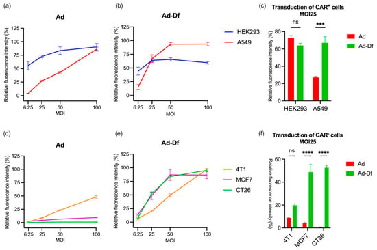
This study evaluated the in vivo therapeutic efficacy of oncolytic serotype 5 adenovirus TAV255 in CAR-deficient tumors. In vitro experiments were performed with cell lines that expressed different levels of CAR (HEK293, A549, CT26, 4T1, and MCF-7). Low CAR cells, such as CT26, [...] Read more.
This study evaluated the in vivo therapeutic efficacy of oncolytic serotype 5 adenovirus TAV255 in CAR-deficient tumors. In vitro experiments were performed with cell lines that expressed different levels of CAR (HEK293, A549, CT26, 4T1, and MCF-7). Low CAR cells, such as CT26, were poorly transduced by Ad in vitro unless the adenovirus was encapsulated in liposomes. However, the CT26 tumor in an immune-competent mouse model responded to the unencapsulated TAV255; 33% of the tumors were induced into complete remission, and mice with complete remission rejected the rechallenge with cancer cell injection. Encapsulation of TAV255 improves its therapeutic efficacy by transducing more CT26 cells, as expected from in vitro results. In a bilateral tumor model, nonencapsulated TAV255 reduced the growth rate of the locally treated tumors but had no effect on the growth rate of the distant tumor site. Conversely, encapsulated TAV255-infected CT26 induced a delayed growth rate of both the primary injected tumor and the distant tumor, consistent with a robust immune response. In vivo, intratumorally injected unencapsulated adenoviruses infect CAR-negative cells with only limited efficiency. However, unencapsulated adenoviruses robustly inhibit the growth of CAR-deficient tumors, an effect that constitutes an ‘in situ vaccination’ by stimulating cytotoxic T cells. Full article
Show Figures

Figure 1

18 pages, 3641 KiB  
Article
X-ray Photoelectron Spectroscopy (XPS) Analysis of Nitrogen Environment in Small Extracellular Vesicle Membranes: A Potential Novel Technique with Application for Cancer Screening
by María Sancho-Albero, Ana Martín-Pardillos, Silvia Irusta, Víctor Sebastián, Vicente Luis Cebolla, Roberto Pazo-Cid, Pilar Martín-Duque and Jesús Santamaría
Cancers 2023, 15(9), 2479; https://doi.org/10.3390/cancers15092479 - 26 Apr 2023
Cited by 4 | Viewed by 2254
Abstract
Small extracellular vesicle (EV) membranes display characteristic protein-lipidic composition features that are related to their cell of origin, providing valuable clues regarding their parental cell composition and real-time state. This could be especially interesting in the case of cancer cell-derived EVs, as their [...] Read more.
Small extracellular vesicle (EV) membranes display characteristic protein-lipidic composition features that are related to their cell of origin, providing valuable clues regarding their parental cell composition and real-time state. This could be especially interesting in the case of cancer cell-derived EVs, as their membranes could serve as valuable tools in liquid biopsy applications and to detect changes in the tumor malignancy. X-Ray Photoelectron Spectroscopy (XPS) is a powerful surface analysis technique able to detect every chemical element present, being also sensitive to their chemical environment. Here we explore the use of XPS as a fast technique to characterize EV membrane composition, with possible application in cancer research. Notably, we have focused on the nitrogen environment as an indicator of the relative abundance of pyridine-type bonding, primary, secondary and tertiary amines. Specifically, we have analyzed how tumoral and healthy cells have different nitrogen chemical environments that can indicate the presence or absence of malignancy. In addition, a collection of human serum samples from cancer patients and healthy donors was also analyzed. The differential XPS analysis of EVs collected from patients confirmed that the patterns of amine evolution could be related to markers of cancer disease, opening the possibility of their use as a non-invasive blood biomarker. Full article
Show Figures

Figure 1

2022

Jump to: 2023, 2021, 2020

17 pages, 4530 KiB  
Article
Molecularly Targeted Photothermal Ablation of Epidermal Growth Factor Receptor-Expressing Cancer Cells with a Polypyrrole–Iron Oxide–Afatinib Nanocomposite
by Lekshmi Rethi, Chinmaya Mutalik, Lekha Rethi, Wei-Hung Chiang, Hsin-Lun Lee, Wen-Yu Pan, Tze-Sen Yang, Jeng-Fong Chiou, Yin-Ju Chen, Er-Yuan Chuang and Long-Sheng Lu
Cancers 2022, 14(20), 5043; https://doi.org/10.3390/cancers14205043 - 14 Oct 2022
Cited by 7 | Viewed by 2234
Abstract
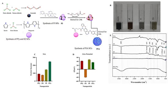
Near-infrared–photothermal therapy (NIR-PTT) is a potential modality for cancer treatment. Directing photothermal effects specifically to cancer cells may enhance the therapeutic index for the best treatment outcome. While epithelial growth factor receptor (EGFR) is commonly overexpressed/genetically altered in human malignancy, it remains unknown [...] Read more.
Near-infrared–photothermal therapy (NIR-PTT) is a potential modality for cancer treatment. Directing photothermal effects specifically to cancer cells may enhance the therapeutic index for the best treatment outcome. While epithelial growth factor receptor (EGFR) is commonly overexpressed/genetically altered in human malignancy, it remains unknown whether targeting EGFR with tyrosine kinase inhibitor (TKI)-conjugated nanoparticles may direct NIR-PTT to cancers with cellular precision. In the present study, we tested this possibility through the fabrication of a polypyrrole–iron oxide–afatinib nanocomposite (PIA-NC). In the PIA-NC, a biocompatible and photothermally conductive polymer (polypyrrole) was conjugated to a TKI (afatinib) that binds to overexpressed wild-type EGFR without overt cytotoxicity. A Fenton catalyst (iron oxide) was further encapsulated in the NC to drive the intracellular ROS surge upon heat activation. Diverse physical and chemical characterization experiments were conducted. Particle internalization, cytotoxicity, ROS production, and apoptosis in EGFR-positive and -negative cell lines were investigated in the presence and absence of NIR. We found that the PIA-NCs were stable with a size of 243 nm and a zeta potential of +35 mV. These PIA-NCs were readily internalized close to the cell membrane by all types of cells used in the study. The Fourier transform infrared spectra showed 3295 cm−1 peaks; substantial O–H stretching was seen, with significant C=C stretching at 1637 cm−1; and a modest appearance of C–O–H bending at 1444 cm−1 confirmed the chemical conjugation of afatinib but not iron oxide to the NC. At a NIR-PTT energy level that has a minimal cytotoxic effect, PIA-NC significantly sensitizes EGFR-overexpressing A549 lung cancer cells to NIR-PTT-induced cytotoxicity at a rate of 70%, but in EGFR-negative 3T3 fibroblasts the rate was 30%. Within 1 min of NIR-PTT, a surge of intracellular ROS was found in PIA-NC-treated A549 cells. This was followed by early induction of cellular apoptosis for 54 ± 0.081% of A549 cells. The number of viable cells was less than a quarter of a percent. Viability levels of A549 cells that had been treated with NIR or PIA were only 50 ± 0.216% and 80 ± 0.216%, respectively. Only 10 ± 0.816% of NIH3T3 cells had undergone necrosis, meaning that 90 ± 0.124% were alive. Viability levels were 65 ± 0.081% and 81 ± 0.2%, respectively, when only NIR and PIA were used. PIA binding was effective against A549 cells but not against NIH3T3 cells. The outcome revealed that higher levels of NC + NIR exposure caused cancer cells to produce more ROS. In summary, our findings proved that a molecularly targeted NC provides an orchestrated platform for cancer cell-specific delivery of NIR-PTT. The geometric proximity design indicates a novel approach to minimizing the off-target biological effects of NIR-PTT. The potential of PIA-NC to be further developed into real-world application warrants further investigation. Full article
Show Figures

Figure 1

16 pages, 2501 KiB  
Article
Biomimetic Red Blood Cell Membrane-Mediated Nanodrugs Loading Ursolic Acid for Targeting NSCLC Therapy
by Ting Wu, Dan Yan, Wenjun Hou, Hui Jiang, Min Wu, Yanling Wang, Gang Chen, Chunming Tang, Yijun Wang and Huae Xu
Cancers 2022, 14(18), 4520; https://doi.org/10.3390/cancers14184520 - 18 Sep 2022
Cited by 4 | Viewed by 2189
Abstract
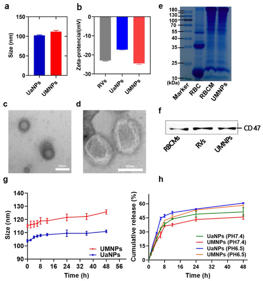
As one of the most common cancers worldwide, non-small-cell lung cancer (NSCLC) treatment always fails owing to the tumor microenvironment and resistance. UA, a traditional Chinese medicine, was reported to have antitumor potential in tumor models in vitro and in vivo, but showed [...] Read more.
As one of the most common cancers worldwide, non-small-cell lung cancer (NSCLC) treatment always fails owing to the tumor microenvironment and resistance. UA, a traditional Chinese medicine, was reported to have antitumor potential in tumor models in vitro and in vivo, but showed impressive results in its potential application for poor water solubility. In this study, a novel biomimetic drug-delivery system based on UA-loaded nanoparticles (UaNPs) with a red blood cell membrane (RBCM) coating was developed. The RBCM-coated UANPs (UMNPs) exhibited improved water solubility, high stability, good biosafety, and efficient tumor accumulation. Importantly, the excellent antitumor efficiency of the UMNPs was confirmed both in vitro and in vivo in cancer models. In addition, we further investigated the antitumor mechanism of UMNPs. The results of Western blotting showed that UMNPs exerted an anticancer effect by inducing the apoptosis and autophagy of NSCLC cells, which makes it superior to free UA. In addition, body weight monitoring, hematoxylin and eosin (HE) analysis, and immunohistochemical (IHC) analysis showed no significant difference between UMNPs and the control group, indicating the safety of UMNPs. Altogether, the preparation of biomimetic UMNPs provides a promising strategy to improve outcomes in NSCLC. Full article
Show Figures

Figure 1

17 pages, 4383 KiB  
Article
Enhanced Antitumoral Activity of Encapsulated BET Inhibitors When Combined with PARP Inhibitors for the Treatment of Triple-Negative Breast and Ovarian Cancers
by Alberto Juan, María del Mar Noblejas-López, Iván Bravo, María Arenas-Moreira, Cristina Blasco-Navarro, Pilar Clemente-Casares, Agustín Lara-Sánchez, Atanasio Pandiella, Carlos Alonso-Moreno and Alberto Ocaña
Cancers 2022, 14(18), 4474; https://doi.org/10.3390/cancers14184474 - 15 Sep 2022
Cited by 5 | Viewed by 2637
Abstract
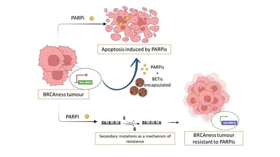
BRCA1/2 protein-deficient or mutated cancers comprise a group of aggressive malignancies. Although PARPis have shown considerably efficacy in their treatment, the widespread use of these agents in clinical practice is restricted by various factors, including the development of acquired resistance due to the [...] Read more.
BRCA1/2 protein-deficient or mutated cancers comprise a group of aggressive malignancies. Although PARPis have shown considerably efficacy in their treatment, the widespread use of these agents in clinical practice is restricted by various factors, including the development of acquired resistance due to the presence of compensatory pathways. BETis can completely disrupt the HR pathway by repressing the expression of BRCA1 and could be aimed at generation combination regimes to overcome PARPi resistance and enhance PARPi efficacy. Due to the poor pharmacokinetic profile and short half-life, the first-in-class BETi JQ1 was loaded into newly developed nanocarrier formulations to improve the effectivity of olaparib for the treatment of BRCAness cancers. First, polylactide polymeric nanoparticles were generated by double emulsion. Moreover, liposomes were prepared by ethanol injection and evaporation solvent method. JQ1-loaded drug delivery systems display optimal hydrodynamic radii between 60 and 120 nm, with a very low polydispersity index (PdI), and encapsulation efficiencies of 92 and 16% for lipid- and polymeric-based formulations, respectively. Formulations show high stability and sustained release. We confirmed that all assayed JQ1 formulations improved antiproliferative activity compared to the free JQ1 in models of ovarian and breast cancers. In addition, synergistic interaction between JQ1 and JQ1-loaded nanocarriers and olaparib evidenced the ability of encapsulated JQ1 to enhance antitumoral activity of PARPis. Full article
Show Figures

Graphical abstract

23 pages, 6964 KiB  
Review
Nanoscale Zeolitic Imidazolate Framework (ZIF)–8 in Cancer Theranostics: Current Challenges and Prospects
by Hongxin Xie, Xinyu Liu, Zhengrong Huang, Liexi Xu, Rui Bai, Fajian He, Mengqin Wang, Linzhi Han, Zhirong Bao, Yuzhou Wu, Conghua Xie and Yan Gong
Cancers 2022, 14(16), 3935; https://doi.org/10.3390/cancers14163935 - 15 Aug 2022
Cited by 41 | Viewed by 5075
Abstract
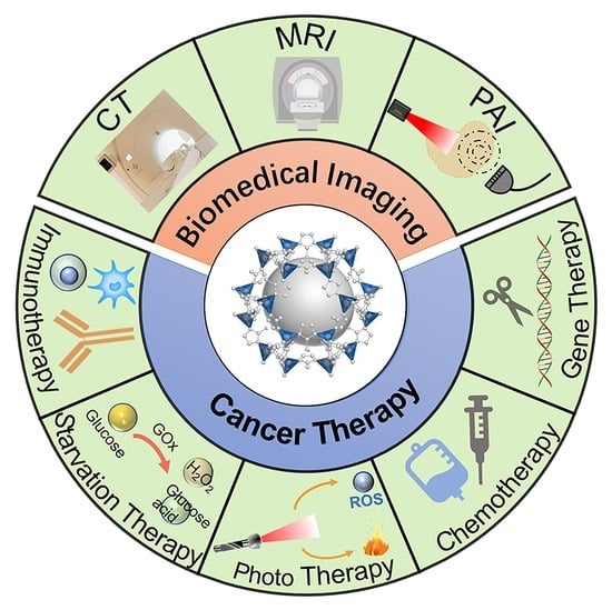
Cancer severely threatens human health and has remained the leading cause of disease–related death for decades. With the rapid advancement of nanomedicine, nanoscale metal–organic frameworks are believed to be potentially applied in the treatment and biomedical imaging for various tumors. Zeolite imidazole framework [...] Read more.
Cancer severely threatens human health and has remained the leading cause of disease–related death for decades. With the rapid advancement of nanomedicine, nanoscale metal–organic frameworks are believed to be potentially applied in the treatment and biomedical imaging for various tumors. Zeolite imidazole framework (ZIF)–8 attracts increasing attention due to its high porosity, large specific surface area, and pH–responsiveness. The designs and modifications of ZIF–8 nanoparticles, as well as the strategy of drug loading, demand a multifaceted and comprehensive understanding of nanomaterial features and tumor characteristics. We searched for studies on ZIF–8–based nanoplatforms in tumor theranostics on Web of Science from 2015 to 2022, mainly focused on the research published in the past 3 years, summarized the progress of their applications in tumor imaging and treatment, and discussed the favorable aspects of ZIF–8 nanoparticles for tumor theranostics as well as the future opportunities and potential challenges. As a kind of metal–organic framework material full of potential, ZIF–8 can be expected to be combined with more therapeutic systems in the future and continue to contribute to all aspects of tumor therapy and diagnosis. Full article
Show Figures

Graphical abstract

22 pages, 3761 KiB  
Article
Nucleolin Overexpression Predicts Patient Prognosis While Providing a Framework for Targeted Therapeutic Intervention in Lung Cancer
by Ângela Valério-Fernandes, Nuno A. Fonseca, Nélio Gonçalves, Ana F. Cruz, Marta I. Pereira, Ana C. Gregório, Vera Moura, Ana F. Ladeirinha, Ana Alarcão, Joana Gonçalves, Antero Abrunhosa, Joana B. Melo, Lina Carvalho, Sérgio Simões and João N. Moreira
Cancers 2022, 14(9), 2217; https://doi.org/10.3390/cancers14092217 - 29 Apr 2022
Cited by 8 | Viewed by 3201
Abstract
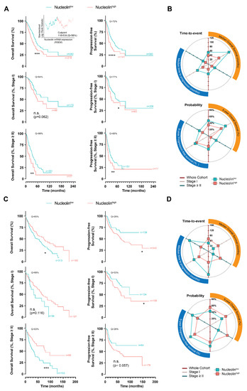
Notwithstanding the advances in the treatment of lung cancer with immune checkpoint inhibitors, the high percentage of non-responders supports the development of novel anticancer treatments. Herein, the expression of the onco-target nucleolin in patient-derived pulmonary carcinomas was characterized, along with the assessment of [...] Read more.
Notwithstanding the advances in the treatment of lung cancer with immune checkpoint inhibitors, the high percentage of non-responders supports the development of novel anticancer treatments. Herein, the expression of the onco-target nucleolin in patient-derived pulmonary carcinomas was characterized, along with the assessment of its potential as a therapeutic target. The clinical prognostic value of nucleolin for human pulmonary carcinomas was evaluated through data mining from the Cancer Genome Atlas project and immunohistochemical detection in human samples. Cell surface expression of nucleolin was evaluated by flow cytometry and subcellular fraction Western blotting in lung cancer cell lines. Nucleolin mRNA overexpression correlated with poor overall survival of lung adenocarcinoma cancer patients and further predicted the disease progression of both lung adenocarcinoma and squamous carcinoma. Furthermore, a third of the cases presented extra-nuclear expression, contrasting with the nucleolar pattern in non-malignant tissues. A two- to twelve-fold improvement in cytotoxicity, subsequent to internalization into the lung cancer cell lines of doxorubicin-loaded liposomes functionalized by the nucleolin-binding F3 peptide, was correlated with the nucleolin cell surface levels and the corresponding extent of cell binding. Overall, the results suggested nucleolin overexpression as a poor prognosis predictor and thus a target for therapeutic intervention in lung cancer. Full article
Show Figures

Figure 1

20 pages, 3593 KiB  
Article
Extracorporeal Removal of Thermosensitive Liposomal Doxorubicin from Systemic Circulation after Tumor Delivery to Reduce Toxicities
by Anjan Motamarry, A. Marissa Wolfe, Krishna K. Ramajayam, Sanket Pattanaik, Thomas Benton, Yuri Peterson, Pegah Faridi, Punit Prakash, Katherine Twombley and Dieter Haemmerich
Cancers 2022, 14(5), 1322; https://doi.org/10.3390/cancers14051322 - 4 Mar 2022
Cited by 4 | Viewed by 4660
Abstract
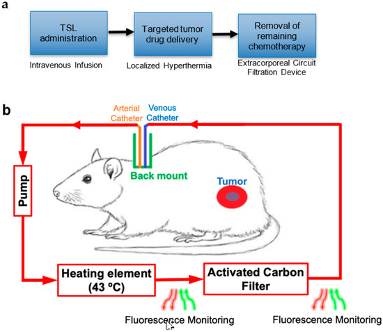
Thermosensitive liposomal doxorubicin (TSL-Dox) combined with localized hyperthermia enables targeted drug delivery. Tumor drug uptake occurs only during hyperthermia. We developed a novel method for removal of systemic TSL-Dox remaining after hyperthermia-triggered delivery to reduce toxicities. The carotid artery and jugular vein of [...] Read more.
Thermosensitive liposomal doxorubicin (TSL-Dox) combined with localized hyperthermia enables targeted drug delivery. Tumor drug uptake occurs only during hyperthermia. We developed a novel method for removal of systemic TSL-Dox remaining after hyperthermia-triggered delivery to reduce toxicities. The carotid artery and jugular vein of Norway brown rats carrying two subcutaneous BN-175 tumors were catheterized. After allowing the animals to recover, TSL-Dox was infused at 7 mg/kg dose. Drug delivery to one of the tumors was performed by inducing 15 min microwave hyperthermia (43 °C). At the end of hyperthermia, an extracorporeal circuit (ECC) comprising a heating module to release drug from TSL-Dox followed by an activated carbon filter to remove free drug was established for 1 h (n = 3). A computational model simulated TSL-Dox pharmacokinetics, including ECC filtration, and predicted cardiac Dox uptake. In animals receiving ECC, we were able to remove 576 ± 65 mg of Dox (29.7 ± 3.7% of the infused dose) within 1 h, with a 2.9-fold reduction of plasma AUC. Fluorescent monitoring enabled real-time quantification of blood concentration and removed drug. Computational modeling predicted that up to 59% of drug could be removed with an ideal filter, and that cardiac uptake can be reduced up to 7×. We demonstrated removal of drug remaining after tumor delivery, reduced plasma AUC, and reduced cardiac uptake, suggesting reduced toxicity. Full article
Show Figures

Figure 1

2021

Jump to: 2023, 2022, 2020

13 pages, 4305 KiB  
Article
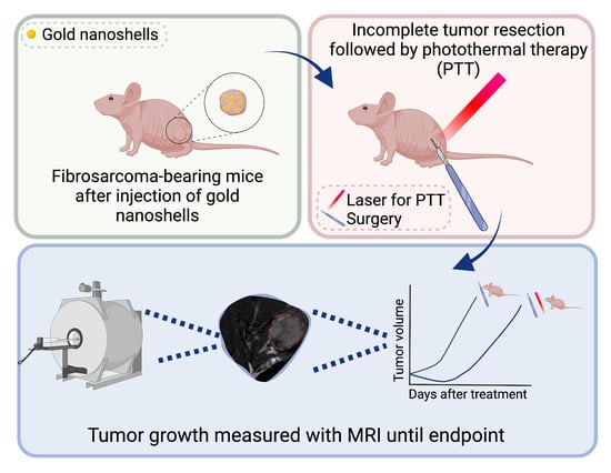
Photothermal Therapy as Adjuvant to Surgery in an Orthotopic Mouse Model of Human Fibrosarcoma
by Marina Simón, Jesper Tranekjær Jørgensen, Fredrik Melander, Thomas Lars Andresen, Anders Christensen and Andreas Kjaer
Cancers 2021, 13(22), 5820; https://doi.org/10.3390/cancers13225820 - 20 Nov 2021
Cited by 9 | Viewed by 2979
Abstract
Surgery is still the first-line treatment for multiple solid cancers. However, recurrence is a common issue, especially when dealing with aggressive tumors or tumors that are difficult to completely remove due to their location. Getting clear surgical margins can be challenging, but treatment [...] Read more.
Surgery is still the first-line treatment for multiple solid cancers. However, recurrence is a common issue, especially when dealing with aggressive tumors or tumors that are difficult to completely remove due to their location. Getting clear surgical margins can be challenging, but treatment strategies combining surgery with other anti-cancer therapies can potentially improve the outcome. Photothermal therapy (PTT) is a technique that relies on photoabsorbing agents, such as gold nanoparticles, to transform light into local hyperthermia. This technique can be used to ablate tumor tissue where the photoabsorbing agent accumulates, sparing healthy surrounding tissue. In this study, we examined the potential of gold nanoparticle-based PTT as an adjuvant treatment to surgery in a mouse model of human fibrosarcoma. For this we performed subtotal tumor resection to mimic a clinical situation where total tumor removal is not achieved, and subsequent PTT was applied on the surgical field. Our results showed that animals undergoing adjuvant PTT after surgery presented sustained delayed tumor growth and improved survival when compared to animals that only underwent surgery. We believe that these findings show the potential of PTT as an adjuvant method to traditional tumor surgery and could pave way to more personalized treatment options. Full article
Show Figures

Graphical abstract

26 pages, 5531 KiB  
Article
Safety and Efficacy Evaluation In Vivo of a Cationic Nucleolipid Nanosystem for the Nanodelivery of a Ruthenium(III) Complex with Superior Anticancer Bioactivity
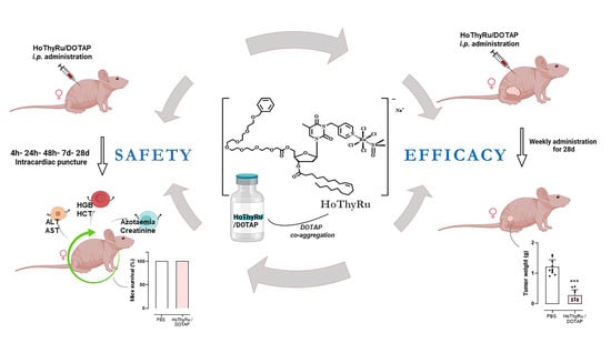
by Marialuisa Piccolo, Maria Grazia Ferraro, Federica Raucci, Claudia Riccardi, Anella Saviano, Irene Russo Krauss, Marco Trifuoggi, Michele Caraglia, Luigi Paduano, Daniela Montesarchio, Francesco Maione, Gabriella Misso, Rita Santamaria and Carlo Irace
Cancers 2021, 13(20), 5164; https://doi.org/10.3390/cancers13205164 - 14 Oct 2021
Cited by 20 | Viewed by 2476
Abstract
Selectivity and efficacy towards target cancer cells, as well as biocompatibility, are current challenges of advanced chemotherapy powering the discovery of unconventional metal-based drugs and the search for novel therapeutic approaches. Among second-generation metal-based chemotherapeutics, ruthenium complexes have demonstrated promising anticancer activity coupled [...] Read more.
Selectivity and efficacy towards target cancer cells, as well as biocompatibility, are current challenges of advanced chemotherapy powering the discovery of unconventional metal-based drugs and the search for novel therapeutic approaches. Among second-generation metal-based chemotherapeutics, ruthenium complexes have demonstrated promising anticancer activity coupled to minimal toxicity profiles and peculiar biochemical features. In this context, our research group has recently focused on a bioactive Ru(III) complex—named AziRu—incorporated into a suite of ad hoc designed nucleolipid nanosystems to ensure its chemical stability and delivery. Indeed, we proved that the structure and properties of decorated nucleolipids can have a major impact on the anticancer activity of the ruthenium core. Moving in this direction, here we describe a preclinical study performed by a mouse xenograft model of human breast cancer to establish safety and efficacy in vivo of a cationic Ru(III)-based nucleolipid formulation, named HoThyRu/DOTAP, endowed with superior antiproliferative activity. The results show a remarkable reduction in tumour with no evidence of animal suffering. Blood diagnostics, as well as biochemical analysis in both acute and chronic treated animal groups, demonstrate a good tolerability profile at the therapeutic regimen, with 100% of mice survival and no indication of toxicity. In addition, ruthenium plasma concentration analysis and tissue bioaccumulation were determined via appropriate sampling and ICP-MS analysis. Overall, this study supports both the efficacy of our Ru-containing nanosystem versus a human breast cancer model and its safety in vivo through well-tolerated animal biological responses, envisaging a possible forthcoming use in clinical trials. Full article
Show Figures

Graphical abstract

23 pages, 65575 KiB  
Article
Doxorubicin-Loaded Mixed Micelles Using Degradable Graft and Diblock Copolymers to Enhance Anticancer Sensitivity
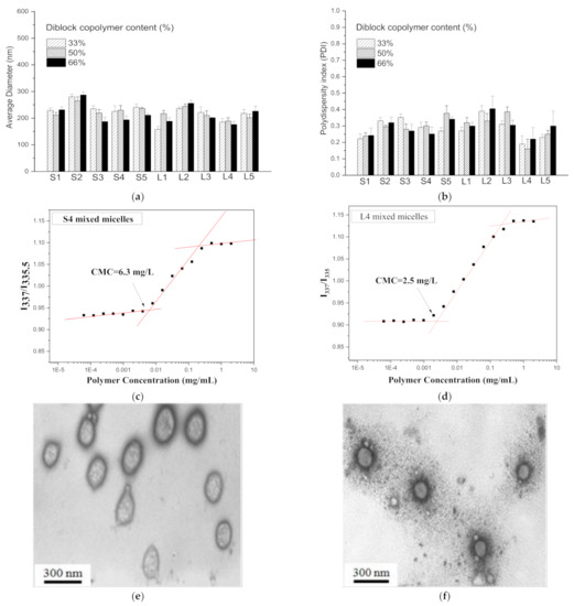
by Yi-Chun Chen, Chang-Jung Chang, Ging-Ho Hsiue and Yi-Ting Chiang
Cancers 2021, 13(15), 3816; https://doi.org/10.3390/cancers13153816 - 29 Jul 2021
Cited by 7 | Viewed by 2462
Abstract
In this study, a graft copolymer, poly(N-(2-hydroxypropyl) methacrylamide dilactate)-co-(N-(2-hydroxypropyl) methacrylamide-co-histidine)-graft-poly(d,l-lactide), and a diblock copolymer, methoxy poly(ethylene glycol)-b-poly(d,l-lactide), were assembled into a mixed micellar system to [...] Read more.
In this study, a graft copolymer, poly(N-(2-hydroxypropyl) methacrylamide dilactate)-co-(N-(2-hydroxypropyl) methacrylamide-co-histidine)-graft-poly(d,l-lactide), and a diblock copolymer, methoxy poly(ethylene glycol)-b-poly(d,l-lactide), were assembled into a mixed micellar system to encapsulate the anticancer drug doxorubicin (Dox). This mixed micellar system possesses the hydrophobic lactide segment of both copolymers, which reinforces its stability in physiological milieus; the histidine molecules appended on the graft copolymer provide the desired pH-responsive behavior to release Dox during internalization in cancer cells. The results demonstrate that the two copolymers were successfully prepared, and their ratios in the mixed micelles were optimized on the basis of the results of the stability tests. Under acidic conditions, the mixed micelles swell and are able to release their payloads. Therefore, the in vitro results indicate that the Dox in the mixed micelles is released effectively in response to the environmental pH of the mimetic internalization process, increasing cancer cells’ sensitivity toward Dox. The mixed micelles display low cytotoxicity due to the degradability of the polymers. The in vivo images show that the high stability of the mixed micelles ensures a high tumor accumulation. This selective tumor accumulation results in an excellent inhibition of in vivo tumor growth and a high rate of apoptosis in cancerous tissues, with low toxicity. This highly stable, mixed micellar system with a pH-dependent drug release, which enables the precise delivery of drugs to the tumor lesions, is feasible to employ clinically in cancer therapy. Full article
Show Figures

Figure 1

11 pages, 1347 KiB  
Communication
Docetaxel Skin Exposure and Micronucleation Contributes to Skin Toxicity Caused by CPC634
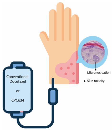
by Florence Atrafi, Ruben A. G. van Eerden, Stijn L. W. Koolen, Peter de Bruijn, Cristianne J. F. Rijcken, Rob Hanssen, Ferry A. L. M. Eskens, Martijn P. Lolkema, Esther Oomen-de Hoop, Jeffrey Damman and Ron H. J. Mathijssen
Cancers 2021, 13(15), 3741; https://doi.org/10.3390/cancers13153741 - 26 Jul 2021
Cited by 10 | Viewed by 2372
Abstract
Docetaxel entrapped nanoparticle CPC634 is associated with dose-related skin toxicity that resembles conventional docetaxel (Cd)-related skin toxicity. This study compared the cutaneous pharmacokinetics and pharmacodynamics of docetaxel and CPC634. In this randomised cross-over study, patients with solid tumours received one cycle of CPC634 [...] Read more.
Docetaxel entrapped nanoparticle CPC634 is associated with dose-related skin toxicity that resembles conventional docetaxel (Cd)-related skin toxicity. This study compared the cutaneous pharmacokinetics and pharmacodynamics of docetaxel and CPC634. In this randomised cross-over study, patients with solid tumours received one cycle of CPC634 and Cd (both at 75 mg/m2). Skin biopsies were taken at baseline and at day 8 of both cycles. Released and total docetaxel (released docetaxel plus entrapped docetaxel) concentrations and histopathological changes in the skin biopsies were evaluated. Twenty patients underwent paired skin biopsies for pharmacokinetic analysis and 10 patients had biopsies available for histopathological assessment. The total skin docetaxel concentration was 369% (95%CI: 229% to 569%, p < 0.001) higher after CPC634 administration compared to Cd while the released docetaxel concentrations were not statistically different (95%CI: −9% to 63%, p = 0.169). The CPC634 released docetaxel concentration in the skin was positively correlated with plasma concentrations (Pearson’s correlation 0.48, p = 0.03). Histopathological examination revealed increased apoptosis, mitotic cells with nuclear atypia, and micronucleation with an enhanced Ki-67 index for both compounds. In conclusion, both CPC634 and Cd treatment result in docetaxel exposure in the skin causing cutaneous anti-mitotic effects such as micronucleation, which could induce an inflammatory reaction leading to skin toxicity. Full article
Show Figures

Graphical abstract

25 pages, 8531 KiB  
Article
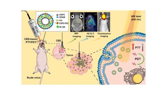
Liposomal IR-780 as a Highly Stable Nanotheranostic Agent for Improved Photothermal/Photodynamic Therapy of Brain Tumors by Convection-Enhanced Delivery
by Yu-Jen Lu, Anilkumar T. S., Chi-Cheng Chuang and Jyh-Ping Chen
Cancers 2021, 13(15), 3690; https://doi.org/10.3390/cancers13153690 - 22 Jul 2021
Cited by 31 | Viewed by 4847
Abstract
As a hydrophobic photosensitizer, IR-780 suffers from poor water solubility and low photostability under near infrared (NIR) light, which severely limits its use during successive NIR laser-assisted photothermal/photodynamic therapy (PTT/PDT). To solve this problem, we fabricate cationic IR-780-loaded liposomes (ILs) by entrapping IR-780 [...] Read more.
As a hydrophobic photosensitizer, IR-780 suffers from poor water solubility and low photostability under near infrared (NIR) light, which severely limits its use during successive NIR laser-assisted photothermal/photodynamic therapy (PTT/PDT). To solve this problem, we fabricate cationic IR-780-loaded liposomes (ILs) by entrapping IR-780 within the lipid bilayer of liposomes. We demonstrate enhanced photostability of IR-780 in ILs with well-preserved photothermal response after three repeated NIR laser exposures, in contrast to the rapid decomposition of free IR-780. The cationic nature of ILs promotes fast endocytosis of liposomal IR-780 by U87MG human glioblastoma cells within 30 min. For PTT/PDT in vitro, ILs treatment plus NIR laser irradiation leads to overexpression of heat shock protein 70 and generation of intracellular reactive oxygen species by U87MG cells, resulting in enhanced cytotoxicity and higher cell apoptosis rate. Using intracranial glioma xenograft in nude mice and administration of ILs by convection enhanced delivery (CED) to overcome blood-brain barrier, liposomal IR-780 could be specifically delivered to the brain tumor, as demonstrated from fluorescence imaging. By providing a highly stable liposomal IR-780, ILs significantly improved anti-cancer efficacy in glioma treatment, as revealed from various diagnostic imaging tools and histological examination. Overall, CED of ILs plus successive laser-assisted PTT/PDT may be an alternative approach for treating brain tumor, which can retard glioma growth and prolong animal survival times from orthotopic brain tumor models. Full article
Show Figures

Graphical abstract

31 pages, 8264 KiB  
Article
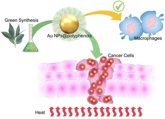
Synergistic Effect Induced by Gold Nanoparticles with Polyphenols Shell during Thermal Therapy: Macrophage Inflammatory Response and Cancer Cell Death Assessment
by Valeria De Matteis, Mariafrancesca Cascione, Loris Rizzello, Daniela Erminia Manno, Claudia Di Guglielmo and Rosaria Rinaldi
Cancers 2021, 13(14), 3610; https://doi.org/10.3390/cancers13143610 - 19 Jul 2021
Cited by 17 | Viewed by 4566
Abstract
Background: In recent decades, gold nanoparticle (Au NP)-based cancer therapy has been heavily debated. The physico-chemical properties of AuNPs can be exploited in photothermal therapy, making them a powerful tool for selectively killing cancer cells. However, the synthetic side products and capping agents [...] Read more.
Background: In recent decades, gold nanoparticle (Au NP)-based cancer therapy has been heavily debated. The physico-chemical properties of AuNPs can be exploited in photothermal therapy, making them a powerful tool for selectively killing cancer cells. However, the synthetic side products and capping agents often induce a strong activation of the inflammatory pathways of macrophages, thus limiting their further applications in vivo. Methods: Here, we described a green method to obtain stable polyphenol-capped AuNPs (Au NPs@polyphenols), as polyphenols are known for their anti-inflammatory and anticancer properties. These NPs were used in human macrophages to test key inflammation-related markers, such as NF-κB, TNF-α, and interleukins-6 and 8. The results were compared with similar NPs obtained by a traditional chemical route (without the polyphenol coating), proving the potential of Au NPs@polyphenols to strongly promote the shutdown of inflammation. This was useful in developing them for use as heat-synergized tools in the thermal treatment of two types of cancer cells, namely, breast cancer (MCF-7) and neuroblastoma (SH-SY5Y) cells. The cell viability, calcium release, oxidative stress, HSP-70 expression, mitochondrial, and DNA damage, as well as cytoskeleton alteration, were evaluated. Results: Our results clearly demonstrate that the combined strategy markedly exerts anticancer effects against the tested cancer cell, while neither of the single treatments (only heat or only NPs) induced significant changes. Conclusions: Au NP@polyphenols may be powerful agents in cancer treatment. Full article
Show Figures

Graphical abstract

10 pages, 10835 KiB  
Article
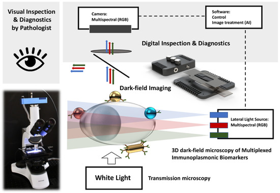
Multiplexed Plasmonic Nano-Labeling for Bioimaging of Cytological Stained Samples
by Paule Marcoux-Valiquette, Cécile Darviot, Lu Wang, Andrée-Anne Grosset, Morteza Hasanzadeh Kafshgari, Mirela Birela, Sergiy Patskovsky, Dominique Trudel and Michel Meunier
Cancers 2021, 13(14), 3509; https://doi.org/10.3390/cancers13143509 - 13 Jul 2021
Cited by 4 | Viewed by 2385
Abstract
Reliable cytopathological diagnosis requires new methods and approaches for the rapid and accurate determination of all cell types. This is especially important when the number of cells is limited, such as in the cytological samples of fine-needle biopsy. Immunoplasmonic-multiplexed- labeling may be one [...] Read more.
Reliable cytopathological diagnosis requires new methods and approaches for the rapid and accurate determination of all cell types. This is especially important when the number of cells is limited, such as in the cytological samples of fine-needle biopsy. Immunoplasmonic-multiplexed- labeling may be one of the emerging solutions to such problems. However, to be accepted and used by the practicing pathologists, new methods must be compatible and complementary with existing cytopathology approaches where counterstaining is central to the correct interpretation of immunolabeling. In addition, the optical detection and imaging setup for immunoplasmonic-multiplexed-labeling must be implemented on the same cytopathological microscope, not interfere with standard H&E imaging, and operate as a second easy-to-use imaging method. In this article, we present multiplex imaging of four types of nanoplasmonic markers on two types of H&E-stained cytological specimens (formalin-fixed paraffin embedded and non-embedded adherent cancer cells) using a specially designed adapter for SI dark-field microscopy. The obtained results confirm the effectiveness of the proposed optical method for quantitative and multiplex identification of various plasmonic NPs, and the possibility of using immunoplasmonic-multiplexed-labeling for cytopathological diagnostics. Full article
Show Figures

Figure 1

19 pages, 4257 KiB  
Review
Polyester Polymeric Nanoparticles as Platforms in the Development of Novel Nanomedicines for Cancer Treatment
by Enrique Niza, Alberto Ocaña, José Antonio Castro-Osma, Iván Bravo and Carlos Alonso-Moreno
Cancers 2021, 13(14), 3387; https://doi.org/10.3390/cancers13143387 - 6 Jul 2021
Cited by 27 | Viewed by 3837
Abstract
Many therapeutic agents have failed in their clinical development, due to the toxic effects associated with non-transformed tissues. In this context, nanotechnology has been exploited to overcome such limitations, and also improve navigation across biological barriers. Amongst the many materials used in nanomedicine, [...] Read more.
Many therapeutic agents have failed in their clinical development, due to the toxic effects associated with non-transformed tissues. In this context, nanotechnology has been exploited to overcome such limitations, and also improve navigation across biological barriers. Amongst the many materials used in nanomedicine, with promising properties as therapeutic carriers, the following one stands out: biodegradable and biocompatible polymers. Polymeric nanoparticles are ideal candidates for drug delivery, given the versatility of raw materials and their feasibility in large-scale production. Furthermore, polymeric nanoparticles show great potential for easy surface modifications to optimize pharmacokinetics, including the half-life in circulation and targeted tissue delivery. Herein, we provide an overview of the current applications of polymeric nanoparticles as platforms in the development of novel nanomedicines for cancer treatment. In particular, we will focus on the raw materials that are widely used for polymeric nanoparticle generation, current methods for formulation, mechanism of action, and clinical investigations. Full article
Show Figures

Graphical abstract

23 pages, 5326 KiB  
Article
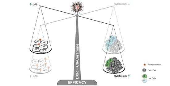
The Enhanced Efficacy of Intracellular Delivery of Doxorubicin/C6-Ceramide Combination Mediated by the F3 Peptide/Nucleolin System Is Supported by the Downregulation of the PI3K/Akt Pathway
by Ana F. Cruz, Mariana B. Caleiras, Nuno A. Fonseca, Nélio Gonçalves, Vera M. Mendes, Susana F. Sampaio, Vera Moura, Joana B. Melo, Ramiro D. Almeida, Bruno Manadas, Sérgio Simões and João N. Moreira
Cancers 2021, 13(12), 3052; https://doi.org/10.3390/cancers13123052 - 18 Jun 2021
Cited by 9 | Viewed by 3298
Abstract
Targeting multiple cellular populations is of high therapeutic relevance for the tackling of solid tumors heterogeneity. Herein, the ability of pegylated and pH-sensitive liposomes, functionalized with the nucleolin-binding F3 peptide and containing doxorubicin (DXR)/C6-ceramide synergistic combination, to target, in vitro, ovarian cancer, including [...] Read more.
Targeting multiple cellular populations is of high therapeutic relevance for the tackling of solid tumors heterogeneity. Herein, the ability of pegylated and pH-sensitive liposomes, functionalized with the nucleolin-binding F3 peptide and containing doxorubicin (DXR)/C6-ceramide synergistic combination, to target, in vitro, ovarian cancer, including ovarian cancer stem cells (CSC), was assessed. The underlying molecular mechanism of action of the nucleolin-mediated intracellular delivery of C6-ceramide to cancer cells was also explored. The assessment of overexpression of surface nucleolin expression by flow cytometry was critical to dissipate differences identified by Western blot in membrane/cytoplasm of SKOV-3, OVCAR-3 and TOV-112D ovarian cancer cell lines. The former was in line with the significant extent of uptake into (bulk) ovarian cancer cells, relative to non-targeted and non-specific counterparts. This pattern of uptake was recapitulated with putative CSC-enriched ovarian SKOV-3 and OVCAR-3 sub-population (EpCAMhigh/CD44high). Co-encapsulation of DXR:C6-ceramide into F3 peptide-targeted liposomes improved cytotoxic activity relative to liposomes containing DXR alone, in an extent that depended on the intrinsic resistance to DXR and on the incubation time. The enhanced cytotoxicity of the targeted combination was mechanistically supported by the downregulation of PI3K/Akt pathway by C6-ceramide, only among the nucleolin-overexpressing cancer cells presenting a basal p-Akt/total Akt ratio lower than 1. Full article
Show Figures

Graphical abstract

17 pages, 8136 KiB  
Article
Biparatopic Protein Nanoparticles for the Precision Therapy of CXCR4+ Cancers
by Olivia Cano-Garrido, Patricia Álamo, Laura Sánchez-García, Aïda Falgàs, Alejandro Sánchez-Chardi, Naroa Serna, Eloi Parladé, Ugutz Unzueta, Mònica Roldán, Eric Voltà-Durán, Isolda Casanova, Antonio Villaverde, Ramón Mangues and Esther Vázquez
Cancers 2021, 13(12), 2929; https://doi.org/10.3390/cancers13122929 - 11 Jun 2021
Cited by 13 | Viewed by 2843
Abstract
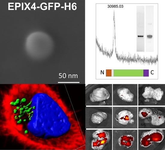
The accumulated molecular knowledge about human cancer enables the identification of multiple cell surface markers as highly specific therapeutic targets. A proper tumor targeting could significantly avoid drug exposure of healthy cells, minimizing side effects, but it is also expected to increase the [...] Read more.
The accumulated molecular knowledge about human cancer enables the identification of multiple cell surface markers as highly specific therapeutic targets. A proper tumor targeting could significantly avoid drug exposure of healthy cells, minimizing side effects, but it is also expected to increase the therapeutic index. Specifically, colorectal cancer has a particularly poor prognosis in late stages, being drug targeting an appropriate strategy to substantially improve the therapeutic efficacy. In this study, we have explored the potential of the human albumin-derived peptide, EPI-X4, as a suitable ligand to target colorectal cancer via the cell surface protein CXCR4, a chemokine receptor overexpressed in cancer stem cells. To explore the potential use of this ligand, self-assembling protein nanoparticles have been generated displaying an engineered EPI-X4 version, which conferred a modest CXCR4 targeting and fast and high level of cell apoptosis in tumor CXCR4+ cells, in vitro and in vivo. In addition, when EPI-X4-based building blocks are combined with biologically inert polypeptides containing the CXCR4 ligand T22, the resulting biparatopic nanoparticles show a dramatically improved biodistribution in mouse models of CXCR4+ human cancer, faster cell internalization and enhanced target cell death when compared to the version based on a single ligand. The generation of biparatopic materials opens exciting possibilities in oncotherapies based on high precision drug delivery based on the receptor CXCR4. Full article
Show Figures

Graphical abstract

26 pages, 2385 KiB  
Review
Nanomedicine to Overcome Multidrug Resistance Mechanisms in Colon and Pancreatic Cancer: Recent Progress
by Raúl Ortíz, Francisco Quiñonero, Beatriz García-Pinel, Marco Fuel, Cristina Mesas, Laura Cabeza, Consolación Melguizo and Jose Prados
Cancers 2021, 13(9), 2058; https://doi.org/10.3390/cancers13092058 - 24 Apr 2021
Cited by 29 | Viewed by 5930
Abstract
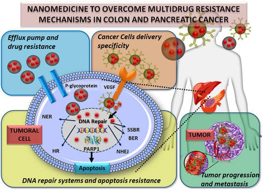
The development of drug resistance is one of the main causes of cancer treatment failure. This phenomenon occurs very frequently in different types of cancer, including colon and pancreatic cancers. However, the underlying molecular mechanisms are not fully understood. In recent years, nanomedicine [...] Read more.
The development of drug resistance is one of the main causes of cancer treatment failure. This phenomenon occurs very frequently in different types of cancer, including colon and pancreatic cancers. However, the underlying molecular mechanisms are not fully understood. In recent years, nanomedicine has improved the delivery and efficacy of drugs, and has decreased their side effects. In addition, it has allowed to design drugs capable of avoiding certain resistance mechanisms of tumors. In this article, we review the main resistance mechanisms in colon and pancreatic cancers, along with the most relevant strategies offered by nanodrugs to overcome this obstacle. These strategies include the inhibition of efflux pumps, the use of specific targets, the development of nanomedicines affecting the environment of cancer-specific tissues, the modulation of DNA repair mechanisms or RNA (miRNA), and specific approaches to damage cancer stem cells, among others. This review aims to illustrate how advanced nanoformulations, including polymeric conjugates, micelles, dendrimers, liposomes, metallic and carbon-based nanoparticles, are allowing to overcome one of the main limitations in the treatment of colon and pancreatic cancers. The future development of nanomedicine opens new horizons for cancer treatment. Full article
Show Figures

Graphical abstract

15 pages, 3236 KiB  
Article
Evaluation of TFR-1 Expression in Feline Mammary Cancer and In Vitro Antitumor Efficacy Study of Doxorubicin-Loaded H-Ferritin Nanocages
by Nicolò Rensi, Alessandro Sammarco, Valentina Moccia, Alessandro Calore, Filippo Torrigiani, Davide Prosperi, Maria Antonietta Rizzuto, Michela Bellini, Raffaella De Maria, Federico Bonsembiante, Silvia Ferro, Rossella Zanetti, Valentina Zappulli and Laura Cavicchioli
Cancers 2021, 13(6), 1248; https://doi.org/10.3390/cancers13061248 - 12 Mar 2021
Viewed by 3396
Abstract
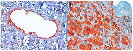
The transferrin receptor 1 (TFR-1) has been found overexpressed in a broad range of solid tumors in humans and is, therefore, attracting great interest in clinical oncology for innovative targeted therapies, including nanomedicine. TFR-1 is recognized by H-Ferritin (HFn) and has been exploited [...] Read more.
The transferrin receptor 1 (TFR-1) has been found overexpressed in a broad range of solid tumors in humans and is, therefore, attracting great interest in clinical oncology for innovative targeted therapies, including nanomedicine. TFR-1 is recognized by H-Ferritin (HFn) and has been exploited to allow selective binding and drug internalization, applying an HFn nanocage loaded with doxorubicin (HFn(DOX)). In veterinary medicine, the role of TFR-1 in animal cancers remains poorly explored, and no attempts to use TFR-1 as a target for drug delivery have been conducted so far. In this study, we determined the TFR-1 expression both in feline mammary carcinomas during tumor progression, as compared to healthy tissue, and, in vitro, in a feline metastatic mammary cancer cell line. The efficacy of HFn(DOX) was compared to treatment with conventional doxorubicin in feline mammary cancer cells. Our results highlighted an increased TFR-1 expression associated with tumor metastatic progression, indicating a more aggressive behavior. Furthermore, it was demonstrated that the use of HFn(DOX) resulted in less proliferation of cells and increased apoptosis when compared to the drug alone. The results of this preliminary study suggest that the use of engineered bionanocages also offers unprecedented opportunities for selective targeted chemotherapy of solid tumors in veterinary medicine. Full article
Show Figures

Figure 1

27 pages, 12838 KiB  
Article
CD44 Targeted Nanomaterials for Treatment of Triple-Negative Breast Cancer
by Ghazal Nabil, Rami Alzhrani, Hashem O. Alsaab, Mohammed Atef, Samaresh Sau, Arun K. Iyer and Hossny El Banna
Cancers 2021, 13(4), 898; https://doi.org/10.3390/cancers13040898 - 20 Feb 2021
Cited by 23 | Viewed by 4116
Abstract
Identified as the second leading cause of cancer-related deaths among American women after lung cancer, breast cancer of all types has been the focus of numerous research studies. Even though triple-negative breast cancer (TNBC) represents 15–20% of the number of breast cancer cases [...] Read more.
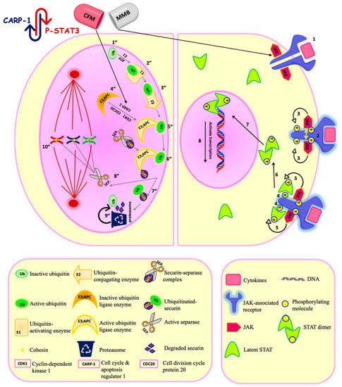
Identified as the second leading cause of cancer-related deaths among American women after lung cancer, breast cancer of all types has been the focus of numerous research studies. Even though triple-negative breast cancer (TNBC) represents 15–20% of the number of breast cancer cases worldwide, its existing therapeutic options are fairly limited. Due to the pivotal role of the presence/absence of specific receptors to luminal A, luminal B, HER-2+, and TNBC in the molecular classification of breast cancer, the lack of these receptors has accounted for the aforementioned limitation. Thereupon, in an attempt to participate in the ongoing research endeavors to overcome such a limitation, the conducted study adopts a combination strategy as a therapeutic paradigm for TNBC, which has proven notable results with respect to both: improving patient outcomes and survivability rates. The study hinges upon an investigation of a promising NPs platform for CD44 mediated theranostic that can be combined with JAK/STAT inhibitors for the treatment of TNBC. The ability of momelotinib (MMB), which is a JAK/STAT inhibitor, to sensitize the TNBC to apoptosis inducer (CFM-4.16) has been evaluated in MDA-MB-231 and MDA-MB-468. MMB + CFM-4.16 combination with a combination index (CI) ≤0.5, has been selected for in vitro and in vivo studies. MMB has been combined with CD44 directed polymeric nanoparticles (PNPs) loaded with CFM-4.16, namely CD44-T-PNPs, which selectively delivered the payload to CD44 overexpressing TNBC with a significant decrease in cell viability associated with a high dose reduction index (DRI). The mechanism underlying their synergism is based on the simultaneous downregulation of P-STAT3 and the up-regulation of CARP-1, which has induced ROS-dependent apoptosis leading to caspase 3/7 elevation, cell shrinkage, DNA damage, and suppressed migration. CD44-T-PNPs showed a remarkable cellular internalization, demonstrated by uptake of a Rhodamine B dye in vitro and S0456 (NIR dye) in vivo. S0456 was conjugated to PNPs to form CD44-T-PNPs/S0456 that simultaneously delivered CFM-4.16 and S0456 parenterally with selective tumor targeting, prolonged circulation, minimized off-target distribution. Full article
Show Figures

Figure 1

36 pages, 4349 KiB  
Review
The Vault Nanoparticle: A Gigantic Ribonucleoprotein Assembly Involved in Diverse Physiological and Pathological Phenomena and an Ideal Nanovector for Drug Delivery and Therapy
by Gianni Frascotti, Elisabetta Galbiati, Matteo Mazzucchelli, Maria Pozzi, Lucia Salvioni, Jacopo Vertemara and Paolo Tortora
Cancers 2021, 13(4), 707; https://doi.org/10.3390/cancers13040707 - 9 Feb 2021
Cited by 25 | Viewed by 6168
Abstract
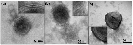
The vault nanoparticle is a eukaryotic ribonucleoprotein complex consisting of 78 individual 97 kDa-“major vault protein” (MVP) molecules that form two symmetrical, cup-shaped, hollow halves. It has a huge size (72.5 × 41 × 41 nm) and an internal cavity, wherein the vault [...] Read more.
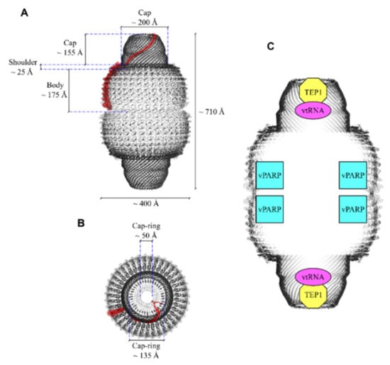
The vault nanoparticle is a eukaryotic ribonucleoprotein complex consisting of 78 individual 97 kDa-“major vault protein” (MVP) molecules that form two symmetrical, cup-shaped, hollow halves. It has a huge size (72.5 × 41 × 41 nm) and an internal cavity, wherein the vault poly(ADP-ribose) polymerase (vPARP), telomerase-associated protein-1 (TEP1), and some small untranslated RNAs are accommodated. Plenty of literature reports on the biological role(s) of this nanocomplex, as well as its involvement in diseases, mostly oncological ones. Nevertheless, much has still to be understood as to how vault participates in normal and pathological mechanisms. In this comprehensive review, current understanding of its biological roles is discussed. By different mechanisms, vault’s individual components are involved in major cellular phenomena, which result in protection against cellular stresses, such as DNA-damaging agents, irradiation, hypoxia, hyperosmotic, and oxidative conditions. These diverse cellular functions are accomplished by different mechanisms, mainly gene expression reprogramming, activation of proliferative/prosurvival signaling pathways, export from the nucleus of DNA-damaging drugs, and import of specific proteins. The cellular functions of this nanocomplex may also result in the onset of pathological conditions, mainly (but not exclusively) tumor proliferation and multidrug resistance. The current understanding of its biological roles in physiological and pathological processes should also provide new hints to extend the scope of its exploitation as a nanocarrier for drug delivery. Full article
Show Figures

Figure 1

2020

Jump to: 2023, 2022, 2021

14 pages, 1963 KiB  
Review
Immunogenicity of Externally Activated Nanoparticles for Cancer Therapy
by Onur Sahin, Ashokkumar Meiyazhagan, Pulickel M. Ajayan and Sunil Krishnan
Cancers 2020, 12(12), 3559; https://doi.org/10.3390/cancers12123559 - 28 Nov 2020
Cited by 8 | Viewed by 2932
Abstract
Nanoparticles activated by external beams, such as ionizing radiation, laser light, or magnetic fields, have attracted significant research interest as a possible modality for treating solid tumors. From producing hyperthermic conditions to generating reactive oxygen species, a wide range of externally activated mechanisms [...] Read more.
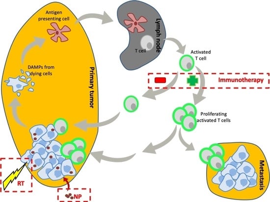
Nanoparticles activated by external beams, such as ionizing radiation, laser light, or magnetic fields, have attracted significant research interest as a possible modality for treating solid tumors. From producing hyperthermic conditions to generating reactive oxygen species, a wide range of externally activated mechanisms have been explored for producing cytotoxicity within tumors with high spatiotemporal control. To further improve tumoricidal effects, recent trends in the literature have focused on stimulating the immune system through externally activated treatment strategies that result in immunogenic cell death. By releasing inflammatory compounds known to initiate an immune response, treatment methods can take advantage of immune system pathways for a durable and robust systemic anti-tumor response. In this review, we discuss recent advancements in radiosensitizing and hyperthermic nanoparticles that have been tuned for promoting immunogenic cell death. Our review covers both preclinical and clinical results, as well as an overview of possible future work. Full article
Show Figures

Graphical abstract

23 pages, 5013 KiB  
Article
Carbon Nanodots for On Demand Chemophotothermal Therapy Combination to Elicit Necroptosis: Overcoming Apoptosis Resistance in Breast Cancer Cell Lines
by Aldo Nicosia, Gennara Cavallaro, Salvatore Costa, Mara Andrea Utzeri, Angela Cuttitta, Gaetano Giammona and Nicolò Mauro
Cancers 2020, 12(11), 3114; https://doi.org/10.3390/cancers12113114 - 25 Oct 2020
Cited by 24 | Viewed by 3436
Abstract
Background: Engineered luminescent carbon nanodots (CDs) are appealing nanomaterials for cancer image-guided photothermal therapy combining near infrared (NIR)–triggered hyperthermia, imaging, and drug delivery in a single platform for efficient killing of cancer cells. This approach would allow eliciting synergistic regulated cell death (RCD) [...] Read more.
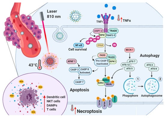
Background: Engineered luminescent carbon nanodots (CDs) are appealing nanomaterials for cancer image-guided photothermal therapy combining near infrared (NIR)–triggered hyperthermia, imaging, and drug delivery in a single platform for efficient killing of cancer cells. This approach would allow eliciting synergistic regulated cell death (RCD) routes such as necroptosis, targeting breast cancer cells refractory to apoptosis, thus overcoming drug resistance. Methods: We report the preparation of CDs bearing biotin as a targeting agent (CDs-PEG-BT), which are able to load high amounts of irinotecan (23.7%) to be released in a pulsed on-demand fashion. CDs-PEG-BT have narrow size distribution, stable red luminescence, and high photothermal conversion in the NIR region, allowing imaging of MDA-MB231 and MCF-7 cancer cells and killing them by photothermal and chemotherapeutic insults. Results: Cellular uptake, viability profiles, and RCD gene expression analyses provided insights about the observed biocompatibility of CDs-PEG-BT, indicating that necroptosis can be induced on-demand after the photothermal activation. Besides, photothermal activation of drug-loaded CDs-PEG-BT implies both necroptosis and apoptosis by the TNFα and RIPK1 pathway. Conclusions: The controlled activation of necroptosis and apoptosis by combining phototherapy and on-demand release of irinotecan is the hallmark of efficient anticancer response in refractory breast cancer cell lines in view of precision medicine applications. Full article
Show Figures

Figure 1

26 pages, 2435 KiB  
Review
Progress in Clinical Trials of Photodynamic Therapy for Solid Tumors and the Role of Nanomedicine
by Hashem O. Alsaab, Maha S. Alghamdi, Albatool S. Alotaibi, Rami Alzhrani, Fatimah Alwuthaynani, Yusuf S. Althobaiti, Atiah H. Almalki, Samaresh Sau and Arun K. Iyer
Cancers 2020, 12(10), 2793; https://doi.org/10.3390/cancers12102793 - 29 Sep 2020
Cited by 94 | Viewed by 7464
Abstract
Current research to find effective anticancer treatments is being performed on photodynamic therapy (PDT) with increasing attention. PDT is a very promising therapeutic way to combine a photosensitive drug with visible light to manage different intense malignancies. PDT has several benefits, including better [...] Read more.
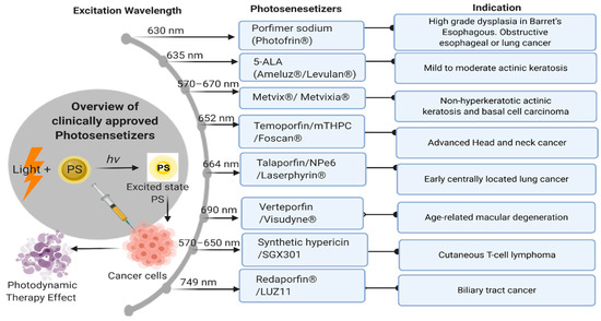
Current research to find effective anticancer treatments is being performed on photodynamic therapy (PDT) with increasing attention. PDT is a very promising therapeutic way to combine a photosensitive drug with visible light to manage different intense malignancies. PDT has several benefits, including better safety and lower toxicity in the treatment of malignant tumors over traditional cancer therapy. This reasonably simple approach utilizes three integral elements: a photosensitizer (PS), a source of light, and oxygen. Upon light irradiation of a particular wavelength, the PS generates reactive oxygen species (ROS), beginning a cascade of cellular death transformations. The positive therapeutic impact of PDT may be limited because several factors of this therapy include low solubilities of PSs, restricting their effective administration, blood circulation, and poor tumor specificity. Therefore, utilizing nanocarrier systems that modulate PS pharmacokinetics (PK) and pharmacodynamics (PD) is a promising approach to bypassing these challenges. In the present paper, we review the latest clinical studies and preclinical in vivo studies on the use of PDT and progress made in the use of nanotherapeutics as delivery tools for PSs to improve their cancer cellular uptake and their toxic properties and, therefore, the therapeutic impact of PDT. We also discuss the effects that photoimmunotherapy (PIT) might have on solid tumor therapeutic strategies. Full article
Show Figures

Figure 1

25 pages, 3321 KiB  
Article
Biomimetic Magnetite Nanoparticles as Targeted Drug Nanocarriers and Mediators of Hyperthermia in an Experimental Cancer Model
by Francesca Oltolina, Ana Peigneux, Donato Colangelo, Nausicaa Clemente, Annarita D’Urso, Guido Valente, Guillermo R. Iglesias, Concepcion Jiménez-Lopez and Maria Prat
Cancers 2020, 12(9), 2564; https://doi.org/10.3390/cancers12092564 - 9 Sep 2020
Cited by 40 | Viewed by 4544
Abstract
Biomimetic magnetic nanoparticles mediated by magnetosome proteins (BMNPs) are potential innovative tools for cancer therapy since, besides being multifunctional platforms, they can be manipulated by an external gradient magnetic field (GMF) and/or an alternating magnetic field (AMF), mediating targeting and hyperthermia, respectively. We [...] Read more.
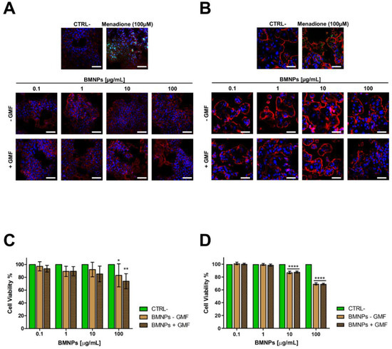
Biomimetic magnetic nanoparticles mediated by magnetosome proteins (BMNPs) are potential innovative tools for cancer therapy since, besides being multifunctional platforms, they can be manipulated by an external gradient magnetic field (GMF) and/or an alternating magnetic field (AMF), mediating targeting and hyperthermia, respectively. We evaluated the cytocompatibility/cytotoxicity of BMNPs and Doxorubicin (DOXO)-BMNPs in the presence/absence of GMF in 4T1 and MCF-7 cells as well as their cellular uptake. We analyzed the biocompatibility and in vivo distribution of BMNPs as well as the effect of DOXO-BMNPs in BALB/c mice bearing 4T1 induced mammary carcinomas after applying GMF and AMF. Results: GMF enhanced the cell uptake of both BMNPs and DOXO-BMNPs and the cytotoxicity of DOXO-BMNPs. BMNPs were biocompatible when injected intravenously in BALB/c mice. The application of GMF on 4T1 tumors after each of the repeated (6×) iv administrations of DOXO-BMNPs enhanced tumor growth inhibition when compared to any other treatment, including that with soluble DOXO. Moreover, injection of DOXO-BMNPs in the tumor combined with application of an AMF resulted in a significant tumor weight reduction. These promising results show the suitability of BMNPs as magnetic nanocarriers for local targeted chemotherapy and as local agents for hyperthermia. Full article
Show Figures

Figure 1

17 pages, 4254 KiB  
Article
Prolonging the Half-Life of Histone Deacetylase Inhibitor Belinostat via 50 nm Scale Liposomal Subcutaneous Delivery System for Peripheral T-Cell Lymphoma
by Meng-Hsuan Cheng, Jun-Yi Weng, Chih-Hung Chuang, Wei-Ting Liao, Yu-Fong Lai, Jia-Yu Liu and Yi-Ping Fang
Cancers 2020, 12(9), 2558; https://doi.org/10.3390/cancers12092558 - 8 Sep 2020
Cited by 7 | Viewed by 2755
Abstract
Lymph node metastasis is an aggressive condition characterized by poor treatment outcomes and low overall survival. Belinostat is a novel histone deacetylase (HDAC) inhibitor approved by the Food and Drug Administration (FDA) for the treatment of relapsed peripheral T-cell lymphoma (PTCL). However, the [...] Read more.
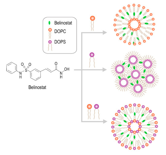
Lymph node metastasis is an aggressive condition characterized by poor treatment outcomes and low overall survival. Belinostat is a novel histone deacetylase (HDAC) inhibitor approved by the Food and Drug Administration (FDA) for the treatment of relapsed peripheral T-cell lymphoma (PTCL). However, the major problem is that belinostat has a short half-life of 1.1 h. In this study, we successfully prepared 50 nm liposomal colloids, which showed a controlled release pattern and excellent pharmacokinetics. The results showed that the particle size of liposomes consisting of dioleoylphosphatidylcholine (DOPC) was larger than that of those consisting of dioleoylglycerophosphoserine (DOPS). In terms of release kinetics of belinostat, the free drug was rapidly released and showed lower area under curve (AUC) exposure for in vivo pharmacokinetics. When liposomal formulations were employed, the release pattern was fitted with Hixson–Crowell models and showed sustained release of belinostat. Moreover, HuT-78 cells were able to take up all the liposomes in a concentration-dependent manner. The safety assessment confirmed hemocompatibility, and the platelet count was increased. Furthermore, the liposomes consisting of DOPC or DOPS had different behavior patterns, and their delivery to lymphatic regions should be thoroughly investigated in the future. Full article
Show Figures

Figure 1

33 pages, 2812 KiB  
Review
Cancer Nanomedicine Special Issue Review Anticancer Drug Delivery with Nanoparticles: Extracellular Vesicles or Synthetic Nanobeads as Therapeutic Tools for Conventional Treatment or Immunotherapy
by Maria Raffaella Zocchi, Francesca Tosetti, Roberto Benelli and Alessandro Poggi
Cancers 2020, 12(7), 1886; https://doi.org/10.3390/cancers12071886 - 13 Jul 2020
Cited by 20 | Viewed by 4965
Abstract
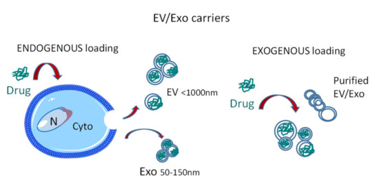
Both natural and synthetic nanoparticles have been proposed as drug carriers in cancer treatment, since they can increase drug accumulation in target tissues, optimizing the therapeutic effect. As an example, extracellular vesicles (EV), including exosomes (Exo), can become drug vehicles through endogenous or [...] Read more.
Both natural and synthetic nanoparticles have been proposed as drug carriers in cancer treatment, since they can increase drug accumulation in target tissues, optimizing the therapeutic effect. As an example, extracellular vesicles (EV), including exosomes (Exo), can become drug vehicles through endogenous or exogenous loading, amplifying the anticancer effects at the tumor site. In turn, synthetic nanoparticles (NP) can carry therapeutic molecules inside their core, improving solubility and stability, preventing degradation, and controlling their release. In this review, we summarize the recent advances in nanotechnology applied for theranostic use, distinguishing between passive and active targeting of these vehicles. In addition, examples of these models are reported: EV as transporters of conventional anticancer drugs; Exo or NP as carriers of small molecules that induce an anti-tumor immune response. Finally, we focus on two types of nanoparticles used to stimulate an anticancer immune response: Exo carried with A Disintegrin And Metalloprotease-10 inhibitors and NP loaded with aminobisphosphonates. The former would reduce the release of decoy ligands that impair tumor cell recognition, while the latter would activate the peculiar anti-tumor response exerted by γδ T cells, creating a bridge between innate and adaptive immunity. Full article
Show Figures

Figure 1

26 pages, 3195 KiB  
Review
Nanomedicine in Non-Small Cell Lung Cancer: From Conventional Treatments to Immunotherapy
by Coral García-Fernández, Cristina Fornaguera and Salvador Borrós
Cancers 2020, 12(6), 1609; https://doi.org/10.3390/cancers12061609 - 18 Jun 2020
Cited by 32 | Viewed by 6294
Abstract
Non-small cell lung cancer (NSCLC) remains the most common cause of cancer-related mortality. The heterogeneous nature of this disease hinders its diagnosis and treatment, requiring continuous advances in research aiming to understand its intricate nature. Consequently, the retrospective analysis of conventional therapies has [...] Read more.
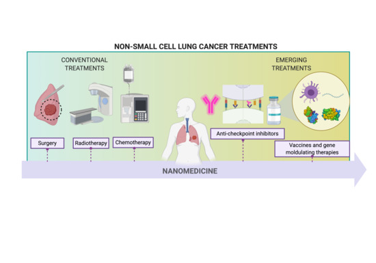
Non-small cell lung cancer (NSCLC) remains the most common cause of cancer-related mortality. The heterogeneous nature of this disease hinders its diagnosis and treatment, requiring continuous advances in research aiming to understand its intricate nature. Consequently, the retrospective analysis of conventional therapies has allowed the introduction of novel tools provided by nanotechnology, leading to considerable improvements in clinical outcomes. Furthermore, the development of novel immunotherapies based on the recently understood interaction of the immune system with the tumor highlights the real possibility of definitively treating NSCLC from its early stages. Novel engineering approaches in nanomedicine will enable to overcome the intrinsic limits of conventional and emerging therapies regarding off-site cytotoxicity, specificity, resistance mechanisms, and administration issues. The convergence point of these therapies with nanotechnology lays the foundation for achieving currently unmet needs. Full article
Show Figures

Graphical abstract

19 pages, 2714 KiB  
Article
Tailored Functionalized Magnetic Nanoparticles to Target Breast Cancer Cells Including Cancer Stem-Like Cells
by Ana Lazaro-Carrillo, Macarena Calero, Antonio Aires, Aitziber L. Cortajarena, Bruno M. Simões, Alfonso Latorre, Álvaro Somoza, Robert B. Clarke, Rodolfo Miranda and Angeles Villanueva
Cancers 2020, 12(6), 1397; https://doi.org/10.3390/cancers12061397 - 29 May 2020
Cited by 16 | Viewed by 4084
Abstract
Nanotechnology-based approaches hold substantial potential to avoid chemoresistance and minimize side effects. In this work, we have used biocompatible iron oxide magnetic nanoparticles (MNPs) called MF66 and functionalized with the antineoplastic drug doxorubicin (DOX) against MDA-MB-231 cells. Electrostatically functionalized MNPs showed effective uptake [...] Read more.
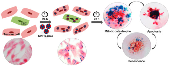
Nanotechnology-based approaches hold substantial potential to avoid chemoresistance and minimize side effects. In this work, we have used biocompatible iron oxide magnetic nanoparticles (MNPs) called MF66 and functionalized with the antineoplastic drug doxorubicin (DOX) against MDA-MB-231 cells. Electrostatically functionalized MNPs showed effective uptake and DOX linked to MNPs was more efficiently retained inside the cells than free DOX, leading to cell inactivation by mitotic catastrophe, senescence and apoptosis. Both effects, uptake and cytotoxicity, were demonstrated by different assays and videomicroscopy techniques. Likewise, covalently functionalized MNPs using three different linkers—disulfide (DOX-S-S-Pyr, called MF66-S-S-DOX), imine (DOX-I-Mal, called MF66-I-DOX) or both (DOX-I-S-S-Pyr, called MF66-S-S-I-DOX)—were also analysed. The highest cell death was detected using a linker sensitive to both pH and reducing environment (DOX-I-S-S-Pyr). The greatest success of this study was to detect also their activity against breast cancer stem-like cells (CSC) from MDA-MB-231 and primary breast cancer cells derived from a patient with a similar genetic profile (triple-negative breast cancer). In summary, these nanoformulations are promising tools as therapeutic agent vehicles, due to their ability to produce efficient internalization, drug delivery, and cancer cell inactivation, even in cancer stem-like cells (CSCs) from patients. Full article
Show Figures

Graphical abstract

18 pages, 3119 KiB  
Article
Nanoemulsions as Delivery Systems for Poly-Chemotherapy Aiming at Melanoma Treatment
by Chiara Dianzani, Chiara Monge, Gianluca Miglio, Loredana Serpe, Katia Martina, Luigi Cangemi, Chiara Ferraris, Silvia Mioletti, Sara Osella, Casimiro Luca Gigliotti, Elena Boggio, Nausicaa Clemente, Umberto Dianzani and Luigi Battaglia
Cancers 2020, 12(5), 1198; https://doi.org/10.3390/cancers12051198 - 9 May 2020
Cited by 35 | Viewed by 4279
Abstract
Aims: Advanced melanoma is characterized by poor outcome. Despite the number of treatments having been increased over the last decade, current pharmacological strategies are only partially effective. Therefore, the improvement of the current systemic therapy is worthy of investigation. Methods: a nanotechnology-based poly-chemotherapy [...] Read more.
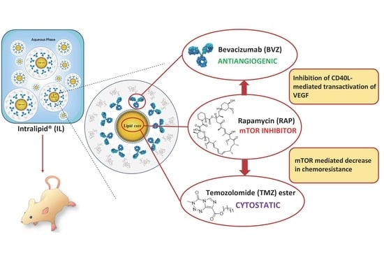
Aims: Advanced melanoma is characterized by poor outcome. Despite the number of treatments having been increased over the last decade, current pharmacological strategies are only partially effective. Therefore, the improvement of the current systemic therapy is worthy of investigation. Methods: a nanotechnology-based poly-chemotherapy was tested at preclinical level. Temozolomide, rapamycin, and bevacizumab were co-loaded as injectable nanoemulsions for total parenteral nutrition (Intralipid®), due to suitable devices, and preliminarily tested in vitro on human and mouse cell models and in vivo on the B16-F10 melanoma mouse model. Results: Drug combination was efficiently loaded in the liquid lipid matrix of Intralipid®, including bevacizumab monoclonal antibody, leading to a fast internalization in tumour cells. An increased cytotoxicity towards melanoma cells, as well as an improved inhibition of tumour relapse, migration, and angiogenesis were demonstrated in cell models for the Intralipid®-loaded drug combinations. In preliminary in vivo studies, the proposed approach was able to reduce tumour growth significantly, compared to controls. A relevant efficacy towards tumour angiogenesis and mitotic index was determined and immune response was involved. Conclusions: In these preliminary studies, Intralipid® proved to be a safe and versatile poly-chemotherapy delivery system for advanced melanoma treatment, by acting on multiple mechanisms. Full article
Show Figures

Graphical abstract

19 pages, 6112 KiB  
Article
Targeted Dual Intervention-Oriented Drug-Encapsulated (DIODE) Nanoformulations for Improved Treatment of Pancreatic Cancer
by Vijay Sagar Madamsetty, Krishnendu Pal, Shamit Kumar Dutta, Enfeng Wang and Debabrata Mukhopadhyay
Cancers 2020, 12(5), 1189; https://doi.org/10.3390/cancers12051189 - 8 May 2020
Cited by 8 | Viewed by 3333
Abstract
Despite recent advancements, effective treatment for pancreatic ductal adenocarcinoma (PDAC) has remained elusive. The overall survival rate in PDAC patients has been dismally low due to resistance to standard therapies. In fact, the failure of monotherapies to provide long-term survival benefits in patients [...] Read more.
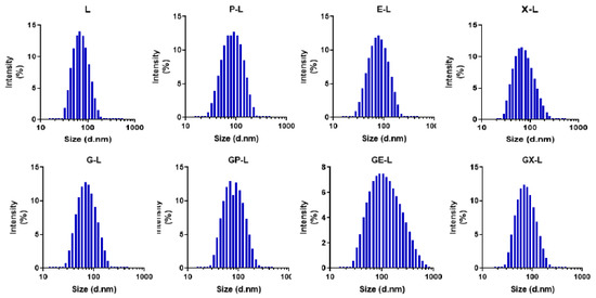
Despite recent advancements, effective treatment for pancreatic ductal adenocarcinoma (PDAC) has remained elusive. The overall survival rate in PDAC patients has been dismally low due to resistance to standard therapies. In fact, the failure of monotherapies to provide long-term survival benefits in patients led to ascension of several combination therapies for PDAC treatment. However, these combination therapies provided modest survival improvements while increasing treatment-related adverse side effects. Hence, recent developments in drug delivery methods hold the potential for enhancing therapeutic benefits by offering cocktail drug loading and minimizing chemotherapy-associated side effects. Nanoformulations-aided deliveries of anticancer agents have been a success in recent years. Yet, improving the tumor-targeted delivery of drugs to PDAC remains a major hurdle. In the present paper, we developed several new tumor-targeted dual intervention-oriented drug-encapsulated (DIODE) liposomes. We successfully formulated liposomes loaded with gemcitabine (G), paclitaxel (P), erlotinib (E), XL-184 (c-Met inhibitor, X), and their combinations (GP, GE, and GX) and evaluated their in vitro and in vivo efficacies. Our novel DIODE liposomal formulations improved median survival in comparison with gemcitabine-loaded liposomes or vehicle. Our findings are suggestive of the importance of the targeted delivery for combination therapies in improving pancreatic cancer treatment. Full article
Show Figures

Figure 1

18 pages, 2392 KiB  
Article
Mesenchymal Stem Cells As Guideposts for Nanoparticle-Mediated Targeted Drug Delivery in Ovarian Cancer
by Buddhadev Layek, Mihir Shetty, Susheel Kumar Nethi, Drishti Sehgal, Timothy K. Starr and Swayam Prabha
Cancers 2020, 12(4), 965; https://doi.org/10.3390/cancers12040965 - 14 Apr 2020
Cited by 21 | Viewed by 4048
Abstract
Nanocarriers have been extensively utilized for the systemic targeting of various solid tumors and their metastases. However, current drug delivery systems, in general, suffer from a lack of selectivity for tumor cells. Here, we develop a novel two-step targeting strategy that relies on [...] Read more.
Nanocarriers have been extensively utilized for the systemic targeting of various solid tumors and their metastases. However, current drug delivery systems, in general, suffer from a lack of selectivity for tumor cells. Here, we develop a novel two-step targeting strategy that relies on the selective accumulation of targetable synthetic receptors (i.e., azide moieties) in tumor tissues, followed by delivery of drug-loaded nanoparticles having a high binding affinity for these receptors. Mesenchymal stem cells (MSCs) were used as vehicles for the tumor-specific accumulation of azide moieties, while dibenzyl cyclooctyne (DBCO) was used as the targeting ligand. Biodistribution and antitumor efficacy studies were performed in both orthotopic metastatic and patient-derived xenograft (PDX) tumor models of ovarian cancer. Our studies show that nanoparticles are retained in tumors at a significantly higher concentration in mice that received azide-labeled MSCs (MSC-Az). Furthermore, we observed significantly reduced tumor growth (p < 0.05) and improved survival in mice receiving MSC-Az along with paclitaxel-loaded DBCO-functionalized nanoparticles compared to controls. These studies demonstrate the feasibility of a two-step targeting strategy for efficient delivery of concentrated chemotherapy for treating solid tumors. Full article
Show Figures

Figure 1

14 pages, 3312 KiB  
Article
In Vivo and In Vitro Anticancer Activity of Doxorubicin-loaded DNA-AuNP Nanocarrier for the Ovarian Cancer Treatment
by Chang-Seuk Lee, Tae Wan Kim, Da Eun Oh, Su Ok Bae, Jaesung Ryu, Hyejeong Kong, Hyeji Jeon, Hee Kyung Seo, Seob Jeon and Tae Hyun Kim
Cancers 2020, 12(3), 634; https://doi.org/10.3390/cancers12030634 - 9 Mar 2020
Cited by 28 | Viewed by 5587
Abstract
In this study, we have determined the anticancer activity of doxorubicin (Dox)-loaded DNA/gold nanoparticle (AuNP) nanocarrier (Dox-DNA-AuNP) for the treatment of ovarian cancer. The anticancer effect of Dox-DNA-AuNP was evaluated in vitro using the EZ-Cytox cell viability assay on three human ovarian cancer [...] Read more.
In this study, we have determined the anticancer activity of doxorubicin (Dox)-loaded DNA/gold nanoparticle (AuNP) nanocarrier (Dox-DNA-AuNP) for the treatment of ovarian cancer. The anticancer effect of Dox-DNA-AuNP was evaluated in vitro using the EZ-Cytox cell viability assay on three human ovarian cancer cell lines, SK-OV-3, HEY A8, and A2780. Dox-DNA-AuNP exhibited outstanding activity with good IC50 values of 4.8, 7.4, and 7.6 nM for SK-OV-3, HEY A8, and A2780, respectively. In vivo evaluation further demonstrated the superior anticancer effects of Dox-DNA-AuNP by inhibiting tumor growth compared to free Dox in an established SK-OV-3 xenograft mice model. Dox-DNA-AuNP showed about a 2.5 times higher tumor growth inhibition rate than free Dox. Furthermore, the immunohistochemical analysis of Ki67 antigen expression showed the lowest number of proliferative cells in the ovarian tumor tissue treated with Dox-DNA-AuNP. These results suggest Dox-DNA-AuNP might be a potential effective agent in ovarian cancer chemotherapy. Full article
Show Figures

Figure 1

Back to TopTop