Regulatory Functions of microRNAs

A topical collection in Cells (ISSN 2073-4409). This collection belongs to the section "Cell Nuclei: Function, Transport and Receptors".

Viewed by 208013

Editors


E-Mail Website
Collection Editor
Department of Human Sciences and Promotion of Quality of Life, Università Telematica San Raffaele Roma, 00166 Rome, Italy
Interests: stem cells; epigenetic mechanisms; noncoding RNAs; cancer stem cells
Special Issues, Collections and Topics in MDPI journals

E-Mail Website
Collection Editor

Topical Collection Information

Dear Colleagues,

MicroRNAs (miRNAs), short non-coding RNAs, have been shown to be involved in all cellular processes and in the pathogenesis of many human disorders, ranging from cancers to autoimmune diseases. However, the role of miRNAs in its detailed mechanisms concerning the initiation and progression of these diseases is still mostly uncharacterized. Therefore, researchers describe their mechanisms of actions, expression patterns and cellular pathways in which they are especially important. This Topical Collection seeks reviews and original papers covering a wide range of topics related to microRNA biology, such as miRNA therapeutics, miRNA regulation in various disorders (cancer, metabolism, autoimmunity or others), interactions between miRNAs and target genes, pathway analyses, and other related topics. Studies involving non-human organisms are also welcome.
Dr. Giorgio Malpeli
Prof. Y-h. Taguchi
Collection Editors

Manuscript Submission Information

Manuscripts should be submitted online at www.mdpi.com by registering and logging in to this website. Once you are registered, click here to go to the submission form. Manuscripts can be submitted until the deadline. All submissions that pass pre-check are peer-reviewed. Accepted papers will be published continuously in the journal (as soon as accepted) and will be listed together on the collection website. Research articles, review articles as well as short communications are invited. For planned papers, a title and short abstract (about 100 words) can be sent to the Editorial Office for announcement on this website.

Submitted manuscripts should not have been published previously, nor be under consideration for publication elsewhere (except conference proceedings papers). All manuscripts are thoroughly refereed through a single-blind peer-review process. A guide for authors and other relevant information for submission of manuscripts is available on the Instructions for Authors page. Cells is an international peer-reviewed open access semimonthly journal published by MDPI.

Please visit the Instructions for Authors page before submitting a manuscript. The Article Processing Charge (APC) for publication in this open access journal is 2700 CHF (Swiss Francs). Submitted papers should be well formatted and use good English. Authors may use MDPI's English editing service prior to publication or during author revisions.

Keywords

  • miRNA regulation
  • biomarker
  • pathway analysis
  • cancer
  • metabolism and diabetes
  • autoimmunity
  • aging
  • target genes
  • expression patterns
Published Papers (38 papers)

2022

Jump to: 2021, 2020, 2019

15 pages, 3238 KiB  
Article
miR-301a Deficiency Attenuates the Macrophage Migration and Phagocytosis through YY1/CXCR4 Pathway
by Jiawei Xu, Lanya Fu, Junyao Deng, Jiaqi Zhang, Ying Zou, Liqiang Liao, Xinrui Ma, Zhenlin Li, Yizhou Xu, Yuantao Xu, Shuyi Xu, Jingmin Liu, Xianghai Wang, Xiaodong Ma and Jiasong Guo
Cells 2022, 11(24), 3952; https://doi.org/10.3390/cells11243952 - 7 Dec 2022
Cited by 9 | Viewed by 3685
Abstract
(1) Background: the miR-301a is well known involving the proliferation and migration of tumor cells. However, the role of miR-301a in the migration and phagocytosis of macrophages is still unclear. (2) Methods: sciatic nerve injury, liver injury models, as well as primary macrophage [...] Read more.
(1) Background: the miR-301a is well known involving the proliferation and migration of tumor cells. However, the role of miR-301a in the migration and phagocytosis of macrophages is still unclear. (2) Methods: sciatic nerve injury, liver injury models, as well as primary macrophage cultures were prepared from the miR-301a knockout (KO) and wild type (WT) mice to assess the macrophage’s migration and phagocytosis capabilities. Targetscan database analysis, Western blotting, siRNA transfection, and CXCR4 inhibition or activation were performed to reveal miR301a’s potential mechanism. (3) Results: the macrophage’s migration and phagocytosis were significantly attenuated by the miR-301a KO both in vivo and in vitro. MiR-301a can target Yin-Yang 1 (YY1), and miR-301a KO resulted in YY1 up-regulation and CXCR4 (YY1′s down-stream molecule) down-regulation. siYY1 increased the expression of CXCR4 and enhanced migration and phagocytosis in KO macrophages. Meanwhile, a CXCR4 inhibitor or agonist could attenuate or accelerate, respectively, the macrophage migration and phagocytosis. (4) Conclusions: current findings indicated that miR-301a plays important roles in a macrophage’s capabilities of migration and phagocytosis through the YY1/CXCR4 pathway. Hence, miR-301a might be a promising therapeutic candidate for inflammatory diseases by adjusting macrophage bio-functions. Full article
Show Figures

Graphical abstract

14 pages, 3532 KiB  
Article
Bta-miR-199a-3p Inhibits LPS-Induced Inflammation in Bovine Mammary Epithelial Cells via the PI3K/AKT/NF-κB Signaling Pathway
by Yuhang Li, Qianqian Ren, Xingping Wang, Zhuoma Luoreng and Dawei Wei
Cells 2022, 11(21), 3518; https://doi.org/10.3390/cells11213518 - 7 Nov 2022
Cited by 9 | Viewed by 2363
Abstract
Mastitis is characterized by inflammatory damage to mammary gland tissue, which could decline milk production and quality and significantly affect the economic benefits of ranching. MicroRNAs (miRNAs), such as miR-199a-3p, are novel therapeutic targets in inflammation, and their regulation is an effective strategy [...] Read more.
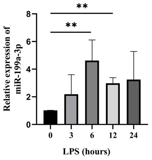
Mastitis is characterized by inflammatory damage to mammary gland tissue, which could decline milk production and quality and significantly affect the economic benefits of ranching. MicroRNAs (miRNAs), such as miR-199a-3p, are novel therapeutic targets in inflammation, and their regulation is an effective strategy for inflammation control. Despite its importance in humans and animals, the molecular mechanism of bovine miR-199a-3p (bta-miR-199a-3p) in dairy cow mastitis and bovine mammary epithelial cell (bMEC) inflammation is unclear. In our study, a bovine mammary epithelial cell line (MAC-T) induced by lipopolysaccharide (LPS) was used as an inflammatory cell model to investigate the molecular mechanism of bta-miR-199a-3p in the MAC-T inflammatory response. bta-miR-199a-3p was up-regulated in the LPS-induced MAC-T cells, while CD2-associated protein (CD2AP) was revealed as its target gene in a double luciferase reporter gene experiment. In addition, the overexpression of bta-miR-199a-3p negatively regulated the expression of CD2AP and the activation of the phosphatidylinositol 3-kinase (PI3K)/AKT/nuclear factor kappa-B (NF-κB) signaling pathway. These subsequently inhibited the secretion of related inflammatory factors (TNF-α, IL-1β, and IL-6) and the expression of apoptotic genes (CASP3 and CASP9), thereby alleviating the LPS-challenged inflammatory response in the MAC-T cells. Silencing of bta-miR-199a-3p, however, reversed the above effects. Thus, bta-miR-199a-3p inhibits LPS-induced inflammation in bMECs by directly targeting CD2AP and regulating the PI3K/AKT/NF-κB signaling pathway. This study reveals the potential regulatory mechanism of bta-miR-199a-3p in bMEC inflammatory immune response and may serve as a useful target for the treatment of mastitis. Full article
Show Figures

Figure 1

12 pages, 1987 KiB  
Article
Ssc-MiR-21-5p and Ssc-MiR-615 Regulates the Proliferation and Apoptosis of Leydig Cells by Targeting SOX5
by Qi Tang, Yanghai Zhang, Linxiu Yue, Hongying Ren and Chuanying Pan
Cells 2022, 11(14), 2253; https://doi.org/10.3390/cells11142253 - 21 Jul 2022
Cited by 12 | Viewed by 2288
Abstract
Leydig cells (LCs) are the predominant cells of androgen production, which plays key roles in spermatogenesis and maintaining male secondary sexual characteristics. Abnormal development of LCs affects androgen levels in vivo, affects fertility and may even lead to infertility. Little is known about [...] Read more.
Leydig cells (LCs) are the predominant cells of androgen production, which plays key roles in spermatogenesis and maintaining male secondary sexual characteristics. Abnormal development of LCs affects androgen levels in vivo, affects fertility and may even lead to infertility. Little is known about the regulation mechanism on LCs’ development and maturation in domestic animals, especially the regulation of non-coding RNAs. In this study, we continued to dig deeper in the previous RNA-seq data of porcine LCs from our group, combined with detecting the expression profiles in different tissues and different types of cells in the testis, to screen out candidate microRNAs (miRNAs) that may affect the regulation of LCs. A total of two miRNAs, ssc-miR-21-5p and ssc-miR-615 (“ssc” is omitted below), were finally determined. After overexpression and interference of miRNAs in vitro, the effects of candidate miRNAs on the proliferation and apoptosis of TM3 (mouse Leydig cell line) were explored. The results showed that miR-21-5p led to a decrease in TM3 cell density and p53 (apoptosis related protein) expression. Meanwhile, miR-21-5p decreased EdU positive cell numbers, but increased TUNEL positive cell numbers, suggesting miR-21-5p could inhibit proliferation and promote apoptosis. Conversely, miR-615 could increase TM3 cell density. Western blot and TUNEL assay indicated miR-615 inhibited apoptosis, but had no effect on proliferation. In addition, Sox5 was identified a potential target gene of these two miRNAs by Dual-Luciferase reporter system assay. Our findings about functions of miRNAs in TM3 and the mapping of miRNAs-target gene regulatory network would provide an important basis for the further elucidation of miRNAs in regulating pig LCs. Full article
Show Figures

Figure 1

2021

Jump to: 2022, 2020, 2019

26 pages, 8186 KiB  
Article
MicroRNA Bta-miR-24-3p Suppressed Galectin-9 Expression through TLR4/NF-ĸB Signaling Pathway in LPS-Stimulated Bovine Endometrial Epithelial Cells
by Ayodele Olaolu Oladejo, Yajuan Li, Wenxiang Shen, Bereket Habte Imam, Xiaohu Wu, Jie Yang, Xiaoyu Ma, Yanan Lv, Wei Jiang, Xuezhi Ding, Shengyi Wang and Zuoting Yan
Cells 2021, 10(12), 3299; https://doi.org/10.3390/cells10123299 - 25 Nov 2021
Cited by 8 | Viewed by 2855
Abstract
Endometritis is a major infectious disease affecting dairy development. MicroRNAs are recognized as critical regulators of the innate immune response. However, the role and mechanism of Bta-miR-24-3p in the development of endometritis are still unclear. This study aimed to investigate the effect of [...] Read more.
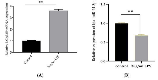
Endometritis is a major infectious disease affecting dairy development. MicroRNAs are recognized as critical regulators of the innate immune response. However, the role and mechanism of Bta-miR-24-3p in the development of endometritis are still unclear. This study aimed to investigate the effect of Bta-miR-24-3p on the inflammatory response triggered by lipopolysaccharide (LPS) and to clarify the possible mechanism. LPS-treated bovine endometrial epithelial cells (BEECs) were cultured to investigate the role of Bta-miR-24-3p. The expression levels of Bta-miR-24-3p were downregulated, and galectin-9 (LGALS9) were measured by quantitative real-time polymerase chain reaction. The LPS-induced inflammatory response was assessed by the elevated secretion of inflammatory cytokines measured by using enzyme-linked immunosorbent assay and quantitative real-time polymerase chain reaction. Activation of nuclear factor-κB (NF-κB) and TLR4 pathway was assessed by Western blot. The interaction between Bta-miR-24-3p and LGALS9 was validated by bioinformatics analysis and a luciferase reporter assay. LPS-induction in BEECs with Bta-miR-24-3p was overexpressed leads inhibition of pro-inflammatory cytokines, LGALS9 expression, and TLR4/NF-ĸB pathway deactivation. Knockdown of LGALS9 inhibited the LPS-induced inflammatory response in BEECs. LGALS9 was validated as a target of Bta-miR-24-3p. Cloned overexpression of LGALS9 failed to alter the effect of Bta-miR-24-3p on the inflammatory response in BEECs. Overall, Bta-miR-24-3p attenuated the LPS-induced inflammatory response via targeting LGALS9. The immunotherapeutic stabilisation of Bta-miR-24-3p could give a therapeutic option for endometritis and other disorders commonly associated with endometritis, suggesting a novel avenue for endometritis treatment. Full article
Show Figures

Figure 1

19 pages, 2306 KiB  
Review
The Role of microRNAs in Pulp Inflammation
by José Luis Muñoz-Carrillo, Silverio Jafet Vázquez-Alcaraz, Jazmín Monserrat Vargas-Barbosa, Luis Guillermo Ramos-Gracia, Israel Alvarez-Barreto, Alejandro Medina-Quiroz and Karla Karina Díaz-Huerta
Cells 2021, 10(8), 2142; https://doi.org/10.3390/cells10082142 - 20 Aug 2021
Cited by 17 | Viewed by 6714
Abstract
The dental pulp can be affected by thermal, physical, chemical, and bacterial phenomena that stimulate the inflammatory response. The pulp tissue produces an immunological, cellular, and vascular reaction in an attempt to defend itself and resolve the affected tissue. The expression of different [...] Read more.
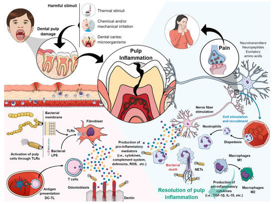
The dental pulp can be affected by thermal, physical, chemical, and bacterial phenomena that stimulate the inflammatory response. The pulp tissue produces an immunological, cellular, and vascular reaction in an attempt to defend itself and resolve the affected tissue. The expression of different microRNAs during pulp inflammation has been previously documented. MicroRNAs (miRNAs) are endogenous small molecules involved in the transcription of genes that regulate the immune system and the inflammatory response. They are present in cellular and physiological functions, as well as in the pathogenesis of human diseases, becoming potential biomarkers for diagnosis, prognosis, monitoring, and safety. Previous studies have evidenced the different roles played by miRNAs in proinflammatory, anti-inflammatory, and immunological phenomena in the dental pulp, highlighting specific key functions of pulp pathology. This systematized review aims to provide an understanding of the role of the different microRNAs detected in the pulp and their effects on the expression of the different target genes that are involved during pulp inflammation. Full article
Show Figures

Figure 1

16 pages, 1160 KiB  
Review
CRISPR/Cas13-Based Approaches for Ultrasensitive and Specific Detection of microRNAs
by Javier T. Granados-Riveron and Guillermo Aquino-Jarquin
Cells 2021, 10(7), 1655; https://doi.org/10.3390/cells10071655 - 1 Jul 2021
Cited by 39 | Viewed by 10925
Abstract
MicroRNAs (miRNAs) have a prominent role in virtually every aspect of cell biology. Due to the small size of mature miRNAs, the high degree of similarity between miRNA family members, and the low abundance of miRNAs in body fluids, miRNA expression profiling is [...] Read more.
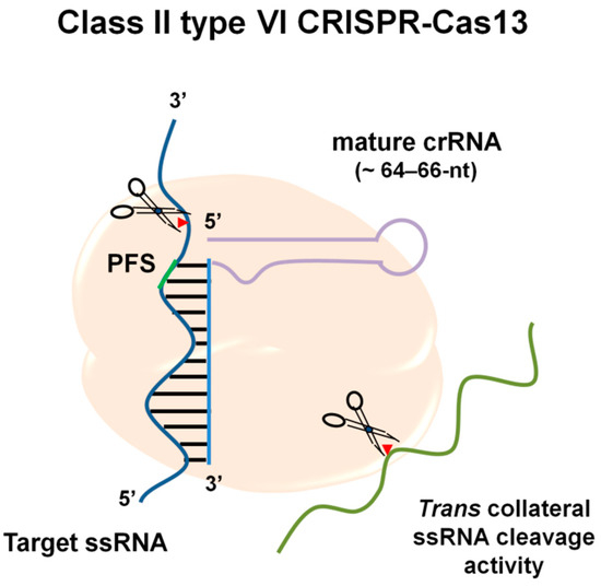
MicroRNAs (miRNAs) have a prominent role in virtually every aspect of cell biology. Due to the small size of mature miRNAs, the high degree of similarity between miRNA family members, and the low abundance of miRNAs in body fluids, miRNA expression profiling is technically challenging. Biosensors based on electrochemical detection for nucleic acids are a novel category of inexpensive and very sensitive diagnostic tools. On the other hand, after recognizing the target sequence, specific CRISPR-associated proteins, including orthologues of Cas12, Cas13, and Cas14, exhibit collateral nonspecific catalytic activities that can be employed for specific and ultrasensitive nucleic acid detection from clinically relevant samples. Recently, several platforms have been developed, connecting the benefits of enzyme-assisted signal amplification and enzyme-free amplification biosensing technologies with CRISPR-based approaches for miRNA detection. Together, they provide high sensitivity, precision, and fewer limitations in diagnosis through efficient sensors at a low cost and a simple miniaturized readout. This review provides an overview of several CRISPR-based biosensing platforms that have been developed and successfully applied for ultrasensitive and specific miRNA detection. Full article
Show Figures

Figure 1

13 pages, 2060 KiB  
Article
ABHD4-Regulating RNA Panel: Novel Biomarkers in Acute Coronary Syndrome Diagnosis
by Sara H. A. Agwa, Sherif Samir Elzahwy, Mahmoud Shawky El Meteini, Hesham Elghazaly, Maha Saad, Aya M. Abd Elsamee, Rania Shamekh and Marwa Matboli
Cells 2021, 10(6), 1512; https://doi.org/10.3390/cells10061512 - 16 Jun 2021
Cited by 10 | Viewed by 3245
Abstract
Background: Acute coronary syndrome (ACS) is a major cause of death all over the world. STEMI represents a type of myocardial infarction with acute ST elevation. We aimed to assess the predictive power of potential RNA panel expression in acute coronary syndrome. Method: [...] Read more.
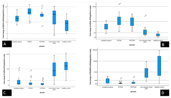
Background: Acute coronary syndrome (ACS) is a major cause of death all over the world. STEMI represents a type of myocardial infarction with acute ST elevation. We aimed to assess the predictive power of potential RNA panel expression in acute coronary syndrome. Method: We used in silico data analysis to retrieve RNAs related to glycerophospholipid metabolism dysregulation and specific to ACS that results in the selection of Alpha/Beta hydrolase fold domain4 (ABHD4) mRNA and its epigenetic regulators (Foxf1 adjacent noncoding developmental regulatory RNA (FENDRR) lncRNA, miRNA-221, and miRNA-197). We assessed the expression of the serum RNA panel in 68 patients with ACS, 21 patients with chest pain due to non-cardiac causes, and 21 healthy volunteers by quantitative real-time polymerase chain reaction. Results: The study data showed significant down regulation in the expression of the serum levels of FENDRR lncRNA and miRNA-221-3p by 120-fold and 22-fold in Unstable angina (UA) in comparison with healthy volunteers, and by 8.6-fold and 2-fold in ST segment elevation myocardial infarction (STEMI) patients versus UA; concomitant upregulation in the expression of ABHD4 mRNA and miRNA-197-5p by 444-fold and 10-fold in UA compared with healthy volunteers, and by 1.54-fold and 4.5-fold in STEMI versus unstable angina. Performance characteristics analysis showed that the ABHD4-regulating RNA panel were potential biomarkers for prediction of ACS. Moreover, there was a significant association between the 2 miRNAs and ABHD4 mRNA and the regulating FENDRR lncRNA. Conclusion: Collectively, ABHD4 mRNA regulating RNA panel based on putative interactions seems to be novel non-invasive biomarkers that could detect ACS early and stratify severity of the condition that could improve health outcome. Full article
Show Figures

Figure 1

16 pages, 2186 KiB  
Article
Regulatory miRNA–mRNA Networks in Parkinson’s Disease
by Bruno Lopes Santos-Lobato, Amanda Ferreira Vidal and Ândrea Ribeiro-dos-Santos
Cells 2021, 10(6), 1410; https://doi.org/10.3390/cells10061410 - 6 Jun 2021
Cited by 20 | Viewed by 4907
Abstract
Parkinson’s disease (PD) is the second-most common neurodegenerative disease, and its pathophysiology is associated with alpha-synuclein accumulation, oxidative stress, mitochondrial dysfunction, and neuroinflammation. MicroRNAs are small non-coding RNAs that regulate gene expression, and many previous studies have described their dysregulation in plasma, CSF, [...] Read more.
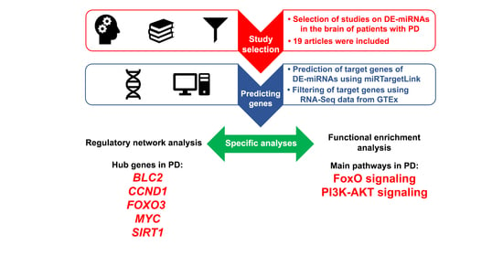
Parkinson’s disease (PD) is the second-most common neurodegenerative disease, and its pathophysiology is associated with alpha-synuclein accumulation, oxidative stress, mitochondrial dysfunction, and neuroinflammation. MicroRNAs are small non-coding RNAs that regulate gene expression, and many previous studies have described their dysregulation in plasma, CSF, and in the brain of patients with PD. In this study, we aimed to provide a regulatory network analysis on differentially expressed miRNAs in the brain of patients with PD. Based on our systematic review with a focus on the substantia nigra and the putamen, we found 99 differentially expressed miRNAs in brain samples from patients with PD, which regulate 135 target genes. Five genes associated with neuronal survival (BCL2, CCND1, FOXO3, MYC, and SIRT1) were modulated by dysregulated miRNAs found in the substantia nigra and the putamen of patients with PD. The functional enrichment analysis found FoxO and PI3K-AKT signaling as pathways related to PD. In conclusion, our comprehensive analysis of brain-related miRNA–mRNA regulatory networks in PD showed that mechanisms involving neuronal survival signaling, such as cell cycle control and regulation of autophagy/apoptosis, may be crucial for the neurodegeneration of PD, being a promising way for novel disease-modifying therapies. Full article
Show Figures

Graphical abstract

23 pages, 8209 KiB  
Article
Horizontal Transfer of miR-643 from Cisplatin-Resistant Cells Confers Chemoresistance to Recipient Drug-Sensitive Cells by Targeting APOL6
by Grace R. Raji, Aswini Poyyakkara, Anjali Kunhi Krishnan, Ashutosh Kumar Maurya, Udeshna Changmai, Sharath S. Shankar and V. B. Sameer Kumar
Cells 2021, 10(6), 1341; https://doi.org/10.3390/cells10061341 - 28 May 2021
Cited by 9 | Viewed by 3083
Abstract
Acquisition of resistance to cisplatin is a major impediment to the success of cisplatin-based combination therapies for cancer. Recent studies indicate that exosomal miRNAs derived from drug-resistant tumour cells can confer resistance properties to recipient cells by a horizontal transfer mechanism. Although the [...] Read more.
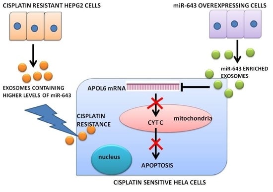
Acquisition of resistance to cisplatin is a major impediment to the success of cisplatin-based combination therapies for cancer. Recent studies indicate that exosomal miRNAs derived from drug-resistant tumour cells can confer resistance properties to recipient cells by a horizontal transfer mechanism. Although the role of horizontal transfer of a few miRNAs has been described, little is known about the concerted action of horizontal transfer of miRNAs in conferring cisplatin resistance. The present study was designed to identify the role of miR-643, which is one of the most significantly increased miRNA in exosomes released from cisplatin-resistant Heptocarcinoma cells, in altering the cisplatin resistance properties of recipient cells. Drug-sensitivity assays involving miR-643 revealed that ectopic expression of miR-643 can desensitise the cells towards cisplatin. Furthermore, we identified APOL6 as a major target of miR-643. Further mechanistic studies showed that miR-643 can modulate APOL6 mRNA and protein levels, leading to a reversal of APOL6-mediated apoptosis. Altogether, our results suggest an APOL6-dependent mechanism for miR-643 mediated cisplatin resistance upon the horizontal transfer across cell types. Full article
Show Figures

Graphical abstract

14 pages, 1003 KiB  
Review
MicroRNAs in Medullary Thyroid Carcinoma: A State of the Art Review of the Regulatory Mechanisms and Future Perspectives
by Francesca Galuppini, Simona Censi, Margherita Moro, Stefano Carraro, Marta Sbaraglia, Maurizio Iacobone, Matteo Fassan, Caterina Mian and Gianmaria Pennelli
Cells 2021, 10(4), 955; https://doi.org/10.3390/cells10040955 - 20 Apr 2021
Cited by 11 | Viewed by 3393
Abstract
Medullary thyroid carcinoma (MTC) is a rare malignant neoplasia with a variable clinical course, with complete remission often difficult to achieve. Genetic alterations lead to fundamental changes not only in hereditary MTC but also in the sporadic form, with close correlations between mutational [...] Read more.
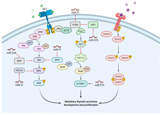
Medullary thyroid carcinoma (MTC) is a rare malignant neoplasia with a variable clinical course, with complete remission often difficult to achieve. Genetic alterations lead to fundamental changes not only in hereditary MTC but also in the sporadic form, with close correlations between mutational status and prognosis. In recent years, microRNAs (miRNAs) have become highly relevant as crucial players in MTC etiology. Current research has focused on their roles in disease carcinogenesis and development, but recent studies have expounded their potential as biomarkers and response predictors to novel biological drugs for advanced MTC. One such element which requires greater investigation is their mechanism of action and the molecular pathways involved in the regulation of gene expression. A more thorough understanding of these mechanisms will help realize the promising potential of miRNAs for MTC therapy and management. Full article
Show Figures

Figure 1

2020

Jump to: 2022, 2021, 2019

21 pages, 5736 KiB  
Article
The Role of miR-21 in Osteoblasts–Osteoclasts Coupling In Vitro
by Agnieszka Smieszek, Klaudia Marcinkowska, Ariadna Pielok, Mateusz Sikora, Lukas Valihrach and Krzysztof Marycz
Cells 2020, 9(2), 479; https://doi.org/10.3390/cells9020479 - 19 Feb 2020
Cited by 46 | Viewed by 5543
Abstract
MiR-21 is being gradually more and more recognized as a molecule regulating bone tissue homeostasis. However, its function is not fully understood due to the dual role of miR-21 on bone-forming and bone-resorbing cells. In this study, we investigated the impact of miR-21 [...] Read more.
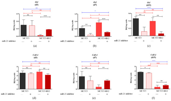
MiR-21 is being gradually more and more recognized as a molecule regulating bone tissue homeostasis. However, its function is not fully understood due to the dual role of miR-21 on bone-forming and bone-resorbing cells. In this study, we investigated the impact of miR-21 inhibition on pre-osteoblastic cells differentiation and paracrine signaling towards pre-osteoclasts using indirect co-culture model of mouse pre-osteoblast (MC3T3) and pre-osteoclast (4B12) cell lines. The inhibition of miR-21 in MC3T3 cells (MC3T3inh21) modulated expression of genes encoding osteogenic markers including collagen type I (Coll-1), osteocalcin (Ocl), osteopontin (Opn), and runt-related transcription factor 2 (Runx-2). Inhibition of miR-21 in osteogenic cultures of MC3T3 also inflected the synthesis of OPN protein which is essential for proper mineralization of extracellular matrix (ECM) and anchoring osteoclasts to the bones. Furthermore, it was shown that in osteoblasts miR-21 regulates expression of factors that are vital for survival of pre-osteoclast, such as receptor activator of nuclear factor κB ligand (RANKL). The pre-osteoclast cultured with MC3T3inh21 cells was characterized by lowered expression of several markers associated with osteoclasts’ differentiation, foremost tartrate-resistant acid phosphatase (Trap) but also receptor activator of nuclear factor-κB ligand (Rank), cathepsin K (Ctsk), carbonic anhydrase II (CaII), and matrix metalloproteinase (Mmp-9). Collectively, our data indicate that the inhibition of miR-21 in MC3T3 cells impairs the differentiation and ECM mineralization as well as influences paracrine signaling leading to decreased viability of pre-osteoclasts. Full article
Show Figures

Figure 1

28 pages, 762 KiB  
Review
miRNAs as Influencers of Cell–Cell Communication in Tumor Microenvironment
by Ilaria Conti, Gabriele Varano, Carolina Simioni, Ilaria Laface, Daniela Milani, Erika Rimondi and Luca M. Neri
Cells 2020, 9(1), 220; https://doi.org/10.3390/cells9010220 - 15 Jan 2020
Cited by 57 | Viewed by 8016
Abstract
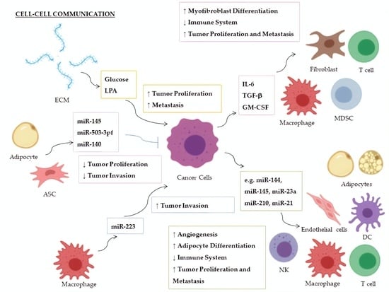
microRNAs (miRNAs) are small noncoding RNAs that regulate gene expression at the posttranscriptional level, inducing the degradation of the target mRNA or translational repression. MiRNAs are involved in the control of a multiplicity of biological processes, and their absence or altered expression has [...] Read more.
microRNAs (miRNAs) are small noncoding RNAs that regulate gene expression at the posttranscriptional level, inducing the degradation of the target mRNA or translational repression. MiRNAs are involved in the control of a multiplicity of biological processes, and their absence or altered expression has been associated with a variety of human diseases, including cancer. Recently, extracellular miRNAs (ECmiRNAs) have been described as mediators of intercellular communication in multiple contexts, including tumor microenvironment. Cancer cells cooperate with stromal cells and elements of the extracellular matrix (ECM) to establish a comfortable niche to grow, to evade the immune system, and to expand. Within the tumor microenvironment, cells release ECmiRNAs and other factors in order to influence and hijack the physiological processes of surrounding cells, fostering tumor progression. Here, we discuss the role of miRNAs in the pathogenesis of multicomplex diseases, such as Alzheimer’s disease, obesity, and cancer, focusing on the contribution of both intracellular miRNAs, and of released ECmiRNAs in the establishment and development of cancer niche. We also review growing evidence suggesting the use of miRNAs as novel targets or potential tools for therapeutic applications. Full article
Show Figures

Graphical abstract

17 pages, 3022 KiB  
Article
Identification of CNS Injury-Related microRNAs as Novel Toll-Like Receptor 7/8 Signaling Activators by Small RNA Sequencing
by Thomas Wallach, Max Wetzel, Paul Dembny, Ori Staszewski, Christina Krüger, Alice Buonfiglioli, Marco Prinz and Seija Lehnardt
Cells 2020, 9(1), 186; https://doi.org/10.3390/cells9010186 - 11 Jan 2020
Cited by 16 | Viewed by 3850
Abstract
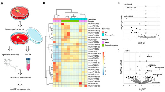
Toll-like receptors (TLRs) belong to pattern recognition receptors, which respond to danger signals such as pathogen-associated molecular patterns or damage-associated molecular patterns. Upon TLR activation in microglia, the major immune cells in the brain, distinct signaling cascades trigger the production of inflammatory molecules, [...] Read more.
Toll-like receptors (TLRs) belong to pattern recognition receptors, which respond to danger signals such as pathogen-associated molecular patterns or damage-associated molecular patterns. Upon TLR activation in microglia, the major immune cells in the brain, distinct signaling cascades trigger the production of inflammatory molecules, being a critical feature in neuroinflammation and neurodegenerative processes. Recently, individual microRNAs (miRNAs) were shown to act as endogenous TLR ligands. Here, we conducted systematic screening for miRNAs as potential TLR7/8 ligands by small RNA sequencing of apoptotic neurons and their corresponding supernatants. Several miRNA species were identified in both supernatants and injured neurons, and 83.3% of the media-enriched miRNAs activated murine and/or human TLR7/8 expressed in HEK293-derived TLR reporter cells. Among the detected extracellular miRNAs, distinct miRNAs such as miR-340-3p and miR-132-5p induced cytokine and chemokine release from microglia and triggered neurotoxicity in vitro. Taken together, our systematic study establishes miRNAs released from injured neurons as new TLR7/8 activators, which contribute to inflammatory and neurodegenerative responses in the central nervous system (CNS). Full article
Show Figures

Figure 1

16 pages, 1690 KiB  
Article
miR-26a is Involved in Glycometabolism and Affects Boar Sperm Viability by Targeting PDHX
by Wencan Wang, Kai Liang, Yu Chang, Mingxia Ran, Yan Zhang, Malik Ahsan Ali, Dinghui Dai, Izhar Hyder Qazi, Ming Zhang, Guangbin Zhou, Jiandong Yang, Christiana Angel and Changjun Zeng
Cells 2020, 9(1), 146; https://doi.org/10.3390/cells9010146 - 8 Jan 2020
Cited by 18 | Viewed by 3708
Abstract
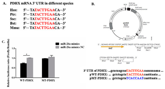
miR-26a is associated with sperm metabolism and can affect sperm motility and apoptosis. However, how miR-26a affects sperm motility remains largely unknown. Our previous study indicated that the PDHX gene is predicted to be a potential target of miR-26a, which is responsible for [...] Read more.
miR-26a is associated with sperm metabolism and can affect sperm motility and apoptosis. However, how miR-26a affects sperm motility remains largely unknown. Our previous study indicated that the PDHX gene is predicted to be a potential target of miR-26a, which is responsible for pyruvate oxidative decarboxylation which is considered as a key step for connecting glycolysis with oxidative phosphorylation. In this study, we first reported a potential relationship between miR-26a and PDHX and their expressions in fresh, frozen-thawed, and epididymal boar sperm. Then, sperm viability and survival were determined after transfection of miR-26a. mRNA and protein expression level of PDHX in the liquid-preserved boar sperm after transfection were also determined by RT-qPCR and Western Blot (WB). Our results showed that expression level of PDHX was significantly increased during sperm transit from epididymal caput to corpus and cauda. Similarly, expression of PDHX was significantly higher (P < 0.05) in fresh sperm as compared to epididymal cauda and frozen-thawed sperm. However, the expression of miR-26a in epididymal corpus sperm was significantly higher (P < 0.05) than that of caput and cauda sperm. Furthermore, after transfection of boar sperm with miR-26a mimic and inhibitor under liquid storage, the lowest and highest sperm viability was observed in miR-26a mimic and inhibitor treatment (P < 0.05), respectively. The protein levels of PDHX, after 24 and 48 h of transfection of miR-26a mimics and inhibitor, were notably decreased and increased (P < 0.05), respectively, as compared to negative control (NC) group. In conclusion, the novel and enticing findings of our study provide a reasonable evidence that miR-26a via PDHX, a link between glycolysis and oxidative phosphorylation, could regulate the glycometabolic pathway which eventually affect boar sperm viability and survival. Full article
Show Figures

Figure 1

25 pages, 3696 KiB  
Review
MicroRNAs: Biological Regulators in Pathogen–Host Interactions
by Stephanie Maia Acuña, Lucile Maria Floeter-Winter and Sandra Marcia Muxel
Cells 2020, 9(1), 113; https://doi.org/10.3390/cells9010113 - 2 Jan 2020
Cited by 68 | Viewed by 8583
Abstract
An inflammatory response is essential for combating invading pathogens. Several effector components, as well as immune cell populations, are involved in mounting an immune response, thereby destroying pathogenic organisms such as bacteria, fungi, viruses, and parasites. In the past decade, microRNAs (miRNAs), a [...] Read more.
An inflammatory response is essential for combating invading pathogens. Several effector components, as well as immune cell populations, are involved in mounting an immune response, thereby destroying pathogenic organisms such as bacteria, fungi, viruses, and parasites. In the past decade, microRNAs (miRNAs), a group of noncoding small RNAs, have emerged as functionally significant regulatory molecules with the significant capability of fine-tuning biological processes. The important role of miRNAs in inflammation and immune responses is highlighted by studies in which the regulation of miRNAs in the host was shown to be related to infectious diseases and associated with the eradication or susceptibility of the infection. Here, we review the biological aspects of microRNAs, focusing on their roles as regulators of gene expression during pathogen–host interactions and their implications in the immune response against Leishmania, Trypanosoma, Toxoplasma, and Plasmodium infectious diseases. Full article
Show Figures

Figure 1

2019

Jump to: 2022, 2021, 2020

15 pages, 1877 KiB  
Article
miR-9-5p Exerts a Dual Role in Cervical Cancer and Targets Transcription Factor TWIST1
by Iris Babion, Annelieke Jaspers, Annina P. van Splunter, Iris A.E. van der Hoorn, Saskia M. Wilting and Renske D.M. Steenbergen
Cells 2020, 9(1), 65; https://doi.org/10.3390/cells9010065 - 26 Dec 2019
Cited by 29 | Viewed by 7738
Abstract
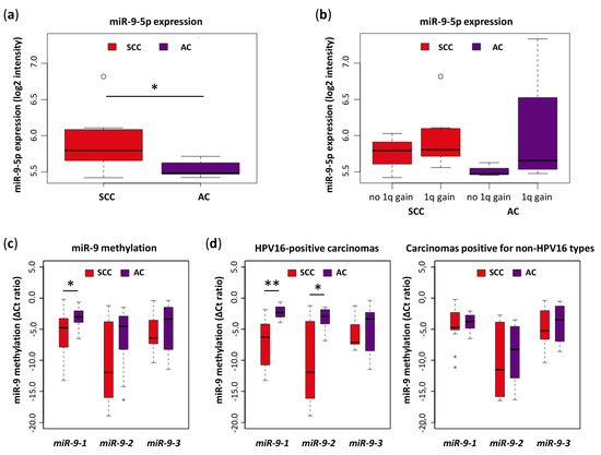
Squamous cell carcinoma (SCC) and adenocarcinoma (AC) represent the major cervical cancer histotypes. Both histotypes are caused by infection with high-risk HPV (hrHPV) and are associated with deregulated microRNA expression. Histotype-dependent expression has been observed for miR-9-5p, showing increased expression in SCC and [...] Read more.
Squamous cell carcinoma (SCC) and adenocarcinoma (AC) represent the major cervical cancer histotypes. Both histotypes are caused by infection with high-risk HPV (hrHPV) and are associated with deregulated microRNA expression. Histotype-dependent expression has been observed for miR-9-5p, showing increased expression in SCC and low expression in AC. Here, we studied the regulation and functionality of miR-9-5p in cervical SCCs and ACs using cervical tissue samples and hrHPV-containing cell lines. Expression and methylation analysis of cervical tissues revealed that low levels of miR-9-5p in ACs are linked to methylation of its precursor genes, particularly miR-9-1. Stratification of tissue samples and hrHPV-containing cell lines suggested that miR-9-5p depends on both histotype and hrHPV type, with higher expression in SCCs and HPV16-positive cells. MiR-9-5p promoted cell viability and anchorage independence in cervical cancer cell lines SiHa (SCC, HPV16) and CaSki (metastasized SCC, HPV16), while it played a tumor suppressive role in HeLa (AC, HPV18). TWIST1, a transcription factor involved in epithelial-to-mesenchymal transition (EMT), was established as a novel miR-9-5p target. Our results show that miR-9-5p plays a dual role in cervical cancer in a histotype- and hrHPV type-dependent manner. MiR-9-5p mediated silencing of TWIST1 suggests two distinct mechanisms towards EMT in cervical cancer. Full article
Show Figures

Figure 1

18 pages, 2053 KiB  
Article
Brain-Derived Extracellular Vesicle microRNA Signatures Associated with In Utero and Postnatal Oxycodone Exposure
by Farah Shahjin, Rahul S. Guda, Victoria L. Schaal, Katherine Odegaard, Alexander Clark, Austin Gowen, Peng Xiao, Steven J. Lisco, Gurudutt Pendyala and Sowmya V. Yelamanchili
Cells 2020, 9(1), 21; https://doi.org/10.3390/cells9010021 - 19 Dec 2019
Cited by 36 | Viewed by 6614
Abstract
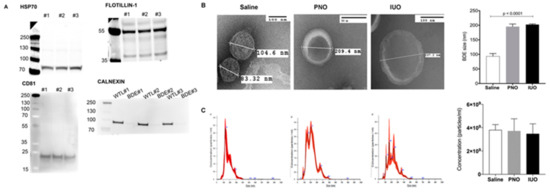
Oxycodone (oxy) is a semi-synthetic opioid commonly used as a pain medication that is also a widely abused prescription drug. While very limited studies have examined the effect of in utero oxy (IUO) exposure on neurodevelopment, a significant gap in knowledge is the [...] Read more.
Oxycodone (oxy) is a semi-synthetic opioid commonly used as a pain medication that is also a widely abused prescription drug. While very limited studies have examined the effect of in utero oxy (IUO) exposure on neurodevelopment, a significant gap in knowledge is the effect of IUO compared with postnatal oxy (PNO) exposure on synaptogenesis—a key process in the formation of synapses during brain development—in the exposed offspring. One relatively unexplored form of cell–cell communication associated with brain development in response to IUO and PNO exposure are extracellular vesicles (EVs). EVs are membrane-bound vesicles that serve as carriers of cargo, such as microRNAs (miRNAs). Using RNA-Seq analysis, we identified distinct brain-derived extracellular vesicle (BDEs) miRNA signatures associated with IUO and PNO exposure, including their gene targets, regulating key functional pathways associated with brain development to be more impacted in the IUO offspring. Further treatment of primary 14-day in vitro (DIV) neurons with IUO BDEs caused a significant reduction in spine density compared to treatment with BDEs from PNO and saline groups. In summary, our studies identified for the first time, key BDE miRNA signatures in IUO- and PNO-exposed offspring, which could impact their brain development as well as synaptic function. Full article
Show Figures

Figure 1

13 pages, 1783 KiB  
Review
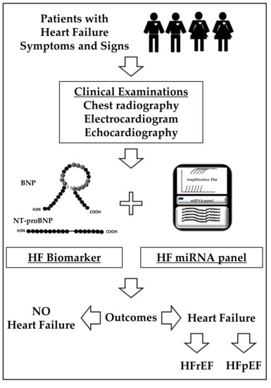
Heart Failure with Reduced Ejection Fraction (HFrEF) and Preserved Ejection Fraction (HFpEF): The Diagnostic Value of Circulating MicroRNAs
by Yei-Tsung Chen, Lee Lee Wong, Oi Wah Liew and Arthur Mark Richards
Cells 2019, 8(12), 1651; https://doi.org/10.3390/cells8121651 - 16 Dec 2019
Cited by 41 | Viewed by 6589
Abstract
Circulating microRNAs offer attractive potential as epigenetic disease biomarkers by virtue of their biological stability and ready accessibility in liquid biopsies. Numerous clinical cohort studies have revealed unique microRNA profiles in different disease settings, suggesting utility as markers with diagnostic and prognostic applications. [...] Read more.
Circulating microRNAs offer attractive potential as epigenetic disease biomarkers by virtue of their biological stability and ready accessibility in liquid biopsies. Numerous clinical cohort studies have revealed unique microRNA profiles in different disease settings, suggesting utility as markers with diagnostic and prognostic applications. Given the complex network of microRNA functions in modulating gene expression and post-transcriptional modifications, the circulating microRNA landscape in disease may reflect pathophysiological status, providing valuable information for delineating distinct subtypes and/or stages of complex diseases. Heart failure (HF) is an increasingly significant global health challenge, imposing major economic liability and health care burden due to high hospitalization, morbidity, and mortality rates. Although HF is defined as a syndrome characterized by symptoms and findings on physical examination, it may be further differentiated based on left ventricular ejection fraction (LVEF) and categorized as HF with reduced ejection fraction (HFrEF) and HF with preserved ejection fraction (HFpEF). The presenting clinical syndromes in HFpEF and HFrEF are similar but mortality differs, being somewhat lower in HFpEF than in HFrEF. However, while HFrEF is responsive to an array of therapies, none has been shown to improve survival in HFpEF. Herein, we review recent HF cohort studies focusing on the distinct microRNA profiles associated with HF subtypes to reveal new insights to underlying mechanisms and explore the possibility of exploiting these differences for diagnostic/prognostic applications. Full article
Show Figures

Figure 1

20 pages, 3424 KiB  
Article
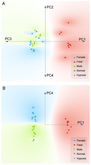
Multi-Omics Integration Reveals Short and Long-Term Effects of Gestational Hypoxia on the Heart Development
by Yu Gao, Chiranjib Dasgupta, Lei Huang, Rui Song, Ziwei Zhang and Lubo Zhang
Cells 2019, 8(12), 1608; https://doi.org/10.3390/cells8121608 - 11 Dec 2019
Cited by 12 | Viewed by 5418
Abstract
Antenatal hypoxia caused epigenetic reprogramming of methylome and transcriptome in the developing heart and increased the risk of heart disease later in life. Herein, we investigated the impact of gestational hypoxia in proteome and metabolome in the hearts of fetus and adult offspring. [...] Read more.
Antenatal hypoxia caused epigenetic reprogramming of methylome and transcriptome in the developing heart and increased the risk of heart disease later in life. Herein, we investigated the impact of gestational hypoxia in proteome and metabolome in the hearts of fetus and adult offspring. Pregnant rats were treated with normoxia or hypoxia (10.5% O2) from day 15 to 21 of gestation. Hearts were isolated from near-term fetuses and 5 month-old offspring, and proteomics and metabolomics profiling was determined. The data demonstrated that antenatal hypoxia altered proteomics and metabolomics profiling in the heart, impacting energy metabolism, lipid metabolism, oxidative stress, and inflammation-related pathways in a developmental and sex dependent manner. Of importance, integrating multi-omics data of transcriptomics, proteomics, and metabolomics profiling revealed reprogramming of the mitochondrion, especially in two clusters: (a) the cluster associated with “mitochondrial translation”/“aminoacyl t-RNA biosynthesis”/“one-carbon pool of folate”/“DNA methylation”; and (b) the cluster with “mitochondrion”/“TCA cycle and respiratory electron transfer”/“acyl-CoA dehydrogenase”/“oxidative phosphorylation”/“complex I”/“troponin myosin cardiac complex”. Our study provides a powerful means of multi-omics data integration and reveals new insights into phenotypic reprogramming of the mitochondrion in the developing heart by fetal hypoxia, contributing to an increase in the heart vulnerability to disease later in life. Full article
Show Figures

Figure 1

28 pages, 595 KiB  
Review
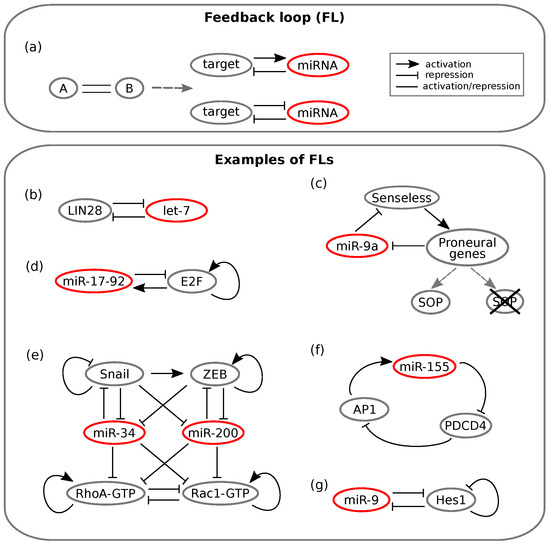
From Endogenous to Synthetic microRNA-Mediated Regulatory Circuits: An Overview
by Elsi Ferro, Chiara Enrico Bena, Silvia Grigolon and Carla Bosia
Cells 2019, 8(12), 1540; https://doi.org/10.3390/cells8121540 - 29 Nov 2019
Cited by 17 | Viewed by 4347
Abstract
MicroRNAs are short non-coding RNAs that are evolutionarily conserved and are pivotal post-transcriptional mediators of gene regulation. Together with transcription factors and epigenetic regulators, they form a highly interconnected network whose building blocks can be classified depending on the number of molecular species [...] Read more.
MicroRNAs are short non-coding RNAs that are evolutionarily conserved and are pivotal post-transcriptional mediators of gene regulation. Together with transcription factors and epigenetic regulators, they form a highly interconnected network whose building blocks can be classified depending on the number of molecular species involved and the type of interactions amongst them. Depending on their topology, these molecular circuits may carry out specific functions that years of studies have related to the processing of gene expression noise. In this review, we first present the different over-represented network motifs involving microRNAs and their specific role in implementing relevant biological functions, reviewing both theoretical and experimental studies. We then illustrate the recent advances in synthetic biology, such as the construction of artificially synthesised circuits, which provide a controlled tool to test experimentally the possible microRNA regulatory tasks and constitute a starting point for clinical applications. Full article
Show Figures

Figure 1

35 pages, 5899 KiB  
Review
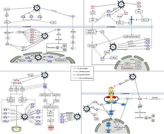
The Regulatory Role of MicroRNA in Hepatitis-B Virus-Associated Hepatocellular Carcinoma (HBV-HCC) Pathogenesis
by Kurt Sartorius, Julia Makarova, Benn Sartorius, Ping An, Cheryl Winkler, Anil Chuturgoon and Anna Kramvis
Cells 2019, 8(12), 1504; https://doi.org/10.3390/cells8121504 - 24 Nov 2019
Cited by 56 | Viewed by 8111
Abstract
The incidence and mortality of hepatitis B virus (HBV)-associated hepatocellular carcinoma (HBV-HCC) is an intractable public health problem in developing countries that is compounded by limited early detection and therapeutic options. Despite the early promise of utilizing the regulatory role of miRNA in [...] Read more.
The incidence and mortality of hepatitis B virus (HBV)-associated hepatocellular carcinoma (HBV-HCC) is an intractable public health problem in developing countries that is compounded by limited early detection and therapeutic options. Despite the early promise of utilizing the regulatory role of miRNA in liver cancer, this field remains largely in the work-in-progress phase. This exploratory review paper adopts a broad focus in order to collate evidence of the regulatory role of miRNA in each stage of the HBV-HCC continuum. This includes the regulatory role of miRNA in early HBV infection, chronic inflammation, fibrosis/cirrhosis, and the onset of HCC. The paper specifically investigates HBV dysregulated miRNA that influence the expression of the host/HBV genome in HBV-HCC pathogenesis and fully acknowledges that this does not cover the full spectrum of dysregulated miRNA. The sheer number of dysregulated miRNA in each phase support a hypothesis that future therapeutic interventions will need to consider incorporating multiple miRNA panels. Full article
Show Figures

Graphical abstract

13 pages, 939 KiB  
Review
Mitochondrial MiRNA in Cardiovascular Function and Disease
by Rui Song, Xiang-Qun Hu and Lubo Zhang
Cells 2019, 8(12), 1475; https://doi.org/10.3390/cells8121475 - 21 Nov 2019
Cited by 50 | Viewed by 6495
Abstract
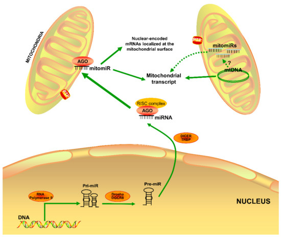
MicroRNAs (miRNAs) are small noncoding RNAs functioning as crucial post-transcriptional regulators of gene expression involved in cardiovascular development and health. Recently, mitochondrial miRNAs (mitomiRs) have been shown to modulate the translational activity of the mitochondrial genome and regulating mitochondrial protein expression and function. [...] Read more.
MicroRNAs (miRNAs) are small noncoding RNAs functioning as crucial post-transcriptional regulators of gene expression involved in cardiovascular development and health. Recently, mitochondrial miRNAs (mitomiRs) have been shown to modulate the translational activity of the mitochondrial genome and regulating mitochondrial protein expression and function. Although mitochondria have been verified to be essential for the development and as a therapeutic target for cardiovascular diseases, we are just beginning to understand the roles of mitomiRs in the regulation of crucial biological processes, including energy metabolism, oxidative stress, inflammation, and apoptosis. In this review, we summarize recent findings regarding how mitomiRs impact on mitochondrial gene expression and mitochondrial function, which may help us better understand the contribution of mitomiRs to both the regulation of cardiovascular function under physiological conditions and the pathogenesis of cardiovascular diseases. Full article
Show Figures

Figure 1

20 pages, 3414 KiB  
Review
The Non-Canonical Aspects of MicroRNAs: Many Roads to Gene Regulation
by Christiaan J. Stavast and Stefan J. Erkeland
Cells 2019, 8(11), 1465; https://doi.org/10.3390/cells8111465 - 19 Nov 2019
Cited by 268 | Viewed by 11042
Abstract
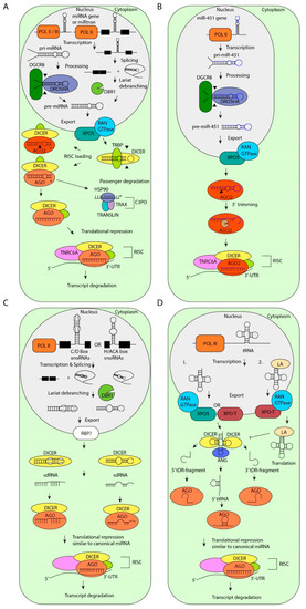
MicroRNAs (miRNAs) are critical regulators of gene expression. As miRNAs are frequently deregulated in many human diseases, including cancer and immunological disorders, it is important to understand their biological functions. Typically, miRNA-encoding genes are transcribed by RNA Polymerase II and generate primary transcripts [...] Read more.
MicroRNAs (miRNAs) are critical regulators of gene expression. As miRNAs are frequently deregulated in many human diseases, including cancer and immunological disorders, it is important to understand their biological functions. Typically, miRNA-encoding genes are transcribed by RNA Polymerase II and generate primary transcripts that are processed by RNase III-endonucleases DROSHA and DICER into small RNAs of approximately 21 nucleotides. All miRNAs are loaded into Argonaute proteins in the RNA-induced silencing complex (RISC) and act as post-transcriptional regulators by binding to the 3′- untranslated region (UTR) of mRNAs. This seed-dependent miRNA binding inhibits the translation and/or promotes the degradation of mRNA targets. Surprisingly, recent data presents evidence for a target-mediated decay mechanism that controls the level of specific miRNAs. In addition, several non-canonical miRNA-containing genes have been recently described and unexpected functions of miRNAs have been identified. For instance, several miRNAs are located in the nucleus, where they are involved in the transcriptional activation or silencing of target genes. These epigenetic modifiers are recruited by RISC and guided by miRNAs to specific loci in the genome. Here, we will review non-canonical aspects of miRNA biology, including novel regulators of miRNA expression and functions of miRNAs in the nucleus. Full article
Show Figures

Figure 1

22 pages, 3059 KiB  
Review
Germinal Centre B Cell Functions and Lymphomagenesis: Circuits Involving MYC and MicroRNAs
by Marcela Cristina Robaina, Luciano Mazzoccoli and Claudete Esteves Klumb
Cells 2019, 8(11), 1365; https://doi.org/10.3390/cells8111365 - 31 Oct 2019
Cited by 13 | Viewed by 6524
Abstract
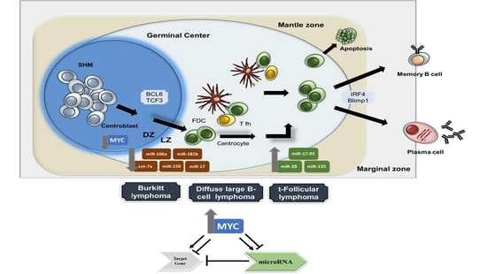
Background: The transcription factor MYC regulates several biological cellular processes, and its target gene network comprises approximately 15% of all human genes, including microRNAs (miRNAs), that also contribute to MYC regulatory activity. Although miRNAs are emerging as key regulators of immune functions, the [...] Read more.
Background: The transcription factor MYC regulates several biological cellular processes, and its target gene network comprises approximately 15% of all human genes, including microRNAs (miRNAs), that also contribute to MYC regulatory activity. Although miRNAs are emerging as key regulators of immune functions, the specific roles of miRNAs in the regulation/dysregulation of germinal centre B-cells and B-cell lymphomas are still being uncovered. The regulatory network that integrates MYC, target genes and miRNAs is a field of intense study, highlighting potential pathways to be explored in the context of future clinical approaches. Methods: The scientific literature that is indexed in PUBMED was consulted for publications involving MYC and miRNAs with validated bioinformatics analyses or experimental protocols. Additionally, seminal studies on germinal centre B-cell functions and lymphomagenesis were reported. Conclusions: This review summarizes the interactions between MYC and miRNAs through regulatory loops and circuits involving target genes in germinal centre B-cell lymphomas with MYC alterations. Moreover, we provide an overview of the understanding of the regulatory networks between MYC and miRNAs, highlighting the potential implication of this approach for the comprehension of germinal centre B-cell lymphoma pathogenesis. Therefore, circuits involving MYC, target genes and miRNAs provide novel insight into lymphomagenesis that could be useful for new improved therapeutic strategies. Full article
Show Figures

Graphical abstract

33 pages, 1903 KiB  
Review
MicroRNAs in Uteroplacental Vascular Dysfunction
by Xiang-Qun Hu and Lubo Zhang
Cells 2019, 8(11), 1344; https://doi.org/10.3390/cells8111344 - 29 Oct 2019
Cited by 27 | Viewed by 5101
Abstract
Pregnancy complications of preeclampsia and intrauterine growth restriction (IUGR) are major causes of maternal and perinatal/neonatal morbidity and mortality. Although their etiologies remain elusive, it is generally accepted that they are secondary to placental insufficiency conferred by both failure in spiral artery remodeling [...] Read more.
Pregnancy complications of preeclampsia and intrauterine growth restriction (IUGR) are major causes of maternal and perinatal/neonatal morbidity and mortality. Although their etiologies remain elusive, it is generally accepted that they are secondary to placental insufficiency conferred by both failure in spiral artery remodeling and uteroplacental vascular malfunction. MicroRNAs (miRNAs) are small no-coding RNA molecules that regulate gene expression at the post-transcriptional level. Increasing evidence suggests that miRNAs participate in virtually all biological processes and are involved in numerous human diseases. Differentially expressed miRNAs in the placenta are typical features of both preeclampsia and IUGR. Dysregulated miRNAs target genes of various signaling pathways in uteroplacental tissues, contributing to the development of both complications. In this review, we provide an overview of how aberrant miRNA expression in preeclampsia and IUGR impacts the expression of genes involved in trophoblast invasion and uteroplacental vascular adaptation. Full article
Show Figures

Figure 1

16 pages, 1234 KiB  
Review
MicroRNAs as a Potential Quality Measurement Tool of Platelet Concentrate Stored in Blood Banks—A Review
by Jersey Heitor da Silva Maués, Caroline de Fátima Aquino Moreira-Nunes and Rommel Mário Rodriguez Burbano
Cells 2019, 8(10), 1256; https://doi.org/10.3390/cells8101256 - 15 Oct 2019
Cited by 17 | Viewed by 3929
Abstract
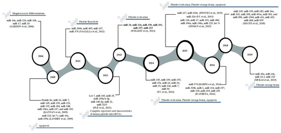
Background: Platelet concentrate (PC) is one of the main products used in a therapeutic transfusion. This blood component requires special storage at blood banks, however, even under good storage conditions, modifications or degradations may occur and are known as platelet storage lesions. Methods: [...] Read more.
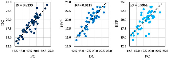
Background: Platelet concentrate (PC) is one of the main products used in a therapeutic transfusion. This blood component requires special storage at blood banks, however, even under good storage conditions, modifications or degradations may occur and are known as platelet storage lesions. Methods: This research was performed on scientific citation databases PubMed/Medline, ScienceDirect, and Web of Science, for publications containing platelet storage lesions. The results obtained mainly reveal the clinical applicability of miRNAs as biomarkers of storage injury and as useful tools for a problem affecting public and private health, the lack of PC bags in countries with few blood donors. The major studies listed in this review identified miRNAs associated with important platelet functions that are relevant in clinical practice as quality biomarkers of PC, such as miR-223, miR-126, miR-10a, miR-150, miR-16, miR-21, miR-326, miR-495, let-7b, let-7c, let-7e, miR-107, miR-10b, miR-145, miR-155, miR-17, miR-191, miR-197, miR-200b, miR-24, miR-331, miR-376. These miRNAs can be used in blood banks to identify platelet injury in PC bags. Conclusion: The studies described in this review relate the functions of miRNAs with molecular mechanisms that result in functional platelet differences, such as apoptosis. Thus, miRNA profiles can be used to measure the quality of storage PC for more than 5 days, identify bags with platelet injury, and distinguish those with functional platelets. Full article
Show Figures

Figure 1

18 pages, 1135 KiB  
Review
The MicroRNA Centrism in the Orchestration of Neuroinflammation in Neurodegenerative Diseases
by Nicoletta Nuzziello and Maria Liguori
Cells 2019, 8(10), 1193; https://doi.org/10.3390/cells8101193 - 2 Oct 2019
Cited by 28 | Viewed by 4792
Abstract
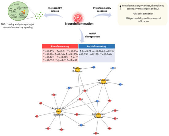
MicroRNAs (miRNAs) are small non-coding RNAs with a unique ability to regulate the transcriptomic profile by binding to complementary regulatory RNA sequences. The ability of miRNAs to enhance (proinflammatory miRNAs) or restrict (anti-inflammatory miRNAs) inflammatory signalling within the central nervous system is an [...] Read more.
MicroRNAs (miRNAs) are small non-coding RNAs with a unique ability to regulate the transcriptomic profile by binding to complementary regulatory RNA sequences. The ability of miRNAs to enhance (proinflammatory miRNAs) or restrict (anti-inflammatory miRNAs) inflammatory signalling within the central nervous system is an area of ongoing research, particularly in the context of disorders that feature neuroinflammation, including neurodegenerative diseases (NDDs). Furthermore, the discovery of competing endogenous RNAs (ceRNAs) has led to an increase in the complexity of miRNA-mediated gene regulation, with a paradigm shift from a unidirectional to a bidirectional regulation, where miRNA acts as both a regulator and is regulated by ceRNAs. Increasing evidence has revealed that ceRNAs, including long non-coding RNAs, circular RNAs, and pseudogenes, can act as miRNA sponges to regulate neuroinflammation in NDDs within complex cross-talk regulatory machinery, which is referred to as ceRNA network (ceRNET). In this review, we discuss the role of miRNAs in neuroinflammatory regulation and the manner in which cellular and vesicular ceRNETs could influence neuroinflammatory dynamics in complex multifactorial diseases, such as NDDs. Full article
Show Figures

Figure 1

18 pages, 3800 KiB  
Article
Understanding the Modus Operandi of MicroRNA Regulatory Clusters
by Arthur C. Oliveira, Luiz A. Bovolenta, Lucas Alves, Lucas Figueiredo, Amanda O. Ribeiro, Vinicius F. Campos, Ney Lemke and Danillo Pinhal
Cells 2019, 8(9), 1103; https://doi.org/10.3390/cells8091103 - 18 Sep 2019
Cited by 11 | Viewed by 4408
Abstract
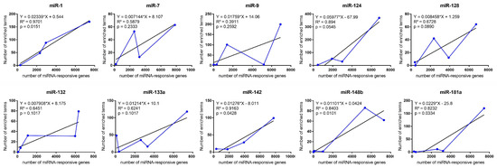
MicroRNAs (miRNAs) are non-coding RNAs that regulate a wide range of biological pathways by post-transcriptionally modulating gene expression levels. Given that even a single miRNA may simultaneously control several genes enrolled in multiple biological functions, one would expect that these tiny RNAs have [...] Read more.
MicroRNAs (miRNAs) are non-coding RNAs that regulate a wide range of biological pathways by post-transcriptionally modulating gene expression levels. Given that even a single miRNA may simultaneously control several genes enrolled in multiple biological functions, one would expect that these tiny RNAs have the ability to properly sort among distinctive cellular processes to drive protein production. To test this hypothesis, we scrutinized previously published microarray datasets and clustered protein-coding gene expression profiles according to the intensity of fold-change levels caused by the exogenous transfection of 10 miRNAs (miR-1, miR-7, miR-9, miR-124, miR-128a, miR-132, miR-133a, miR-142, miR-148b, miR-181a) in a human cell line. Through an in silico functional enrichment analysis, we discovered non-randomic regulatory patterns, proper of each cluster identified. We demonstrated that miRNAs are capable of equivalently modulate the expression signatures of target genes in regulatory clusters according to the biological function they are assigned to. Moreover, target prediction analysis applied to ten vertebrate species, suggest that such miRNA regulatory modus operandi is evolutionarily conserved within vertebrates. Overall, we discovered a complex regulatory cluster-module strategy driven by miRNAs, which relies on the controlled intensity of the repression over distinct targets under specific biological contexts. Our discovery helps to clarify the mechanisms underlying the functional activity of miRNAs and makes it easier to take the fastest and most accurate path in the search for the functions of miRNAs in any distinct biological process of interest. Full article
Show Figures

Figure 1

19 pages, 4027 KiB  
Article
miR-263b Controls Circadian Behavior and the Structural Plasticity of Pacemaker Neurons by Regulating the LIM-Only Protein Beadex
by Xiaoge Nian, Wenfeng Chen, Weiwei Bai, Zhangwu Zhao and Yong Zhang
Cells 2019, 8(8), 923; https://doi.org/10.3390/cells8080923 - 18 Aug 2019
Cited by 14 | Viewed by 4368
Abstract
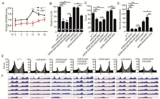
Circadian clocks drive rhythmic physiology and behavior to allow adaption to daily environmental changes. In Drosophila, the small ventral lateral neurons (sLNvs) are primary pacemakers that control circadian rhythms. Circadian changes are observed in the dorsal axonal projections of the sLNvs, but [...] Read more.
Circadian clocks drive rhythmic physiology and behavior to allow adaption to daily environmental changes. In Drosophila, the small ventral lateral neurons (sLNvs) are primary pacemakers that control circadian rhythms. Circadian changes are observed in the dorsal axonal projections of the sLNvs, but their physiological importance and the underlying mechanism are unclear. Here, we identified miR-263b as an important regulator of circadian rhythms and structural plasticity of sLNvs in Drosophila. Depletion of miR-263b (miR-263bKO) in flies dramatically impaired locomotor rhythms under constant darkness. Indeed, miR-263b is required for the structural plasticity of sLNvs. miR-263b regulates circadian rhythms through inhibition of expression of the LIM-only protein Beadex (Bx). Consistently, overexpression of Bx or loss-of-function mutation (BxhdpR26) phenocopied miR-263bKO and miR-263b overexpression in behavior and molecular characteristics. In addition, mutating the miR-263b binding sites in the Bx 3′ UTR using CRISPR/Cas9 recapitulated the circadian phenotypes of miR-263bKO flies. Together, these results establish miR-263b as an important regulator of circadian locomotor behavior and structural plasticity. Full article
Show Figures

Figure 1

13 pages, 796 KiB  
Review
MicroRNAs in Obesity and Related Metabolic Disorders
by Jean-François Landrier, Adel Derghal and Lourdes Mounien
Cells 2019, 8(8), 859; https://doi.org/10.3390/cells8080859 - 9 Aug 2019
Cited by 140 | Viewed by 9525
Abstract
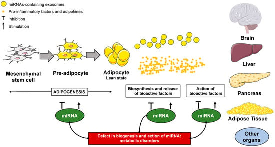
Metabolic disorders are characterized by the inability to properly use and/or store energy. The burdens of metabolic disease, such as obesity or diabetes, are believed to arise through a complex interplay between genetics and epigenetics predisposition, environment and nutrition. Therefore, understanding the molecular [...] Read more.
Metabolic disorders are characterized by the inability to properly use and/or store energy. The burdens of metabolic disease, such as obesity or diabetes, are believed to arise through a complex interplay between genetics and epigenetics predisposition, environment and nutrition. Therefore, understanding the molecular mechanisms for the onset of metabolic disease will provide new insights for prevention and treatment. There is growing concern about the dysregulation of micro-RNAs (miRNAs) in metabolic diseases. MiRNAs are short non-coding RNA molecules that post-transcriptionally repress the expression of genes by binding to untranslated regions and coding sequences of the target mRNAs. This review aims to provide recent data about the potential involvement of miRNAs in metabolic diseases, particularly obesity and type 2 diabetes. Full article
Show Figures

Figure 1

14 pages, 2932 KiB  
Article
MiR-200-3p Is Potentially Involved in Cell Cycle Arrest by Regulating Cyclin A during Aestivation in Apostichopus japonicus
by Shanshan Wang, Muyan Chen, Yingchao Yin and Kenneth B. Storey
Cells 2019, 8(8), 843; https://doi.org/10.3390/cells8080843 - 6 Aug 2019
Cited by 12 | Viewed by 3529
Abstract
The sea cucumber (Apostichopus japonicus) has become a good model organism for studying environmentally induced aestivation in marine invertebrates. We hypothesized that mechanisms that arrest energy-expensive cell cycle activity would contribute significantly to establishing the hypometabolic state during aestivation. Cyclin A [...] Read more.
The sea cucumber (Apostichopus japonicus) has become a good model organism for studying environmentally induced aestivation in marine invertebrates. We hypothesized that mechanisms that arrest energy-expensive cell cycle activity would contribute significantly to establishing the hypometabolic state during aestivation. Cyclin A is a core and particularly interesting cell cycle regulator that functions in both the S phase and in mitosis. In the present study, negative relationships between miR-200-3p and AjCA expressions were detected at both the transcriptional and the translational levels during aestivation in A. japonicus. Dual-luciferase reporter assays confirmed the targeted location of the miR-200-3p binding site within the AjCA gene transcript. Furthermore, gain- and loss-of-function experiments were conducted in vivo with sea cucumbers to verify the interaction between miR-200-3p and AjCA in intestine tissue by qRT-PCR and Western blotting. The results show that the overexpression of miR-200-3p mimics suppressed AjCA transcript levels and translated protein production, whereas transfection with a miR-200-3p inhibitor enhanced both AjCA mRNA and AjCA protein in A. japonicus intestine. Our findings suggested a potential mechanism that reversibly arrests cell cycle progression during aestivation, which may center on miR-200-3p inhibitory control over the translation of cyclin A mRNA transcripts. Full article
Show Figures

Figure 1

17 pages, 11248 KiB  
Article
Differential Inhibition of Target Gene Expression by Human microRNAs
by Peng Li, Yi Chen, Conslata Awino Juma, Chengyong Yang, Jinfeng Huang, Xiaoxiao Zhang and Yan Zeng
Cells 2019, 8(8), 791; https://doi.org/10.3390/cells8080791 - 30 Jul 2019
Cited by 16 | Viewed by 4140
Abstract
microRNAs (miRNAs) exert their functions by repressing the expression of their target genes, but most miRNA target genes are unknown, and the degree to which a miRNA differentially inhibits the expression of its targets is underappreciated. We selected human miR-1, miR-122, and miR-124 [...] Read more.
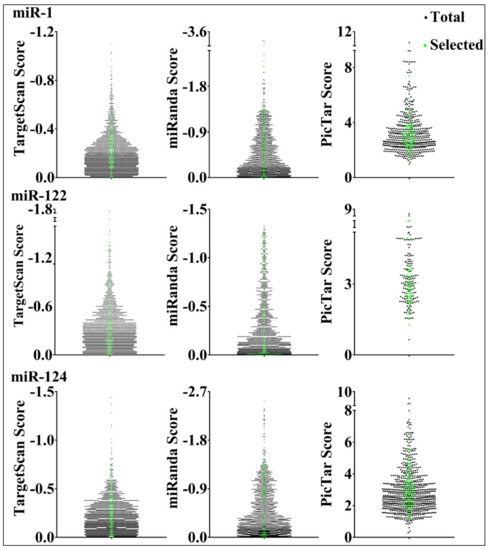
microRNAs (miRNAs) exert their functions by repressing the expression of their target genes, but most miRNA target genes are unknown, and the degree to which a miRNA differentially inhibits the expression of its targets is underappreciated. We selected human miR-1, miR-122, and miR-124 as representatives to investigate the reliability of miRNA target predictions and examine how miRNAs suppress their targets. We constructed miRNA target gene reporter libraries based on prediction programs TargetScan, miRanda, and PicTar, and performed large-scale reporter assays to directly evaluate whether and how strongly a predicted target gene is repressed by its miRNA. We then performed statistical analyses to examine parameters that contributed to the miRNA inhibition of target genes. We found that the three programs have approximately 72–85% success rates in predicting genuine targets and that the miRNA inhibition of different targets varies in extent. We also identified parameters that could predict the degrees of miRNA repression, and further showed that differential miR-124 repression might contribute to differential gene expression in vivo. Our studies systematically investigated hundreds of miRNA target genes, shed light on factors influencing miRNA functions, and suggested a new mechanism by which differential target repression by miRNAs regulates endogenous gene expression. Full article
Show Figures

Figure 1

11 pages, 1605 KiB  
Article
Physical Exercise Modulates miR-21-5p, miR-129-5p, miR-378-5p, and miR-188-5p Expression in Progenitor Cells Promoting Osteogenesis
by Maria Teresa Valenti, Michela Deiana, Samuele Cheri, Monica Dotta, Francesco Zamboni, Daniele Gabbiani, Federico Schena, Luca Dalle Carbonare and Monica Mottes
Cells 2019, 8(7), 742; https://doi.org/10.3390/cells8070742 - 19 Jul 2019
Cited by 47 | Viewed by 6269
Abstract
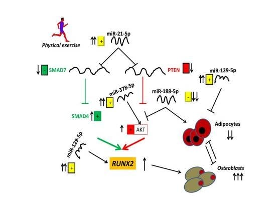
Physical exercise is known to promote beneficial effects on overall health, counteracting risks related to degenerative diseases. MicroRNAs (miRNAs), short non-coding RNAs affecting the expression of a cell’s transcriptome, can be modulated by different stimuli. Yet, the molecular effects on osteogenic differentiation triggered [...] Read more.
Physical exercise is known to promote beneficial effects on overall health, counteracting risks related to degenerative diseases. MicroRNAs (miRNAs), short non-coding RNAs affecting the expression of a cell’s transcriptome, can be modulated by different stimuli. Yet, the molecular effects on osteogenic differentiation triggered by miRNAs upon physical exercise are not completely understood. In this study, we recruited 20 male amateur runners participating in a half marathon. Runners’ sera, collected before (PRE RUN) and after (POST RUN) the run, were added to cultured human mesenchymal stromal cells. We then investigated their effects on the modulation of selected miRNAs and the consequential effects on osteogenic differentiation. Our results showed an increased expression of miRNAs promoting osteogenic differentiation (miR-21-5p, miR-129-5p, and miR-378-5p) and a reduced expression of miRNAs involved in the adipogenic differentiation of progenitor cells (miR-188-5p). In addition, we observed the downregulation of PTEN and SMAD7 expression along with increased AKT/pAKT and SMAD4 protein levels in MSCs treated with POST RUN sera. The consequent upregulation of RUNX2 expression was also proven, highlighting the molecular mechanisms by which miR-21-5p promotes osteogenic differentiation. In conclusion, our work proposes novel data, which demonstrate how miRNAs may regulate the osteogenic commitment of progenitor cells in response to physical exercise. Full article
Show Figures

Graphical abstract

19 pages, 8236 KiB  
Article
Identifying microRNAs and Their Editing Sites in Macaca mulatta
by Qingyi Wang, Zhigang Zhao, Xiaotuo Zhang, Chenyu Lu, Shuchao Ren, Shipeng Li, Junqiang Guo, Peiran Liao, Bingbing Jiang and Yun Zheng
Cells 2019, 8(7), 682; https://doi.org/10.3390/cells8070682 - 5 Jul 2019
Cited by 9 | Viewed by 3840
Abstract
MicroRNAs (miRNAs) are small non-coding RNAs that are critical in post-transcriptional regulation. Macaca mulatta is an important nonhuman primate that is often used in basic and translational researches. However, the annotation of miRNAs in Macaca mulatta is far from complete, and there are [...] Read more.
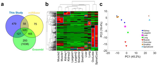
MicroRNAs (miRNAs) are small non-coding RNAs that are critical in post-transcriptional regulation. Macaca mulatta is an important nonhuman primate that is often used in basic and translational researches. However, the annotation of miRNAs in Macaca mulatta is far from complete, and there are no reports of miRNA editing events in Macaca mulatta, although editing may affect the biogenesis or functions of the miRNAs. To improve miRNA annotation and to reveal editing events of miRNAs in Macaca mulatta, we generated 12 small RNA profiles from eight tissues and performed comprehensive analysis of these profiles. We identified 479 conserved pre-miRNAs that have not been reported in Macaca mulatta and 17 species specific miRNAs. Furthermore, we identified 3386 editing sites with significant editing levels from 471 pre-miRNAs after analyzing the 12 self-generated and 58 additional published sRNA-seq profiles from 17 different types of organs or tissues. In addition to 16 conserved A-to-I editing sites, we identified five conserved C-to-U editing sites in miRNAs of Macaca mulatta and Homo sapiens. We also identified 11 SNPs in the miRNAs of Macaca mulatta. The analysis of the potential targets of 69 miRNAs with editing or mutation events in their seed regions suggest that these editing or mutation events severely changed their targets and their potential functions. These results significantly increase our understanding of miRNAs and their mutation/editing events in Macaca mulatta. Full article
Show Figures

Figure 1

19 pages, 4193 KiB  
Article
The Inhibition on MDFIC and PI3K/AKT Pathway Caused by miR-146b-3p Triggers Suppression of Myoblast Proliferation and Differentiation and Promotion of Apoptosis
by Weiling Huang, Lijin Guo, Minxing Zhao, Dexiang Zhang, Haiping Xu and Qinghua Nie
Cells 2019, 8(7), 656; https://doi.org/10.3390/cells8070656 - 29 Jun 2019
Cited by 37 | Viewed by 5420
Abstract
Accumulating studies report that microRNAs (miRNAs) are actively involved in skeletal myogenesis. Previously, our study revealed that miR-146b-3p was related to the growth of skeletal muscle. Here, we further report that miR-146b-3p is essential for the proliferation, differentiation, and apoptosis of chicken myoblast. [...] Read more.
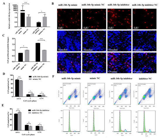
Accumulating studies report that microRNAs (miRNAs) are actively involved in skeletal myogenesis. Previously, our study revealed that miR-146b-3p was related to the growth of skeletal muscle. Here, we further report that miR-146b-3p is essential for the proliferation, differentiation, and apoptosis of chicken myoblast. Elevated expression of miR-146b-3p can dramatically suppress proliferation and differentiation, and facilitate apoptosis of chicken myoblast. Besides, we identified two target genes of miR-146b-3p, AKT1 and MDFIC, and found that miR-146b-3p can inhibit the PI3K/AKT pathway. Our study also showed that both AKT1 and MDFIC can promote the proliferation and differentiation while inhibit the apoptosis of myoblast in chicken. Overall, our results demonstrate that miR-146b-3p, directly suppressing PI3K/AKT pathway and MDFIC, acts in the proliferation, differentiation, and apoptosis of myoblast in chicken. Full article
Show Figures

Figure 1

17 pages, 1738 KiB  
Article
miR-26a-5p is a Stable Reference Gene for miRNA Studies in Chondrocytes from Developing Human Cartilage
by Enrico Ragni, Paola De Luca, Antongiulio Marmotti and Laura de Girolamo
Cells 2019, 8(6), 631; https://doi.org/10.3390/cells8060631 - 22 Jun 2019
Cited by 6 | Viewed by 3613
Abstract
miRNAs are emerging as key regulators of complex biological systems in several developmental processes. qRT-PCR is a powerful tool to quantitatively assess the profiles and modulation of miRNA expression. In the emerging field of cartilage maturation studies, from precursor to hypertrophic chondrocytes, few [...] Read more.
miRNAs are emerging as key regulators of complex biological systems in several developmental processes. qRT-PCR is a powerful tool to quantitatively assess the profiles and modulation of miRNA expression. In the emerging field of cartilage maturation studies, from precursor to hypertrophic chondrocytes, few data about miRNA regulation are available, and no consensus on the best reference gene (RG) has been reached. This is a crucial pitfall since reliable outcomes depend on proper data normalization. The aim of this work was to identify reliable and stable miRNA RGs, basing the analysis on available high throughput qRT-PCR miRNA data (from the NCBI Gene Expression Omnibus database, GSE49152) obtained from human embryonic cartilage tissues enriched in the precursor, differentiated, and hypertrophic chondrocytes. Four normalization approaches were used, and the stability was quantified by combining BestKeeper, delta-Ct, geNorm, and NormFinder statistical tools. An integrated approach allowed to identify miR-26a-5p as the most stable RG and miR-212-3p as the worst one. RNU44, used in original dataset analysis, performed as second best RG. Applications of different normalization strategies significantly impacted the profiles and modulation of miRNA expression. Herein presented results point out the crucial need of a consensus on data normalization studies aimed at dissecting miRNA role in human cartilage development, to avoid the postulation of unreliable biological conclusions. Full article
Show Figures

Figure 1

13 pages, 648 KiB  
Article
MiR-34b-5p Mediates the Proliferation and Differentiation of Myoblasts by Targeting IGFBP2
by Zhijun Wang, Xiaocui Zhang, Zhenhui Li, Bahareldin Ali Abdalla, Yangfeng Chen and Qinghua Nie
Cells 2019, 8(4), 360; https://doi.org/10.3390/cells8040360 - 17 Apr 2019
Cited by 24 | Viewed by 5516
Abstract
As key post-transcriptional regulators, microRNAs (miRNAs) play an indispensable role in skeletal muscle development. Our previous study suggested that miR-34b-5p and IGFBP2 could have a potential role in skeletal muscle growth. Our goal in this study is to explore the function and regulatory [...] Read more.
As key post-transcriptional regulators, microRNAs (miRNAs) play an indispensable role in skeletal muscle development. Our previous study suggested that miR-34b-5p and IGFBP2 could have a potential role in skeletal muscle growth. Our goal in this study is to explore the function and regulatory mechanism of miR-34b-5p and IGFBP2 in myogenesis. In this study, the dual-luciferase reporter assay and Western blot analysis showed that IGFBP2 is a direct target of miR-34b-5p. Flow cytometric analysis and EdU assay showed that miR-34b-5p could repress the cell cycle progression of myoblasts, and miR-34b-5p could promote the formation of myotubes by promoting the expression of MyHC. On the contrary, the overexpression of IGFBP2 significantly facilitated the proliferation of myoblasts and hampered the formation of myotubes. Together, our results indicate that miR-34b-5p could mediate the proliferation and differentiation of myoblasts by targeting IGFBP2. Full article
Show Figures

Figure 1

19 pages, 1595 KiB  
Review
MicroRNAs as Potential Pharmaco-Targets in Ischemia-Reperfusion Injury Compounded by Diabetes
by Hassan Dehaini, Hussein Awada, Ahmed El-Yazbi, Fouad A. Zouein, Khodr Issa, Assaad A. Eid, Maryam Ibrahim, Adnan Badran, Elias Baydoun, Gianfranco Pintus and Ali H. Eid
Cells 2019, 8(2), 152; https://doi.org/10.3390/cells8020152 - 12 Feb 2019
Cited by 41 | Viewed by 5299
Abstract
Background: Ischemia-Reperfusion (I/R) injury is the tissue damage that results from re-oxygenation of ischemic tissues. There are many players that contribute to I/R injury. One of these factors is the family of microRNAs (miRNAs), which are currently being heavily studied. This review aims [...] Read more.
Background: Ischemia-Reperfusion (I/R) injury is the tissue damage that results from re-oxygenation of ischemic tissues. There are many players that contribute to I/R injury. One of these factors is the family of microRNAs (miRNAs), which are currently being heavily studied. This review aims to critically summarize the latest papers that attributed roles of certain miRNAs in I/R injury, particularly in diabetic conditions and dissect their potential as novel pharmacologic targets in the treatment and management of diabetes. Methods: PubMed was searched for publications containing microRNA and I/R, in the absence or presence of diabetes. All papers that provided sufficient evidence linking miRNA with I/R, especially in the context of diabetes, were selected. Several miRNAs are found to be either pro-apoptotic, as in the case of miR-34a, miR-144, miR-155, and miR-200, or anti-apoptotic, as in the case of miR-210, miR-21, and miR-146a. Here, we further dissect the evidence that shows diverse cell-context dependent effects of these miRNAs, particularly in cardiomyocytes, endothelial, or leukocytes. We also provide insight into cases where the possibility of having two miRNAs working together to intensify a given response is noted. Conclusions: This review arrives at the conclusion that the utilization of miRNAs as translational agents or pharmaco-targets in treating I/R injury in diabetic patients is promising and becoming increasingly clearer. Full article
Show Figures

Graphical abstract

Back to TopTop