Molecular and Cellular Mechanisms of Parkinson's Disease

A topical collection in Cells (ISSN 2073-4409). This collection belongs to the section "Cellular Aging".

Viewed by 58103

Editor


E-Mail Website
Collection Editor
Parkinson-Klinik Ortenau GmbH, 77709 Wolfach, Germany
Interests: botulinum toxin; dystonia; cervical dystonia; sialorrhea; Parkinson’s disease; Blepharospasm
Special Issues, Collections and Topics in MDPI journals

Topical Collection Information

Dear Colleagues,

Parkinson’s disease is a disease for which we have attained a massive gain in knowledge within the last few decades, and for which the number of publications has increased as in hardly any other area of medicine. We are fortunate to have many effective medications at our disposal for this disease. The central issue here has been the substantia nigra, and the deficiency in levodopa has been the pivotal focus for therapy. In the last two decades, however, we have learned that emphasis on these concepts offers only limited benefits and may in fact be detrimental for further progress in research. To be precise, the substantia nigra is neither the origin nor the end point of the degenerative process, and furthermore dopaminergic therapy is merely a symptomatic intervention. Of course, all further progress is a factor of our basic hypotheses, but these have to be substantiated as to their validity. In Parkinson’s diagnostics and therapy, we have arrived at something of a stalemate: many new findings are truly spectacular, but not yet the final answer to all our questions. As clinicians, of course, we tend to target quick solutions, but so do our patients, and these quick solutions are still very illusive. For this reason, we have to return to the situation at the historical start of work on Parkinson’s and take a good look at current gaps in knowledge in order to work them out scientifically. For this reason, we have decided to put out a Topical Collection entitled “Molecular and Cellular Mechanisms of Parkinson’s Disease”. We are deliberately not working on a new and comprehensive model in this Topical Collection, but intend to outline the current status of different aspects so that the large spectrum of findings can be seen and so that some hints can be gleaned as to just where we might profitably pursue new fields of research and where we should avoid some potential dead-ends. Put metaphorically, if I have become lost in a city it is more helpful to return to the starting point and not just to rely on keeping to the obviously wrong route taken. So, we want to meet at the market and trade our current awareness.

Prof. Wolfgang Jost
Collection Editor

Manuscript Submission Information

Manuscripts should be submitted online at www.mdpi.com by registering and logging in to this website. Once you are registered, click here to go to the submission form. Manuscripts can be submitted until the deadline. All submissions that pass pre-check are peer-reviewed. Accepted papers will be published continuously in the journal (as soon as accepted) and will be listed together on the collection website. Research articles, review articles as well as short communications are invited. For planned papers, a title and short abstract (about 100 words) can be sent to the Editorial Office for announcement on this website.

Submitted manuscripts should not have been published previously, nor be under consideration for publication elsewhere (except conference proceedings papers). All manuscripts are thoroughly refereed through a single-blind peer-review process. A guide for authors and other relevant information for submission of manuscripts is available on the Instructions for Authors page. Cells is an international peer-reviewed open access semimonthly journal published by MDPI.

Please visit the Instructions for Authors page before submitting a manuscript. The Article Processing Charge (APC) for publication in this open access journal is 2700 CHF (Swiss Francs). Submitted papers should be well formatted and use good English. Authors may use MDPI's English editing service prior to publication or during author revisions.

Keywords

  • Parkinson’s disease
  • Alpha-synuclein
  • Genetics
  • Inflammation

Published Papers (15 papers)

2024

Jump to: 2023, 2021, 2020

15 pages, 567 KiB  
Review
Dysregulated Coagulation in Parkinson’s Disease
by Xinqing Wang, Wenxin Li, Xinyue Zhao, Ning Hu, Xi Wang, Xilin Xiao, Kai Yang and Taolei Sun
Cells 2024, 13(22), 1874; https://doi.org/10.3390/cells13221874 - 13 Nov 2024
Viewed by 362
Abstract
Parkinson’s disease (PD), a prevalent neurodegenerative disorder characterized by dopaminergic neuron degeneration and α-synuclein accumulation, has been increasingly associated with coagulation dysfunction. This review synthesizes emerging evidence linking dysregulated coagulation to PD pathophysiology. We examine the alterations in coagulation parameters, including elevated fibrinogen [...] Read more.
Parkinson’s disease (PD), a prevalent neurodegenerative disorder characterized by dopaminergic neuron degeneration and α-synuclein accumulation, has been increasingly associated with coagulation dysfunction. This review synthesizes emerging evidence linking dysregulated coagulation to PD pathophysiology. We examine the alterations in coagulation parameters, including elevated fibrinogen levels, impaired fibrinolysis, and platelet dysfunction, which collectively contribute to a hypercoagulable state in PD patients. Epidemiological studies have revealed a higher incidence of thrombotic events, such as deep vein thrombosis (DVT) and stroke, among PD patients, suggesting significant comorbidity between PD and coagulation disorders. This review explores the potential pathophysiological mechanisms underlying this association, focusing on the roles of inflammation and oxidative stress. Additionally, we discuss the limitations of current research and propose future directions. This comprehensive analysis underscores the importance of understanding the coagulation–neurodegeneration axis in PD, which may lead to novel diagnostic and therapeutic strategies for this debilitating condition. Full article
Show Figures

Figure 1

2023

Jump to: 2024, 2021, 2020

22 pages, 2026 KiB  
Review
Apathy in Parkinson’s Disease: Clinical Patterns and Neurobiological Basis
by Matthieu Béreau, Vincent Van Waes, Mathieu Servant, Eloi Magnin, Laurent Tatu and Mathieu Anheim
Cells 2023, 12(12), 1599; https://doi.org/10.3390/cells12121599 - 10 Jun 2023
Cited by 13 | Viewed by 4779
Abstract
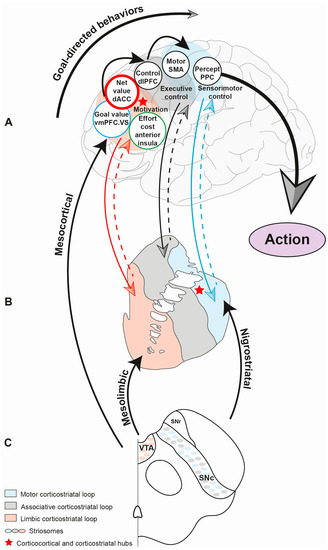
Apathy is commonly defined as a loss of motivation leading to a reduction in goal-directed behaviors. This multidimensional syndrome, which includes cognitive, emotional and behavioral components, is one of the most prevalent neuropsychiatric features of Parkinson’s disease (PD). It has been established that [...] Read more.
Apathy is commonly defined as a loss of motivation leading to a reduction in goal-directed behaviors. This multidimensional syndrome, which includes cognitive, emotional and behavioral components, is one of the most prevalent neuropsychiatric features of Parkinson’s disease (PD). It has been established that the prevalence of apathy increases as PD progresses. However, the pathophysiology and anatomic substrate of this syndrome remain unclear. Apathy seems to be underpinned by impaired anatomical structures that link the prefrontal cortex with the limbic system. It can be encountered in the prodromal stage of the disease and in fluctuating PD patients receiving bilateral chronic subthalamic nucleus stimulation. In these stages, apathy may be considered as a disorder of motivation that embodies amotivational behavioral syndrome, is underpinned by combined dopaminergic and serotonergic denervation and is dopa-responsive. In contrast, in advanced PD patients, apathy may be considered as cognitive apathy that announces cognitive decline and PD dementia, is underpinned by diffuse neurotransmitter system dysfunction and Lewy pathology spreading and is no longer dopa-responsive. In this review, we discuss the clinical patterns of apathy and their treatment, the neurobiological basis of apathy, the potential role of the anatomical structures involved and the pathways in motivational and cognitive apathy. Full article
Show Figures

Figure 1

2021

Jump to: 2024, 2023, 2020

18 pages, 4747 KiB  
Article
Motor Cortex Stimulation Reversed Hypernociception, Increased Serotonin in Raphe Neurons, and Caused Inhibition of Spinal Astrocytes in a Parkinson’s Disease Rat Model
by Ana Carolina P. Campos, Miriã B. Berzuíno, Gabriela R. Barbosa, Helena M. R. C. Freire, Patricia S. Lopes, Danielle V. Assis, Erich T. Fonoff and Rosana L. Pagano
Cells 2021, 10(5), 1158; https://doi.org/10.3390/cells10051158 - 11 May 2021
Cited by 6 | Viewed by 3645
Abstract
Persistent pain is a prevalent symptom of Parkinson’s disease (PD), which is related to the loss of monoamines and neuroinflammation. Motor cortex stimulation (MCS) inhibits persistent pain by activating the descending analgesic pathways; however, its effectiveness in the control of PD-induced pain remains [...] Read more.
Persistent pain is a prevalent symptom of Parkinson’s disease (PD), which is related to the loss of monoamines and neuroinflammation. Motor cortex stimulation (MCS) inhibits persistent pain by activating the descending analgesic pathways; however, its effectiveness in the control of PD-induced pain remains unclear. Here, we evaluated the analgesic efficacy of MCS together with serotonergic and spinal glial modulation in an experimental PD (ePD) rat model. Wistar rats with unilateral striatal 6-OHDA and MCS were assessed for behavioral immobility and nociceptive responses. The immunoreactivity of dopamine in the substantia nigra and serotonin in the nucleus raphe magnus (NRM) and the neuronal, astrocytic, and microglial activation in the dorsal horn of the spinal cord were evaluated. MCS, without interfering with dopamine loss, reversed ePD-induced immobility and hypernociception. This response was accompanied by an exacerbated increase in serotonin in the NRM and a decrease in neuronal and astrocytic hyperactivation in the spinal cord, without inhibiting ePD-induced microglial hypertrophy and hyperplasia. Taken together, MCS induces analgesia in the ePD model, while restores the descending serotonergic pathway with consequent inhibition of spinal neurons and astrocytes, showing the role of MCS in PD-induced pain control. Full article
Show Figures

Figure 1

17 pages, 15425 KiB  
Article
A Propagated Skeleton Approach to High Throughput Screening of Neurite Outgrowth for In Vitro Parkinson’s Disease Modelling
by Justus Schikora, Nina Kiwatrowski, Nils Förster, Leonie Selbach, Friederike Ostendorf, Frida Pallapies, Britta Hasse, Judith Metzdorf, Ralf Gold, Axel Mosig and Lars Tönges
Cells 2021, 10(4), 931; https://doi.org/10.3390/cells10040931 - 17 Apr 2021
Cited by 9 | Viewed by 5535
Abstract
Neuronal models of neurodegenerative diseases such as Parkinson’s Disease (PD) are extensively studied in pathological and therapeutical research with neurite outgrowth being a core feature. Screening of neurite outgrowth enables characterization of various stimuli and therapeutic effects after lesion. In this study, we [...] Read more.
Neuronal models of neurodegenerative diseases such as Parkinson’s Disease (PD) are extensively studied in pathological and therapeutical research with neurite outgrowth being a core feature. Screening of neurite outgrowth enables characterization of various stimuli and therapeutic effects after lesion. In this study, we describe an autonomous computational assay for a high throughput skeletonization approach allowing for quantification of neurite outgrowth in large data sets from fluorescence microscopic imaging. Development and validation of the assay was conducted with differentiated SH-SY5Y cells and primary mesencephalic dopaminergic neurons (MDN) treated with the neurotoxic lesioning compound Rotenone. Results of manual annotation using NeuronJ and automated data were shown to correlate strongly (R2-value 0.9077 for SH-SY5Y cells and R2-value 0.9297 for MDN). Pooled linear regressions of results from SH-SY5Y cell image data could be integrated into an equation formula (y=0.5410·x+1792; y=0.8789·x+0.09191 for normalized results) with y depicting automated and x depicting manual data. This automated neurite length algorithm constitutes a valuable tool for modelling of neurite outgrowth that can be easily applied to evaluate therapeutic compounds with high throughput approaches. Full article
Show Figures

Figure 1

11 pages, 2621 KiB  
Article
Chronic–Progressive Dopaminergic Deficiency Does Not Induce Midbrain Neurogenesis
by Mareike Fauser, Francisco Pan-Montojo, Christian Richter, Philipp J. Kahle, Sigrid C. Schwarz, Johannes Schwarz, Alexander Storch and Andreas Hermann
Cells 2021, 10(4), 775; https://doi.org/10.3390/cells10040775 - 31 Mar 2021
Cited by 3 | Viewed by 2626
Abstract
Background: Consecutive adult neurogenesis is a well-known phenomenon in the ventricular–subventricular zone of the lateral wall of the lateral ventricles (V–SVZ) and has been controversially discussed in so-called “non-neurogenic” brain areas such as the periventricular regions (PVRs) of the aqueduct and the fourth [...] Read more.
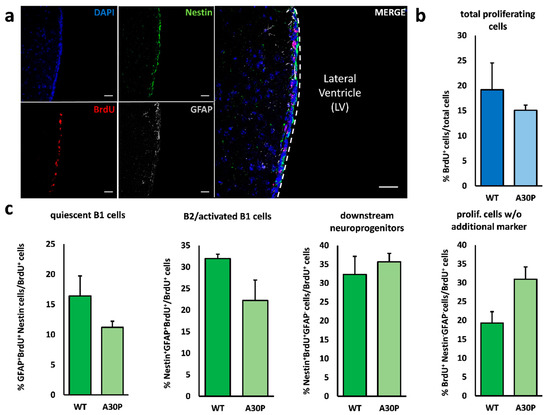
Background: Consecutive adult neurogenesis is a well-known phenomenon in the ventricular–subventricular zone of the lateral wall of the lateral ventricles (V–SVZ) and has been controversially discussed in so-called “non-neurogenic” brain areas such as the periventricular regions (PVRs) of the aqueduct and the fourth ventricle. Dopamine is a known modulator of adult neural stem cell (aNSC) proliferation and dopaminergic neurogenesis in the olfactory bulb, though a possible interplay between local dopaminergic neurodegeneration and induction of aNSC proliferation in mid/hindbrain PVRs is currently enigmatic. Objective/Hypothesis: To analyze the influence of chronic–progressive dopaminergic neurodegeneration on both consecutive adult neurogenesis in the PVRs of the V–SVZ and mid/hindbrain aNSCs in two mechanistically different transgenic animal models of Parkinson´s disease (PD). Methods: We used Thy1-m[A30P]h α synuclein mice and Leu9′Ser hypersensitive α4* nAChR mice to assess the influence of midbrain dopaminergic neuronal loss on neurogenic activity in the PVRs of the V–SVZ, the aqueduct and the fourth ventricle. Results: In both animal models, overall proliferative activity in the V–SVZ was not altered, though the proportion of B2/activated B1 cells on all proliferating cells was reduced in the V–SVZ in Leu9′Ser hypersensitive α4* nAChR mice. Putative aNSCs in the mid/hindbrain PVRs are known to be quiescent in vivo in healthy controls, and dopaminergic deficiency did not induce proliferative activity in these regions in both disease models. Conclusions: Our data do not support an activation of endogenous aNSCs in mid/hindbrain PVRs after local dopaminergic neurodegeneration. Spontaneous endogenous regeneration of dopaminergic cell loss through resident aNSCs is therefore unlikely. Full article
Show Figures

Figure 1

21 pages, 9242 KiB  
Article
Parkinson’s Disease Causative Mutation in Vps35 Disturbs Tetherin Trafficking to Cell Surfaces and Facilitates Virus Spread
by Yingzhuo Ding, Yan Li, Gaurav Chhetri, Xiaoxin Peng, Jing Wu, Zejian Wang, Bo Zhao, Wenjuan Zhao and Xueyi Li
Cells 2021, 10(4), 746; https://doi.org/10.3390/cells10040746 - 28 Mar 2021
Cited by 6 | Viewed by 3156
Abstract
Parkinson’s disease (PD) is the most common neurodegenerative movement disorder, characterized by progressive loss of dopaminergic neurons in the substantia nigra, intraneuronal deposition of misfolded proteins known as Lewy bodies, and chronic neuroinflammation. PD can arise from monogenic mutations, but in most cases, [...] Read more.
Parkinson’s disease (PD) is the most common neurodegenerative movement disorder, characterized by progressive loss of dopaminergic neurons in the substantia nigra, intraneuronal deposition of misfolded proteins known as Lewy bodies, and chronic neuroinflammation. PD can arise from monogenic mutations, but in most cases, the etiology is unclear. Viral infection is gaining increasing attentions as a trigger of PD. In this study, we investigated whether the PD-causative 620 aspartate (D) to asparagine (N) mutation in the vacuolar protein sorting 35 ortholog (Vps35) precipitated herpes simplex virus (HSV) infection. We observed that ectopic expression of Vps35 significantly reduced the proliferation and release of HSV-1 virions; the D620N mutation rendered Vps35 a partial loss of such inhibitory effects. Tetherin is a host cell protein capable of restricting the spread of encapsulated viruses including HSV-1 and SARS-Cov-2, both of which are implicated in the development of parkinsonism. Compared with cells overexpressing wildtype Vps35, cells expressing mutant Vps35 with D620N had less Tetherin on cell surfaces. Real-time and static cell imaging revealed that Tetherin recycled through Vps35-positive endosomes. Expression of Vps35 with D620N reduced endosomal dynamics and frequency of motile Tetherin-containing vesicles, a sign of defective production of recycling carriers. Our study suggests that the D620N mutation in Vps35 hinders Tetherin trafficking to cell surfaces and facilitates virus spread. Full article
Show Figures

Figure 1

29 pages, 1103 KiB  
Review
Mechanisms of Neurodegeneration in Various Forms of Parkinsonism—Similarities and Differences
by Dariusz Koziorowski, Monika Figura, Łukasz M. Milanowski, Stanisław Szlufik, Piotr Alster, Natalia Madetko and Andrzej Friedman
Cells 2021, 10(3), 656; https://doi.org/10.3390/cells10030656 - 16 Mar 2021
Cited by 33 | Viewed by 6946
Abstract
Parkinson’s disease (PD), dementia with Lewy body (DLB), progressive supranuclear palsy (PSP), corticobasal degeneration (CBD) and multiple system atrophy (MSA) belong to a group of neurodegenerative diseases called parkinsonian syndromes. They share several clinical, neuropathological and genetic features. Neurodegenerative diseases are characterized by [...] Read more.
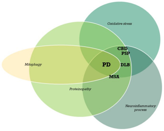
Parkinson’s disease (PD), dementia with Lewy body (DLB), progressive supranuclear palsy (PSP), corticobasal degeneration (CBD) and multiple system atrophy (MSA) belong to a group of neurodegenerative diseases called parkinsonian syndromes. They share several clinical, neuropathological and genetic features. Neurodegenerative diseases are characterized by the progressive dysfunction of specific populations of neurons, determining clinical presentation. Neuronal loss is associated with extra- and intracellular accumulation of misfolded proteins. The parkinsonian diseases affect distinct areas of the brain. PD and MSA belong to a group of synucleinopathies that are characterized by the presence of fibrillary aggregates of α-synuclein protein in the cytoplasm of selected populations of neurons and glial cells. PSP is a tauopathy associated with the pathological aggregation of the microtubule associated tau protein. Although PD is common in the world’s aging population and has been extensively studied, the exact mechanisms of the neurodegeneration are still not fully understood. Growing evidence indicates that parkinsonian disorders to some extent share a genetic background, with two key components identified so far: the microtubule associated tau protein gene (MAPT) and the α-synuclein gene (SNCA). The main pathways of parkinsonian neurodegeneration described in the literature are the protein and mitochondrial pathways. The factors that lead to neurodegeneration are primarily environmental toxins, inflammatory factors, oxidative stress and traumatic brain injury. Full article
Show Figures

Figure 1

22 pages, 514 KiB  
Review
The Nigral Coup in Parkinson’s Disease by α-Synuclein and Its Associated Rebels
by Jeswinder Sian-Hulsmann and Peter Riederer
Cells 2021, 10(3), 598; https://doi.org/10.3390/cells10030598 - 9 Mar 2021
Cited by 21 | Viewed by 3633
Abstract
The risk of Parkinson’s disease increases with age. However, the etiology of the illness remains obscure. It appears highly likely that the neurodegenerative processes involve an array of elements that influence each other. In addition, genetic, endogenous, or exogenous toxins need to be [...] Read more.
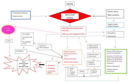
The risk of Parkinson’s disease increases with age. However, the etiology of the illness remains obscure. It appears highly likely that the neurodegenerative processes involve an array of elements that influence each other. In addition, genetic, endogenous, or exogenous toxins need to be considered as viable partners to the cellular degeneration. There is compelling evidence that indicate the key involvement of modified α-synuclein (Lewy bodies) at the very core of the pathogenesis of the disease. The accumulation of misfolded α-synuclein may be a consequence of some genetic defect or/and a failure of the protein clearance system. Importantly, α-synuclein pathology appears to be a common denominator for many cellular deleterious events such as oxidative stress, mitochondrial dysfunction, dopamine synaptic dysregulation, iron dyshomeostasis, and neuroinflammation. These factors probably employ a common apoptotic/or autophagic route in the final stages to execute cell death. The misfolded α-synuclein inclusions skillfully trigger or navigate these processes and thus amplify the dopamine neuron fatalities. Although the process of neuroinflammation may represent a secondary event, nevertheless, it executes a fundamental role in neurodegeneration. Some viral infections produce parkinsonism and exhibit similar characteristic neuropathological changes such as a modest brain dopamine deficit and α-synuclein pathology. Thus, viral infections may heighten the risk of developing PD. Alternatively, α-synuclein pathology may induce a dysfunctional immune system. Thus, sporadic Parkinson’s disease is caused by multifactorial trigger factors and metabolic disturbances, which need to be considered for the development of potential drugs in the disorder. Full article
Show Figures

Figure 1

14 pages, 9267 KiB  
Article
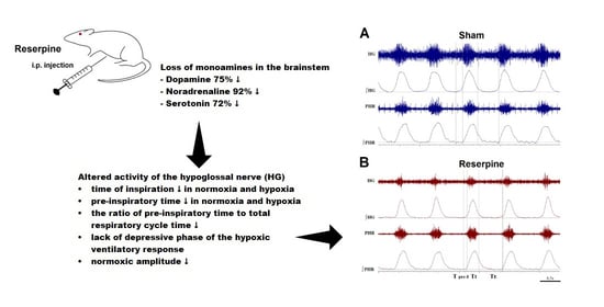
Deficiency of Biogenic Amines Modulates the Activity of Hypoglossal Nerve in the Reserpine Model of Parkinson’s Disease
by Monika Jampolska, Kryspin Andrzejewski, Małgorzata Zaremba, Ilona Joniec-Maciejak and Katarzyna Kaczyńska
Cells 2021, 10(3), 531; https://doi.org/10.3390/cells10030531 - 2 Mar 2021
Cited by 3 | Viewed by 2657
Abstract
The underlying cause of respiratory impairments appearing in Parkinson’s disease (PD) is still far from being elucidated. To better understand the pathogenesis of respiratory disorders appearing in PD, we studied hypoglossal (HG) and phrenic (PHR) motoneuron dysfunction in a rat model evoked with [...] Read more.
The underlying cause of respiratory impairments appearing in Parkinson’s disease (PD) is still far from being elucidated. To better understand the pathogenesis of respiratory disorders appearing in PD, we studied hypoglossal (HG) and phrenic (PHR) motoneuron dysfunction in a rat model evoked with reserpine administration. After reserpine, a decrease in the baseline amplitude and minute HG activity was noted, and no depressive phase of the hypoxic ventilatory response was observed. The pre-inspiratory time of HG activity along with the ratio of pre-inspiratory time to total respiratory cycle time and the ratio of pre-inspiratory to inspiratory amplitude were significantly reduced during normoxia, hypoxia, and recovery compared to sham rats. We suggest that the massive depletion of not only dopamine, but above all noradrenaline and serotonin in the brainstem observed in our study, has an impact on the pre-inspiratory activity of the HG. The shortening of the pre-inspiratory activity of the HG in the reserpine model may indicate a serious problem with maintaining the correct diameter of the upper airways in the preparation phase for inspiratory effort and explain the development of obstructive sleep apnea in some PD patients. Therapies involving the supplementation of amine depletion other than dopamine should be considered. Full article
Show Figures

Graphical abstract

10 pages, 281 KiB  
Commentary
An Introspective Approach: A Lifetime of Parkinson’s Disease Research and Not Much to Show for It Yet?
by Gordon W. Arbuthnott
Cells 2021, 10(3), 513; https://doi.org/10.3390/cells10030513 - 28 Feb 2021
Cited by 2 | Viewed by 2256
Abstract
I feel part of a massive effort to understand what is wrong with motor systems in the brain relating to Parkinson’s disease. Today, the symptoms of the disease can be modified slightly, but dopamine neurons still die; the disease progression continues inexorably. Maybe [...] Read more.
I feel part of a massive effort to understand what is wrong with motor systems in the brain relating to Parkinson’s disease. Today, the symptoms of the disease can be modified slightly, but dopamine neurons still die; the disease progression continues inexorably. Maybe the next research phase will bring the power of modern genetics to bear on halting, or better, preventing cell death. The arrival of accessible human neuron assemblies in organoids perhaps will provide a better access to the processes underlying neuronal demise. Full article
18 pages, 653 KiB  
Review
The Role of DJ-1 in Cellular Metabolism and Pathophysiological Implications for Parkinson’s Disease
by Pauline Mencke, Ibrahim Boussaad, Chiara D. Romano, Toshimori Kitami, Carole L. Linster and Rejko Krüger
Cells 2021, 10(2), 347; https://doi.org/10.3390/cells10020347 - 7 Feb 2021
Cited by 35 | Viewed by 5769
Abstract
DJ-1 is a multifunctional protein associated with pathomechanisms implicated in different chronic diseases including neurodegeneration, cancer and diabetes. Several of the physiological functions of DJ-1 are not yet fully understood; however, in the last years, there has been increasing evidence for a potential [...] Read more.
DJ-1 is a multifunctional protein associated with pathomechanisms implicated in different chronic diseases including neurodegeneration, cancer and diabetes. Several of the physiological functions of DJ-1 are not yet fully understood; however, in the last years, there has been increasing evidence for a potential role of DJ-1 in the regulation of cellular metabolism. Here, we summarize the current knowledge on specific functions of DJ-1 relevant to cellular metabolism and their role in modulating metabolic pathways. Further, we illustrate pathophysiological implications of the metabolic effects of DJ-1 in the context of neurodegeneration in Parkinson´s disease. Full article
Show Figures

Figure 1

20 pages, 4899 KiB  
Article
Seeding Propensity and Characteristics of Pathogenic αSyn Assemblies in Formalin-Fixed Human Tissue from the Enteric Nervous System, Olfactory Bulb, and Brainstem in Cases Staged for Parkinson’s Disease
by Alexis Fenyi, Charles Duyckaerts, Luc Bousset, Heiko Braak, Kelly Del Tredici, Ronald Melki and on behalf of the Brainbank Neuro-CEB Neuropathology Network
Cells 2021, 10(1), 139; https://doi.org/10.3390/cells10010139 - 12 Jan 2021
Cited by 16 | Viewed by 4840
Abstract
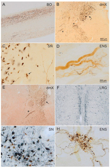
We investigated α-synuclein’s (αSyn) seeding activity in tissue from the brain and enteric nervous system. Specifically, we assessed the seeding propensity of pathogenic αSyn in formalin-fixed tissue from the gastric cardia and five brain regions of 29 individuals (12 Parkinson’s disease, 8 incidental [...] Read more.
We investigated α-synuclein’s (αSyn) seeding activity in tissue from the brain and enteric nervous system. Specifically, we assessed the seeding propensity of pathogenic αSyn in formalin-fixed tissue from the gastric cardia and five brain regions of 29 individuals (12 Parkinson’s disease, 8 incidental Lewy body disease, 9 controls) using a protein misfolding cyclic amplification assay. The structural characteristics of the resultant αSyn assemblies were determined by limited proteolysis and transmission electron microscopy. We show that fixed tissue from Parkinson’s disease (PD) and incidental Lewy body disease (ILBD) seeds the aggregation of monomeric αSyn into fibrillar assemblies. Significant variations in the characteristics of fibrillar assemblies derived from different regions even within the same individual were observed. This finding suggests that fixation stabilizes seeds with an otherwise limited seeding propensity, that yield assemblies with different intrinsic structures (i.e., strains). The lag phase preceding fibril assembly for patients ≥80 was significantly shorter than in other age groups, suggesting the existence of increased numbers of seeds or a higher seeding potential of pathogenic αSyn with time. Seeding activity did not diminish in late-stage disease. No statistically significant difference in the seeding efficiency of specific regions was found, nor was there a relationship between seeding efficiency and the load of pathogenic αSyn in a particular region at a given neuropathological stage. Full article
Show Figures

Figure 1

2020

Jump to: 2024, 2023, 2021

13 pages, 3159 KiB  
Article
Changes in Striatal Medium Spiny Neuron Morphology Resulting from Dopamine Depletion Are Reversible
by Victoria Sofie Witzig, Daniel Komnig and Björn H. Falkenburger
Cells 2020, 9(11), 2441; https://doi.org/10.3390/cells9112441 - 9 Nov 2020
Cited by 15 | Viewed by 4419
Abstract
The classical motor symptoms of Parkinson’s disease (PD) are caused by degeneration of dopaminergic neurons in the substantia nigra, which is followed by secondary dendritic pruning and spine loss at striatal medium spiny neurons (MSN). We hypothesize that these morphological changes at MSN [...] Read more.
The classical motor symptoms of Parkinson’s disease (PD) are caused by degeneration of dopaminergic neurons in the substantia nigra, which is followed by secondary dendritic pruning and spine loss at striatal medium spiny neurons (MSN). We hypothesize that these morphological changes at MSN underlie at least in part long-term motor complications in PD patients. In order to define the potential benefits and limitations of dopamine substitution, we tested in a mouse model whether dendritic pruning and spine loss can be reversible when dopaminergic axon terminals regenerate. In order to induce degeneration of nigrostriatal dopaminergic neurons we used the toxicity of 1-methyl-4-phenyl-1,2,3,6-tetrahydropyridine (MPTP) in C57BL/6J mice; 30 mg/kg MPTP was applied i.p. on five consecutive days. In order to assess the consequences of dopamine depletion, mice were analyzed 21 days after the last injection. In order to test reversibility of MSN changes we exploited the property of this model that striatal axon terminals regenerate by sprouting within 90 days and analyzed a second cohort 90 days after MPTP. Degeneration of dopaminergic neurons was confirmed by counting TH-positive neurons in the substantia nigra and by analyzing striatal catecholamines. Striatal catecholamine recovered 90 days after MPTP. MSN morphology was visualized by Golgi staining and quantified as total dendritic length, number of dendritic branch points, and density of dendritic spines. All morphological parameters of striatal MSN were reduced 21 days after MPTP. Statistical analysis indicated that dendritic pruning and the reduction of spine density represent two distinct responses to dopamine depletion. Ninety days after MPTP, all morphological changes recovered. Our findings demonstrate that morphological changes in striatal MSN resulting from dopamine depletion are reversible. They suggest that under optimal conditions, symptomatic dopaminergic therapy might be able to prevent maladaptive plasticity and long-term motor complications in PD patients. Full article
Show Figures

Graphical abstract

3 pages, 824 KiB  
Editorial
Is It Meanwhile Biomedical Sciences or Still “Ars Medica”?
by Wolfgang H. Jost
Cells 2020, 9(10), 2313; https://doi.org/10.3390/cells9102313 - 17 Oct 2020
Viewed by 1809
Abstract
Scientific work is usually quite time-intensive and frequently replete with frustrations [...] Full article
Show Figures

Figure 1

21 pages, 3327 KiB  
Article
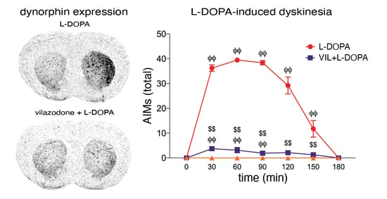
The Multimodal Serotonergic Agent Vilazodone Inhibits L-DOPA-Induced Gene Regulation in Striatal Projection Neurons and Associated Dyskinesia in an Animal Model of Parkinson’s Disease
by Feras Altwal, Connor Moon, Anthony R. West and Heinz Steiner
Cells 2020, 9(10), 2265; https://doi.org/10.3390/cells9102265 - 9 Oct 2020
Cited by 14 | Viewed by 3506
Abstract
Levodopa (L-DOPA) treatment in Parkinson’s disease is limited by the emergence of L-DOPA-induced dyskinesia. Such dyskinesia is associated with aberrant gene regulation in neurons of the striatum, which is caused by abnormal dopamine release from serotonin terminals. Previous work showed that modulating the [...] Read more.
Levodopa (L-DOPA) treatment in Parkinson’s disease is limited by the emergence of L-DOPA-induced dyskinesia. Such dyskinesia is associated with aberrant gene regulation in neurons of the striatum, which is caused by abnormal dopamine release from serotonin terminals. Previous work showed that modulating the striatal serotonin innervation with selective serotonin reuptake inhibitors (SSRIs) or 5-HT1A receptor agonists could attenuate L-DOPA-induced dyskinesia. We investigated the effects of a novel serotonergic agent, vilazodone, which combines SSRI and 5-HT1A partial agonist properties, on L-DOPA-induced behavior and gene regulation in the striatum in an animal model of Parkinson’s disease. After unilateral dopamine depletion by 6-hydroxydopamine (6-OHDA), rats received repeated L-DOPA treatment (5 mg/kg) alone or in combination with vilazodone (10 mg/kg) for 3 weeks. Gene regulation was then mapped throughout the striatum using in situ hybridization histochemistry. Vilazodone suppressed the development of L-DOPA-induced dyskinesia and turning behavior but did not interfere with the prokinetic effects of L-DOPA (forelimb stepping). L-DOPA treatment drastically increased the expression of dynorphin (direct pathway), 5-HT1B, and zif268 mRNA in the striatum ipsilateral to the lesion. These effects were inhibited by vilazodone. In contrast, vilazodone had no effect on enkephalin expression (indirect pathway) or on gene expression in the intact striatum. Thus, vilazodone inhibited L-DOPA-induced gene regulation selectively in the direct pathway of the dopamine-depleted striatum, molecular changes that are considered critical for L-DOPA-induced dyskinesia. These findings position vilazodone, an approved antidepressant, as a potential adjunct medication for the treatment of L-DOPA-induced motor side effects. Full article
Show Figures

Graphical abstract

Back to TopTop