Lamins and Laminopathies

A topical collection in Cells (ISSN 2073-4409). This collection belongs to the section "Cell Nuclei: Function, Transport and Receptors".

Viewed by 367213

Editor


E-Mail Website
Collection Editor
Ludwig Boltzmann Institute of Osteology, 1st Medical Department of Hanusch Hospital, Heinrich Collin Str. 30, A-1140 Vienna, Austria
Interests: nuclear lamins; LAP2alpha; nuclear structure; nuclear envelope disassembly and assembly; chromatin organization; cell cycle regulation; premature aging; osteogenesis
Special Issues, Collections and Topics in MDPI journals

Topical Collection Information

Dear Colleagues,

More then 20 years ago, the interest in lamins increased significantly within the scientific community with the discovery that mutations in the gene encoding A-type lamins (LMNA) could lead to human diseases. Up to now there have been reports of nearly 500 distinct mutations in LMNA, associated with over 10 distinct human diseases, which are now generally termed laminopathies and range from muscular dystrophies, cardiomyopathies and lipodystrophies to premature aging diseases, such as Hutchinson-Gilford progeria syndrome.

Nuclear lamins are type V intermediate filament (IF) proteins and the main constituents of the nuclear lamina, a filamentous meshwork underlying the inner nuclear membrane and anchoring chromatin to the nuclear envelope (NE). Beside their structural role determining nuclear shape and stability, lamins are involved in various essential cellular functions, such as gene expression, chromatin organization, DNA replication and repair, cell proliferation and differentiation, and mechanosensing. This Topical Collection gives the opportunity to publish original research, as well as review articles, in an Open Access format in the exciting and still growing field of nuclear lamins and laminopathies. We aim to become a platform for lamin researchers and to build a comprehensive collection of articles covering the various aspects of lamins. Therefore we welcome contributions ranging from the molecular structure and properties of lamins, their involvement in specific cellular processes up to the molecular mechanisms underlying the distinct laminopathies in humans and mouse models.

Dr. Thomas Dechat
Collection Editor

Manuscript Submission Information

Manuscripts should be submitted online at www.mdpi.com by registering and logging in to this website. Once you are registered, click here to go to the submission form. Manuscripts can be submitted until the deadline. All submissions that pass pre-check are peer-reviewed. Accepted papers will be published continuously in the journal (as soon as accepted) and will be listed together on the collection website. Research articles, review articles as well as short communications are invited. For planned papers, a title and short abstract (about 100 words) can be sent to the Editorial Office for announcement on this website.

Submitted manuscripts should not have been published previously, nor be under consideration for publication elsewhere (except conference proceedings papers). All manuscripts are thoroughly refereed through a single-blind peer-review process. A guide for authors and other relevant information for submission of manuscripts is available on the Instructions for Authors page. Cells is an international peer-reviewed open access semimonthly journal published by MDPI.

Please visit the Instructions for Authors page before submitting a manuscript. The Article Processing Charge (APC) for publication in this open access journal is 2700 CHF (Swiss Francs). Submitted papers should be well formatted and use good English. Authors may use MDPI's English editing service prior to publication or during author revisions.


Keywords

  • nuclear lamins
  • lamin-binding proteins
  • chromatin organization
  • gene expression
  • mechanosensing
  • laminopathies
  • nuclear envelope
  • cell proliferation and differentiation
  • DNA repair
  • mouse models

Related Special Issue

Published Papers (61 papers)

2024

Jump to: 2023, 2022, 2021, 2020, 2019, 2018, 2017, 2016

34 pages, 13933 KiB  
Article
LMNA-Related Dilated Cardiomyopathy: Single-Cell Transcriptomics during Patient-Derived iPSC Differentiation Support Cell Type and Lineage-Specific Dysregulation of Gene Expression and Development for Cardiomyocytes and Epicardium-Derived Cells with Lamin A/C Haploinsufficiency
by Michael V. Zaragoza, Thuy-Anh Bui, Halida P. Widyastuti, Mehrsa Mehrabi, Zixuan Cang, Yutong Sha, Anna Grosberg and Qing Nie
Cells 2024, 13(17), 1479; https://doi.org/10.3390/cells13171479 - 3 Sep 2024
Viewed by 1336
Abstract
LMNA-related dilated cardiomyopathy (DCM) is an autosomal-dominant genetic condition with cardiomyocyte and conduction system dysfunction often resulting in heart failure or sudden death. The condition is caused by mutation in the Lamin A/C (LMNA) gene encoding Type-A nuclear lamin proteins [...] Read more.
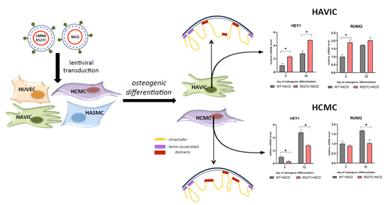
LMNA-related dilated cardiomyopathy (DCM) is an autosomal-dominant genetic condition with cardiomyocyte and conduction system dysfunction often resulting in heart failure or sudden death. The condition is caused by mutation in the Lamin A/C (LMNA) gene encoding Type-A nuclear lamin proteins involved in nuclear integrity, epigenetic regulation of gene expression, and differentiation. The molecular mechanisms of the disease are not completely understood, and there are no definitive treatments to reverse progression or prevent mortality. We investigated possible mechanisms of LMNA-related DCM using induced pluripotent stem cells derived from a family with a heterozygous LMNA c.357-2A>G splice-site mutation. We differentiated one LMNA-mutant iPSC line derived from an affected female (Patient) and two non-mutant iPSC lines derived from her unaffected sister (Control) and conducted single-cell RNA sequencing for 12 samples (four from Patients and eight from Controls) across seven time points: Day 0, 2, 4, 9, 16, 19, and 30. Our bioinformatics workflow identified 125,554 cells in raw data and 110,521 (88%) high-quality cells in sequentially processed data. Unsupervised clustering, cell annotation, and trajectory inference found complex heterogeneity: ten main cell types; many possible subtypes; and lineage bifurcation for cardiac progenitors to cardiomyocytes (CMs) and epicardium-derived cells (EPDCs). Data integration and comparative analyses of Patient and Control cells found cell type and lineage-specific differentially expressed genes (DEGs) with enrichment, supporting pathway dysregulation. Top DEGs and enriched pathways included 10 ZNF genes and RNA polymerase II transcription in pluripotent cells (PP); BMP4 and TGF Beta/BMP signaling, sarcomere gene subsets and cardiogenesis, CDH2 and EMT in CMs; LMNA and epigenetic regulation, as well as DDIT4 and mTORC1 signaling in EPDCs. Top DEGs also included XIST and other X-linked genes, six imprinted genes (SNRPN, PWAR6, NDN, PEG10, MEG3, MEG8), and enriched gene sets related to metabolism, proliferation, and homeostasis. We confirmed Lamin A/C haploinsufficiency by allelic expression and Western blot. Our complex Patient-derived iPSC model for Lamin A/C haploinsufficiency in PP, CM, and EPDC provided support for dysregulation of genes and pathways, many previously associated with Lamin A/C defects, such as epigenetic gene expression, signaling, and differentiation. Our findings support disruption of epigenomic developmental programs, as proposed in other LMNA disease models. We recognized other factors influencing epigenetics and differentiation; thus, our approach needs improvement to further investigate this mechanism in an iPSC-derived model. Full article
Show Figures

Graphical abstract

13 pages, 19803 KiB  
Article
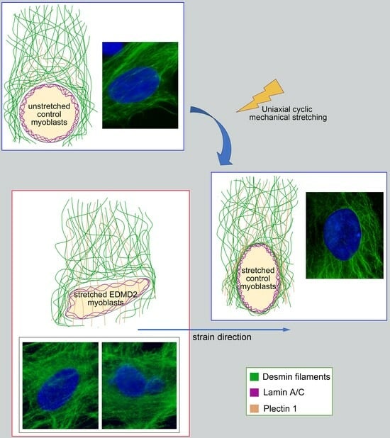
Desmin and Plectin Recruitment to the Nucleus and Nuclei Orientation Are Lost in Emery-Dreifuss Muscular Dystrophy Myoblasts Subjected to Mechanical Stimulation
by Vittoria Cenni, Camilla Evangelisti, Spartaco Santi, Patrizia Sabatelli, Simona Neri, Marco Cavallo, Giovanna Lattanzi and Elisabetta Mattioli
Cells 2024, 13(2), 162; https://doi.org/10.3390/cells13020162 - 16 Jan 2024
Viewed by 1634
Abstract
In muscle cells subjected to mechanical stimulation, LINC complex and cytoskeletal proteins are basic to preserve cellular architecture and maintain nuclei orientation and positioning. In this context, the role of lamin A/C remains mostly elusive. This study demonstrates that in human myoblasts subjected [...] Read more.
In muscle cells subjected to mechanical stimulation, LINC complex and cytoskeletal proteins are basic to preserve cellular architecture and maintain nuclei orientation and positioning. In this context, the role of lamin A/C remains mostly elusive. This study demonstrates that in human myoblasts subjected to mechanical stretching, lamin A/C recruits desmin and plectin to the nuclear periphery, allowing a proper spatial orientation of the nuclei. Interestingly, in Emery-Dreifuss Muscular Dystrophy (EDMD2) myoblasts exposed to mechanical stretching, the recruitment of desmin and plectin to the nucleus and nuclear orientation were impaired, suggesting that a functional lamin A/C is crucial for the response to mechanical strain. While describing a new mechanism of action headed by lamin A/C, these findings show a structural alteration that could be involved in the onset of the muscle defects observed in muscular laminopathies. Full article
Show Figures

Graphical abstract

2023

Jump to: 2024, 2022, 2021, 2020, 2019, 2018, 2017, 2016

16 pages, 10237 KiB  
Article
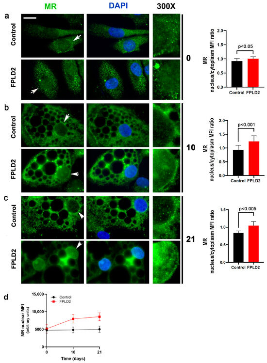
Mineralocorticoid Receptor Antagonism Prevents Type 2 Familial Partial Lipodystrophy Brown Adipocyte Dysfunction
by Elisa Schena, Elisabetta Mattioli, Chiara Peres, Laura Zanotti, Paolo Morselli, Patricia Iozzo, Maria Angela Guzzardi, Chiara Bernardini, Monica Forni, Salvatore Nesci, Massimiliano Caprio, Carolina Cecchetti, Uberto Pagotto, Elena Gabusi, Luca Cattini, Gina Lisignoli, William Blalock, Alessandra Gambineri and Giovanna Lattanzi
Cells 2023, 12(22), 2586; https://doi.org/10.3390/cells12222586 - 7 Nov 2023
Cited by 1 | Viewed by 1508
Abstract
Type-2 Familial Partial Lipodystrophy (FPLD2), a rare lipodystrophy caused by LMNA mutations, is characterized by a loss of subcutaneous fat from the trunk and limbs and excess accumulation of adipose tissue in the neck and face. Several studies have reported that the mineralocorticoid [...] Read more.
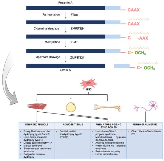
Type-2 Familial Partial Lipodystrophy (FPLD2), a rare lipodystrophy caused by LMNA mutations, is characterized by a loss of subcutaneous fat from the trunk and limbs and excess accumulation of adipose tissue in the neck and face. Several studies have reported that the mineralocorticoid receptor (MR) plays an essential role in adipose tissue differentiation and functionality. We previously showed that brown preadipocytes isolated from a FPLD2 patient’s neck aberrantly differentiate towards the white lineage. As this condition may be related to MR activation, we suspected altered MR dynamics in FPLD2. Despite cytoplasmic MR localization in control brown adipocytes, retention of MR was observed in FPLD2 brown adipocyte nuclei. Moreover, overexpression of wild-type or mutated prelamin A caused GFP-MR recruitment to the nuclear envelope in HEK293 cells, while drug-induced prelamin A co-localized with endogenous MR in human preadipocytes. Based on in silico analysis and in situ protein ligation assays, we could suggest an interaction between prelamin A and MR, which appears to be inhibited by mineralocorticoid receptor antagonism. Importantly, the MR antagonist spironolactone redirected FPLD2 preadipocyte differentiation towards the brown lineage, avoiding the formation of enlarged and dysmorphic lipid droplets. Finally, beneficial effects on brown adipose tissue activity were observed in an FPLD2 patient undergoing spironolactone treatment. These findings identify MR as a new lamin A interactor and a new player in lamin A-linked lipodystrophies. Full article
Show Figures

Figure 1

18 pages, 3213 KiB  
Article
Cellular and Genomic Features of Muscle Differentiation from Isogenic Fibroblasts and Myoblasts
by Louise Benarroch, Julia Madsen-Østerbye, Mohamed Abdelhalim, Kamel Mamchaoui, Jessica Ohana, Anne Bigot, Vincent Mouly, Gisèle Bonne, Anne T. Bertrand and Philippe Collas
Cells 2023, 12(15), 1995; https://doi.org/10.3390/cells12151995 - 3 Aug 2023
Cited by 2 | Viewed by 2882
Abstract
The ability to recapitulate muscle differentiation in vitro enables the exploration of mechanisms underlying myogenesis and muscle diseases. However, obtaining myoblasts from patients with neuromuscular diseases or from healthy subjects poses ethical and procedural challenges that limit such investigations. An alternative consists in [...] Read more.
The ability to recapitulate muscle differentiation in vitro enables the exploration of mechanisms underlying myogenesis and muscle diseases. However, obtaining myoblasts from patients with neuromuscular diseases or from healthy subjects poses ethical and procedural challenges that limit such investigations. An alternative consists in converting skin fibroblasts into myogenic cells by forcing the expression of the myogenic regulator MYOD. Here, we directly compared cellular phenotype, transcriptome, and nuclear lamina-associated domains (LADs) in myo-converted human fibroblasts and myotubes differentiated from myoblasts. We used isogenic cells from a 16-year-old donor, ruling out, for the first time to our knowledge, genetic factors as a source of variations between the two myogenic models. We show that myo-conversion of fibroblasts upregulates genes controlling myogenic pathways leading to multinucleated cells expressing muscle cell markers. However, myotubes are more advanced in myogenesis than myo-converted fibroblasts at the phenotypic and transcriptomic levels. While most LADs are shared between the two cell types, each also displays unique domains of lamin A/C interactions. Furthermore, myotube-specific LADs are more gene-rich and less heterochromatic than shared LADs or LADs unique to myo-converted fibroblasts, and they uniquely sequester developmental genes. Thus, myo-converted fibroblasts and myotubes retain cell type-specific features of radial and functional genome organization. Our results favor a view of myo-converted fibroblasts as a practical model to investigate the phenotypic and genomic properties of muscle cell differentiation in normal and pathological contexts, but also highlight current limitations in using fibroblasts as a source of myogenic cells. Full article
Show Figures

Figure 1

22 pages, 19445 KiB  
Article
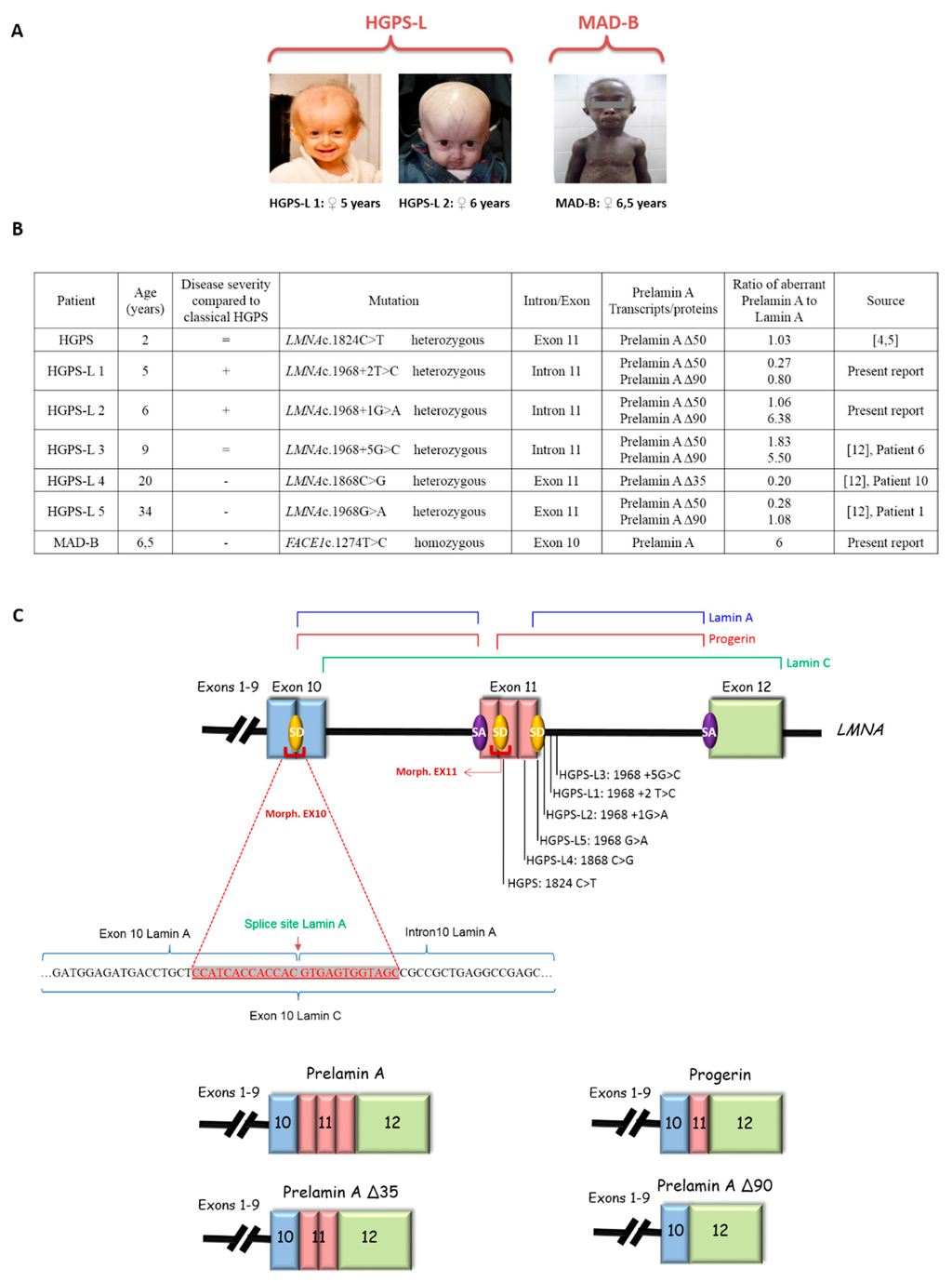
Impact of Combined Baricitinib and FTI Treatment on Adipogenesis in Hutchinson–Gilford Progeria Syndrome and Other Lipodystrophic Laminopathies
by Ramona Hartinger, Eva-Maria Lederer, Elisa Schena, Giovanna Lattanzi and Karima Djabali
Cells 2023, 12(10), 1350; https://doi.org/10.3390/cells12101350 - 9 May 2023
Cited by 8 | Viewed by 2629
Abstract
Hutchinson–Gilford progeria syndrome (HGPS) is a rare genetic disease that causes premature aging symptoms, such as vascular diseases, lipodystrophy, loss of bone mineral density, and alopecia. HGPS is mostly linked to a heterozygous and de novo mutation in the LMNA gene (c.1824 C [...] Read more.
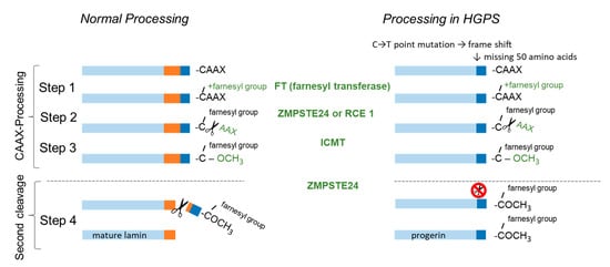
Hutchinson–Gilford progeria syndrome (HGPS) is a rare genetic disease that causes premature aging symptoms, such as vascular diseases, lipodystrophy, loss of bone mineral density, and alopecia. HGPS is mostly linked to a heterozygous and de novo mutation in the LMNA gene (c.1824 C > T; p.G608G), resulting in the production of a truncated prelamin A protein called “progerin”. Progerin accumulation causes nuclear dysfunction, premature senescence, and apoptosis. Here, we examined the effects of baricitinib (Bar), an FDA-approved JAK/STAT inhibitor, and a combination of Bar and lonafarnib (FTI) treatment on adipogenesis using skin-derived precursors (SKPs). We analyzed the effect of these treatments on the differentiation potential of SKPs isolated from pre-established human primary fibroblast cultures. Compared to mock-treated HGPS SKPs, Bar and Bar + FTI treatments improved the differentiation of HGPS SKPs into adipocytes and lipid droplet formation. Similarly, Bar and Bar + FTI treatments improved the differentiation of SKPs derived from patients with two other lipodystrophic diseases: familial partial lipodystrophy type 2 (FPLD2) and mandibuloacral dysplasia type B (MADB). Overall, the results show that Bar treatment improves adipogenesis and lipid droplet formation in HGPS, FPLD2, and MADB, indicating that Bar + FTI treatment might further ameliorate HGPS pathologies compared to lonafarnib treatment alone. Full article
Show Figures

Figure 1

24 pages, 3735 KiB  
Article
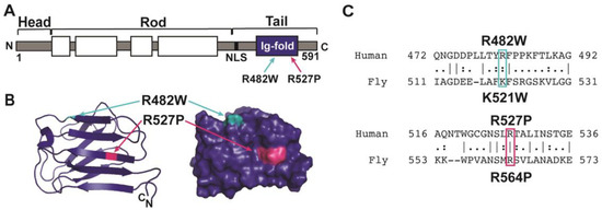
Drosophila Models Reveal Properties of Mutant Lamins That Give Rise to Distinct Diseases
by Sydney G. Walker, Christopher J. Langland, Jill Viles, Laura A. Hecker and Lori L. Wallrath
Cells 2023, 12(8), 1142; https://doi.org/10.3390/cells12081142 - 12 Apr 2023
Cited by 1 | Viewed by 2197
Abstract
Mutations in the LMNA gene cause a collection of diseases known as laminopathies, including muscular dystrophies, lipodystrophies, and early-onset aging syndromes. The LMNA gene encodes A-type lamins, lamins A/C, intermediate filaments that form a meshwork underlying the inner nuclear membrane. Lamins have a [...] Read more.
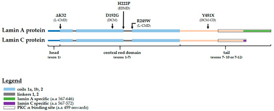
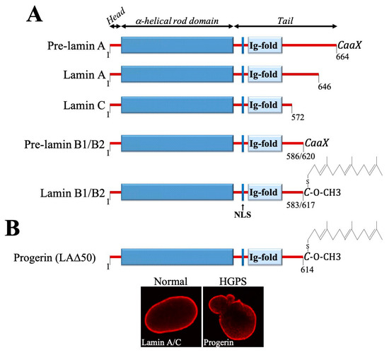
Mutations in the LMNA gene cause a collection of diseases known as laminopathies, including muscular dystrophies, lipodystrophies, and early-onset aging syndromes. The LMNA gene encodes A-type lamins, lamins A/C, intermediate filaments that form a meshwork underlying the inner nuclear membrane. Lamins have a conserved domain structure consisting of a head, coiled-coil rod, and C-terminal tail domain possessing an Ig-like fold. This study identified differences between two mutant lamins that cause distinct clinical diseases. One of the LMNA mutations encodes lamin A/C p.R527P and the other codes lamin A/C p.R482W, which are typically associated with muscular dystrophy and lipodystrophy, respectively. To determine how these mutations differentially affect muscle, we generated the equivalent mutations in the Drosophila Lamin C (LamC) gene, an orthologue of human LMNA. The muscle-specific expression of the R527P equivalent showed cytoplasmic aggregation of LamC, a reduced larval muscle size, decreased larval motility, and cardiac defects resulting in a reduced adult lifespan. By contrast, the muscle-specific expression of the R482W equivalent caused an abnormal nuclear shape without a change in larval muscle size, larval motility, and adult lifespan compared to controls. Collectively, these studies identified fundamental differences in the properties of mutant lamins that cause clinically distinct phenotypes, providing insights into disease mechanisms. Full article
Show Figures

Figure 1

19 pages, 2482 KiB  
Article
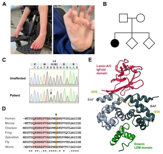
A De Novo Sequence Variant in Barrier-to-Autointegration Factor Is Associated with Dominant Motor Neuronopathy
by Agathe Marcelot, Felipe Rodriguez-Tirado, Philippe Cuniasse, Mei-ling Joiner, Simona Miron, Alexey A. Soshnev, Mimi Fang, Miles A. Pufall, Katherine D. Mathews, Steven A. Moore, Sophie Zinn-Justin and Pamela K. Geyer
Cells 2023, 12(6), 847; https://doi.org/10.3390/cells12060847 - 9 Mar 2023
Cited by 2 | Viewed by 2487
Abstract
Barrier-to-autointegration factor (BAF) is an essential component of the nuclear lamina. Encoded by BANF1, this DNA binding protein contributes to the regulation of gene expression, cell cycle progression, and nuclear integrity. A rare recessive BAF variant, Ala12Thr, causes the premature aging syndrome, [...] Read more.
Barrier-to-autointegration factor (BAF) is an essential component of the nuclear lamina. Encoded by BANF1, this DNA binding protein contributes to the regulation of gene expression, cell cycle progression, and nuclear integrity. A rare recessive BAF variant, Ala12Thr, causes the premature aging syndrome, Néstor–Guillermo progeria syndrome (NGPS). Here, we report the first dominant pathogenic BAF variant, Gly16Arg, identified in a patient presenting with progressive neuromuscular weakness. Although disease variants carry nearby amino acid substitutions, cellular and biochemical properties are distinct. In contrast to NGPS, Gly16Arg patient fibroblasts show modest changes in nuclear lamina structure and increases in repressive marks associated with heterochromatin. Structural studies reveal that the Gly16Arg substitution introduces a salt bridge between BAF monomers, reducing the conformation ensemble available to BAF. We show that this structural change increases the double-stranded DNA binding affinity of BAF Gly16Arg. Together, our findings suggest that BAF Gly16Arg has an increased chromatin occupancy that leads to epigenetic changes and impacts nuclear functions. These observations provide a new example of how a missense mutation can change a protein conformational equilibrium to cause a dominant disease and extend our understanding of mechanisms by which BAF function impacts human health. Full article
Show Figures

Figure 1

22 pages, 1796 KiB  
Review
Epigenetics in LMNA-Related Cardiomyopathy
by Yinuo Wang and Gergana Dobreva
Cells 2023, 12(5), 783; https://doi.org/10.3390/cells12050783 - 1 Mar 2023
Cited by 17 | Viewed by 4639
Abstract
Mutations in the gene for lamin A/C (LMNA) cause a diverse range of diseases known as laminopathies. LMNA-related cardiomyopathy is a common inherited heart disease and is highly penetrant with a poor prognosis. In the past years, numerous investigations using mouse models, [...] Read more.
Mutations in the gene for lamin A/C (LMNA) cause a diverse range of diseases known as laminopathies. LMNA-related cardiomyopathy is a common inherited heart disease and is highly penetrant with a poor prognosis. In the past years, numerous investigations using mouse models, stem cell technologies, and patient samples have characterized the phenotypic diversity caused by specific LMNA variants and contributed to understanding the molecular mechanisms underlying the pathogenesis of heart disease. As a component of the nuclear envelope, LMNA regulates nuclear mechanostability and function, chromatin organization, and gene transcription. This review will focus on the different cardiomyopathies caused by LMNA mutations, address the role of LMNA in chromatin organization and gene regulation, and discuss how these processes go awry in heart disease. Full article
Show Figures

Figure 1

26 pages, 4688 KiB  
Article
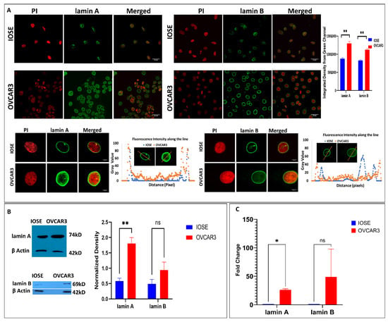
Elevated Levels of Lamin A Promote HR and NHEJ-Mediated Repair Mechanisms in High-Grade Ovarian Serous Carcinoma Cell Line
by Duhita Sengupta, Asima Mukhopadhyay and Kaushik Sengupta
Cells 2023, 12(5), 757; https://doi.org/10.3390/cells12050757 - 27 Feb 2023
Cited by 1 | Viewed by 2269
Abstract
Extensive research for the last two decades has significantly contributed to understanding the roles of lamins in the maintenance of nuclear architecture and genome organization which is drastically modified in neoplasia. It must be emphasized that alteration in lamin A/C expression and distribution [...] Read more.
Extensive research for the last two decades has significantly contributed to understanding the roles of lamins in the maintenance of nuclear architecture and genome organization which is drastically modified in neoplasia. It must be emphasized that alteration in lamin A/C expression and distribution is a consistent event during tumorigenesis of almost all tissues of human bodies. One of the important signatures of a cancer cell is its inability to repair DNA damage which befalls several genomic events that transform the cells to be sensitive to chemotherapeutic agents. This genomic and chromosomal instability is the most common feature found in cases of high-grade ovarian serous carcinoma. Here, we report elevated levels of lamins in OVCAR3 cells (high-grade ovarian serous carcinoma cell line) in comparison to IOSE (immortalised ovarian surface epithelial cells) and, consequently, altered damage repair machinery in OVCAR3. We have analysed the changes in global gene expression as a sequel to DNA damage induced by etoposide in ovarian carcinoma where lamin A is particularly elevated in expression and reported some differentially expressed genes associated with pathways conferring cellular proliferation and chemoresistance. We hereby establish the role of elevated lamin A in neoplastic transformation in the context of high-grade ovarian serous cancer through a combination of HR and NHEJ mechanisms. Full article
Show Figures

Figure 1

23 pages, 2044 KiB  
Systematic Review
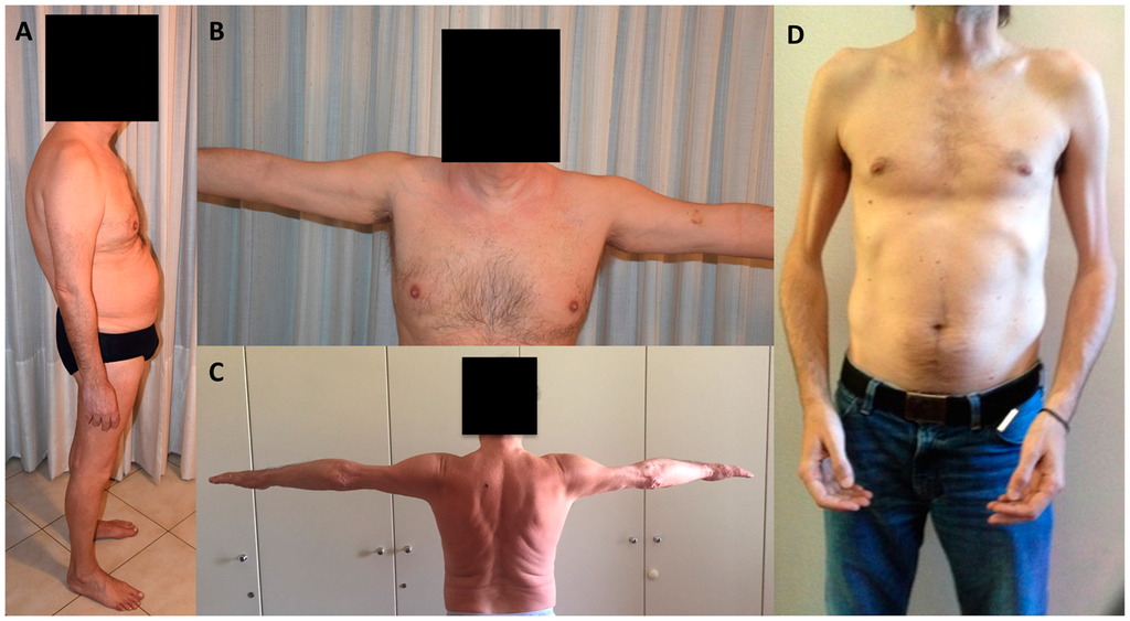
Clinical Spectrum of LMNA-Associated Type 2 Familial Partial Lipodystrophy: A Systematic Review
by Antia Fernandez-Pombo, Everardo Josue Diaz-Lopez, Ana I. Castro, Sofia Sanchez-Iglesias, Silvia Cobelo-Gomez, Teresa Prado-Moraña and David Araujo-Vilar
Cells 2023, 12(5), 725; https://doi.org/10.3390/cells12050725 - 24 Feb 2023
Cited by 15 | Viewed by 5369
Abstract
Type 2 familial partial lipodystrophy (FPLD2) is a laminopathic lipodystrophy due to pathogenic variants in the LMNA gene. Its rarity implies that it is not well-known. The aim of this review was to explore the published data regarding the clinical characterisation of this [...] Read more.
Type 2 familial partial lipodystrophy (FPLD2) is a laminopathic lipodystrophy due to pathogenic variants in the LMNA gene. Its rarity implies that it is not well-known. The aim of this review was to explore the published data regarding the clinical characterisation of this syndrome in order to better describe FPLD2. For this purpose, a systematic review through a search on PubMed until December 2022 was conducted and the references of the retrieved articles were also screened. A total of 113 articles were included. FPLD2 is characterised by the loss of fat starting around puberty in women, affecting limbs and trunk, and its accumulation in the face, neck and abdominal viscera. This adipose tissue dysfunction conditions the development of metabolic complications associated with insulin resistance, such as diabetes, dyslipidaemia, fatty liver disease, cardiovascular disease, and reproductive disorders. However, a great degree of phenotypical variability has been described. Therapeutic approaches are directed towards the associated comorbidities, and recent treatment modalities have been explored. A comprehensive comparison between FPLD2 and other FPLD subtypes can also be found in the present review. This review aimed to contribute towards augmenting knowledge of the natural history of FPLD2 by bringing together the main clinical research in this field. Full article
Show Figures

Figure 1

27 pages, 5570 KiB  
Article
Abnormal Cellular Phenotypes Induced by Three TMPO/LAP2 Variants Identified in Men with Cardiomyopathies
by Nathalie Vadrot, Flavie Ader, Maryline Moulin, Marie Merlant, Françoise Chapon, Estelle Gandjbakhch, Fabien Labombarda, Pascale Maragnes, Patricia Réant, Caroline Rooryck, Vincent Probst, Erwan Donal, Pascale Richard, Ana Ferreiro and Brigitte Buendia
Cells 2023, 12(2), 337; https://doi.org/10.3390/cells12020337 - 16 Jan 2023
Viewed by 2516
Abstract
A single missense variant of the TMPO/LAP2α gene, encoding LAP2 proteins, has been associated with cardiomyopathy in two brothers. To further evaluate its role in cardiac muscle, we included TMPO in our cardiomyopathy diagnostic gene panel. A screening of ~5000 patients revealed [...] Read more.
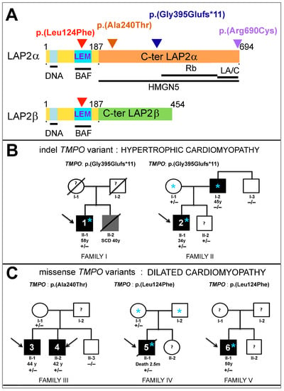
A single missense variant of the TMPO/LAP2α gene, encoding LAP2 proteins, has been associated with cardiomyopathy in two brothers. To further evaluate its role in cardiac muscle, we included TMPO in our cardiomyopathy diagnostic gene panel. A screening of ~5000 patients revealed three novel rare TMPO heterozygous variants in six males diagnosed with hypertrophic or dilated cardiomypathy. We identified in different cellular models that (1) the frameshift variant LAP2α p.(Gly395Glufs*11) induced haploinsufficiency, impeding cell proliferation and/or producing a truncated protein mislocalized in the cytoplasm; (2) the C-ter missense variant LAP2α p.(Ala240Thr) led to a reduced proximity events between LAP2α and the nucleosome binding protein HMGN5; and (3) the LEM-domain missense variant p.(Leu124Phe) decreased both associations of LAP2α/β with the chromatin-associated protein BAF and inhibition of the E2F1 transcription factor activity which is known to be dependent on Rb, partner of LAP2α. Additionally, the LAP2α expression was lower in the left ventricles of male mice compared to females. In conclusion, our study reveals distinct altered properties of LAP2 induced by these TMPO/LAP2 variants, leading to altered cell proliferation, chromatin structure or gene expression-regulation pathways, and suggests a potential sex-dependent role of LAP2 in myocardial function and disease. Full article
Show Figures

Figure 1

2022

Jump to: 2024, 2023, 2021, 2020, 2019, 2018, 2017, 2016

24 pages, 2879 KiB  
Systematic Review
Genotype-Phenotype Correlations in Human Diseases Caused by Mutations of LINC Complex-Associated Genes: A Systematic Review and Meta-Summary
by Emily C. Storey and Heidi R. Fuller
Cells 2022, 11(24), 4065; https://doi.org/10.3390/cells11244065 - 15 Dec 2022
Cited by 13 | Viewed by 3041
Abstract
Mutations in genes encoding proteins associated with the linker of nucleoskeleton and cytoskeleton (LINC) complex within the nuclear envelope cause different diseases with varying phenotypes including skeletal muscle, cardiac, metabolic, or nervous system pathologies. There is some understanding of the structure of LINC [...] Read more.
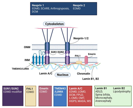
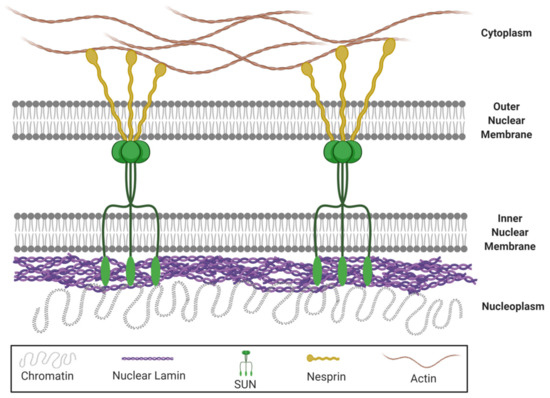
Mutations in genes encoding proteins associated with the linker of nucleoskeleton and cytoskeleton (LINC) complex within the nuclear envelope cause different diseases with varying phenotypes including skeletal muscle, cardiac, metabolic, or nervous system pathologies. There is some understanding of the structure of LINC complex-associated proteins and how they interact, but it is unclear how mutations in genes encoding them can cause the same disease, and different diseases with different phenotypes. Here, published mutations in LINC complex-associated proteins were systematically reviewed and analyzed to ascertain whether patterns exist between the genetic sequence variants and clinical phenotypes. This revealed LMNA is the only LINC complex-associated gene in which mutations commonly cause distinct conditions, and there are no clear genotype-phenotype correlations. Clusters of LMNA variants causing striated muscle disease are located in exons 1 and 6, and metabolic disease-associated LMNA variants are frequently found in the tail of lamin A/C. Additionally, exon 6 of the emerin gene, EMD, may be a mutation “hot-spot”, and diseases related to SYNE1, encoding nesprin-1, are most often caused by nonsense type mutations. These results provide insight into the diverse roles of LINC-complex proteins in human disease and provide direction for future gene-targeted therapy development. Full article
Show Figures

Figure 1

18 pages, 4759 KiB  
Article
Loss of Mature Lamin A/C Triggers a Shift in Intracellular Metabolic Homeostasis via AMPKα Activation
by Ying Zhou, Jia-Jie Yang, Yuan Cheng, Ge-Xuan Feng, Rong-Hui Yang, Yuan Yuan, Li-Yong Wang, Miao Wang and Lu Kong
Cells 2022, 11(24), 3988; https://doi.org/10.3390/cells11243988 - 9 Dec 2022
Cited by 1 | Viewed by 2262
Abstract
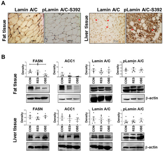
The roles of lamin A/C in adipocyte differentiation and skeletal muscle lipid metabolism are associated with familial partial lipodystrophy of Dunnigan (FPLD). We confirmed that LMNA knockdown (KD) in mouse adipose-derived mesenchymal stem cells (AD-MSCs) prevented adipocyte maturation. Importantly, in in vitro experiments, [...] Read more.
The roles of lamin A/C in adipocyte differentiation and skeletal muscle lipid metabolism are associated with familial partial lipodystrophy of Dunnigan (FPLD). We confirmed that LMNA knockdown (KD) in mouse adipose-derived mesenchymal stem cells (AD-MSCs) prevented adipocyte maturation. Importantly, in in vitro experiments, we discovered a significant increase in phosphorylated lamin A/C levels at serine 22 or 392 sites (pLamin A/C-S22/392) accompanying increased lipid synthesis in a liver cell line (7701 cells) and two hepatocellular carcinoma (HCC) cell lines (HepG2 and MHCC97-H cells). Moreover, HCC cells did not survive after LMNA knockout (KO) or even KD. Evidently, the functions of lamin A/C differ between the liver and adipose tissue. To date, the mechanism of hepatocyte lipid metabolism mediated by nuclear lamin A/C remains unclear. Our in-depth study aimed to identify the molecular connection between lamin A/C and pLamin A/C, hepatic lipid metabolism and liver cancer. Gain- and loss-of-function experiments were performed to investigate functional changes and the related molecular pathways in 7701 cells. Adenosine 5’ monophosphate-activated protein kinase α (AMPKα) was activated when abnormalities in functional lamin A/C were observed following lamin A/C depletion or farnesyltransferase inhibitor (FTI) treatment. Active AMPKα directly phosphorylated acetyl-CoA-carboxylase 1 (ACC1) and subsequently inhibited lipid synthesis but induced glycolysis in both HCC cells and normal cells. According to the mass spectrometry analysis, lamin A/C potentially regulated AMPKα activation through its chaperone proteins, ATPase or ADP/ATP transporter 2. Lonafarnib (an FTI) combined with low-glucose conditions significantly decreased the proliferation of the two HCC cell lines more efficiently than lonafarnib alone by inhibiting glycolysis or the maturation of prelamin A. Full article
Show Figures

Figure 1

2021

Jump to: 2024, 2023, 2022, 2020, 2019, 2018, 2017, 2016

14 pages, 2274 KiB  
Article
Muscle Enriched Lamin Interacting Protein (Mlip) Binds Chromatin and Is Required for Myoblast Differentiation
by Elmira Ahmady, Alexandre Blais and Patrick G. Burgon
Cells 2021, 10(3), 615; https://doi.org/10.3390/cells10030615 - 10 Mar 2021
Cited by 11 | Viewed by 2858
Abstract
Muscle-enriched A-type lamin-interacting protein (Mlip) is a recently discovered Amniota gene that encodes proteins of unknown biological function. Here we report Mlip’s direct interaction with chromatin, and it may function as a transcriptional co-factor. Chromatin immunoprecipitations with microarray analysis demonstrated [...] Read more.
Muscle-enriched A-type lamin-interacting protein (Mlip) is a recently discovered Amniota gene that encodes proteins of unknown biological function. Here we report Mlip’s direct interaction with chromatin, and it may function as a transcriptional co-factor. Chromatin immunoprecipitations with microarray analysis demonstrated a propensity for Mlip to associate with genomic regions in close proximity to genes that control tissue-specific differentiation. Gel mobility shift assays confirmed that Mlip protein complexes with genomic DNA. Blocking Mlip expression in C2C12 myoblasts down-regulates myogenic regulatory factors (MyoD and MyoG) and subsequently significantly inhibits myogenic differentiation and the formation of myotubes. Collectively our data demonstrate that Mlip is required for C2C12 myoblast differentiation into myotubes. Mlip may exert this role as a transcriptional regulator of a myogenic program that is unique to amniotes. Full article
Show Figures

Figure 1

21 pages, 8620 KiB  
Article
The SC-35 Splicing Factor Interacts with RNA Pol II and A-Type Lamin Depletion Weakens This Interaction
by Soňa Legartová, Paolo Fagherazzi, Lenka Stixová, Aleš Kovařík, Ivan Raška and Eva Bártová
Cells 2021, 10(2), 297; https://doi.org/10.3390/cells10020297 - 1 Feb 2021
Cited by 2 | Viewed by 4033
Abstract
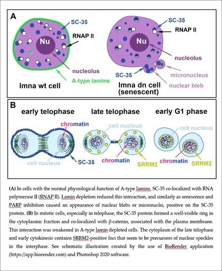
The essential components of splicing are the splicing factors accumulated in nuclear speckles; thus, we studied how DNA damaging agents and A-type lamin depletion affect the properties of these regions, positive on the SC-35 protein. We observed that inhibitor of PARP (poly [...] Read more.
The essential components of splicing are the splicing factors accumulated in nuclear speckles; thus, we studied how DNA damaging agents and A-type lamin depletion affect the properties of these regions, positive on the SC-35 protein. We observed that inhibitor of PARP (poly (ADP-ribose) polymerase), and more pronouncedly inhibitors of RNA polymerases, caused DNA damage and increased the SC-35 protein level. Interestingly, nuclear blebs, induced by PARP inhibitor and observed in A-type lamin-depleted or senescent cells, were positive on both the SC-35 protein and another component of the spliceosome, SRRM2. In the interphase cell nuclei, SC-35 interacted with the phosphorylated form of RNAP II, which was A-type lamin-dependent. In mitotic cells, especially in telophase, the SC-35 protein formed a well-visible ring in the cytoplasmic fraction and colocalized with β-catenin, associated with the plasma membrane. The antibody against the SRRM2 protein showed that nuclear speckles are already established in the cytoplasm of the late telophase and at the stage of early cytokinesis. In addition, we observed the occurrence of splicing factors in the nuclear blebs and micronuclei, which are also sites of both transcription and splicing. This conclusion supports the fact that splicing proceeds transcriptionally. According to our data, this process is A-type lamin-dependent. Lamin depletion also reduces the interaction between SC-35 and β-catenin in mitotic cells. Full article
Show Figures

Graphical abstract

2020

Jump to: 2024, 2023, 2022, 2021, 2019, 2018, 2017, 2016

20 pages, 4452 KiB  
Article
Protein Kinase C Alpha Cellular Distribution, Activity, and Proximity with Lamin A/C in Striated Muscle Laminopathies
by Hannah A. Nicolas, Anne T. Bertrand, Sarah Labib, Musfira Mohamed-Uvaize, Pierrette M. Bolongo, Wen Yu Wu, Zofia T. Bilińska, Gisèle Bonne, Marie-Andrée Akimenko and Frédérique Tesson
Cells 2020, 9(11), 2388; https://doi.org/10.3390/cells9112388 - 31 Oct 2020
Cited by 5 | Viewed by 3710
Abstract
Striated muscle laminopathies are cardiac and skeletal muscle conditions caused by mutations in the lamin A/C gene (LMNA). LMNA codes for the A-type lamins, which are nuclear intermediate filaments that maintain the nuclear structure and nuclear processes such as gene expression. [...] Read more.
Striated muscle laminopathies are cardiac and skeletal muscle conditions caused by mutations in the lamin A/C gene (LMNA). LMNA codes for the A-type lamins, which are nuclear intermediate filaments that maintain the nuclear structure and nuclear processes such as gene expression. Protein kinase C alpha (PKC-α) interacts with lamin A/C and with several lamin A/C partners involved in striated muscle laminopathies. To determine PKC-α’s involvement in muscular laminopathies, PKC-α’s localization, activation, and interactions with the A-type lamins were examined in various cell types expressing pathogenic lamin A/C mutations. The results showed aberrant nuclear PKC-α cellular distribution in mutant cells compared to WT. PKC-α activation (phos-PKC-α) was decreased or unchanged in the studied cells expressing LMNA mutations, and the activation of its downstream targets, ERK 1/2, paralleled PKC-α activation alteration. Furthermore, the phos-PKC-α-lamin A/C proximity was altered. Overall, the data showed that PKC-α localization, activation, and proximity with lamin A/C were affected by certain pathogenic LMNA mutations, suggesting PKC-α involvement in striated muscle laminopathies. Full article
Show Figures

Figure 1

14 pages, 3897 KiB  
Article
Increased Lamin B1 Levels Promote Cell Migration by Altering Perinuclear Actin Organization
by Andrea Fracchia, Tal Asraf, Mali Salmon-Divon and Gabi Gerlitz
Cells 2020, 9(10), 2161; https://doi.org/10.3390/cells9102161 - 24 Sep 2020
Cited by 16 | Viewed by 5512
Abstract
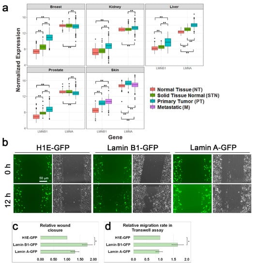
Cell migration requires reposition and reshaping of the cell nucleus. The nuclear lamina is highly important for migration of both primary and cancer cells. B-type lamins are important for proper migration of epicardial cells and neurons and increased lamin B to lamin A [...] Read more.
Cell migration requires reposition and reshaping of the cell nucleus. The nuclear lamina is highly important for migration of both primary and cancer cells. B-type lamins are important for proper migration of epicardial cells and neurons and increased lamin B to lamin A ratio accelerates cancer cell migration through confined spaces. Moreover, a positive association between lamin B1 levels and tumor formation and progression is found in various cancer types. Still, the molecular mechanism by which B-type lamins promote cell migration is not fully understood. To better understand this mechanism, we tested the effects of lamin B1 on perinuclear actin organization. Here we show that induction of melanoma cell migration leads to the formation of a cytosolic Linker of Nucleoskeleton and Cytoskeleton (LINC) complex-independent perinuclear actin rim, which has not been detected in migrating cells, yet. Significantly, increasing the levels of lamin B1 but not the levels of lamin A prevented perinuclear actin rim formation while accelerated the cellular migration rate. To interfere with the perinuclear actin rim, we generated a chimeric protein that is localized to the outer nuclear membrane and cleaves perinuclear actin filaments in a specific manner without disrupting other cytosolic actin filaments. Using this tool, we found that disruption of the perinuclear actin rim accelerated the cellular migration rate in a similar manner to lamin B1 over-expression. Taken together, our results suggest that increased lamin B1 levels can accelerate cell migration by inhibiting the association of the nuclear envelope with actin filaments that may reduce nuclear movement and deformability. Full article
Show Figures

Figure 1

10 pages, 1687 KiB  
Article
ZMPSTE24 Is Associated with Elevated Inflammation and Progerin mRNA
by Moritz Messner, Santhosh Kumar Ghadge, Thomas Maurer, Michael Graber, Simon Staggl, Sarah Christine Maier, Gerhard Pölzl and Marc-Michael Zaruba
Cells 2020, 9(9), 1981; https://doi.org/10.3390/cells9091981 - 28 Aug 2020
Cited by 6 | Viewed by 2907
Abstract
Lamins are important filaments forming the inner nuclear membrane. Lamin A is processed by zinc metalloproteinase (ZMPSTE24). Failure to cleave a truncated form of prelamin A—also called progerin—causes Hutchinson–Gilford progeria syndrome a well-known premature aging disease. Minor levels of progerin are readily expressed [...] Read more.
Lamins are important filaments forming the inner nuclear membrane. Lamin A is processed by zinc metalloproteinase (ZMPSTE24). Failure to cleave a truncated form of prelamin A—also called progerin—causes Hutchinson–Gilford progeria syndrome a well-known premature aging disease. Minor levels of progerin are readily expressed in the blood of healthy individuals due to alternative splicing. Previously, we found an association of increased progerin mRNA with overweight and chronic inflammation (hs-CRP). Here, we aimed to elucidate correlations of ZMPSTE24, lamin A/C and progerin with the inflammatory marker hs-CRP. In this retrospective, cross-sectional study we analyzed blood samples from 110 heart failure patients for quantitative mRNA expression of ZMPSTE24, lamin A/C, progerin and hs-CRP protein. Spearman correlations and linear regression analyses including adjustments for age, gender and ejection fraction showed a significant positive correlation of lnprogerin with lnZMPSTE24 (n = 110; r = 0.33; p = 0.0004) and lnlamin A/C (n = 110; r = 0.82, p < 0.0001), whereas no association was observed between lnlamin A/C and lnZMPSTE24 expression. Further analyses showed a significant positive correlation of lnhs-CRP with lnZMPSTE24 (n = 110; r = 0.21; p = 0.01) and lnlamin A/C (n = 110; r = 0.24; p = 0.03). We conclude that chronic inflammation is associated with increased expression of ZMPSTE24 and lamin A/C mRNA. Both markers also positively correlate with increased expression of the premature aging marker progerin which may be linked to cardiovascular aging. Full article
Show Figures

Figure 1

22 pages, 2398 KiB  
Review
Structural and Mechanical Aberrations of the Nuclear Lamina in Disease
by Merel Stiekema, Marc A. M. J. van Zandvoort, Frans C. S. Ramaekers and Jos L. V. Broers
Cells 2020, 9(8), 1884; https://doi.org/10.3390/cells9081884 - 11 Aug 2020
Cited by 28 | Viewed by 6849
Abstract
The nuclear lamins are the major components of the nuclear lamina in the nuclear envelope. Lamins are involved in numerous functions, including a role in providing structural support to the cell and the mechanosensing of the cell. Mutations in the genes encoding for [...] Read more.
The nuclear lamins are the major components of the nuclear lamina in the nuclear envelope. Lamins are involved in numerous functions, including a role in providing structural support to the cell and the mechanosensing of the cell. Mutations in the genes encoding for lamins lead to the rare diseases termed laminopathies. However, not only laminopathies show alterations in the nuclear lamina. Deregulation of lamin expression is reported in multiple cancers and several viral infections lead to a disrupted nuclear lamina. The structural and mechanical effects of alterations in the nuclear lamina can partly explain the phenotypes seen in disease, such as muscular weakness in certain laminopathies and transmigration of cancer cells. However, a lot of answers to questions about the relation between changes in the nuclear lamina and disease development remain elusive. Here, we review the current understandings of the contribution of the nuclear lamina in the structural support and mechanosensing of healthy and diseased cells. Full article
Show Figures

Figure 1

14 pages, 2762 KiB  
Article
In Vivo Assembly of a Dictyostelium Lamin Mutant Induced by Light, Mechanical Stress, and pH
by Marianne Grafe, Phillip Hofmann, Petros Batsios, Irene Meyer and Ralph Gräf
Cells 2020, 9(8), 1834; https://doi.org/10.3390/cells9081834 - 4 Aug 2020
Cited by 4 | Viewed by 2501
Abstract
We expressed Dictyostelium lamin (NE81) lacking both a functional nuclear localization signal and a CAAX-box for C-terminal lipid modification. This lamin mutant assembled into supramolecular, three-dimensional clusters in the cytosol that disassembled at the onset of mitosis and re-assembled in late telophase, thus [...] Read more.
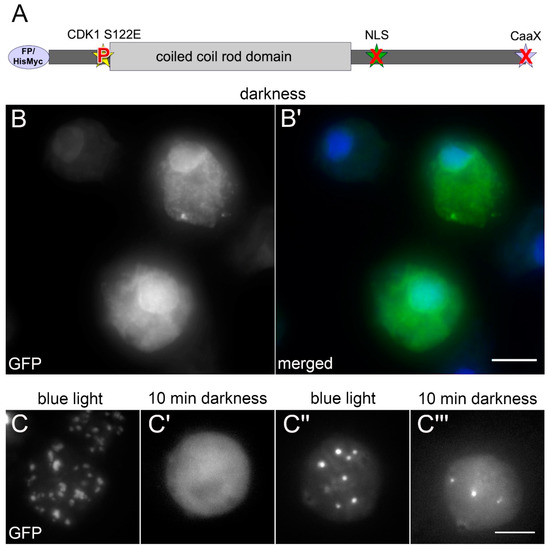
We expressed Dictyostelium lamin (NE81) lacking both a functional nuclear localization signal and a CAAX-box for C-terminal lipid modification. This lamin mutant assembled into supramolecular, three-dimensional clusters in the cytosol that disassembled at the onset of mitosis and re-assembled in late telophase, thus mimicking the behavior of the endogenous protein. As disassembly is regulated by CDK1-mediated phosphorylation at serine 122, we generated a phosphomimetic S122E mutant called GFP-NE81-S122E-ΔNLSΔCLIM. Surprisingly, during imaging, the fusion protein assembled into cytosolic clusters, similar to the protein lacking the phosphomimetic mutation. Clusters disassembled again in the darkness. Assembly could be induced with blue but not green or near ultraviolet light, and it was independent of the fusion tag. Assembly similarly occurred upon cell flattening. Earlier reports and own observations suggested that both blue light and cell flattening could result in a decrease of intracellular pH. Indeed, keeping the cells at low pH also reversibly induced cluster formation. Our results indicate that lamin assembly can be induced by various stress factors and that these are transduced via intracellular acidification. Although these effects have been shown in a phosphomimetic CDK1 mutant of the Dictyostelium lamin, they are likely relevant also for wild-type lamin. Full article
Show Figures

Figure 1

24 pages, 915 KiB  
Review
Modeling of Cell Nuclear Mechanics: Classes, Components, and Applications
by Chad M. Hobson and Andrew D. Stephens
Cells 2020, 9(7), 1623; https://doi.org/10.3390/cells9071623 - 6 Jul 2020
Cited by 17 | Viewed by 4926
Abstract
Cell nuclei are paramount for both cellular function and mechanical stability. These two roles of nuclei are intertwined as altered mechanical properties of nuclei are associated with altered cell behavior and disease. To further understand the mechanical properties of cell nuclei and guide [...] Read more.
Cell nuclei are paramount for both cellular function and mechanical stability. These two roles of nuclei are intertwined as altered mechanical properties of nuclei are associated with altered cell behavior and disease. To further understand the mechanical properties of cell nuclei and guide future experiments, many investigators have turned to mechanical modeling. Here, we provide a comprehensive review of mechanical modeling of cell nuclei with an emphasis on the role of the nuclear lamina in hopes of spurring future growth of this field. The goal of this review is to provide an introduction to mechanical modeling techniques, highlight current applications to nuclear mechanics, and give insight into future directions of mechanical modeling. There are three main classes of mechanical models—schematic, continuum mechanics, and molecular dynamics—which provide unique advantages and limitations. Current experimental understanding of the roles of the cytoskeleton, the nuclear lamina, and the chromatin in nuclear mechanics provide the basis for how each component is subsequently treated in mechanical models. Modeling allows us to interpret assay-specific experimental results for key parameters and quantitatively predict emergent behaviors. This is specifically powerful when emergent phenomena, such as lamin-based strain stiffening, can be deduced from complimentary experimental techniques. Modeling differences in force application, geometry, or composition can additionally clarify seemingly conflicting experimental results. Using these approaches, mechanical models have informed our understanding of relevant biological processes such as migration, nuclear blebbing, nuclear rupture, and cell spreading and detachment. There remain many aspects of nuclear mechanics for which additional mechanical modeling could provide immediate insight. Although mechanical modeling of cell nuclei has been employed for over a decade, there are still relatively few models for any given biological phenomenon. This implies that an influx of research into this realm of the field has the potential to dramatically shape both future experiments and our current understanding of nuclear mechanics, function, and disease. Full article
Show Figures

Figure 1

14 pages, 1083 KiB  
Article
Cytokine Profile in Striated Muscle Laminopathies: New Promising Biomarkers for Disease Prediction
by Cristina Cappelletti, Irene Tramacere, Paola Cavalcante, Elisa Schena, Luisa Politano, Nicola Carboni, Alessandra Gambineri, Adele D’Amico, Lucia Ruggiero, Giulia Ricci, Gabriele Siciliano, Giuseppe Boriani, Tiziana Enrica Mongini, Liliana Vercelli, Elena Biagini, Matteo Ziacchi, Maria Rosaria D’Apice, Giovanna Lattanzi, Renato Mantegazza, Lorenzo Maggi and Pia Bernasconiadd Show full author list remove Hide full author list
Cells 2020, 9(6), 1532; https://doi.org/10.3390/cells9061532 - 23 Jun 2020
Cited by 7 | Viewed by 3315
Abstract
Laminopathies are a wide and heterogeneous group of rare human diseases caused by mutations of the LMNA gene or related nuclear envelope genes. The variety of clinical phenotypes and the wide spectrum of histopathological changes among patients carrying an identical mutation in the [...] Read more.
Laminopathies are a wide and heterogeneous group of rare human diseases caused by mutations of the LMNA gene or related nuclear envelope genes. The variety of clinical phenotypes and the wide spectrum of histopathological changes among patients carrying an identical mutation in the LMNA gene make the prognostic process rather difficult, and classical genetic screens appear to have limited predictive value for disease development. The aim of this study was to evaluate whether a comprehensive profile of circulating cytokines may be a useful tool to differentiate and stratify disease subgroups, support clinical follow-ups and contribute to new therapeutic approaches. Serum levels of 51 pro- and anti-inflammatory molecules, including cytokines, chemokines and growth factors, were quantified by a Luminex multiple immune-assay in 53 patients with muscular laminopathy (Musc-LMNA), 10 with non-muscular laminopathy, 22 with other muscular disorders and in 35 healthy controls. Interleukin-17 (IL-17), granulocyte colony-stimulating factor (G-CSF) and transforming growth factor beta (TGF-β2) levels significantly discriminated Musc-LMNA from controls; interleukin-1β (IL-1β), interleukin-4 (IL-4) and interleukin-8 (IL-8) were differentially expressed in Musc-LMNA patients compared to those with non-muscular laminopathies, whereas IL-17 was significantly higher in Musc-LMNA patients with muscular and cardiac involvement. These findings support the hypothesis of a key role of the immune system in Musc-LMNA and emphasize the potential use of cytokines as biomarkers for these disorders. Full article
Show Figures

Figure 1

24 pages, 6066 KiB  
Article
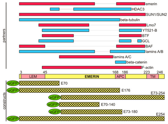
EDMD-Causing Emerin Mutant Myogenic Progenitors Exhibit Impaired Differentiation Using Similar Mechanisms
by Ashvin Iyer and James M. Holaska
Cells 2020, 9(6), 1463; https://doi.org/10.3390/cells9061463 - 15 Jun 2020
Cited by 5 | Viewed by 3436
Abstract
Mutations in the gene encoding emerin (EMD) cause Emery–Dreifuss muscular dystrophy (EDMD1), an inherited disorder characterized by progressive skeletal muscle wasting, irregular heart rhythms and contractures of major tendons. The skeletal muscle defects seen in EDMD are caused by failure of [...] Read more.
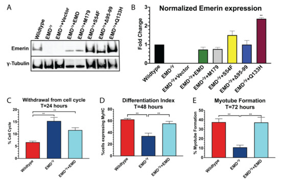
Mutations in the gene encoding emerin (EMD) cause Emery–Dreifuss muscular dystrophy (EDMD1), an inherited disorder characterized by progressive skeletal muscle wasting, irregular heart rhythms and contractures of major tendons. The skeletal muscle defects seen in EDMD are caused by failure of muscle stem cells to differentiate and regenerate the damaged muscle. However, the underlying mechanisms remain poorly understood. Most EDMD1 patients harbor nonsense mutations and have no detectable emerin protein. There are three EDMD-causing emerin mutants (S54F, Q133H, and Δ95–99) that localize correctly to the nuclear envelope and are expressed at wildtype levels. We hypothesized these emerin mutants would share in the disruption of key molecular pathways involved in myogenic differentiation. We generated myogenic progenitors expressing wildtype emerin and each EDMD1-causing emerin mutation (S54F, Q133H, Δ95–99) in an emerin-null (EMD−/y) background. S54F, Q133H, and Δ95–99 failed to rescue EMD−/y myogenic differentiation, while wildtype emerin efficiently rescued differentiation. RNA sequencing was done to identify pathways and networks important for emerin regulation of myogenic differentiation. This analysis significantly reduced the number of pathways implicated in EDMD1 muscle pathogenesis. Full article
Show Figures

Figure 1

13 pages, 1945 KiB  
Review
Targeted Regulation of Nuclear Lamins by Ubiquitin and Ubiquitin-Like Modifiers
by Michael Blank
Cells 2020, 9(6), 1340; https://doi.org/10.3390/cells9061340 - 27 May 2020
Cited by 5 | Viewed by 4269
Abstract
Nuclear lamins (NLs) are essential components of the animal cell nucleus involved in the regulation of a plethora of molecular and cellular processes. These include the nuclear envelope assembly and stability, mechanotransduction and chromatin organization, transcription, DNA replication, damage repair, and genomic integrity [...] Read more.
Nuclear lamins (NLs) are essential components of the animal cell nucleus involved in the regulation of a plethora of molecular and cellular processes. These include the nuclear envelope assembly and stability, mechanotransduction and chromatin organization, transcription, DNA replication, damage repair, and genomic integrity maintenance. Mutations in NLs can lead to the development of a wide range of distinct disease phenotypes, laminopathies, consisting of cardiac, neuromuscular, metabolic and premature aging syndromes. In addition, alterations in the expression of nuclear lamins were associated with different types of neoplastic diseases. Despite the importance and critical roles that NLs play in the diverse cellular activities, we only recently started to uncover the complexity of regulatory mechanisms governing their expression, localization and functions. This integrative review summarizes and discusses the recent findings on the emerging roles of ubiquitin and ubiquitin-like modifiers (ULMs) in the regulation of NLs, highlighting the intriguing molecular associations and cross-talks occurring between NLs and these regulatory molecules under physiological conditions and in the disease states. Full article
Show Figures

Figure 1

25 pages, 2156 KiB  
Review
Crucial Role of Lamin A/C in the Migration and Differentiation of MSCs in Bone
by Natividad Alcorta-Sevillano, Iratxe Macías, Clara I. Rodríguez and Arantza Infante
Cells 2020, 9(6), 1330; https://doi.org/10.3390/cells9061330 - 26 May 2020
Cited by 30 | Viewed by 5741
Abstract
Lamin A/C, intermediate filament proteins from the nuclear lamina encoded by the LMNA gene, play a central role in mediating the mechanosignaling of cytoskeletal forces into nucleus. In fact, this mechanotransduction process is essential to ensure the proper functioning of other tasks also [...] Read more.
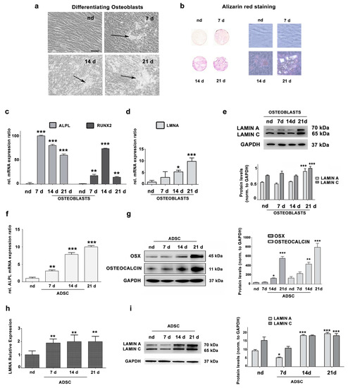
Lamin A/C, intermediate filament proteins from the nuclear lamina encoded by the LMNA gene, play a central role in mediating the mechanosignaling of cytoskeletal forces into nucleus. In fact, this mechanotransduction process is essential to ensure the proper functioning of other tasks also mediated by lamin A/C: the structural support of the nucleus and the regulation of gene expression. In this way, lamin A/C is fundamental for the migration and differentiation of mesenchymal stem cells (MSCs), the progenitors of osteoblasts, thus affecting bone homeostasis. Bone formation is a complex process regulated by chemical and mechanical cues, coming from the surrounding extracellular matrix. MSCs respond to signals modulating the expression levels of lamin A/C, and therefore, adapting their nuclear shape and stiffness. To promote cell migration, MSCs need soft nuclei with low lamin A content. Conversely, during osteogenic differentiation, lamin A/C levels are known to be increased. Several LMNA mutations present a negative impact in the migration and osteogenesis of MSCs, affecting bone tissue homeostasis and leading to pathological conditions. This review aims to describe these concepts by discussing the latest state-of-the-art in this exciting area, focusing on the relationship between lamin A/C in MSCs’ function and bone tissue from both, health and pathological points of view. Full article
Show Figures

Figure 1

33 pages, 3823 KiB  
Review
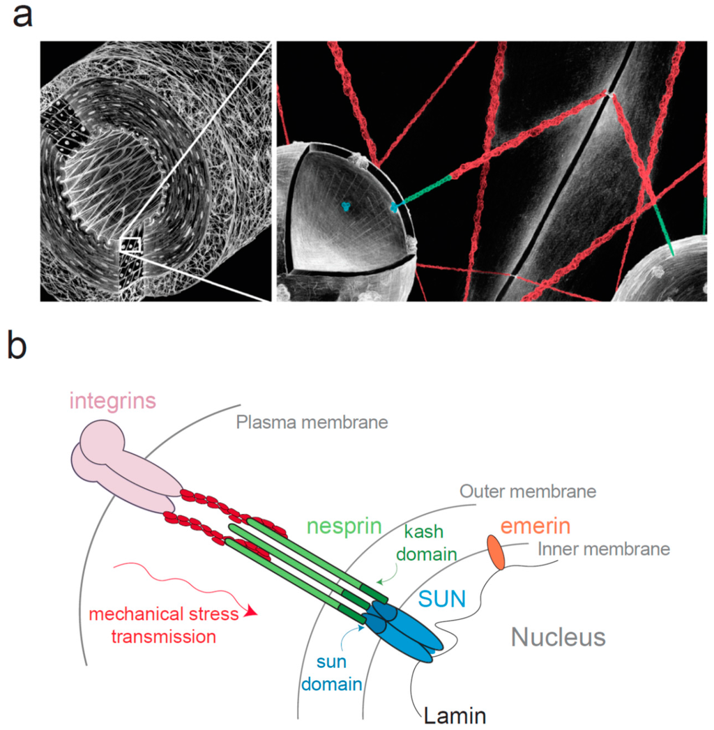
Lamin A/C Mechanotransduction in Laminopathies
by Francesca Donnaloja, Federica Carnevali, Emanuela Jacchetti and Manuela Teresa Raimondi
Cells 2020, 9(5), 1306; https://doi.org/10.3390/cells9051306 - 24 May 2020
Cited by 44 | Viewed by 9804
Abstract
Mechanotransduction translates forces into biological responses and regulates cell functionalities. It is implicated in several diseases, including laminopathies which are pathologies associated with mutations in lamins and lamin-associated proteins. These pathologies affect muscle, adipose, bone, nerve, and skin cells and range from muscular [...] Read more.
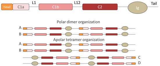
Mechanotransduction translates forces into biological responses and regulates cell functionalities. It is implicated in several diseases, including laminopathies which are pathologies associated with mutations in lamins and lamin-associated proteins. These pathologies affect muscle, adipose, bone, nerve, and skin cells and range from muscular dystrophies to accelerated aging. Although the exact mechanisms governing laminopathies and gene expression are still not clear, a strong correlation has been found between cell functionality and nuclear behavior. New theories base on the direct effect of external force on the genome, which is indeed sensitive to the force transduced by the nuclear lamina. Nuclear lamina performs two essential functions in mechanotransduction pathway modulating the nuclear stiffness and governing the chromatin remodeling. Indeed, A-type lamin mutation and deregulation has been found to affect the nuclear response, altering several downstream cellular processes such as mitosis, chromatin organization, DNA replication-transcription, and nuclear structural integrity. In this review, we summarize the recent findings on the molecular composition and architecture of the nuclear lamina, its role in healthy cells and disease regulation. We focus on A-type lamins since this protein family is the most involved in mechanotransduction and laminopathies. Full article
Show Figures

Figure 1

16 pages, 8139 KiB  
Article
Progerin Expression Induces Inflammation, Oxidative Stress and Senescence in Human Coronary Endothelial Cells
by Guillaume Bidault, Marie Garcia, Jacqueline Capeau, Romain Morichon, Corinne Vigouroux and Véronique Béréziat
Cells 2020, 9(5), 1201; https://doi.org/10.3390/cells9051201 - 12 May 2020
Cited by 44 | Viewed by 5720
Abstract
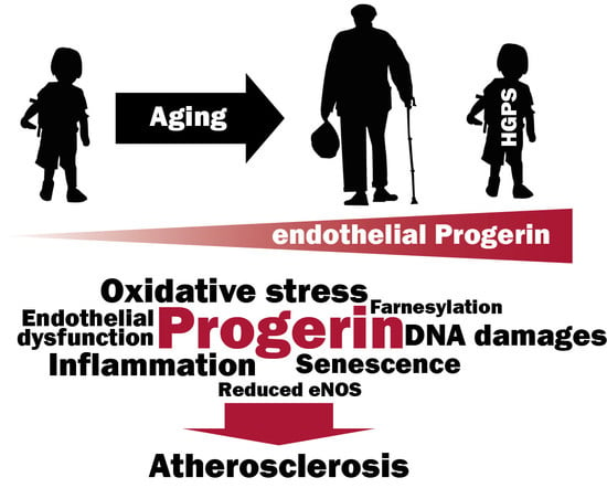
Hutchinson–Gilford progeria syndrome (HGPS) is a rare premature aging disorder notably characterized by precocious and deadly atherosclerosis. Almost 90% of HGPS patients carry a LMNA p.G608G splice variant that leads to the expression of a permanently farnesylated abnormal form of prelamin-A, referred to [...] Read more.
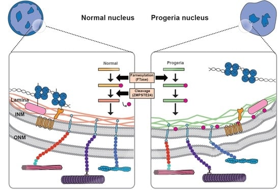
Hutchinson–Gilford progeria syndrome (HGPS) is a rare premature aging disorder notably characterized by precocious and deadly atherosclerosis. Almost 90% of HGPS patients carry a LMNA p.G608G splice variant that leads to the expression of a permanently farnesylated abnormal form of prelamin-A, referred to as progerin. Endothelial dysfunction is a key determinant of atherosclerosis, notably during aging. Previous studies have shown that progerin accumulates in HGPS patients’ endothelial cells but also during vascular physiological aging. However, whether progerin expression in human endothelial cells can recapitulate features of endothelial dysfunction is currently unknown. Herein, we evaluated the direct impact of exogenously expressed progerin and wild-type lamin-A on human endothelial cell function and senescence. Our data demonstrate that progerin, but not wild-type lamin-A, overexpression induces endothelial cell dysfunction, characterized by increased inflammation and oxidative stress together with persistent DNA damage, increased cell cycle arrest protein expression and cellular senescence. Inhibition of progerin prenylation using a pravastatin–zoledronate combination partly prevents these defects. Our data suggest a direct proatherogenic role of progerin in human endothelial cells, which could contribute to HGPS-associated early atherosclerosis and also potentially be involved in physiological endothelial aging participating to age-related cardiometabolic diseases. Full article
Show Figures

Graphical abstract

19 pages, 3162 KiB  
Article
Spatiotemporal Mislocalization of Nuclear Membrane-Associated Proteins in γ-Irradiation-Induced Senescent Cells
by Alena Svobodová Kovaříková, Eva Bártová, Aleš Kovařík and Emilie Lukášová
Cells 2020, 9(4), 999; https://doi.org/10.3390/cells9040999 - 17 Apr 2020
Cited by 3 | Viewed by 3519
Abstract
Cellular senescence, induced by genotoxic or replication stress, is accompanied by defects in nuclear morphology and nuclear membrane-heterochromatin disruption. In this work, we analyzed cytological and molecular changes in the linker of nucleoskeleton and cytoskeleton (LINC) complex proteins in senescence triggered by γ-irradiation. [...] Read more.
Cellular senescence, induced by genotoxic or replication stress, is accompanied by defects in nuclear morphology and nuclear membrane-heterochromatin disruption. In this work, we analyzed cytological and molecular changes in the linker of nucleoskeleton and cytoskeleton (LINC) complex proteins in senescence triggered by γ-irradiation. We used human mammary carcinoma and osteosarcoma cell lines, both original and shRNA knockdown clones targeting lamin B receptor (LBR) and leading to LBR and lamin B (LB1) reduction. The expression status and integrity of LINC complex proteins (nesprin-1, SUN1, SUN2), lamin A/C, and emerin were analyzed by immunodetection using confocal microscopy and Western blot. The results show frequent mislocalization of these proteins from the nuclear membrane to cytoplasm and micronuclei and, in some cases, their fragmentation and amplification. The timing of these changes clearly preceded the onset of senescence. The LBR deficiency triggered neither senescence nor changes in the LINC protein distribution before irradiation. However, the cytological changes following irradiation were more pronounced in shRNA knockdown cells compared to original cell lines. We conclude that mislocalization of LINC complex proteins is a significant characteristic of cellular senescence phenotypes and may influence complex events at the nuclear membrane, including trafficking and heterochromatin attachment. Full article
Show Figures

Figure 1

15 pages, 3407 KiB  
Article
Lamin A and Prelamin A Counteract Migration of Osteosarcoma Cells
by Camilla Evangelisti, Francesca Paganelli, Gaia Giuntini, Elisabetta Mattioli, Alessandra Cappellini, Giulia Ramazzotti, Irene Faenza, Maria Cristina Maltarello, Alberto M. Martelli, Katia Scotlandi, Francesca Chiarini and Giovanna Lattanzi
Cells 2020, 9(3), 774; https://doi.org/10.3390/cells9030774 - 22 Mar 2020
Cited by 15 | Viewed by 4251
Abstract
A type lamins are fundamental components of the nuclear lamina. Changes in lamin A expression correlate with malignant transformation in several cancers. However, the role of lamin A has not been explored in osteosarcoma (OS). Here, we wanted to investigate the role of [...] Read more.
A type lamins are fundamental components of the nuclear lamina. Changes in lamin A expression correlate with malignant transformation in several cancers. However, the role of lamin A has not been explored in osteosarcoma (OS). Here, we wanted to investigate the role of lamin A in normal osteoblasts (OBs) and OS cells. Thus, we studied the expression of lamin A/C in OS cells compared to OBs and evaluated the effects of lamin A overexpression in OS cell lines. We show that, while lamin A expression increases during osteoblast differentiation, all examined OS cell lines express lower lamin A levels relative to differentiated OBs. The condition of low LMNA expression confers to OS cells a significant increase in migration potential, while overexpression of lamin A reduces migration ability of OS cells. Moreover, overexpression of unprocessable prelamin A also reduces cell migration. In agreement with the latter finding, OS cells which accumulate the highest prelamin A levels upon inhibition of lamin A maturation by statins, had significantly reduced migration ability. Importantly, OS cells subjected to statin treatment underwent apoptotic cell death in a RAS-independent, lamin A-dependent manner. Our results show that pro-apoptotic effects of statins and statin inhibitory effect on OS cell migration are comparable to those obtained by prelamin A accumulation and further suggest that modulation of lamin A expression and post-translational processing can be a tool to decrease migration potential in OS cells. Full article
Show Figures

Figure 1

16 pages, 9331 KiB  
Article
Silencing of Euchromatic Transposable Elements as a Consequence of Nuclear Lamina Dysfunction
by Valeria Cavaliere, Giovanna Lattanzi and Davide Andrenacci
Cells 2020, 9(3), 625; https://doi.org/10.3390/cells9030625 - 5 Mar 2020
Cited by 6 | Viewed by 3631
Abstract
Transposable elements (TEs) are mobile genomic sequences that are normally repressed to avoid proliferation and genome instability. Gene silencing mechanisms repress TEs by RNA degradation or heterochromatin formation. Heterochromatin maintenance is therefore important to keep TEs silent. Loss of heterochromatic domains has been [...] Read more.
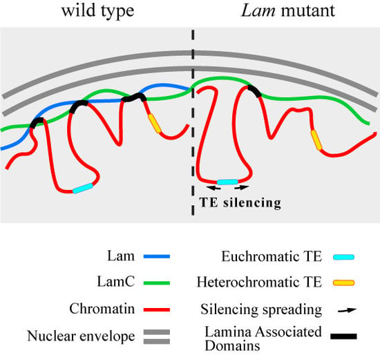
Transposable elements (TEs) are mobile genomic sequences that are normally repressed to avoid proliferation and genome instability. Gene silencing mechanisms repress TEs by RNA degradation or heterochromatin formation. Heterochromatin maintenance is therefore important to keep TEs silent. Loss of heterochromatic domains has been linked to lamin mutations, which have also been associated with derepression of TEs. In fact, lamins are structural components of the nuclear lamina (NL), which is considered a pivotal structure in the maintenance of heterochromatin domains at the nuclear periphery in a silent state. Here, we show that a lethal phenotype associated with Lamin loss-of-function mutations is influenced by Drosophila gypsy retrotransposons located in euchromatic regions, suggesting that NL dysfunction has also effects on active TEs located in euchromatic loci. In fact, expression analysis of different long terminal repeat (LTR) retrotransposons and of one non-LTR retrotransposon located near active genes shows that Lamin inactivation determines the silencing of euchromatic TEs. Furthermore, we show that the silencing effect on euchromatic TEs spreads to the neighboring genomic regions, with a repressive effect on nearby genes. We propose that NL dysfunction may have opposed regulatory effects on TEs that depend on their localization in active or repressed regions of the genome. Full article
Show Figures

Graphical abstract

17 pages, 2028 KiB  
Article
Comparative Interactome Analysis of Emerin, MAN1 and LEM2 Reveals a Unique Role for LEM2 in Nucleotide Excision Repair
by Bernhard Moser, José Basílio, Josef Gotzmann, Andreas Brachner and Roland Foisner
Cells 2020, 9(2), 463; https://doi.org/10.3390/cells9020463 - 18 Feb 2020
Cited by 15 | Viewed by 4862
Abstract
LAP2-Emerin-MAN1 (LEM) domain-containing proteins represent an abundant group of inner nuclear membrane proteins involved in diverse nuclear functions, but their functional redundancies remain unclear. Here, using the biotinylation-dependent proximity approach, we report proteome-wide comparative interactome analysis of the two structurally related LEM proteins [...] Read more.
LAP2-Emerin-MAN1 (LEM) domain-containing proteins represent an abundant group of inner nuclear membrane proteins involved in diverse nuclear functions, but their functional redundancies remain unclear. Here, using the biotinylation-dependent proximity approach, we report proteome-wide comparative interactome analysis of the two structurally related LEM proteins MAN1 (LEMD3) and LEM2 (LEMD2), and the more distantly related emerin (EMD). While over 60% of the relatively small group of MAN1 and emerin interactors were also found in the LEM2 interactome, the latter included a large number of candidates (>85%) unique for LEM2. The interacting partners unique for emerin support and provide further insight into the previously reported role of emerin in centrosome positioning, and the MAN1-specific interactors suggest a role of MAN1 in ribonucleoprotein complex assembly. Interestingly, the LEM2-specific interactome contained several proteins of the nucleotide excision repair pathway. Accordingly, LEM2-depleted cells, but not MAN1- and emerin-depleted cells, showed impaired proliferation following ultraviolet-C (UV-C) irradiation and prolonged accumulation of γH2AX, similar to cells deficient in the nucleotide excision repair protein DNA damage-binding protein 1 (DDB1). These findings indicate impaired DNA damage repair in LEM2-depleted cells. Overall, this interactome study identifies new potential interaction partners of emerin, MAN1 and particularly LEM2, and describes a novel potential involvement of LEM2 in nucleotide excision repair at the nuclear periphery. Full article
Show Figures

Figure 1

13 pages, 3516 KiB  
Article
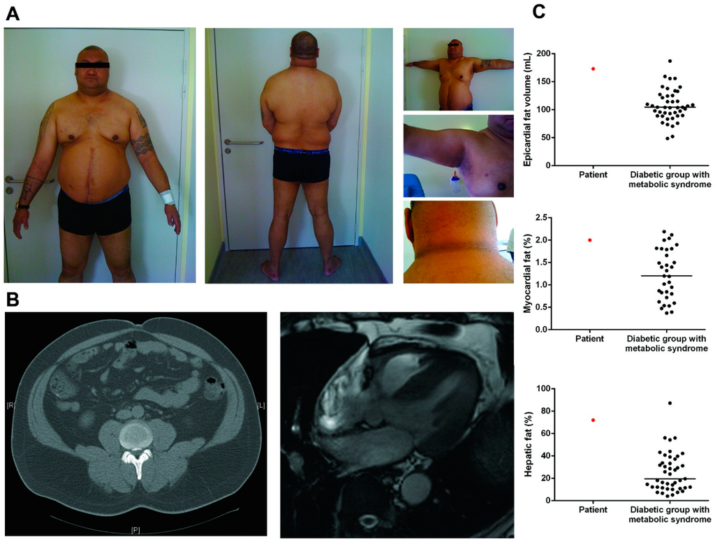
Unraveling LMNA Mutations in Metabolic Syndrome: Cellular Phenotype and Clinical Pitfalls
by Camille Desgrouas, Alice-Anaïs Varlet, Anne Dutour, Damien Galant, Françoise Merono, Nathalie Bonello-Palot, Patrice Bourgeois, Adèle Lasbleiz, Cathy Petitjean, Patricia Ancel, Nicolas Levy, Catherine Badens and Bénédicte Gaborit
Cells 2020, 9(2), 310; https://doi.org/10.3390/cells9020310 - 28 Jan 2020
Cited by 13 | Viewed by 3464
Abstract
This study details the clinical and cellular phenotypes associated with two missense heterozygous mutations in LMNA, c.1745G>T p.(Arg582Leu), and c.1892G>A p.(Gly631Asp), in two patients with early onset of diabetes mellitus, hypertriglyceridemia and non-alcoholic fatty liver disease. In these two patients, subcutaneous adipose [...] Read more.
This study details the clinical and cellular phenotypes associated with two missense heterozygous mutations in LMNA, c.1745G>T p.(Arg582Leu), and c.1892G>A p.(Gly631Asp), in two patients with early onset of diabetes mellitus, hypertriglyceridemia and non-alcoholic fatty liver disease. In these two patients, subcutaneous adipose tissue was persistent, at least on the abdomen, and the serum leptin level remained in the normal range. Cellular studies showed elevated nuclear anomalies, an accelerated senescence rate and a decrease of replication capacity in patient cells. In cellular models, the overexpression of mutated prelamin A phenocopied misshapen nuclei, while the partial reduction of lamin A expression in patient cells significantly improved nuclear morphology. Altogether, these results suggest a link between lamin A mutant expression and senescence associated phenotypes. Transcriptome analysis of the whole subcutaneous adipose tissue from the two patients and three controls, paired for age and sex using RNA sequencing, showed the up regulation of genes implicated in immunity and the down regulation of genes involved in development and cell differentiation in patient adipose tissue. Therefore, our results suggest that some mutations in LMNA are associated with severe metabolic phenotypes without subcutaneous lipoatrophy, and are associated with nuclear misshaping. Full article
Show Figures

Figure 1

17 pages, 3535 KiB  
Article
SUN1/2 Are Essential for RhoA/ROCK-Regulated Actomyosin Activity in Isolated Vascular Smooth Muscle Cells
by Lauren Porter, Rose-Marie Minaisah, Sultan Ahmed, Seema Ali, Rosemary Norton, Qiuping Zhang, Elisa Ferraro, Chris Molenaar, Mark Holt, Susan Cox, Samuel Fountain, Catherine Shanahan and Derek Warren
Cells 2020, 9(1), 132; https://doi.org/10.3390/cells9010132 - 6 Jan 2020
Cited by 19 | Viewed by 5947
Abstract
Vascular smooth muscle cells (VSMCs) are the predominant cell type in the blood vessel wall. Changes in VSMC actomyosin activity and morphology are prevalent in cardiovascular disease. The actin cytoskeleton actively defines cellular shape and the LInker of Nucleoskeleton and Cytoskeleton (LINC) complex, [...] Read more.
Vascular smooth muscle cells (VSMCs) are the predominant cell type in the blood vessel wall. Changes in VSMC actomyosin activity and morphology are prevalent in cardiovascular disease. The actin cytoskeleton actively defines cellular shape and the LInker of Nucleoskeleton and Cytoskeleton (LINC) complex, comprised of nesprin and the Sad1p, UNC-84 (SUN)-domain family members SUN1/2, has emerged as a key regulator of actin cytoskeletal organisation. Although SUN1 and SUN2 function is partially redundant, they possess specific functions and LINC complex composition is tailored for cell-type-specific functions. We investigated the importance of SUN1 and SUN2 in regulating actomyosin activity and cell morphology in VSMCs. We demonstrate that siRNA-mediated depletion of either SUN1 or SUN2 altered VSMC spreading and impaired actomyosin activity and RhoA activity. Importantly, these findings were recapitulated using aortic VSMCs isolated from wild-type and SUN2 knockout (SUN2 KO) mice. Inhibition of actomyosin activity, using the rho-associated, coiled-coil-containing protein kinase1/2 (ROCK1/2) inhibitor Y27632 or blebbistatin, reduced SUN2 mobility in the nuclear envelope and decreased the association between SUN2 and lamin A, confirming that SUN2 dynamics and interactions are influenced by actomyosin activity. We propose that the LINC complex exists in a mechanical feedback circuit with RhoA to regulate VSMC actomyosin activity and morphology. Full article
Show Figures

Figure 1

2019

Jump to: 2024, 2023, 2022, 2021, 2020, 2018, 2017, 2016

27 pages, 723 KiB  
Review
Cellular and Animal Models of Striated Muscle Laminopathies
by Hannah A. Nicolas, Marie-Andrée Akimenko and Frédérique Tesson
Cells 2019, 8(4), 291; https://doi.org/10.3390/cells8040291 - 29 Mar 2019
Cited by 5 | Viewed by 5317
Abstract
The lamin A/C (LMNA) gene codes for nuclear intermediate filaments constitutive of the nuclear lamina. LMNA has 12 exons and alternative splicing of exon 10 results in two major isoforms—lamins A and C. Mutations found throughout the LMNA gene cause a [...] Read more.
The lamin A/C (LMNA) gene codes for nuclear intermediate filaments constitutive of the nuclear lamina. LMNA has 12 exons and alternative splicing of exon 10 results in two major isoforms—lamins A and C. Mutations found throughout the LMNA gene cause a group of diseases collectively known as laminopathies, of which the type, diversity, penetrance and severity of phenotypes can vary from one individual to the other, even between individuals carrying the same mutation. The majority of the laminopathies affect cardiac and/or skeletal muscles. The underlying molecular mechanisms contributing to such tissue-specific phenotypes caused by mutations in a ubiquitously expressed gene are not yet well elucidated. This review will explore the different phenotypes observed in established models of striated muscle laminopathies and their respective contributions to advancing our understanding of cardiac and skeletal muscle-related laminopathies. Potential future directions for developing effective treatments for patients with lamin A/C mutation-associated cardiac and/or skeletal muscle conditions will be discussed. Full article
Show Figures

Figure 1

15 pages, 5098 KiB  
Review
Lamina Associated Domains and Gene Regulation in Development and Cancer
by Silke J. A. Lochs, Samy Kefalopoulou and Jop Kind
Cells 2019, 8(3), 271; https://doi.org/10.3390/cells8030271 - 21 Mar 2019
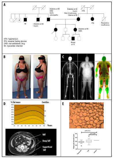
Cited by 44 | Viewed by 11565
Abstract
The nuclear lamina (NL) is a thin meshwork of filaments that lines the inner nuclear membrane, thereby providing a platform for chromatin binding and supporting genome organization. Genomic regions contacting the NL are lamina associated domains (LADs), which contain thousands of genes that [...] Read more.
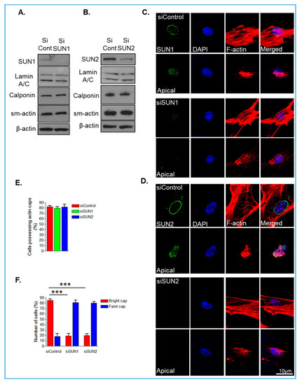
The nuclear lamina (NL) is a thin meshwork of filaments that lines the inner nuclear membrane, thereby providing a platform for chromatin binding and supporting genome organization. Genomic regions contacting the NL are lamina associated domains (LADs), which contain thousands of genes that are lowly transcribed, and enriched for repressive histone modifications. LADs are dynamic structures that shift spatial positioning in accordance with cell-type specific gene expression changes during differentiation and development. Furthermore, recent studies have linked the disruption of LADs and alterations in the epigenome with the onset of diseases such as cancer. Here we focus on the role of LADs and the NL in gene regulation during development and cancer. Full article
Show Figures

Figure 1

17 pages, 22472 KiB  
Article
Tissue-Specific Influence of Lamin A Mutations on Notch Signaling and Osteogenic Phenotype of Primary Human Mesenchymal Cells
by Kseniya Perepelina, Polina Klauzen, Anna Kostareva and Anna Malashicheva
Cells 2019, 8(3), 266; https://doi.org/10.3390/cells8030266 - 21 Mar 2019
Cited by 13 | Viewed by 4539
Abstract
Lamin A is involved in many cellular functions due to its ability to bind chromatin and transcription factors and affect their properties. Mutations of LMNA gene encoding lamin A affect the differentiation capacity of stem cells, but the mechanisms of this influence remain [...] Read more.
Lamin A is involved in many cellular functions due to its ability to bind chromatin and transcription factors and affect their properties. Mutations of LMNA gene encoding lamin A affect the differentiation capacity of stem cells, but the mechanisms of this influence remain largely unclear. We and others have reported recently an interaction of lamin A with Notch pathway, which is among the main developmental regulators of cellular identity. The aim of this study was to explore the influence of LMNA mutations on the proosteogenic response of human cells of mesenchymal origin and to further explore the interaction of LMNA with Notch pathway. Mutations R527C and R471C in LMNA are associated with mandibuloacral dysplasia type A, a highly penetrant disease with a variety of abnormalities involving bone development. We used lentiviral constructs bearing mutations R527C and R471C and explored its influence on proosteogenic phenotype expression and Notch pathway activity in four types of human cells: umbilical vein endothelial cells (HUVEC), cardiac mesenchymal cells (HCMC), aortic smooth muscle cells (HASMC), and aortic valve interstitial cells (HAVIC). The proosteogenic response of the cells was induced by the addition of either LPS or specific effectors of osteogenic differentiation to the culture medium; phenotype was estimated by the expression of osteogenic markers by qPCR; activation of Notch was assessed by expression of Notch-related and Notch-responsive genes by qPCR and by activation of a luciferase CSL-reporter construct. Overall, we observed different reactivity of all four cell lineages to the stimulation with either LPS or osteogenic factors. R527C had a stronger influence on the proosteogenic phenotype. We observed the inhibiting action of LMNA R527C on osteogenic differentiation in HCMC in the presence of activated Notch signaling, while LMNA R527C caused the activation of osteogenic differentiation in HAVIC in the presence of activated Notch signaling. Our results suggest that the effect of a LMNA mutation is strongly dependent not only on a specific mutation itself, but also might be influenced by the intrinsic molecular context of a cell lineage. Full article
Show Figures

Graphical abstract

30 pages, 9362 KiB  
Article
Emerin Is Required for Proper Nucleus Reassembly after Mitosis: Implications for New Pathogenetic Mechanisms for Laminopathies Detected in EDMD1 Patients
by Magda Dubińska-Magiera, Katarzyna Kozioł, Magdalena Machowska, Katarzyna Piekarowicz, Daria Filipczak and Ryszard Rzepecki
Cells 2019, 8(3), 240; https://doi.org/10.3390/cells8030240 - 13 Mar 2019
Cited by 15 | Viewed by 7118
Abstract
Emerin is an essential LEM (LAP2, Emerin, MAN1) domain protein in metazoans and an integral membrane protein associated with inner and outer nuclear membranes. Mutations in the human EMD gene coding for emerin result in the rare genetic disorder: Emery–Dreifuss muscular dystrophy type [...] Read more.
Emerin is an essential LEM (LAP2, Emerin, MAN1) domain protein in metazoans and an integral membrane protein associated with inner and outer nuclear membranes. Mutations in the human EMD gene coding for emerin result in the rare genetic disorder: Emery–Dreifuss muscular dystrophy type 1 (EDMD1). This disease belongs to a broader group called laminopathies—a heterogeneous group of rare genetic disorders affecting tissues of mesodermal origin. EDMD1 phenotype is characterized by progressive muscle wasting, contractures of the elbow and Achilles tendons, and cardiac conduction defects. Emerin is involved in many cellular and intranuclear processes through interactions with several partners: lamins; barrier-to-autointegration factor (BAF), β-catenin, actin, and tubulin. Our study demonstrates the presence of the emerin fraction which associates with mitotic spindle microtubules and centrosomes during mitosis and colocalizes during early mitosis with lamin A/C, BAF, and membranes at the mitotic spindle. Transfection studies with cells expressing EGFP-emerin protein demonstrate that the emerin fusion protein fraction also localizes to centrosomes and mitotic spindle microtubules during mitosis. Transient expression of emerin deletion mutants revealed that the resulting phenotypes vary and are mutant dependent. The most frequent phenotypes include aberrant nuclear shape, tubulin network mislocalization, aberrant mitosis, and mislocalization of centrosomes. Emerin deletion mutants demonstrated different chromatin binding capacities in an in vitro nuclear assembly assay and chromatin-binding properties correlated with the strength of phenotypic alteration in transfected cells. Aberrant tubulin staining and microtubule network phenotype appearance depended on the presence of the tubulin binding region in the expressed deletion mutants. We believe that the association with tubulin might help to “deliver” emerin and associated membranes to decondensing chromatin. Preliminary analyses of cells from Polish patients with EDMD1 revealed that for several mutations thought to be null for emerin protein, a truncated emerin protein was present. We infer that the EDMD1 phenotype may be strengthened by the toxicity of truncated emerin expressed in patients with certain nonsense mutations in EMD. Full article
Show Figures

Figure 1

22 pages, 2472 KiB  
Review
Deciphering Nuclear Mechanobiology in Laminopathy
by Jungwon Hah and Dong-Hwee Kim
Cells 2019, 8(3), 231; https://doi.org/10.3390/cells8030231 - 11 Mar 2019
Cited by 27 | Viewed by 8835
Abstract
Extracellular mechanical stimuli are translated into biochemical signals inside the cell via mechanotransduction. The nucleus plays a critical role in mechanoregulation, which encompasses mechanosensing and mechanotransduction. The nuclear lamina underlying the inner nuclear membrane not only maintains the structural integrity, but also connects [...] Read more.
Extracellular mechanical stimuli are translated into biochemical signals inside the cell via mechanotransduction. The nucleus plays a critical role in mechanoregulation, which encompasses mechanosensing and mechanotransduction. The nuclear lamina underlying the inner nuclear membrane not only maintains the structural integrity, but also connects the cytoskeleton to the nuclear envelope. Lamin mutations, therefore, dysregulate the nuclear response, resulting in abnormal mechanoregulations, and ultimately, disease progression. Impaired mechanoregulations even induce malfunction in nuclear positioning, cell migration, mechanosensation, as well as differentiation. To know how to overcome laminopathies, we need to understand the mechanisms of laminopathies in a mechanobiological way. Recently, emerging studies have demonstrated the varying defects from lamin mutation in cellular homeostasis within mechanical surroundings. Therefore, this review summarizes recent findings highlighting the role of lamins, the architecture of nuclear lamina, and their disease relevance in the context of nuclear mechanobiology. We will also provide an overview of the differentiation of cellular mechanics in laminopathy. Full article
Show Figures

Graphical abstract

13 pages, 1110 KiB  
Review
The Nuclear Lamina as an Organizer of Chromosome Architecture
by Yuri Y. Shevelyov and Sergey V. Ulianov
Cells 2019, 8(2), 136; https://doi.org/10.3390/cells8020136 - 8 Feb 2019
Cited by 66 | Viewed by 13947
Abstract
The nuclear lamina (NL) is a meshwork of lamins and lamin-associated proteins adjoining the inner side of the nuclear envelope. In early embryonic cells, the NL mainly suppresses background transcription, whereas, in differentiated cell types, its disruption affects gene expression more severely. Normally, [...] Read more.
The nuclear lamina (NL) is a meshwork of lamins and lamin-associated proteins adjoining the inner side of the nuclear envelope. In early embryonic cells, the NL mainly suppresses background transcription, whereas, in differentiated cell types, its disruption affects gene expression more severely. Normally, the NL serves as a backbone for multiple chromatin anchoring sites, thus shaping the spatial organization of chromosomes in the interphase nucleus. However, upon cell senescence, aging, or in some types of terminally differentiated cells and lamin-associated diseases, the loss of NL-chromatin tethering causes drastic alterations in chromosome architecture. Here, we provide an overview of the recent advances in the field of NL-chromatin interactions, focusing on their impact on chromatin positioning, compaction, repression, and spatial organization. Full article
Show Figures

Figure 1

28 pages, 13162 KiB  
Article
Host Vesicle Fusion Protein VAPB Contributes to the Nuclear Egress Stage of Herpes Simplex Virus Type-1 (HSV-1) Replication
by Natalia Saiz-Ros, Rafal Czapiewski, Ilaria Epifano, Andrew Stevenson, Selene K. Swanson, Charles R. Dixon, Dario B. Zamora, Marion McElwee, Swetha Vijayakrishnan, Christine A. Richardson, Li Dong, David A. Kelly, Lior Pytowski, Martin W. Goldberg, Laurence Florens, Sheila V. Graham and Eric C. Schirmer
Cells 2019, 8(2), 120; https://doi.org/10.3390/cells8020120 - 3 Feb 2019
Cited by 13 | Viewed by 7566
Abstract
The primary envelopment/de-envelopment of Herpes viruses during nuclear exit is poorly understood. In Herpes simplex virus type-1 (HSV-1), proteins pUL31 and pUL34 are critical, while pUS3 and some others contribute; however, efficient membrane fusion may require additional host proteins. We postulated that vesicle [...] Read more.
The primary envelopment/de-envelopment of Herpes viruses during nuclear exit is poorly understood. In Herpes simplex virus type-1 (HSV-1), proteins pUL31 and pUL34 are critical, while pUS3 and some others contribute; however, efficient membrane fusion may require additional host proteins. We postulated that vesicle fusion proteins present in the nuclear envelope might facilitate primary envelopment and/or de-envelopment fusion with the outer nuclear membrane. Indeed, a subpopulation of vesicle-associated membrane protein-associated protein B (VAPB), a known vesicle trafficking protein, was present in the nuclear membrane co-locating with pUL34. VAPB knockdown significantly reduced both cell-associated and supernatant virus titers. Moreover, VAPB depletion reduced cytoplasmic accumulation of virus particles and increased levels of nuclear encapsidated viral DNA. These results suggest that VAPB is an important player in the exit of primary enveloped HSV-1 virions from the nucleus. Importantly, VAPB knockdown did not alter pUL34, calnexin or GM-130 localization during infection, arguing against an indirect effect of VAPB on cellular vesicles and trafficking. Immunogold-labelling electron microscopy confirmed VAPB presence in nuclear membranes and moreover associated with primary enveloped HSV-1 particles. These data suggest that VAPB could be a cellular component of a complex that facilitates UL31/UL34/US3-mediated HSV-1 nuclear egress. Full article
Show Figures

Figure 1

2018

Jump to: 2024, 2023, 2022, 2021, 2020, 2019, 2017, 2016

12 pages, 2825 KiB  
Article
Samp1 Mislocalization in Emery-Dreifuss Muscular Dystrophy
by Elisabetta Mattioli, Marta Columbaro, Mohammed Hakim Jafferali, Elisa Schena, Einar Hallberg and Giovanna Lattanzi
Cells 2018, 7(10), 170; https://doi.org/10.3390/cells7100170 - 15 Oct 2018
Cited by 8 | Viewed by 4386
Abstract
LMNA linked-Emery-Dreifuss muscular dystrophy (EDMD2) is a rare disease characterized by muscle weakness, muscle wasting, and cardiomyopathy with conduction defects. The mutated protein lamin A/C binds several nuclear envelope components including the Linker of Nucleoskeleton and Cytoskeleton (LINC) complex and the inner nuclear [...] Read more.
LMNA linked-Emery-Dreifuss muscular dystrophy (EDMD2) is a rare disease characterized by muscle weakness, muscle wasting, and cardiomyopathy with conduction defects. The mutated protein lamin A/C binds several nuclear envelope components including the Linker of Nucleoskeleton and Cytoskeleton (LINC) complex and the inner nuclear membrane protein Samp1 (Spindle Associated Membrane Protein 1). Considering that Samp1 is upregulated during muscle cell differentiation and it is involved in nuclear movement, we hypothesized that it could be part of the protein platform formed by LINC proteins and prelamin A at the myotube nuclear envelope and, as previously demonstrated for those proteins, could be affected in EDMD2. Our results show that Samp1 is uniformly distributed at the nuclear periphery of normal human myotubes and committed myoblasts, but its anchorage at the nuclear poles is related to the presence of farnesylated prelamin A and it is disrupted by the loss of prelamin A farnesylation. Moreover, Samp1 is absent from the nuclear poles in EDMD2 myotubes, which shows that LMNA mutations associated with muscular dystrophy, due to reduced prelamin A levels in muscle cell nuclei, impair Samp1 anchorage. Conversely, SUN1 pathogenetic mutations do not alter Samp1 localization in myotubes, which suggests that Samp1 lies upstream of SUN1 in nuclear envelope protein complexes. The hypothesis that Samp1 is part of the protein platform that regulates microtubule nucleation from the myotube nuclear envelope in concert with pericentrin and LINC components warrants future investigation. As a whole, our data identify Samp1 as a new contributor to EDMD2 pathogenesis and our data are relevant to the understanding of nuclear clustering occurring in laminopathic muscle. Full article
Show Figures

Figure 1

17 pages, 563 KiB  
Review
Lamins in Lung Cancer: Biomarkers and Key Factors for Disease Progression through miR-9 Regulation?
by Julien Guinde, Diane Frankel, Sophie Perrin, Valérie Delecourt, Nicolas Lévy, Fabrice Barlesi, Philippe Astoul, Patrice Roll and Elise Kaspi
Cells 2018, 7(7), 78; https://doi.org/10.3390/cells7070078 - 16 Jul 2018
Cited by 16 | Viewed by 5827
Abstract
Lung cancer represents the primary cause of cancer death in the world. Malignant cells identification and characterization are crucial for the diagnosis and management of patients with primary or metastatic cancers. In this context, the identification of new biomarkers is essential to improve [...] Read more.
Lung cancer represents the primary cause of cancer death in the world. Malignant cells identification and characterization are crucial for the diagnosis and management of patients with primary or metastatic cancers. In this context, the identification of new biomarkers is essential to improve the differential diagnosis between cancer subtypes, to select the most appropriate therapy, and to establish prognostic correlations. Nuclear abnormalities are hallmarks of carcinoma cells and are used as cytological diagnostic criteria of malignancy. Lamins (divided into A- and B-types) are localized in the nuclear matrix comprising nuclear lamina, where they act as scaffolding protein, involved in many nuclear functions, with regulatory effects on the cell cycle and differentiation, senescence and apoptosis. Previous studies have suggested that lamins are involved in tumor development and progression with opposite results concerning their prognostic role. This review provides an overview of lamins expression in lung cancer and the relevance of these findings for disease diagnosis and prognosis. Furthermore, we discuss the link between A-type lamins expression in lung carcinoma cells and nuclear deformability, epithelial to mesenchymal transition, and metastatic potential, and which mechanisms could regulate A-type lamins expression in lung cancer, such as the microRNA miR-9. Full article
Show Figures

Figure 1

21 pages, 6956 KiB  
Article
OGT (O-GlcNAc Transferase) Selectively Modifies Multiple Residues Unique to Lamin A
by Dan N. Simon, Amanda Wriston, Qiong Fan, Jeffrey Shabanowitz, Alyssa Florwick, Tejas Dharmaraj, Sherket B. Peterson, Yosef Gruenbaum, Cathrine R. Carlson, Line M. Grønning-Wang, Donald F. Hunt and Katherine L. Wilson
Cells 2018, 7(5), 44; https://doi.org/10.3390/cells7050044 - 17 May 2018
Cited by 15 | Viewed by 7489
Abstract
The LMNA gene encodes lamins A and C with key roles in nuclear structure, signaling, gene regulation, and genome integrity. Mutations in LMNA cause over 12 diseases (‘laminopathies’). Lamins A and C are identical for their first 566 residues. However, they form separate [...] Read more.
The LMNA gene encodes lamins A and C with key roles in nuclear structure, signaling, gene regulation, and genome integrity. Mutations in LMNA cause over 12 diseases (‘laminopathies’). Lamins A and C are identical for their first 566 residues. However, they form separate filaments in vivo, with apparently distinct roles. We report that lamin A is β-O-linked N-acetylglucosamine-(O-GlcNAc)-modified in human hepatoma (Huh7) cells and in mouse liver. In vitro assays with purified O-GlcNAc transferase (OGT) enzyme showed robust O-GlcNAcylation of recombinant mature lamin A tails (residues 385–646), with no detectable modification of lamin B1, lamin C, or ‘progerin’ (Δ50) tails. Using mass spectrometry, we identified 11 O-GlcNAc sites in a ‘sweet spot’ unique to lamin A, with up to seven sugars per peptide. Most sites were unpredicted by current algorithms. Double-mutant (S612A/T643A) lamin A tails were still robustly O-GlcNAc-modified at seven sites. By contrast, O-GlcNAcylation was undetectable on tails bearing deletion Δ50, which causes Hutchinson–Gilford progeria syndrome, and greatly reduced by deletion Δ35. We conclude that residues deleted in progeria are required for substrate recognition and/or modification by OGT in vitro. Interestingly, deletion Δ35, which does not remove the majority of identified O-GlcNAc sites, does remove potential OGT-association motifs (lamin A residues 622–625 and 639–645) homologous to that in mouse Tet1. These biochemical results are significant because they identify a novel molecular pathway that may profoundly influence lamin A function. The hypothesis that lamin A is selectively regulated by OGT warrants future testing in vivo, along with two predictions: genetic variants may contribute to disease by perturbing OGT-dependent regulation, and nutrient or other stresses might cause OGT to misregulate wildtype lamin A. Full article
Show Figures

Graphical abstract

21 pages, 3854 KiB  
Article
Autophagic Removal of Farnesylated Carboxy-Terminal Lamin Peptides
by Xiang Lu and Karima Djabali
Cells 2018, 7(4), 33; https://doi.org/10.3390/cells7040033 - 23 Apr 2018
Cited by 17 | Viewed by 7339
Abstract
The mammalian nuclear lamina proteins—prelamin A- and B-type lamins—are post-translationally modified by farnesylation, endoproteolysis, and carboxymethylation at a carboxy-terminal CAAX (C, cysteine; a, aliphatic amino acid; X, any amino acid) motif. However, prelamin A processing into mature lamin A is a unique process [...] Read more.
The mammalian nuclear lamina proteins—prelamin A- and B-type lamins—are post-translationally modified by farnesylation, endoproteolysis, and carboxymethylation at a carboxy-terminal CAAX (C, cysteine; a, aliphatic amino acid; X, any amino acid) motif. However, prelamin A processing into mature lamin A is a unique process because it results in the production of farnesylated and carboxymethylated peptides. In cells from patients with Hutchinson–Gilford progeria syndrome, the mutant prelamin A protein, progerin, cannot release its prenylated carboxyl-terminal moiety and therefore remains permanently associated with the nuclear envelope (NE), causing severe nuclear alterations and a dysmorphic morphology. To obtain a better understanding of the abnormal interaction and retention of progerin in the NE, we analyzed the spatiotemporal distribution of the EGFP fusion proteins with or without a nuclear localization signal (NLS) and a functional CAAX motif in HeLa cells transfected with a series of plasmids that encode the carboxy-terminal ends of progerin and prelamin A. The farnesylated carboxy-terminal fusion peptides bind to the NE and induce the formation of abnormally shaped nuclei. In contrast, the unfarnesylated counterparts exhibit a diffuse localization in the nucleoplasm, without obvious NE deformation. High levels of farnesylated prelamin A and progerin carboxy-terminal peptides induce nucleophagic degradation of the toxic protein, including several nuclear components and chromatin. However, SUN1, a constituent of the linker of nucleoskeleton and cytoskeleton (LINC) complex, is excluded from these autophagic NE protrusions. Thus, nucleophagy requires NE flexibility, as indicated by SUN1 delocalization from the elongated NE–autophagosome complex. Full article
Show Figures

Figure 1

9 pages, 533 KiB  
Review
Consequences of Lamin B1 and Lamin B Receptor Downregulation in Senescence
by Emilie Lukášová, Aleš Kovařík and Stanislav Kozubek
Cells 2018, 7(2), 11; https://doi.org/10.3390/cells7020011 - 6 Feb 2018
Cited by 43 | Viewed by 10649
Abstract
Anchoring of heterochromatin to the nuclear envelope appears to be an important process ensuring the spatial organization of the chromatin structure and genome function in eukaryotic nuclei. Proteins of the inner nuclear membrane (INM) mediating these interactions are able to recognize lamina-associated heterochromatin [...] Read more.
Anchoring of heterochromatin to the nuclear envelope appears to be an important process ensuring the spatial organization of the chromatin structure and genome function in eukaryotic nuclei. Proteins of the inner nuclear membrane (INM) mediating these interactions are able to recognize lamina-associated heterochromatin domains (termed LAD) and simultaneously bind either lamin A/C or lamin B1. One of these proteins is the lamin B receptor (LBR) that binds lamin B1 and tethers heterochromatin to the INM in embryonic and undifferentiated cells. It is replaced by lamin A/C with specific lamin A/C binding proteins at the beginning of cell differentiation and in differentiated cells. Our functional experiments in cancer cell lines show that heterochromatin in cancer cells is tethered to the INM by LBR, which is downregulated together with lamin B1 at the onset of cell transition to senescence. The downregulation of these proteins in senescent cells leads to the detachment of centromeric repetitive sequences from INM, their relocation to the nucleoplasm, and distension. In cells, the expression of LBR and LB1 is highly coordinated as evidenced by the reduction of both proteins in LBR shRNA lines. The loss of the constitutive heterochromatin structure containing LADs results in changes in chromatin architecture and genome function and can be the reason for the permanent loss of cell proliferation in senescence. Full article
Show Figures

Figure 1

2017

Jump to: 2024, 2023, 2022, 2021, 2020, 2019, 2018, 2016

2231 KiB  
Review
Venture from the Interior—Herpesvirus pUL31 Escorts Capsids from Nucleoplasmic Replication Compartments to Sites of Primary Envelopment at the Inner Nuclear Membrane
by Susanne M. Bailer
Cells 2017, 6(4), 46; https://doi.org/10.3390/cells6040046 - 25 Nov 2017
Cited by 15 | Viewed by 6295
Abstract
Herpesviral capsid assembly is initiated in the nucleoplasm of the infected cell. Size constraints require that newly formed viral nucleocapsids leave the nucleus by an evolutionarily conserved vescular transport mechanism called nuclear egress. Mature capsids released from the nucleoplasm are engaged in a [...] Read more.
Herpesviral capsid assembly is initiated in the nucleoplasm of the infected cell. Size constraints require that newly formed viral nucleocapsids leave the nucleus by an evolutionarily conserved vescular transport mechanism called nuclear egress. Mature capsids released from the nucleoplasm are engaged in a membrane-mediated budding process, composed of primary envelopment at the inner nuclear membrane and de-envelopment at the outer nuclear membrane. Once in the cytoplasm, the capsids receive their secondary envelope for maturation into infectious virions. Two viral proteins conserved throughout the herpesvirus family, the integral membrane protein pUL34 and the phosphoprotein pUL31, form the nuclear egress complex required for capsid transport from the infected nucleus to the cytoplasm. Formation of the nuclear egress complex results in budding of membrane vesicles revealing its function as minimal virus-encoded membrane budding and scission machinery. The recent structural analysis unraveled details of the heterodimeric nuclear egress complex and the hexagonal coat it forms at the inside of budding vesicles to drive primary envelopment. With this review, I would like to present the capsid-escort-model where pUL31 associates with capsids in nucleoplasmic replication compartments for escort to sites of primary envelopment thereby coupling capsid maturation and nuclear egress. Full article
Show Figures

Figure 1

5662 KiB  
Article
Dupuytren’s and Ledderhose Diseases in a Family with LMNA-Related Cardiomyopathy and a Novel Variant in the ASTE1 Gene
by Michael V. Zaragoza, Cecilia H. H. Nguyen, Halida P. Widyastuti, Linda A. McCarthy and Anna Grosberg
Cells 2017, 6(4), 40; https://doi.org/10.3390/cells6040040 - 1 Nov 2017
Cited by 7 | Viewed by 14593
Abstract
Dupuytren’s disease (palmar fibromatosis) involves nodules in fascia of the hand that leads to flexion contractures. Ledderhose disease (plantar fibromatosis) is similar with nodules of the foot. While clinical aspects are well-described, genetic mechanisms are unknown. We report a family with cardiac disease [...] Read more.
Dupuytren’s disease (palmar fibromatosis) involves nodules in fascia of the hand that leads to flexion contractures. Ledderhose disease (plantar fibromatosis) is similar with nodules of the foot. While clinical aspects are well-described, genetic mechanisms are unknown. We report a family with cardiac disease due to a heterozygous LMNA mutation (c.736C>T, p.Gln246Stop) with palmar/plantar fibromatosis and investigate the hypothesis that a second rare DNA variant increases the risk for fibrotic disease in LMNA mutation carriers. The proband and six family members were evaluated for the cardiac and hand/feet phenotypes and tested for the LMNA mutation. Fibroblast RNA studies revealed monoallelic expression of the normal LMNA allele and reduced lamin A/C mRNAs consistent with LMNA haploinsufficiency. A novel, heterozygous missense variant (c.230T>C, p.Val77Ala) in the Asteroid Homolog 1 (ASTE1) gene was identified as a potential risk factor in fibrotic disease using exome sequencing and family studies of five family members: four LMNA mutation carriers with fibromatosis and one individual without the LMNA mutation and no fibromatosis. With a possible role in epidermal growth factor receptor signaling, ASTE1 may contribute to the increased risk for palmar/plantar fibromatosis in patients with Lamin A/C haploinsufficiency. Full article
Show Figures

Figure 1

8206 KiB  
Article
Expression Profiling of Differentiating Emerin-Null Myogenic Progenitor Identifies Molecular Pathways Implicated in Their Impaired Differentiation
by Ashvin Iyer, Adam J. Koch and James M. Holaska
Cells 2017, 6(4), 38; https://doi.org/10.3390/cells6040038 - 22 Oct 2017
Cited by 9 | Viewed by 9358
Abstract
Mutations in the gene encoding emerin cause Emery-Dreifuss muscular dystrophy (EDMD), a disorder causing progressive skeletal muscle wasting, irregular heart rhythms and contractures of major tendons. RNA sequencing was performed on differentiating wildtype and emerin-null myogenic progenitors to identify molecular pathways implicated in [...] Read more.
Mutations in the gene encoding emerin cause Emery-Dreifuss muscular dystrophy (EDMD), a disorder causing progressive skeletal muscle wasting, irregular heart rhythms and contractures of major tendons. RNA sequencing was performed on differentiating wildtype and emerin-null myogenic progenitors to identify molecular pathways implicated in EDMD, 340 genes were uniquely differentially expressed during the transition from day 0 to day 1 in wildtype cells. 1605 genes were uniquely expressed in emerin-null cells; 1706 genes were shared among both wildtype and emerin-null cells. One thousand and forty-seven transcripts showed differential expression during the transition from day 1 to day 2. Four hundred and thirty-one transcripts showed altered expression in both wildtype and emerin-null cells. Two hundred and ninety-five transcripts were differentially expressed only in emerin-null cells and 321 transcripts were differentially expressed only in wildtype cells. DAVID, STRING and Ingenuity Pathway Analysis identified pathways implicated in impaired emerin-null differentiation, including cell signaling, cell cycle checkpoints, integrin signaling, YAP/TAZ signaling, stem cell differentiation, and multiple muscle development and myogenic differentiation pathways. Functional enrichment analysis showed biological functions associated with the growth of muscle tissue and myogenesis of skeletal muscle were inhibited. The large number of differentially expressed transcripts upon differentiation induction suggests emerin functions during transcriptional reprograming of progenitors to committed myoblasts. Full article
Show Figures

Figure 1

626 KiB  
Review
A Perspective on the Experimental Techniques for Studying Lamins
by Ilaria Pecorari, Daniele Borin and Orfeo Sbaizero
Cells 2017, 6(4), 33; https://doi.org/10.3390/cells6040033 - 10 Oct 2017
Cited by 3 | Viewed by 5147
Abstract
Lamins are type V intermediate filaments that collectively form a meshwork underneath the inner nuclear membrane, called nuclear lamina. Furthermore, they are also present in the nucleoplasm. Lamins are experiencing a growing interest, since a wide range of diseases are induced by mutations [...] Read more.
Lamins are type V intermediate filaments that collectively form a meshwork underneath the inner nuclear membrane, called nuclear lamina. Furthermore, they are also present in the nucleoplasm. Lamins are experiencing a growing interest, since a wide range of diseases are induced by mutations in the gene coding for A-type lamins, globally known as laminopathies. Moreover, it has been demonstrated that lamins are involved in other pathological conditions, like cancer. The role of lamins has been studied from several perspectives, exploiting different techniques and procedures. This multidisciplinary approach has contributed to resolving the unique features of lamins and has provided a thorough insight in their role in living organisms. Yet, there are still many unanswered questions, which constantly generate research in the field. The present work is aimed to review some interesting experimental techniques performed so far to study lamins. Scientists can take advantage of this collection for their novel investigations, being aware of the already pursued and consolidated methodologies. Hopefully, advances in these research directions will provide insights to achieve better diagnostic procedures and effective therapeutic options. Full article
Show Figures

Figure 1

2420 KiB  
Review
Lamin B Receptor: Interplay between Structure, Function and Localization
by Eleni Nikolakaki, Ilias Mylonis and Thomas Giannakouros
Cells 2017, 6(3), 28; https://doi.org/10.3390/cells6030028 - 31 Aug 2017
Cited by 41 | Viewed by 16412
Abstract
Lamin B receptor (LBR) is an integral protein of the inner nuclear membrane, containing a hydrophilic N-terminal end protruding into the nucleoplasm, eight hydrophobic segments that span the membrane and a short, nucleoplasmic C-terminal tail. Two seemingly unrelated functions have been [...] Read more.
Lamin B receptor (LBR) is an integral protein of the inner nuclear membrane, containing a hydrophilic N-terminal end protruding into the nucleoplasm, eight hydrophobic segments that span the membrane and a short, nucleoplasmic C-terminal tail. Two seemingly unrelated functions have been attributed to LBR. Its N-terminal domain tethers heterochromatin to the nuclear periphery, thus contributing to the shape of interphase nuclear architecture, while its transmembrane domains exhibit sterol reductase activity. Mutations within the transmembrane segments result in defects in cholesterol synthesis and are associated with diseases such as the Pelger–Huët anomaly and Greenberg skeletal dysplasia, whereas no such harmful mutations related to the anchoring properties of LBR have been reported so far. Recent evidence suggests a dynamic regulation of LBR expression levels, structural organization, localization and function, in response to various signals. The molecular mechanisms underlying this dynamic behavior have not yet been fully unraveled. Here, we provide an overview of the current knowledge of the interplay between the structure, function and localization of LBR, and hint at the interconnection of the two distinct functions of LBR. Full article
Show Figures

Figure 1

4932 KiB  
Article
Distinct Fiber Type Signature in Mouse Muscles Expressing a Mutant Lamin A Responsible for Congenital Muscular Dystrophy in a Patient
by Alice Barateau, Nathalie Vadrot, Onnik Agbulut, Patrick Vicart, Sabrina Batonnet-Pichon and Brigitte Buendia
Cells 2017, 6(2), 10; https://doi.org/10.3390/cells6020010 - 24 Apr 2017
Cited by 4 | Viewed by 6738
Abstract
Specific mutations in LMNA, which encodes nuclear intermediate filament proteins lamins A/C, affect skeletal muscle tissues. Early-onset LMNA myopathies reveal different alterations of muscle fibers, including fiber type disproportion or prominent dystrophic and/or inflammatory changes. Recently, we identified the p.R388P LMNA mutation [...] Read more.
Specific mutations in LMNA, which encodes nuclear intermediate filament proteins lamins A/C, affect skeletal muscle tissues. Early-onset LMNA myopathies reveal different alterations of muscle fibers, including fiber type disproportion or prominent dystrophic and/or inflammatory changes. Recently, we identified the p.R388P LMNA mutation as responsible for congenital muscular dystrophy (L-CMD) and lipodystrophy. Here, we asked whether viral-mediated expression of mutant lamin A in murine skeletal muscles would be a pertinent model to reveal specific muscle alterations. We found that the total amount and size of muscle fibers as well as the extent of either inflammation or muscle regeneration were similar to wildtype or mutant lamin A. In contrast, the amount of fast oxidative muscle fibers containing myosin heavy chain IIA was lower upon expression of mutant lamin A, in correlation with lower expression of genes encoding transcription factors MEF2C and MyoD. These data validate this in vivo model for highlighting distinct muscle phenotypes associated with different lamin contexts. Additionally, the data suggest that alteration of muscle fiber type identity may contribute to the mechanisms underlying physiopathology of L-CMD related to R388P mutant lamin A. Full article
Show Figures

Figure 1

16850 KiB  
Article
Microinjection of Antibodies Targeting the Lamin A/C Histone-Binding Site Blocks Mitotic Entry and Reveals Separate Chromatin Interactions with HP1, CenpB and PML
by Charles R. Dixon, Melpomeni Platani, Alexandr A. Makarov and Eric C. Schirmer
Cells 2017, 6(2), 9; https://doi.org/10.3390/cells6020009 - 25 Mar 2017
Cited by 9 | Viewed by 7598
Abstract
Lamins form a scaffold lining the nucleus that binds chromatin and contributes to spatial genome organization; however, due to the many other functions of lamins, studies knocking out or altering the lamin polymer cannot clearly distinguish between direct and indirect effects. To overcome [...] Read more.
Lamins form a scaffold lining the nucleus that binds chromatin and contributes to spatial genome organization; however, due to the many other functions of lamins, studies knocking out or altering the lamin polymer cannot clearly distinguish between direct and indirect effects. To overcome this obstacle, we specifically targeted the mapped histone-binding site of A/C lamins by microinjecting antibodies specific to this region predicting that this would make the genome more mobile. No increase in chromatin mobility was observed; however, interestingly, injected cells failed to go through mitosis, while control antibody-injected cells did. This effect was not due to crosslinking of the lamin polymer, as Fab fragments also blocked mitosis. The lack of genome mobility suggested other lamin-chromatin interactions. To determine what these might be, mini-lamin A constructs were expressed with or without the histone-binding site that assembled into independent intranuclear structures. HP1, CenpB and PML proteins accumulated at these structures for both constructs, indicating that other sites supporting chromatin interactions exist on lamin A. Together, these results indicate that lamin A-chromatin interactions are highly redundant and more diverse than generally acknowledged and highlight the importance of trying to experimentally separate their individual functions. Full article
Show Figures

Figure 1

864 KiB  
Review
Implications for Diverse Functions of the LINC Complexes Based on the Structure
by Miki Hieda
Cells 2017, 6(1), 3; https://doi.org/10.3390/cells6010003 - 26 Jan 2017
Cited by 24 | Viewed by 7624
Abstract
The linker of nucleoskeleton and cytoskeleton (LINC) complex is composed of the outer and inner nuclear membrane protein families Klarsicht, Anc-1, and Syne homology (KASH), and Sad1 and UNC-84 (SUN) homology domain proteins. Increasing evidence has pointed to diverse functions of the LINC [...] Read more.
The linker of nucleoskeleton and cytoskeleton (LINC) complex is composed of the outer and inner nuclear membrane protein families Klarsicht, Anc-1, and Syne homology (KASH), and Sad1 and UNC-84 (SUN) homology domain proteins. Increasing evidence has pointed to diverse functions of the LINC complex, such as in nuclear migration, nuclear integrity, chromosome movement and pairing during meiosis, and mechanotransduction to the genome. In metazoan cells, the nuclear envelope possesses the nuclear lamina, which is a thin meshwork of intermediate filaments known as A-type and B-type lamins and lamin binding proteins. Both of lamins physically interact with the inner nuclear membrane spanning SUN proteins. The nuclear lamina has also been implicated in various functions, including maintenance of nuclear integrity, mechanotransduction, cellular signalling, and heterochromatin dynamics. Thus, it is clear that the LINC complex and nuclear lamins perform diverse but related functions. However, it is unknown whether the LINC complex–lamins interactions are involved in these diverse functions, and their regulation mechanism has thus far been elusive. Recent structural analysis suggested a dynamic nature of the LINC complex component, thus providing an explanation for LINC complex organization. This review, elaborating on the integration of crystallographic and biochemical data, helps to integrate this research to gain a better understanding of the diverse functions of the LINC complex. Full article
Show Figures

Figure 1

2016

Jump to: 2024, 2023, 2022, 2021, 2020, 2019, 2018, 2017

2694 KiB  
Article
Prelamin A Accumulation Attenuates Rac1 Activity and Increases the Intrinsic Migrational Persistence of Aged Vascular Smooth Muscle Cells
by Lauren J. Porter, Mark R. Holt, Daniel Soong, Catherine M. Shanahan and Derek T. Warren
Cells 2016, 5(4), 41; https://doi.org/10.3390/cells5040041 - 15 Nov 2016
Cited by 12 | Viewed by 6410
Abstract
Vascular smooth muscle cell (VSMC) motility is essential during both physiological and pathological vessel remodeling. Although ageing has emerged as a major risk factor in the development of cardiovascular disease, our understanding of the impact of ageing on VSMC motility remains limited. Prelamin [...] Read more.
Vascular smooth muscle cell (VSMC) motility is essential during both physiological and pathological vessel remodeling. Although ageing has emerged as a major risk factor in the development of cardiovascular disease, our understanding of the impact of ageing on VSMC motility remains limited. Prelamin A accumulation is known to drive VSMC ageing and we show that presenescent VSMCs, that have accumulated prelamin A, display increased focal adhesion dynamics, augmented migrational velocity/persistence and attenuated Rac1 activity. Importantly, prelamin A accumulation in proliferative VSMCs, induced by depletion of the prelamin A processing enzyme FACE1, recapitulated the focal adhesion, migrational persistence and Rac1 phenotypes observed in presenescent VSMCs. Moreover, lamin A/C-depleted VSMCs also display reduced Rac1 activity, suggesting that prelamin A influences Rac1 activity by interfering with lamin A/C function at the nuclear envelope. Taken together, these data demonstrate that lamin A/C maintains Rac1 activity in VSMCs and prelamin A disrupts lamin A/C function to reduce Rac1 activity and induce migrational persistence during VSMC ageing. Full article
Show Figures

Figure 1

583 KiB  
Review
Implications and Assessment of the Elastic Behavior of Lamins in Laminopathies
by Subarna Dutta, Maitree Bhattacharyya and Kaushik Sengupta
Cells 2016, 5(4), 37; https://doi.org/10.3390/cells5040037 - 14 Oct 2016
Cited by 6 | Viewed by 5463
Abstract
Lamins are mechanosensitive and elastic components of the nuclear lamina that respond to external mechanical cues by altering gene regulation in a feedback mechanism. Numerous mutations in A-type lamins cause a plethora of diverse diseases collectively termed as laminopathies, the majority of which [...] Read more.
Lamins are mechanosensitive and elastic components of the nuclear lamina that respond to external mechanical cues by altering gene regulation in a feedback mechanism. Numerous mutations in A-type lamins cause a plethora of diverse diseases collectively termed as laminopathies, the majority of which are characterized by irregularly shaped, fragile, and plastic nuclei. These nuclei are challenged to normal mechanotransduction and lead to disease phenotypes. Here, we review our current understanding of the nucleocytoskeleton coupling in mechanotransduction mediated by lamins. We also present an up-to-date understanding of the methods used to determine laminar elasticity both at the bulk and single molecule level. Full article
Show Figures

Figure 1

885 KiB  
Review
Skeletal Muscle Laminopathies: A Review of Clinical and Molecular Features
by Lorenzo Maggi, Nicola Carboni and Pia Bernasconi
Cells 2016, 5(3), 33; https://doi.org/10.3390/cells5030033 - 11 Aug 2016
Cited by 71 | Viewed by 11554
Abstract
LMNA-related disorders are caused by mutations in the LMNA gene, which encodes for the nuclear envelope proteins, lamin A and C, via alternative splicing. Laminopathies are associated with a wide range of disease phenotypes, including neuromuscular, cardiac, metabolic disorders and premature aging [...] Read more.
LMNA-related disorders are caused by mutations in the LMNA gene, which encodes for the nuclear envelope proteins, lamin A and C, via alternative splicing. Laminopathies are associated with a wide range of disease phenotypes, including neuromuscular, cardiac, metabolic disorders and premature aging syndromes. The most frequent diseases associated with mutations in the LMNA gene are characterized by skeletal and cardiac muscle involvement. This review will focus on genetics and clinical features of laminopathies affecting primarily skeletal muscle. Although only symptomatic treatment is available for these patients, many achievements have been made in clarifying the pathogenesis and improving the management of these diseases. Full article
Show Figures

Figure 1

2301 KiB  
Article
Antisense-Based Progerin Downregulation in HGPS-Like Patients’ Cells
by Karim Harhouri, Claire Navarro, Camille Baquerre, Nathalie Da Silva, Catherine Bartoli, Frank Casey, Guedenon Koffi Mawuse, Yassamine Doubaj, Nicolas Lévy and Annachiara De Sandre-Giovannoli
Cells 2016, 5(3), 31; https://doi.org/10.3390/cells5030031 - 11 Jul 2016
Cited by 33 | Viewed by 8555
Abstract
Progeroid laminopathies, including Hutchinson-Gilford Progeria Syndrome (HGPS, OMIM #176670), are premature and accelerated aging diseases caused by defects in nuclear A-type Lamins. Most HGPS patients carry a de novo point mutation within exon 11 of the LMNA gene encoding A-type Lamins. This mutation [...] Read more.
Progeroid laminopathies, including Hutchinson-Gilford Progeria Syndrome (HGPS, OMIM #176670), are premature and accelerated aging diseases caused by defects in nuclear A-type Lamins. Most HGPS patients carry a de novo point mutation within exon 11 of the LMNA gene encoding A-type Lamins. This mutation activates a cryptic splice site leading to the deletion of 50 amino acids at its carboxy-terminal domain, resulting in a truncated and permanently farnesylated Prelamin A called Prelamin A Δ50 or Progerin. Some patients carry other LMNA mutations affecting exon 11 splicing and are named “HGPS-like” patients. They also produce Progerin and/or other truncated Prelamin A isoforms (Δ35 and Δ90) at the transcriptional and/or protein level. The results we present show that morpholino antisense oligonucleotides (AON) prevent pathogenic LMNA splicing, markedly reducing the accumulation of Progerin and/or other truncated Prelamin A isoforms (Prelamin A Δ35, Prelamin A Δ90) in HGPS-like patients’ cells. Finally, a patient affected with Mandibuloacral Dysplasia type B (MAD-B, carrying a homozygous mutation in ZMPSTE24, encoding an enzyme involved in Prelamin A maturation, leading to accumulation of wild type farnesylated Prelamin A), was also included in this study. These results provide preclinical proof of principle for the use of a personalized antisense approach in HGPS-like and MAD-B patients, who may therefore be eligible for inclusion in a therapeutic trial based on this approach, together with classical HGPS patients. Full article
Show Figures

Figure 1

1107 KiB  
Review
Under Pressure: Mechanical Stress Management in the Nucleus
by Néjma Belaadi, Julien Aureille and Christophe Guilluy
Cells 2016, 5(2), 27; https://doi.org/10.3390/cells5020027 - 14 Jun 2016
Cited by 45 | Viewed by 11298
Abstract
Cells are constantly adjusting to the mechanical properties of their surroundings, operating a complex mechanochemical feedback, which hinges on mechanotransduction mechanisms. Whereas adhesion structures have been shown to play a central role in mechanotransduction, it now emerges that the nucleus may act as [...] Read more.
Cells are constantly adjusting to the mechanical properties of their surroundings, operating a complex mechanochemical feedback, which hinges on mechanotransduction mechanisms. Whereas adhesion structures have been shown to play a central role in mechanotransduction, it now emerges that the nucleus may act as a mechanosensitive structure. Here, we review recent advances demonstrating that mechanical stress emanating from the cytoskeleton can activate pathways in the nucleus which eventually impact both its structure and the transcriptional machinery. Full article
Show Figures

Figure 1

2334 KiB  
Article
A Heterozygous ZMPSTE24 Mutation Associated with Severe Metabolic Syndrome, Ectopic Fat Accumulation, and Dilated Cardiomyopathy
by Damien Galant, Bénédicte Gaborit, Camille Desgrouas, Ines Abdesselam, Monique Bernard, Nicolas Levy, Françoise Merono, Catherine Coirault, Patrice Roll, Arnaud Lagarde, Nathalie Bonello-Palot, Patrice Bourgeois, Anne Dutour and Catherine Badens
Cells 2016, 5(2), 21; https://doi.org/10.3390/cells5020021 - 25 Apr 2016
Cited by 25 | Viewed by 8506
Abstract
ZMPSTE24 encodes the only metalloprotease, which transforms prelamin into mature lamin A. Up to now, mutations in ZMPSTE24 have been linked to Restrictive Dermopathy (RD), Progeria or Mandibulo-Acral Dysplasia (MAD). We report here the phenotype of a patient referred for severe metabolic syndrome [...] Read more.
ZMPSTE24 encodes the only metalloprotease, which transforms prelamin into mature lamin A. Up to now, mutations in ZMPSTE24 have been linked to Restrictive Dermopathy (RD), Progeria or Mandibulo-Acral Dysplasia (MAD). We report here the phenotype of a patient referred for severe metabolic syndrome and cardiomyopathy, carrying a mutation in ZMPSTE24. The patient presented with a partial lipodystrophic syndrome associating hypertriglyceridemia, early onset type 2 diabetes, and android obesity with truncal and abdominal fat accumulation but without subcutaneous lipoatrophy. Other clinical features included acanthosis nigricans, liver steatosis, dilated cardiomyopathy, and high myocardial and hepatic triglycerides content. Mutated fibroblasts from the patient showed increased nuclear shape abnormalities and premature senescence as demonstrated by a decreased Population Doubling Level, an increased beta-galactosidase activity and a decreased BrdU incorporation rate. Reduced prelamin A expression by siRNA targeted toward LMNA transcripts resulted in decreased nuclear anomalies. We show here that a central obesity without subcutaneous lipoatrophy is associated with a laminopathy due to a heterozygous missense mutation in ZMPSTE24. Given the high prevalence of metabolic syndrome and android obesity in the general population, and in the absence of familial study, the causative link between mutation and phenotype cannot be formally established. Nevertheless, altered lamina architecture observed in mutated fibroblasts are responsible for premature cellular senescence and could contribute to the phenotype observed in this patient. Full article
Show Figures

Figure 1

5038 KiB  
Article
Src1 is a Protein of the Inner Nuclear Membrane Interacting with the Dictyostelium Lamin NE81
by Petros Batsios, Xiang Ren, Otto Baumann, Denis A. Larochelle and Ralph Gräf
Cells 2016, 5(1), 13; https://doi.org/10.3390/cells5010013 - 18 Mar 2016
Cited by 16 | Viewed by 9205
Abstract
The nuclear envelope (NE) consists of the outer and inner nuclear membrane (INM), whereby the latter is bound to the nuclear lamina. Src1 is a Dictyostelium homologue of the helix-extension-helix family of proteins, which also includes the human lamin-binding protein MAN1. Both endogenous [...] Read more.
The nuclear envelope (NE) consists of the outer and inner nuclear membrane (INM), whereby the latter is bound to the nuclear lamina. Src1 is a Dictyostelium homologue of the helix-extension-helix family of proteins, which also includes the human lamin-binding protein MAN1. Both endogenous Src1 and GFP-Src1 are localized to the NE during the entire cell cycle. Immuno-electron microscopy and light microscopy after differential detergent treatment indicated that Src1 resides in the INM. FRAP experiments with GFP-Src1 cells suggested that at least a fraction of the protein could be stably engaged in forming the nuclear lamina together with the Dictyostelium lamin NE81. Both a BioID proximity assay and mis-localization of soluble, truncated mRFP-Src1 at cytosolic clusters consisting of an intentionally mis-localized mutant of GFP-NE81 confirmed an interaction of Src1 and NE81. Expression GFP-Src11–646, a fragment C-terminally truncated after the first transmembrane domain, disrupted interaction of nuclear membranes with the nuclear lamina, as cells formed protrusions of the NE that were dependent on cytoskeletal pulling forces. Protrusions were dependent on intact microtubules but not actin filaments. Our results indicate that Src1 is required for integrity of the NE and highlight Dictyostelium as a promising model for the evolution of nuclear architecture. Full article
Show Figures

Figure 1

4295 KiB  
Article
Matefin/SUN-1 Phosphorylation on Serine 43 Is Mediated by CDK-1 and Required for Its Localization to Centrosomes and Normal Mitosis in C. elegans Embryos
by Noam Zuela and Yosef Gruenbaum
Cells 2016, 5(1), 8; https://doi.org/10.3390/cells5010008 - 24 Feb 2016
Cited by 9 | Viewed by 6896
Abstract
Matefin/SUN-1 is an evolutionary conserved C. elegans inner nuclear membrane SUN-domain protein. By creating a bridge with the KASH-domain protein ZYG-12, it connects the nucleus to cytoplasmic filaments and organelles. Matefin/SUN-1 is expressed in the germline where it undergoes specific phosphorylation at its [...] Read more.
Matefin/SUN-1 is an evolutionary conserved C. elegans inner nuclear membrane SUN-domain protein. By creating a bridge with the KASH-domain protein ZYG-12, it connects the nucleus to cytoplasmic filaments and organelles. Matefin/SUN-1 is expressed in the germline where it undergoes specific phosphorylation at its N-terminal domain, which is required for germline development and homologous chromosome pairing. The maternally deposited matefin/SUN-1 is then essential for embryonic development. Here, we show that in embryos, serine 43 of matefin/SUN-1 (S43) is phosphorylated in a CDK-1 dependent manner and is localized throughout the cell cycle mostly to centrosomes. By generating animals expressing phosphodead S43A and phosphomimetic S43E mutations, we show that phosphorylation of S43 is required to maintain centrosome integrity and function, as well as for the localization of ZYG-12 and lamin. Expression of S43E in early embryos also leads to an increase in chromatin structural changes, decreased progeny and to almost complete embryonic lethality. Down regulation of emerin further increases the occurrence of chromatin organization abnormalities, indicating possible collaborative roles for these proteins that is regulated by S43 phosphorylation. Taken together, these results support a role for phosphorylation of serine 43 in matefin/SUN-1 in mitosis. Full article
Show Figures

Figure 1

Back to TopTop