Emerging Cancer Target Genes

A topical collection in Cells (ISSN 2073-4409). This collection belongs to the section "Cell and Gene Therapy".

Viewed by 136096

Editor


E-Mail Website
Collection Editor
Department of Medicine, University Hospital Frankfurt, Goethe University, Frankfurt am Main, Germany
Interests: acute myeloid leukemia; cell biology; metabolism; molecular biology; genetics; gene editing
Special Issues, Collections and Topics in MDPI journals

Topical Collection Information

Dear Colleagues,

With the development of a multitude of new high-throughput techniques in recent years, basic research has reached an unprecedented pace with the result that an incredible amount of data can be produced year by year, which far exceeds the capacity of researchers to fully validate it. This includes identifying a large number of candidate target genes that could potentially be used in therapy for the treatment of patients.Novel therapeutic options are urgently sought, particularly in cancer research, which very often focuses on entities in which diagnosis means a highly restricted quality of life and greatly shortened survival. This Topical Collection of Cells invites contributions that have experimentally identified such new targets, such as novel oncogenes or tumor suppressor genes, cancer-inducing or -repressing long noncoding RNAs or cellular signaling and metabolic pathways influencing tumorigenesis. Additionally, we are interested in new therapeutic options, such as new drugs or drugs with improved bioavailability, drug combinations with proven synergistic activity or new innovative strategies to fight tumors. The identification of each of these targets offers the opportunity to develop and refine therapies, or to combine therapies to achieve the greatest possible success for the patient.

Dr. Frank Schnütgen
Collection Editor

Manuscript Submission Information

Manuscripts should be submitted online at www.mdpi.com by registering and logging in to this website. Once you are registered, click here to go to the submission form. Manuscripts can be submitted until the deadline. All submissions that pass pre-check are peer-reviewed. Accepted papers will be published continuously in the journal (as soon as accepted) and will be listed together on the collection website. Research articles, review articles as well as short communications are invited. For planned papers, a title and short abstract (about 100 words) can be sent to the Editorial Office for announcement on this website.

Submitted manuscripts should not have been published previously, nor be under consideration for publication elsewhere (except conference proceedings papers). All manuscripts are thoroughly refereed through a single-blind peer-review process. A guide for authors and other relevant information for submission of manuscripts is available on the Instructions for Authors page. Cells is an international peer-reviewed open access semimonthly journal published by MDPI.

Please visit the Instructions for Authors page before submitting a manuscript. The Article Processing Charge (APC) for publication in this open access journal is 2700 CHF (Swiss Francs). Submitted papers should be well formatted and use good English. Authors may use MDPI's English editing service prior to publication or during author revisions.

Keywords

  • Cancer
  • Cancer therapy
  • Genetic screening
  • Therapeutic targets
  • Metabolism
  • Transcriptomics
Published Papers (35 papers)

2024

Jump to: 2023, 2022, 2021

2 pages, 687 KiB  
Correction
Correction: Park et al. ABCA1-Mediated EMT Promotes Papillary Thyroid Cancer Malignancy through the ERK/Fra-1/ZEB1 Pathway. Cells 2023, 12, 274
by Ji-Hye Park, Jae-Kyung Myung, Sun-Joo Lee, Hyewon Kim, Soyeon Kim, Seung-Bum Lee, Hyosun Jang, Won-Il Jang, Sunhoo Park, Hyunwon Yang, Sehwan Shim and Min-Jung Kim
Cells 2024, 13(22), 1821; https://doi.org/10.3390/cells13221821 - 5 Nov 2024
Viewed by 311
Abstract
Error in Figure [...] Full article
Show Figures

Figure 6

2023

Jump to: 2024, 2022, 2021

16 pages, 1941 KiB  
Article
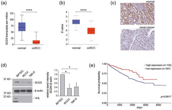
SCD5 Regulation by VHL Affects Cell Proliferation and Lipid Homeostasis in ccRCC
by Athina Ganner, Antonia Philipp, Simon Lagies, Laura Wingendorf, Lu Wang, Felicitas Pilz, Thomas Welte, Kelli Grand, Soeren S. Lienkamp, Marinella Klein, Bernd Kammerer, Ian J. Frew, Gerd Walz and Elke Neumann-Haefelin
Cells 2023, 12(6), 835; https://doi.org/10.3390/cells12060835 - 8 Mar 2023
Cited by 2 | Viewed by 2384
Abstract
Clear cell renal cell carcinoma (ccRCC) is the most common histological subtype of renal cancer, and inactivation of the VHL tumor suppressor gene is found in almost all cases of hereditary and sporadic ccRCCs. CcRCC is associated with the reprogramming of fatty acid [...] Read more.
Clear cell renal cell carcinoma (ccRCC) is the most common histological subtype of renal cancer, and inactivation of the VHL tumor suppressor gene is found in almost all cases of hereditary and sporadic ccRCCs. CcRCC is associated with the reprogramming of fatty acid metabolism, and stearoyl-CoA desaturases (SCDs) are the main enzymes controlling fatty acid composition in cells. In this study, we report that mRNA and protein expression of the stearoyl-CoA desaturase SCD5 is downregulated in VHL-deficient cell lines. Similarly, in C. elegans vhl-1 mutants, FAT-7/SCD5 activity is repressed, supporting an evolutionary conservation. SCD5 regulation by VHL depends on HIF, and loss of SCD5 promotes cell proliferation and a metabolic shift towards ceramide production. In summary, we identify a novel regulatory function of VHL in relation to SCD5 and fatty acid metabolism, and propose a new mechanism of how loss of VHL may contribute to ccRCC tumor formation and progression. Full article
Show Figures

Figure 1

18 pages, 13603 KiB  
Article
The Potential of PSMA as a Vascular Target in TNBC
by Amelie Heesch, Lars Ortmanns, Jochen Maurer, Elmar Stickeler, Sabri E. M. Sahnoun, Felix M. Mottaghy and Agnieszka Morgenroth
Cells 2023, 12(4), 551; https://doi.org/10.3390/cells12040551 - 8 Feb 2023
Cited by 11 | Viewed by 3237
Abstract
Recent studies proving prostate-specific membrane antigen (PSMA) expression on triple-negative breast cancer (TNBC) cells and adjacent endothelial cells suggest PSMA as a promising target for therapy of until now not-targetable cancer entities. In this study, PSMA and its isoform expression were analyzed in [...] Read more.
Recent studies proving prostate-specific membrane antigen (PSMA) expression on triple-negative breast cancer (TNBC) cells and adjacent endothelial cells suggest PSMA as a promising target for therapy of until now not-targetable cancer entities. In this study, PSMA and its isoform expression were analyzed in different TNBC cells, breast cancer stem cells (BCSCs), and tumor-associated endothelial cells. PSMA expression was detected in 91% of the investigated TNBC cell lines. The PSMA splice isoforms were predominantly found in the BCSCs. Tumor-conditioned media from two TNBC cell lines, BT-20 (high full-length PSMA expression, PSMAΔ18 expression) and Hs578T (low full-length PSMA expression, no isoform expression), showed significant pro-angiogenic effect with induction of tube formation in endothelial cells. All TNBC cell lines induced PSMA expression in human umbilical vein endothelial cells (HUVEC). Significant uptake of radiolabeled ligand [68Ga]Ga-PSMA was detected in BCSC1 (4.2%), corresponding to the high PSMA expression. Moreover, hypoxic conditions increased the uptake of radiolabeled ligand [177Lu]Lu-PSMA in MDA-MB-231 (0.4% vs. 3.4%, under hypoxia and normoxia, respectively) and MCF-10A (0.3% vs. 3.0%, under normoxia and hypoxia, respectively) significantly (p < 0.001). [177Lu]Lu-PSMA-induced apoptosis rates were highest in BT-20 and MDA-MB-231 associated endothelial cells. Together, these findings demonstrate the potential of PSMA-targeted therapy in TNBC. Full article
Show Figures

Figure 1

13 pages, 5722 KiB  
Article
ABCA1-Mediated EMT Promotes Papillary Thyroid Cancer Malignancy through the ERK/Fra-1/ZEB1 Pathway
by Ji-Hye Park, Jae-Kyung Myung, Sun-Joo Lee, Hyewon Kim, Soyeon Kim, Seung-Bum Lee, Hyosun Jang, Won-Il Jang, Sunhoo Park, Hyunwon Yang, Sehwan Shim and Min-Jung Kim
Cells 2023, 12(2), 274; https://doi.org/10.3390/cells12020274 - 10 Jan 2023
Cited by 6 | Viewed by 2561 | Correction
Abstract
Papillary thyroid cancer (PTC) is the most prevalent histological type of thyroid cancer (TC) worldwide. Although tumor metastasis occurs in regional lymph nodes, distant metastasis (DM) may also occur. Radioactive iodine (RAI) therapy is an effective treatment for TC; however, resistance to RAI [...] Read more.
Papillary thyroid cancer (PTC) is the most prevalent histological type of thyroid cancer (TC) worldwide. Although tumor metastasis occurs in regional lymph nodes, distant metastasis (DM) may also occur. Radioactive iodine (RAI) therapy is an effective treatment for TC; however, resistance to RAI occurs in patients with DM. Therefore, in this study, we investigated the efficacy of DM-related biomarkers as therapeutic targets for PTC therapy. ABCA1 expression was higher in aggressive BCPAP cells than in other PTC cells in terms of migration and invasion capacity. The knockdown of ABCA1 substantially decreased the expression of the epithelial–mesenchymal transition (EMT) marker, N-cadherin, and EMT regulator (ZEB1), resulting in suppressed migration and invasion of BCPAP cells. ABCA1 knockdown also reduced ERK activity and Fra-1 expression, which correlated with the effects of an ERK inhibitor or siRNA-mediated inhibition of ERK or Fra-1 expression. Furthermore, ABCA1-knocked-down BCPAP cells suppressed cell migration and invasion by reducing Fra-1 recruitment to Zeb1 promoter; lung metastasis was not observed in mice injected with ABCA1-knocked-down cells. Overall, our findings suggest that ABCA1 regulates lung metastasis in TC cells. Full article
Show Figures

Figure 1

2022

Jump to: 2024, 2023, 2021

18 pages, 4188 KiB  
Article
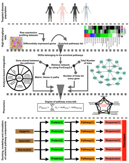
An Approach for Systems-Level Understanding of Prostate Cancer from High-Throughput Data Integration to Pathway Modeling and Simulation
by Mohammad Mobashir, S. Pauliina Turunen, Mohammad Asrar Izhari, Ibraheem Mohammed Ashankyty, Thomas Helleday and Kaisa Lehti
Cells 2022, 11(24), 4121; https://doi.org/10.3390/cells11244121 - 19 Dec 2022
Cited by 11 | Viewed by 2897
Abstract
To understand complex diseases, high-throughput data are generated at large and multiple levels. However, extracting meaningful information from large datasets for comprehensive understanding of cell phenotypes and disease pathophysiology remains a major challenge. Despite tremendous advances in understanding molecular mechanisms of cancer and [...] Read more.
To understand complex diseases, high-throughput data are generated at large and multiple levels. However, extracting meaningful information from large datasets for comprehensive understanding of cell phenotypes and disease pathophysiology remains a major challenge. Despite tremendous advances in understanding molecular mechanisms of cancer and its progression, current knowledge appears discrete and fragmented. In order to render this wealth of data more integrated and thus informative, we have developed a GECIP toolbox to investigate the crosstalk and the responsible genes’/proteins’ connectivity of enriched pathways from gene expression data. To implement this toolbox, we used mainly gene expression datasets of prostate cancer, and the three datasets were GSE17951, GSE8218, and GSE1431. The raw samples were processed for normalization, prediction of differentially expressed genes, and the prediction of enriched pathways for the differentially expressed genes. The enriched pathways have been processed for crosstalk degree calculations for which number connections per gene, the frequency of genes in the pathways, sharing frequency, and the connectivity have been used. For network prediction, protein–protein interaction network database FunCoup2.0 was used, and cytoscape software was used for the network visualization. In our results, we found that there were enriched pathways 27, 45, and 22 for GSE17951, GSE8218, and GSE1431, respectively, and 11 pathways in common between all of them. From the crosstalk results, we observe that focal adhesion and PI3K pathways, both experimentally proven central for cellular output upon perturbation of numerous individual/distinct signaling pathways, displayed highest crosstalk degree. Moreover, we also observe that there were more critical pathways which appear to be highly significant, and these pathways are HIF1a, hippo, AMPK, and Ras. In terms of the pathways’ components, GSK3B, YWHAE, HIF1A, ATP1A3, and PRKCA are shared between the aforementioned pathways and have higher connectivity with the pathways and the other pathway components. Finally, we conclude that the focal adhesion and PI3K pathways are the most critical pathways, and since for many other pathways, high-rank enrichment did not translate to high crosstalk degree, the global impact of one pathway on others appears distinct from enrichment. Full article
Show Figures

Figure 1

22 pages, 7512 KiB  
Article
Systematic Analysis of a Pyroptosis-Related Signature to Predict the Prognosis and Immune Microenvironment of Lower-Grade Glioma
by Yongze He, Yuxiang Cai, Jinsheng Liu, Haixia Ding, Xiang Li, Sufang Tian and Zhiqiang Li
Cells 2022, 11(24), 3980; https://doi.org/10.3390/cells11243980 - 9 Dec 2022
Cited by 2 | Viewed by 1766
Abstract
Current treatments for lower-grade glioma (LGG) do not effectively improve life expectancy rates, and this is a major global health concern. Improving our knowledge of this disease will ultimately help to improve prevention, accurate prognosis, and treatment strategies. Pyroptosis is an inflammatory form [...] Read more.
Current treatments for lower-grade glioma (LGG) do not effectively improve life expectancy rates, and this is a major global health concern. Improving our knowledge of this disease will ultimately help to improve prevention, accurate prognosis, and treatment strategies. Pyroptosis is an inflammatory form of regulated cell death, which plays an important role in tumor progression and occurrence. There is still a lack of effective markers to evaluate the prognosis of LGG patients. We collected paraffin-embedded tissue samples and prognostic information from 85 patients with low-grade gliomas and fabricated them into a tissue microarray. Combining data from public databases, we explored the relationship between pyroptosis-related genes (PRGs) and the prognoses of patients with LGG and investigated their correlations with the tumor microenvironment (TME) by means of machine learning, single-cell, immunohistochemical, nomogram, GSEA, and Cox regression analyses. We developed a six-gene PRG-based prognostic model, and the results have identified CASP4 as an effective marker for LGG prognosis predictions. Furthermore, the effects on immune cell infiltration may also provide guidance for future immunotherapy strategies. Full article
Show Figures

Figure 1

17 pages, 4107 KiB  
Article
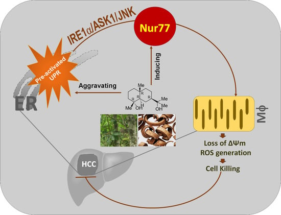
Orphan Nuclear Receptor Nur77 Mediates the Lethal Endoplasmic Reticulum Stress and Therapeutic Efficacy of Cryptomeridiol in Hepatocellular Carcinoma
by Xudan Li, Quancheng Chen, Jie Liu, Shenjin Lai, Minda Zhang, Tidong Zhen, Hongyu Hu, Xiang Gao, Alice S. T. Wong and Jin-Zhang Zeng
Cells 2022, 11(23), 3870; https://doi.org/10.3390/cells11233870 - 1 Dec 2022
Cited by 6 | Viewed by 2771
Abstract
Hepatocellular carcinoma (HCC) commonly possesses chronical elevation of IRE1α-ASK1 signaling. Orphan nuclear receptor Nur77, a promising therapeutic target in various cancer types, is frequently silenced in HCC. In this study, we show that cryptomeridiol (Bkh126), a naturally occurring sesquiterpenoid derivative isolated from traditional [...] Read more.
Hepatocellular carcinoma (HCC) commonly possesses chronical elevation of IRE1α-ASK1 signaling. Orphan nuclear receptor Nur77, a promising therapeutic target in various cancer types, is frequently silenced in HCC. In this study, we show that cryptomeridiol (Bkh126), a naturally occurring sesquiterpenoid derivative isolated from traditional Chinese medicine Magnolia officinalis, has therapeutic efficacy in HCC by aggravating the pre-activated UPR and activating the silenced Nur77. Mechanistically, Nur77 is induced to sense IRE1α-ASK1-JNK signaling and translocate to the mitochondria, which leads to the loss of mitochondrial membrane potential (Δψm). The Bkh126-induced aggravation of ER stress and mitochondrial dysfunction result in increased cytotoxic product of reactive oxygen species (ROS). The in vivo anti-HCC activity of Bkh126 is superior to that of sorafenib, currently used to treat advanced HCC. Our study shows that Bkh126 induces Nur77 to connect ER stress to mitochondria-mediated cell killing. The identification of Nur77 as a molecular target of Bhk126 provides a basis for improving the leads for the further development of anti-HCC drugs. Full article
Show Figures

Graphical abstract

16 pages, 8933 KiB  
Article
Visfatin-Induced Inhibition of miR-1264 Facilitates PDGF-C Synthesis in Chondrosarcoma Cells and Enhances Endothelial Progenitor Cell Angiogenesis
by Chang-Yu Song, Sunny Li-Yun Chang, Chih-Yang Lin, Chun-Hao Tsai, Shang-Yu Yang, Yi-Chin Fong, Yu-Wen Huang, Shih-Wei Wang, Wei-Cheng Chen and Chih-Hsin Tang
Cells 2022, 11(21), 3470; https://doi.org/10.3390/cells11213470 - 2 Nov 2022
Cited by 9 | Viewed by 2147
Abstract
New treatments for chondrosarcoma are extremely important. Chondrosarcoma is a primary malignant bone tumor with a very unfavorable prognosis. High-grade chondrosarcoma has a high potential to metastasize to any organ in the body. Platelet-derived growth factor (PDGF) is a potent angiogenic factor that [...] Read more.
New treatments for chondrosarcoma are extremely important. Chondrosarcoma is a primary malignant bone tumor with a very unfavorable prognosis. High-grade chondrosarcoma has a high potential to metastasize to any organ in the body. Platelet-derived growth factor (PDGF) is a potent angiogenic factor that promotes tumor angiogenesis and metastasis. The adipocytokine visfatin promotes metastatic potential of chondrosarcoma; however, the role of visfatin in angiogenesis in human chondrosarcoma is unclear. We report that the levels of PDGF-C expression were positively correlated with tumor stages, significantly higher than the levels of expression in normal cartilage. Visfatin increased PDGF-C expression and endothelial progenitor cell (EPC) angiogenesis through the PI3K/Akt/mTOR signaling pathway, and dose-dependently down-regulated the synthesis of miR-1264, which targets the 3′-UTR of PDGF-C. Additionally, we discovered inhibition of visfatin or PDGF-C in chondrosarcoma tumors significantly reduced tumor angiogenesis and size. Our results indicate that visfatin inhibits miR-1264 production through the PI3K/Akt/mTOR signaling cascade, and thereby promotes PDGF-C expression and chondrosarcoma angiogenesis. Visfatin may be worth targeting in the treatment of chondrosarcoma angiogenesis. Full article
Show Figures

Figure 1

16 pages, 4708 KiB  
Article
Clinical and Prognostic Value of PPIA, SQSTM1, and CCL20 in Hepatocellular Carcinoma Patients by Single-Cell Transcriptome Analysis
by Lisha Mou, Chenyang Jia, Zijing Wu, Boyang Xin, Carmen Alicia Liang Zhen, Bailiang Wang, Yong Ni and Zuhui Pu
Cells 2022, 11(19), 3078; https://doi.org/10.3390/cells11193078 - 30 Sep 2022
Cited by 11 | Viewed by 2681
Abstract
Hepatocellular carcinoma (HCC) is the most malignant and poor-prognosis subtype of primary liver cancer. The scRNA-seq approach provides unique insight into tumor cell behavior at the single-cell level. Cytokine signaling in the immune system plays an important role in tumorigenesis and has both [...] Read more.
Hepatocellular carcinoma (HCC) is the most malignant and poor-prognosis subtype of primary liver cancer. The scRNA-seq approach provides unique insight into tumor cell behavior at the single-cell level. Cytokine signaling in the immune system plays an important role in tumorigenesis and has both pro-tumorigenic and anti-tumorigenic functions. A biomarker of cytokine signaling in immune-related genes (CSIRG) is urgently required to assess HCC patient diagnosis and treatment. By analyzing the expression profiles of HCC single cells, TCGA, and ICGC data, we discovered that three important CSIRG (PPIA, SQSTM1, and CCL20) were linked to the overall survival of HCC patients. Cancer status and three hub CSIRG were taken into account while creating a risk nomogram. The nomogram had a high level of predictability and accuracy. Based on the CSIRG risk score, a distinct pattern of somatic tumor mutational burden (TMB) was detected between the two groups. The enrichment of the pyrimidine metabolism pathway, purine metabolism pathway, and lysosome pathway in HCC was linked to the CSIRG high-risk scores. Overall, scRNA-seq and bulk RNA-seq were used to create a strong CSIRG signature for HCC diagnosis. Full article
Show Figures

Figure 1

20 pages, 1198 KiB  
Review
Regulating the Expression of HIF-1α or lncRNA: Potential Directions for Cancer Therapy
by Minghui Zhang, Yu Zhang, Yubo Ding, Jialu Huang, Jingwei Yao, Zhuoyi Xie, Yufan Lv and Jianhong Zuo
Cells 2022, 11(18), 2811; https://doi.org/10.3390/cells11182811 - 8 Sep 2022
Cited by 11 | Viewed by 3154
Abstract
Previous studies have shown that tumors under a hypoxic environment can induce an important hypoxia-responsive element, hypoxia-induced factor-1α (HIF-1α), which can increase tumor migration, invasion, and metastatic ability by promoting epithelial-to-mesenchymal transition (EMT) in tumor cells. Currently, with the deeper knowledge of long [...] Read more.
Previous studies have shown that tumors under a hypoxic environment can induce an important hypoxia-responsive element, hypoxia-induced factor-1α (HIF-1α), which can increase tumor migration, invasion, and metastatic ability by promoting epithelial-to-mesenchymal transition (EMT) in tumor cells. Currently, with the deeper knowledge of long noncoding RNAs (lncRNAs), more and more functions of lncRNAs have been discovered. HIF-1α can regulate hypoxia-responsive lncRNAs under hypoxic conditions, and changes in the expression level of lncRNAs can regulate the production of EMT transcription factors and signaling pathway transduction, thus promoting EMT progress. In conclusion, this review summarizes the regulation of the EMT process by HIF-1α and lncRNAs and discusses their relationship with tumorigenesis. Since HIF-1α plays an important role in tumor progression, we also summarize the current drugs that inhibit tumor progression by modulating HIF-1α. Full article
Show Figures

Figure A1

15 pages, 1199 KiB  
Review
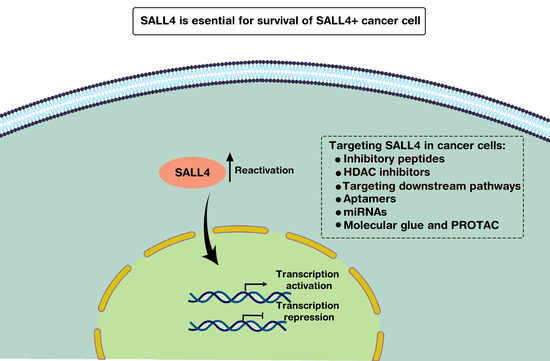
SALL4: An Intriguing Therapeutic Target in Cancer Treatment
by Shiva Moein, Daniel G. Tenen, Giovanni Amabile and Li Chai
Cells 2022, 11(16), 2601; https://doi.org/10.3390/cells11162601 - 20 Aug 2022
Cited by 15 | Viewed by 4301
Abstract
Spalt-Like Transcription Factor 4 (SALL4) is a critical factor for self-renewal ability and pluripotency of stem cells. On the other hand, various reports show tight relation of SALL4 to cancer occurrence and metastasis. SALL4 exerts its effects not only by inducing gene expression [...] Read more.
Spalt-Like Transcription Factor 4 (SALL4) is a critical factor for self-renewal ability and pluripotency of stem cells. On the other hand, various reports show tight relation of SALL4 to cancer occurrence and metastasis. SALL4 exerts its effects not only by inducing gene expression but also repressing a large cluster of genes through interaction with various epigenetic modifiers. Due to high expression of SALL4 in cancer cells and its silence in almost all adult tissues, it is an ideal target for cancer therapy. However, targeting SALL4 meets various challenges. SALL4 is a transcription factor and designing appropriate drug to inhibit this intra-nucleus component is challenging. On the other hand, due to lack of our knowledge on structure of the protein and the suitable active sites, it becomes more difficult to reach the appropriate drugs against SALL4. In this review, we have focused on approaches applied yet to target this oncogene and discuss the potential of degrader systems as new therapeutics to target oncogenes. Full article
Show Figures

Graphical abstract

17 pages, 14105 KiB  
Article
Gremlin-1 Promotes Colorectal Cancer Cell Metastasis by Activating ATF6 and Inhibiting ATF4 Pathways
by Ruohan Li, Huaixiang Zhou, Mingzhe Li, Qiuyan Mai, Zhang Fu, Youheng Jiang, Changxue Li, Yunfei Gao, Yunping Fan, Kaiming Wu, Clive Da Costa, Xia Sheng, Yulong He and Ningning Li
Cells 2022, 11(14), 2136; https://doi.org/10.3390/cells11142136 - 7 Jul 2022
Cited by 14 | Viewed by 3938
Abstract
Cancer cell survival, function and fate strongly depend on endoplasmic reticulum (ER) proteostasis. Although previous studies have implicated the ER stress signaling network in all stages of cancer development, its role in cancer metastasis remains to be elucidated. In this study, we investigated [...] Read more.
Cancer cell survival, function and fate strongly depend on endoplasmic reticulum (ER) proteostasis. Although previous studies have implicated the ER stress signaling network in all stages of cancer development, its role in cancer metastasis remains to be elucidated. In this study, we investigated the role of Gremlin-1 (GREM1), a secreted protein, in the invasion and metastasis of colorectal cancer (CRC) cells in vitro and in vivo. Firstly, public datasets showed a positive correlation between high expression of GREM1 and a poor prognosis for CRC. Secondly, GREM1 enhanced motility and invasion of CRC cells by epithelial–mesenchymal transition (EMT). Thirdly, GREM1 upregulated expression of activating transcription factor 6 (ATF6) and downregulated that of ATF4, and modulation of the two key players of the unfolded protein response (UPR) was possibly through activation of PI3K/AKT/mTOR and antagonization of BMP2 signaling pathways, respectively. Taken together, our results demonstrate that GREM1 is an invasion-promoting factor via regulation of ATF6 and ATF4 expression in CRC cells, suggesting GREM1 may be a potential pharmacological target for colorectal cancer treatment. Full article
Show Figures

Figure 1

25 pages, 7323 KiB  
Article
Prognostic and Immunotherapeutic Roles of KRAS in Pan-Cancer
by Kaixin Yang, Chengyun Li, Yang Liu, Xueyan Gu, Longchang Jiang and Lei Shi
Cells 2022, 11(9), 1427; https://doi.org/10.3390/cells11091427 - 22 Apr 2022
Cited by 4 | Viewed by 3267
Abstract
KRAS is one well-established tumor-driver gene associated with cancer initiation, development, and progression. Nonetheless, comparative studies of the relevance of KRAS across diverse tumors remain sparse. We explored the KRAS expression and prognostic values in diverse cancer types via multiple web-based bioinformatics tools, [...] Read more.
KRAS is one well-established tumor-driver gene associated with cancer initiation, development, and progression. Nonetheless, comparative studies of the relevance of KRAS across diverse tumors remain sparse. We explored the KRAS expression and prognostic values in diverse cancer types via multiple web-based bioinformatics tools, including cBioPortal, Oncomine, PrognoScan, Kaplan–Meier Plotter, etc. We found that KRAS is highly expressed in various malignancies compared to normal cohorts (BRCA, CHOL, ESCA, HNSC, LIHC, LUAD, LUSC, and STAD) and less expressed in COAD, KIRC, READ, and THCA than in normal samples. We observed the dysregulation of the DNA methylation of KRAS in cancers and discovered that numerous oncogenic and tumor-suppressive transcription factors bind the KRAS promoter region. Pan-cancer analysis also showed that a high level of KRAS is associated with poor outcomes. Additionally, KRAS is remarkably correlated with the level of immune cell infiltration and tumorigenic gene signatures. In conclusion, our findings reveal novel insights into KRAS expression and its biological functions in diverse cancer types, indicating that KRAS could serve as a prognostic biomarker and is associated with immune infiltrates. Full article
Show Figures

Figure 1

19 pages, 4169 KiB  
Article
Comprehensive Analysis of CDK1-Associated ceRNA Network Revealing the Key Pathways LINC00460/LINC00525-Hsa-Mir-338-FAM111/ZWINT as Prognostic Biomarkers in Lung Adenocarcinoma Combined with Experiments
by Wen Li, Shan-Shan Feng, Hao Wu, Jing Deng, Wang-Yan Zhou, Ming-Xi Jia, Yi Shi, Liang Ma, Xiao-Xi Zeng, Zavuga Zuberi, Da Fu, Xiang Liu and Zhu Chen
Cells 2022, 11(7), 1220; https://doi.org/10.3390/cells11071220 - 4 Apr 2022
Cited by 4 | Viewed by 2920
Abstract
Lung adenocarcinoma (LUAD) is the leading cause of cancer deaths worldwide, and effective biomarkers are still lacking for early detection and prognosis prediction. Here, based on gene expression profiles of LUAD patients from The Cancer Genome Atlas (TCGA), 806 long non-coding RNAs (lncRNAs), [...] Read more.
Lung adenocarcinoma (LUAD) is the leading cause of cancer deaths worldwide, and effective biomarkers are still lacking for early detection and prognosis prediction. Here, based on gene expression profiles of LUAD patients from The Cancer Genome Atlas (TCGA), 806 long non-coding RNAs (lncRNAs), 122 microRNAs (miRNAs) and 1269 mRNAs associated with CDK1 were identified. The regulatory axis of LINC00460/LINC00525-hsa-mir-338-FAM111B/ZWINT was determined according to the correlation between gene expression and patient prognosis. The abnormal up-regulation of FAM111B/ZWINT in LUAD was related to hypomethylation. Furthermore, immune infiltration analysis suggested FAM111B/ZWINT could affect the development and prognosis of cancer by regulating the LUAD immune microenvironment. EMT feature analysis suggested that FAM111B/ZWINT promoted tumor spread through the EMT process. Functional analysis showed FAM111B/ZWINT was involved in cell cycle events such as DNA replication and chromosome separation. We analyzed the HERB and GSCALite databases to identify potential target medicines that may play a role in the treatment of LUAD. Finally, the expression of LINC00460/LINC00525-hsa-mir-338-FAM111B/ZWINT axis was verified in LUAD cells by RT-qPCR, and these results were consistent with bioinformatics analysis. Overall, we constructed a CDK1-related ceRNA network and revealed the LINC00460/LINC00525-hsa-mir-338-FAM111/ZWINT pathways as potential diagnostic biomarkers or therapeutic targets of LUAD. Full article
Show Figures

Figure 1

16 pages, 1668 KiB  
Article
RNAi-Mediated Screen of Primary AML Cells Nominates MDM4 as a Therapeutic Target in NK-AML with DNMT3A Mutations
by Olga Alexandra Sidorova, Shady Sayed, Maciej Paszkowski-Rogacz, Michael Seifert, Aylin Camgöz, Ingo Roeder, Martin Bornhäuser, Christian Thiede and Frank Buchholz
Cells 2022, 11(5), 854; https://doi.org/10.3390/cells11050854 - 2 Mar 2022
Cited by 5 | Viewed by 3690
Abstract
DNA-methyltransferase 3A (DNMT3A) mutations belong to the most frequent genetic aberrations found in adult acute myeloid leukemia (AML). Recent evidence suggests that these mutations arise early in leukemogenesis, marking leukemic progenitors and stem cells, and persist through consolidation chemotherapy, providing a [...] Read more.
DNA-methyltransferase 3A (DNMT3A) mutations belong to the most frequent genetic aberrations found in adult acute myeloid leukemia (AML). Recent evidence suggests that these mutations arise early in leukemogenesis, marking leukemic progenitors and stem cells, and persist through consolidation chemotherapy, providing a pool for AML relapse. Currently, there are no therapeutic approaches directed specifically against this cell population. To unravel therapeutically actionable targets in mutant DNMT3A-driven AML cells, we have performed a focused RNAi screen in a panel of 30 primary AML samples, all carrying a DNMT3A R882 mutation. As one of the strongest hits, we identified MDM4 as a gene essential for proliferation of primary DNMT3AWT/R882X AML cells. We analyzed a publicly available RNA-Seq dataset of primary normal karyotype (NK) AML samples and found a trend towards MDM4 transcript overexpression particularly in DNMT3A-mutant samples. Moreover, we found that the MDM2/4 inhibitor ALRN-6924 impairs growth of DNMT3AWT/R882X primary cells in vitro by inducing cell cycle arrest through upregulation of p53 target genes. Our results suggest that MDM4 inhibition is a potential target in NK-AML patients bearing DNMT3A R882X mutations. Full article
Show Figures

Graphical abstract

18 pages, 34487 KiB  
Article
Metastatic Tumor Cell-Specific FABP7 Promotes NSCLC Metastasis via Inhibiting β-Catenin Degradation
by Qiaorui Bai, Xia Yang, Quanfeng Li, Weizhong Chen, Han Tian, Rong Lian, Ximeng Liu, Shuang Wang and Yi Yang
Cells 2022, 11(5), 805; https://doi.org/10.3390/cells11050805 - 25 Feb 2022
Cited by 7 | Viewed by 3122
Abstract
Metastasis accounts for 90% of cancer-related deaths and represents a prominent malignant feature in non-small cell lung cancer (NSCLC), while tumor cell-specific mechanisms and molecules pivotal for the metastatic capacity remain unclear. By analyzing single-cell RNA sequencing data, we found that fatty acid [...] Read more.
Metastasis accounts for 90% of cancer-related deaths and represents a prominent malignant feature in non-small cell lung cancer (NSCLC), while tumor cell-specific mechanisms and molecules pivotal for the metastatic capacity remain unclear. By analyzing single-cell RNA sequencing data, we found that fatty acid binding protein 7 (FABP7) was specifically up-regulated in tumor cells of metastatic NSCLC patients and might be a prognostic indicator for poor survival. Experimental studies based on NSCLC cell lines showed that FABP7 promoted the metastatic competencies of NSCLC cells in vitro and in vivo. Mechanistically, we demonstrated that FABP7 was important to canonical Wnt signaling activation and competitively inhibited the interaction between β-catenin and components of its cytoplasmic degradation complex, thereby repressing the phosphorylation-dependent ubiquitination and degradation of β-catenin. Our present study identifies FABP7 as a metastatic tumor cell-specific pro-metastatic gene and uncovers a previously unknown regulatory mechanism underlying Wnt hyperactivation via FABP7-impaired cytoplasmic β-catenin degradation, implicating a novel molecule in regulating NSCLC metastasis. Full article
Show Figures

Figure 1

21 pages, 1883 KiB  
Review
Recent Advances in Ovarian Cancer: Therapeutic Strategies, Potential Biomarkers, and Technological Improvements
by Salima Akter, Md. Ataur Rahman, Mohammad Nazmul Hasan, Hajara Akhter, Priya Noor, Rokibul Islam, Yoonhwa Shin, MD. Hasanur Rahman, Md. Shamim Gazi, Md Nazmul Huda, Nguyen Minh Nam, Jinwook Chung, Sunhee Han, Bonglee Kim, Insug Kang, Joohun Ha, Wonchae Choe, Tae Gyu Choi and Sung Soo Kim
Cells 2022, 11(4), 650; https://doi.org/10.3390/cells11040650 - 13 Feb 2022
Cited by 49 | Viewed by 8575
Abstract
Aggressive and recurrent gynecological cancers are associated with worse prognosis and a lack of effective therapeutic response. Ovarian cancer (OC) patients are often diagnosed in advanced stages, when drug resistance, angiogenesis, relapse, and metastasis impact survival outcomes. Currently, surgical debulking, radiotherapy, and/or chemotherapy [...] Read more.
Aggressive and recurrent gynecological cancers are associated with worse prognosis and a lack of effective therapeutic response. Ovarian cancer (OC) patients are often diagnosed in advanced stages, when drug resistance, angiogenesis, relapse, and metastasis impact survival outcomes. Currently, surgical debulking, radiotherapy, and/or chemotherapy remain the mainstream treatment modalities; however, patients suffer unwanted side effects and drug resistance in the absence of targeted therapies. Hence, it is urgent to decipher the complex disease biology and identify potential biomarkers, which could greatly contribute to making an early diagnosis or predicting the response to specific therapies. This review aims to critically discuss the current therapeutic strategies for OC, novel drug-delivery systems, and potential biomarkers in the context of genetics and molecular research. It emphasizes how the understanding of disease biology is related to the advancement of technology, enabling the exploration of novel biomarkers that may be able to provide more accurate diagnosis and prognosis, which would effectively translate into targeted therapies, ultimately improving patients’ overall survival and quality of life. Full article
Show Figures

Figure 1

26 pages, 5040 KiB  
Article
Identification of the Cysteine Protease Legumain as a Potential Chronic Hypoxia-Specific Multiple Myeloma Target Gene
by Ada-Sophia Clees, Verena Stolp, Björn Häupl, Dominik C. Fuhrmann, Frank Wempe, Marcel Seibert, Sarah Weber, Antje Banning, Ritva Tikkanen, Richard Williams, Bernhard Brüne, Hubert Serve, Frank Schnütgen, Ivana von Metzler and Nina Kurrle
Cells 2022, 11(2), 292; https://doi.org/10.3390/cells11020292 - 15 Jan 2022
Cited by 4 | Viewed by 3828
Abstract
Multiple myeloma (MM) is the second most common hematologic malignancy, which is characterized by clonal proliferation of neoplastic plasma cells in the bone marrow. This microenvironment is characterized by low oxygen levels (1–6% O2), known as hypoxia. For MM cells, hypoxia [...] Read more.
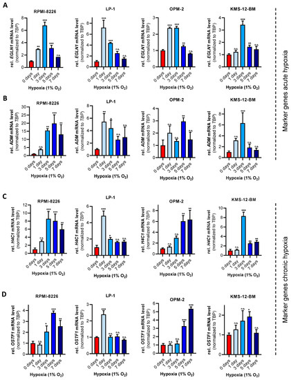
Multiple myeloma (MM) is the second most common hematologic malignancy, which is characterized by clonal proliferation of neoplastic plasma cells in the bone marrow. This microenvironment is characterized by low oxygen levels (1–6% O2), known as hypoxia. For MM cells, hypoxia is a physiologic feature that has been described to promote an aggressive phenotype and to confer drug resistance. However, studies on hypoxia are scarce and show little conformity. Here, we analyzed the mRNA expression of previously determined hypoxia markers to define the temporal adaptation of MM cells to chronic hypoxia. Subsequent analyses of the global proteome in MM cells and the stromal cell line HS-5 revealed hypoxia-dependent regulation of proteins, which directly or indirectly upregulate glycolysis. In addition, chronic hypoxia led to MM-specific regulation of nine distinct proteins. One of these proteins is the cysteine protease legumain (LGMN), the depletion of which led to a significant growth disadvantage of MM cell lines that is enhanced under hypoxia. Thus, herein, we report a methodologic strategy to examine MM cells under physiologic hypoxic conditions in vitro and to decipher and study previously masked hypoxia-specific therapeutic targets such as the cysteine protease LGMN. Full article
Show Figures

Figure 1

2021

Jump to: 2024, 2023, 2022

13 pages, 2843 KiB  
Article
DSTYK Enhances Chemoresistance in Triple-Negative Breast Cancer Cells
by Stella C. Ogbu, Samuel Rojas, John Weaver, Phillip R. Musich, Jinyu Zhang, Zhi Q. Yao and Yong Jiang
Cells 2022, 11(1), 97; https://doi.org/10.3390/cells11010097 - 29 Dec 2021
Cited by 10 | Viewed by 3318
Abstract
Breast cancer, as the most prevalent cancer in women, is responsible for more than 15% of new cancer cases and about 6.9% of all cancer-related death in the US. A major cause of therapeutic failure in breast cancer is the development of resistance [...] Read more.
Breast cancer, as the most prevalent cancer in women, is responsible for more than 15% of new cancer cases and about 6.9% of all cancer-related death in the US. A major cause of therapeutic failure in breast cancer is the development of resistance to chemotherapy, especially for triple-negative breast cancer (TNBC). Therefore, how to overcome chemoresistance is the major challenge to improve the life expectancy of breast cancer patients. Our studies demonstrate that TNBC cells surviving the chronic treatment of chemotherapeutic drugs show significantly higher expression of the dual serine/threonine and tyrosine protein kinase (DSTYK) than non-treated parental cells. In our in vitro cellular models, DSTYK knockout via the CRISPR/Cas9-mediated technique results in apoptotic cell death of chemoresistant cells upon drug treatment. Moreover, DSTYK knockout promotes chemotherapeutic drug-induced tumor cell death in an orthotopic mouse model. These findings suggest that DSTYK exerts an important and previously unknown role in promoting chemoresistance. Our studies provide fundamental insight into the role of DSTYK in chemoresistance in TNBC cells and lay the foundation for the development of new strategies targeting DSTYK for improving TNBC therapy. Full article
Show Figures

Figure 1

28 pages, 37763 KiB  
Article
Somatic Mutational Profile of High-Grade Serous Ovarian Carcinoma and Triple-Negative Breast Carcinoma in Young and Elderly Patients: Similarities and Divergences
by Pedro Adolpho de Menezes Pacheco Serio, Gláucia Fernanda de Lima Pereira, Maria Lucia Hirata Katayama, Rosimeire Aparecida Roela, Simone Maistro and Maria Aparecida Azevedo Koike Folgueira
Cells 2021, 10(12), 3586; https://doi.org/10.3390/cells10123586 - 20 Dec 2021
Cited by 11 | Viewed by 4064
Abstract
Background: Triple-negative breast cancer (TNBC) and High-Grade Serous Ovarian Cancer (HGSOC) are aggressive malignancies that share similarities; however, different ages of onset may reflect distinct tumor behaviors. Thus, our aim was to compare somatic mutations in potential driver genes in 109 TNBC and [...] Read more.
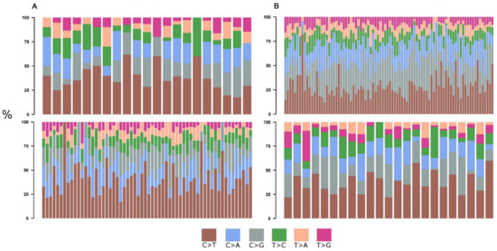
Background: Triple-negative breast cancer (TNBC) and High-Grade Serous Ovarian Cancer (HGSOC) are aggressive malignancies that share similarities; however, different ages of onset may reflect distinct tumor behaviors. Thus, our aim was to compare somatic mutations in potential driver genes in 109 TNBC and 81 HGSOC from young (Y ≤ 40 years) and elderly (E ≥ 75 years) patients. Methods: Open access mutational data (WGS or WES) were collected for TNBC and HGSOC patients. Potential driver genes were those that were present in the Cancer Gene Census—CGC, the Candidate Cancer Gene Database—CCGD, or OncoKB and those that were considered pathogenic in variant effect prediction tools. Results: Mutational signature 3 (homologous repair defects) was the only gene that was represented in all four subgroups. The median number of mutated CGCs per sample was similar in HGSOC (Y:3 vs. E:4), but it was higher in elderly TNBC than it was in young TNBC (Y:3 vs. E:6). At least 90% of the samples from TNBC and HGSOC from Y and E patients presented at least one known affected TSG. Besides TP53, which was mutated in 67–83% of the samples, the affected TSG in TP53 wild-type samples were NF1 (yHGSOC and yTNBC), PHF6 (eHGSOC and yTNBC), PTEN, PIK3R1 and ZHFX3 (yTNBC), KMT2C, ARID1B, TBX3, and ATM (eTNBC). A few samples only presented one affected oncogene (but no TSG): KRAS and TSHR in eHGSOC and RAC1 and PREX2 (a regulator of RAC1) in yTNBC. At least ⅔ of the tumors presented mutated oncogenes associated with tumor suppressor genes; the Ras and/or PIK3CA signaling pathways were altered in 15% HGSOC and 20–35% TNBC (Y vs. E); DNA repair genes were mutated in 19–33% of the HGSOC tumors but were more frequently mutated in E-TNBC (56%). However, in HGSOC, 9.5% and 3.3% of the young and elderly patients, respectively, did not present any tumors with an affected CGC nor did 4.65% and none of the young and elderly TNBC patients. Conclusion: Most HGSOC and TNBC from young and elderly patients present an affected TSG, mainly TP53, as well as mutational signature 3; however, a few tumors only present an affected oncogene or no affected cancer-causing genes. Full article
Show Figures

Figure 1

17 pages, 4896 KiB  
Article
Gene Expression Profiles Identify Biomarkers of Resistance to Decitabine in Myelodysplastic Syndromes
by Seungyoun Kim, Dong-Yeop Shin, Dayeon Kim, Somi Oh, Junshik Hong, Inho Kim and Eunju Kim
Cells 2021, 10(12), 3494; https://doi.org/10.3390/cells10123494 - 10 Dec 2021
Cited by 4 | Viewed by 3781
Abstract
Myelodysplastic syndrome (MDS) is a clonal hematopoietic stem cell disease characterized by inefficient hematopoiesis and the potential development of acute leukemia. Among the most notable advances in the treatment of MDS is the hypomethylating agent, decitabine (5-aza-2′deoxycytidine). Although decitabine is well known as [...] Read more.
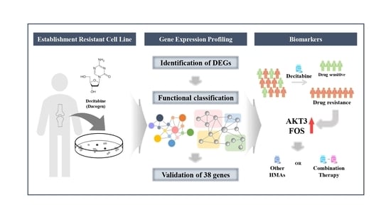
Myelodysplastic syndrome (MDS) is a clonal hematopoietic stem cell disease characterized by inefficient hematopoiesis and the potential development of acute leukemia. Among the most notable advances in the treatment of MDS is the hypomethylating agent, decitabine (5-aza-2′deoxycytidine). Although decitabine is well known as an effective method for treating MDS patients, only a subset of patients respond and a tolerance often develops, leading to treatment failure. Moreover, decitabine treatment is costly and causes unnecessary toxicity. Therefore, clarifying the mechanism of decitabine resistance is important for improving its therapeutic efficacy. To this end, we established a decitabine-resistant F-36P cell line from the parental F-36P leukemia cell line, and applied a genetic approach employing next-generation sequencing, various experimental techniques, and bioinformatics tools to determine differences in gene expression and relationships among genes. Thirty-eight candidate genes encoding proteins involved in decitabine-resistant-related pathways, including immune checkpoints, the regulation of myeloid cell differentiation, and PI3K-Akt signaling, were identified. Interestingly, two of the candidate genes, AKT3 and FOS, were overexpressed in MDS patients with poor prognoses. On the basis of these results, we are pursuing development of a gene chip for diagnosing decitabine resistance in MDS patients, with the goal of ultimately improving the power to predict treatment strategies and the prognosis of MDS patients. Full article
Show Figures

Graphical abstract

20 pages, 6028 KiB  
Article
Modulation of FLT3-ITD Localization and Targeting of Distinct Downstream Signaling Pathways as Potential Strategies to Overcome FLT3-Inhibitor Resistance
by Maximilian Fleischmann, Mike Fischer, Ulf Schnetzke, Colin Fortner, Joanna Kirkpatrick, Florian H. Heidel, Andreas Hochhaus and Sebastian Scholl
Cells 2021, 10(11), 2992; https://doi.org/10.3390/cells10112992 - 3 Nov 2021
Cited by 6 | Viewed by 3437
Abstract
OBJECTIVES: Internal tandem duplications (ITDs) of the Fms-like tyrosine kinase 3 (FLT3) represent the most frequent molecular aberrations in acute myeloid leukemia (AML) and are associated with an inferior prognosis. The pattern of downstream activation by this constitutively activated receptor tyrosine kinase is [...] Read more.
OBJECTIVES: Internal tandem duplications (ITDs) of the Fms-like tyrosine kinase 3 (FLT3) represent the most frequent molecular aberrations in acute myeloid leukemia (AML) and are associated with an inferior prognosis. The pattern of downstream activation by this constitutively activated receptor tyrosine kinase is influenced by the localization of FLT3-ITD depending on its glycosylation status. Different pharmacological approaches can affect FLT3-ITD-driven oncogenic pathways by the modulation of FLT3-ITD localization. AIMS: The objective of this study was to investigate the effects of N-glycosylation inhibitors (tunicamycin or 2-deoxy-D-glucose) or the histone deacetylase inhibitor valproic acid (VPA) on FLT3-ITD localization and downstream activity. We sought to determine the potential differences between the distinct FLT3-ITD variants, particularly concerning their susceptibility towards combined treatment by addressing either N-glycosylation and the heat shock protein 90 (HSP90) by 17-AAG, or by targeting the PI3K/AKT/mTOR pathway by rapamycin after treatment with VPA. METHODS: Murine Ba/F3 leukemia cell lines were stably transfected with distinct FLT3-ITD variants resulting in IL3-independent growth. These Ba/F3 FLT3-ITD cell lines or FLT3-ITD-expressing human MOLM13 cells were exposed to tunicamycin, 2-deoxy-D-glucose or VPA, and 17-AAG or rapamycin, and characterized in terms of downstream signaling by immunoblotting. FLT3 surface expression, apoptosis, and metabolic activity were analyzed by flow cytometry or an MTS assay. Proteome analysis by liquid chromatography–tandem mass spectrometry was performed to assess differential protein expression. RESULTS: The susceptibility of FLT3-ITD-expressing cells to 17-AAG after pre-treatment with tunicamycin or 2-deoxy-D-glucose was demonstrated. Importantly, in Ba/F3 cells that were stably expressing distinct FLT3-ITD variants that were located either in the juxtamembrane domain (JMD) or in the tyrosine kinase 1 domain (TKD1), response to the sequential treatments with tunicamycin and 17-AAG varied between individual FLT3-ITD motifs without dependence on the localization of the ITD. In all of the FLT3-ITD cell lines that were investigated, incubation with tunicamycin was accompanied by intracellular retention of FLT3-ITD due to the inhibition of glycosylation. In contrast, treatment of Ba/F3-FLT3-ITD cells with VPA was associated with a significant increase of FLT3-ITD surface expression depending on FLT3 protein synthesis. The allocation of FLT3 to different cellular compartments that was induced by tunicamycin, 2-deoxy-D-glucose, or VPA resulted in the activation of distinct downstream signaling pathways. Whole proteome analyses of Ba/F3 FLT3-ITD cells revealed up-regulation of the relevant chaperone proteins (e.g., calreticulin, calnexin, HSP90beta1) that are directly involved in the stabilization of FLT3-ITD or in its retention in the ER compartment. CONCLUSION: The allocation of FLT3-ITD to different cellular compartments and targeting distinct downstream signaling pathways by combined treatment with N-glycosylation and HSP90 inhibitors or VPA and rapamycin might represent new therapeutic strategies to overcome resistance towards tyrosine kinase inhibitors in FLT3-ITD-positive AML. The treatment approaches addressing N-glycosylation of FLT3-ITD appear to depend on patient-specific FLT3-ITD sequences, potentially affecting the efficacy of such pharmacological strategies. Full article
Show Figures

Figure 1

23 pages, 1848 KiB  
Review
A Role for the Bone Marrow Microenvironment in Drug Resistance of Acute Myeloid Leukemia
by Seyed Mohammadreza Bolandi, Mahdi Pakjoo, Peyman Beigi, Mohammad Kiani, Ali Allahgholipour, Negar Goudarzi, Jamshid S. Khorashad and Anna M. Eiring
Cells 2021, 10(11), 2833; https://doi.org/10.3390/cells10112833 - 21 Oct 2021
Cited by 19 | Viewed by 4855
Abstract
Acute myeloid leukemia (AML) is a heterogeneous disease with a poor prognosis and remarkable resistance to chemotherapeutic agents. Understanding resistance mechanisms against currently available drugs helps to recognize the therapeutic obstacles. Various mechanisms of resistance to chemotherapy or targeted inhibitors have been described [...] Read more.
Acute myeloid leukemia (AML) is a heterogeneous disease with a poor prognosis and remarkable resistance to chemotherapeutic agents. Understanding resistance mechanisms against currently available drugs helps to recognize the therapeutic obstacles. Various mechanisms of resistance to chemotherapy or targeted inhibitors have been described for AML cells, including a role for the bone marrow niche in both the initiation and persistence of the disease, and in drug resistance of the leukemic stem cell (LSC) population. The BM niche supports LSC survival through direct and indirect interactions among the stromal cells, hematopoietic stem/progenitor cells, and leukemic cells. Additionally, the BM niche mediates changes in metabolic and signal pathway activation due to the acquisition of new mutations or selection and expansion of a minor clone. This review briefly discusses the role of the BM microenvironment and metabolic pathways in resistance to therapy, as discovered through AML clinical studies or cell line and animal models. Full article
Show Figures

Figure 1

15 pages, 3658 KiB  
Article
Distinct Clinical Impact and Biological Function of Angiopoietin and Angiopoietin-like Proteins in Human Breast Cancer
by Hui Yang, Melody Zhang, Xuan-Yu Mao, Hang Chang, Jesus Perez-Losada and Jian-Hua Mao
Cells 2021, 10(10), 2590; https://doi.org/10.3390/cells10102590 - 29 Sep 2021
Cited by 9 | Viewed by 2822
Abstract
Secreted angiopoietin/angiopoietin-like (ANGPT/ANGPTL) proteins are involved in many biological processes. However, the role of these proteins in human breast cancers (BCs) remains largely unclear. Here, we conducted integrated omics analyses to evaluate the clinical impact of ANGPT/ANGPTL [...] Read more.
Secreted angiopoietin/angiopoietin-like (ANGPT/ANGPTL) proteins are involved in many biological processes. However, the role of these proteins in human breast cancers (BCs) remains largely unclear. Here, we conducted integrated omics analyses to evaluate the clinical impact of ANGPT/ANGPTL proteins and to elucidate their biological functions. In BCs, we identified rare mutations in ANGPT/ANGPTL genes, frequent gains of ANGPT1, ANGPT4, and ANGPTL1, and frequent losses of ANGPT2, ANGPTL5, and ANGPTL7, but observed that ANGPTL1, 2, and 4 were robustly downregulated in multiple datasets. The expression levels of ANGPTL1, 5, and 8 were positively correlated with overall survival (OS), while the expression levels of ANGPTL4 were negatively correlated with OS. Additionally, the expression levels of ANGPTL1 and 7 were positively correlated with distant metastasis-free survival (DMFS), while the expression levels of ANGPT2 and ANGPTL4 were negatively correlated with DMFS. The prognostic impacts of ANGPT/ANGPTL genes depended on the molecular subtypes and on clinical factors. We discovered that various ANGPT/ANGPTL genes were co-expressed with various genes involved in different pathways. Finally, with the exception of ANGPTL3, the remaining genes showed significant correlations with cancer-associated fibroblasts, endothelial cells, and microenvironment score, whereas only ANGPTL6 was significantly correlated with immune score. Our findings provide strong evidence for the distinct clinical impact and biological function of ANGPT/ANGPTL proteins, but the question of whether some of them could be potential therapeutic targets still needs further investigation in BCs. Full article
Show Figures

Figure 1

12 pages, 1558 KiB  
Article
Targeting Asparagine and Serine Metabolism in Germinal Centre-Derived B Cells Non-Hodgkin Lymphomas (B-NHL)
by Zuhal Eraslan, Grigorios Papatzikas, Jean-Baptiste Cazier, Farhat L. Khanim and Ulrich L. Günther
Cells 2021, 10(10), 2589; https://doi.org/10.3390/cells10102589 - 29 Sep 2021
Cited by 6 | Viewed by 2684
Abstract
BL and DLBCL are subtypes of B-cell lymphomas that arise from germinal centre B lymphocytes. Differentiation between BL and DLBCL is critical and can be challenging, as these two types of cancer share the same morphological, immunophenotypic, and genetic characteristics. In this study, [...] Read more.
BL and DLBCL are subtypes of B-cell lymphomas that arise from germinal centre B lymphocytes. Differentiation between BL and DLBCL is critical and can be challenging, as these two types of cancer share the same morphological, immunophenotypic, and genetic characteristics. In this study, we have examined metabolism in BL and DLBCL lymphomas and found distinctive differences in serine metabolism. We show that BL cells consume significantly more extracellular asparagine than DLBCL cells. Using a tracer-based approach, we find that asparagine regulates the serine uptake and serine synthesis in BL and DLBCL cells. Calculation of Differentially Expressed Genes (DEGs) from RNAseq datasets of BL and DLBCL patients show that BL cancers express the genes involved in serine synthesis at a higher level than DLBCL. Remarkably, combined use of an inhibitor of serine biosynthesis pathway and an anticancer drug asparaginase increases the sensitivity of BL cells to extracellular asparagine deprivation without inducing a change in the sensitivity of DLBCL cells to asparaginase. In summary, our study unravels metabolic differences between BL and DLBCL with diagnostic potential which may also open new avenues for treatment. Full article
Show Figures

Figure 1

20 pages, 4374 KiB  
Article
A Preclinical Investigation of GBM-N019 as a Potential Inhibitor of Glioblastoma via Exosomal mTOR/CDK6/STAT3 Signaling
by Alexander T. H. Wu, Hsu-Shan Huang, Ya-Ting Wen, Bashir Lawal, Ntlotlang Mokgautsi, Thanh-Tuan Huynh, Michael Hsiao and Li Wei
Cells 2021, 10(9), 2391; https://doi.org/10.3390/cells10092391 - 11 Sep 2021
Cited by 6 | Viewed by 3844
Abstract
Glioblastoma (GBM) is one of the most aggressive brain malignancies with high incidences of developing treatment resistance, resulting in poor prognoses. Glioma stem cell (GSC)-derived exosomes are important players that contribute to GBM tumorigenesis and aggressive properties. Herein, we investigated the inhibitory roles [...] Read more.
Glioblastoma (GBM) is one of the most aggressive brain malignancies with high incidences of developing treatment resistance, resulting in poor prognoses. Glioma stem cell (GSC)-derived exosomes are important players that contribute to GBM tumorigenesis and aggressive properties. Herein, we investigated the inhibitory roles of GBM-N019, a novel small molecule on the transfer of aggressive and invasive properties through the delivery of oncogene-loaded exosomes from GSCs to naïve and non-GSCs. Our results indicated that GBM-N019 significantly downregulated the expressions of the mammalian target of rapamycin (mTOR), signal transducer and activator of transcription 3 (STAT3), and cyclin-dependent kinase 6 (CDK6) signaling networks with concomitant inhibitory activities against viability, clonogenicity, and migratory abilities of U251 and U87MG cells. Treatments with GBM-N019 halted the exosomal transfer of protein kinase B (Akt), mTOR, p-mTOR, and Ras-related protein RAB27A to the naïve U251 and U87MG cells, and rescued the cells from invasive and stemness properties that were associated with activation of these oncogenes. GBM-N019 also synergized with and enhanced the anti-GBM activities of palbociclib in vitro and in vivo. In conclusion, our results suggested that GBM-N019 possesses good translational relevance as a potential anti-glioblastoma drug candidate worthy of consideration for clinical trials against recurrent glioblastomas. Full article
Show Figures

Figure 1

16 pages, 2986 KiB  
Article
26S Proteasome Non-ATPase Regulatory Subunits 1 (PSMD1) and 3 (PSMD3) as Putative Targets for Cancer Prognosis and Therapy
by Andres J. Rubio, Alfonso E. Bencomo-Alvarez, James E. Young, Vanessa V. Velazquez, Joshua J. Lara, Mayra A. Gonzalez and Anna M. Eiring
Cells 2021, 10(9), 2390; https://doi.org/10.3390/cells10092390 - 11 Sep 2021
Cited by 15 | Viewed by 4281
Abstract
Ever since the ubiquitin proteasome system was characterized, efforts have been made to manipulate its function to abrogate the progression of cancer. As a result, the anti-cancer drugs bortezomib, carfilzomib, and ixazomib targeting the 26S proteasome were developed to treat multiple myeloma, mantle [...] Read more.
Ever since the ubiquitin proteasome system was characterized, efforts have been made to manipulate its function to abrogate the progression of cancer. As a result, the anti-cancer drugs bortezomib, carfilzomib, and ixazomib targeting the 26S proteasome were developed to treat multiple myeloma, mantle cell lymphoma, and diffuse large B-cell lymphoma, among others. Despite success, adverse side effects and drug resistance are prominent, raising the need for alternative therapeutic options. We recently demonstrated that knockdown of the 19S regulatory components, 26S proteasome non-ATPase subunits 1 (PSMD1) and 3 (PSMD3), resulted in increased apoptosis of chronic myeloid leukemia (CML) cells, but had no effect on normal controls, suggesting they may be good targets for therapy. Therefore, we hypothesized that PSMD1 and PSMD3 are potential targets for anti-cancer therapeutics and that their relevance stretches beyond CML to other types of cancers. In the present study, we analyzed PSMD1 and PSMD3 mRNA and protein expression in cancerous tissue versus normal controls using data from The Cancer Genome Atlas (TCGA) and the Clinical Proteomic Tumor Analysis Consortium (CPTAC), comparing expression with overall survival. Altogether, our data suggest that PSMD1 and PSMD3 may be novel putative targets for cancer prognosis and therapy that are worthy of future investigation. Full article
Show Figures

Figure 1

30 pages, 2365 KiB  
Article
Phosphorylation-Mediated Molecular Pathway Changes in Human Pituitary Neuroendocrine Tumors Identified by Quantitative Phosphoproteomics
by Jiajia Li, Siqi Wen, Biao Li, Na Li and Xianquan Zhan
Cells 2021, 10(9), 2225; https://doi.org/10.3390/cells10092225 - 27 Aug 2021
Cited by 7 | Viewed by 3076
Abstract
To investigate the biological role of protein phosphorylation in human nonfunctional pituitary neuroendocrine tumors (NF-PitNETs), proteins extracted from NF-PitNET and control tissues were analyzed with tandem mass tag (TMT)-based quantitative proteomics coupled with TiO2 enrichment of phosphopeptides. A total of 595 differentially [...] Read more.
To investigate the biological role of protein phosphorylation in human nonfunctional pituitary neuroendocrine tumors (NF-PitNETs), proteins extracted from NF-PitNET and control tissues were analyzed with tandem mass tag (TMT)-based quantitative proteomics coupled with TiO2 enrichment of phosphopeptides. A total of 595 differentially phosphorylated proteins (DPPs) with 1412 phosphosites were identified in NF-PitNETs compared to controls (p < 0.05). KEGG pathway network analysis of 595 DPPs identified nine statistically significant signaling pathways, including the spliceosome pathway, the RNA transport pathway, proteoglycans in cancer, SNARE interactions in vesicular transport, platelet activation, bacterial invasion of epithelial cells, tight junctions, vascular smooth muscle contraction, and protein processing in the endoplasmic reticulum. GO analysis revealed that these DPPs were involved in multiple cellular components (CCs), biological processes (BPs), and molecule functions (MFs). The kinase analysis of 595 DPPs identified seven kinases, including GRP78, WSTF, PKN2, PRP4, LOK, NEK1, and AMPKA1, and the substrate of these kinases could provide new ideas for seeking drug targets for NF-PitNETs. The randomly selected DPP calnexin was further confirmed with immunoprecipitation (IP) and Western blot (WB). These findings provide the first DPP profiling, phosphorylation-mediated molecular network alterations, and the key kinase profiling in NF-PitNET pathogenesis, which are a precious resource for understanding the biological roles of protein phosphorylation in NF-PitNET pathogenesis and discovering effective phosphoprotein biomarkers and therapeutic targets and drugs for the management of NF-PitNETs. Full article
Show Figures

Figure 1

14 pages, 37236 KiB  
Article
Prognostic Value of a Glycolytic Signature and Its Regulation by Y-Box-Binding Protein 1 in Triple-Negative Breast Cancer
by Yi-Wen Lai, Wen-Jing Hsu, Wen-Ying Lee, Cheng-Hsun Chen, Ying-Huei Tsai, Jia-Zih Dai, Ching-Chieh Yang and Cheng-Wei Lin
Cells 2021, 10(8), 1890; https://doi.org/10.3390/cells10081890 - 26 Jul 2021
Cited by 14 | Viewed by 3089
Abstract
Triple-negative breast cancer (TNBC) is the most malignant subtype of breast cancer as it shows a high capacity for metastasis and poor prognoses. Metabolic reprogramming is one of the hallmarks of cancer, and aberrant glycolysis was reported to be upregulated in TNBC. Thus, [...] Read more.
Triple-negative breast cancer (TNBC) is the most malignant subtype of breast cancer as it shows a high capacity for metastasis and poor prognoses. Metabolic reprogramming is one of the hallmarks of cancer, and aberrant glycolysis was reported to be upregulated in TNBC. Thus, identifying metabolic biomarkers for diagnoses and investigating cross-talk between glycolysis and invasiveness could potentially enable the development of therapeutics for patients with TNBC. In order to determine novel and reliable metabolic biomarkers for predicting clinical outcomes of TNBC, we analyzed transcriptome levels of glycolysis-related genes in various subtypes of breast cancer from public databases and identified a distinct glycolysis gene signature, which included ENO1, SLC2A6, LDHA, PFKP, PGAM1, and GPI, that was elevated and associated with poorer prognoses of TNBC patients. Notably, we found a transcription factor named Y-box-binding protein 1 (YBX1) to be strongly associated with this glycolysis gene signature, and it was overexpressed in TNBC. A mechanistic study further validated that YBX1 was upregulated in TNBC cell lines, and knockdown of YBX1 suppressed expression of those glycolytic genes. Moreover, YBX1 expression was positively associated with epithelial-to-mesenchymal transition (EMT) genes in breast cancer patients, and suppression of YBX1 downregulated expressions of EMT-related genes and tumor migration and invasion in MDA-MB-231 and BT549 TNBC cells. Our data revealed an YBX1-glycolysis-EMT network as an attractive diagnostic marker and metabolic target in TNBC patients. Full article
Show Figures

Figure 1

15 pages, 2309 KiB  
Article
Identification of a New Transcriptional Co-Regulator of STEAP1 in Ewing’s Sarcoma
by Fatu Badiane Markey, Brigette Romero, Vijay Parashar and Mona Batish
Cells 2021, 10(6), 1300; https://doi.org/10.3390/cells10061300 - 24 May 2021
Cited by 6 | Viewed by 2999
Abstract
Ewing’s sarcoma (ES) is caused by a chromosomal translocation leading to the formation of the fused EWSFLI1 gene, which codes for an aberrant transcription factor EWSFLI1. The transcriptional targets of EWSFLI1 have been viewed as promising and novel drug targets in the treatment [...] Read more.
Ewing’s sarcoma (ES) is caused by a chromosomal translocation leading to the formation of the fused EWSFLI1 gene, which codes for an aberrant transcription factor EWSFLI1. The transcriptional targets of EWSFLI1 have been viewed as promising and novel drug targets in the treatment of ES. One such target is six transmembrane epithelial antigen of the prostate 1 (STEAP1), a transmembrane protein that is upregulated by EWSFLI1 in ES. STEAP1 is a hallmark of tumor invasiveness and an indicator of tumor responsiveness to therapy. EWSFLI1 binds to the STEAP1 promoter region, but the mechanism of action by which it upregulates STEAP1 expression in ES is not entirely understood. Upon analysis of the STEAP1 promoter, we predicted two binding sites for NKX2.2, another crucial transcription factor involved in ES pathogenesis. We confirmed the interaction of NKX2.2 with the STEAP1 promoter using chromatin immunoprecipitation (ChIP) analysis. We used single-molecule RNA imaging, biochemical, and genetic studies to identify the novel role of NKX2.2 in regulating STEAP1 expression in ES. Our results show that NKX2.2 is a co-regulator of STEAP1 expression and functions by interacting with the STEAP1 promoter at sites proximal to the reported EWSFLI1 sites. The co-operative interaction of NKX2.2 with EWSFLI1 in regulating STEAP1 holds potential as a new target for therapeutic interventions for ES. Full article
Show Figures

Figure 1

27 pages, 4677 KiB  
Review
Modelling the Functions of Polo-Like Kinases in Mice and Their Applications as Cancer Targets with a Special Focus on Ovarian Cancer
by Monika Kressin, Daniela Fietz, Sven Becker and Klaus Strebhardt
Cells 2021, 10(5), 1176; https://doi.org/10.3390/cells10051176 - 12 May 2021
Cited by 13 | Viewed by 4481
Abstract
Polo-like kinases (PLKs) belong to a five-membered family of highly conserved serine/threonine kinases (PLK1-5) that play differentiated and essential roles as key mitotic kinases and cell cycle regulators and with this in proliferation and cellular growth. Besides, evidence is accumulating for complex and [...] Read more.
Polo-like kinases (PLKs) belong to a five-membered family of highly conserved serine/threonine kinases (PLK1-5) that play differentiated and essential roles as key mitotic kinases and cell cycle regulators and with this in proliferation and cellular growth. Besides, evidence is accumulating for complex and vital non-mitotic functions of PLKs. Dysregulation of PLKs is widely associated with tumorigenesis and by this, PLKs have gained increasing significance as attractive targets in cancer with diagnostic, prognostic and therapeutic potential. PLK1 has proved to have strong clinical relevance as it was found to be over-expressed in different cancer types and linked to poor patient prognosis. Targeting the diverse functions of PLKs (tumor suppressor, oncogenic) are currently at the center of numerous investigations in particular with the inhibition of PLK1 and PLK4, respectively in multiple cancer trials. Functions of PLKs and the effects of their inhibition have been extensively studied in cancer cell culture models but information is rare on how these drugs affect benign tissues and organs. As a step further towards clinical application as cancer targets, mouse models therefore play a central role. Modelling PLK function in animal models, e.g., by gene disruption or by treatment with small molecule PLK inhibitors offers promising possibilities to unveil the biological significance of PLKs in cancer maintenance and progression and give important information on PLKs’ applicability as cancer targets. In this review we aim at summarizing the approaches of modelling PLK function in mice so far with a special glimpse on the significance of PLKs in ovarian cancer and of orthotopic cancer models used in this fatal malignancy. Full article
Show Figures

Figure 1

12 pages, 662 KiB  
Review
Microtubule-Associated Protein ATIP3, an Emerging Target for Personalized Medicine in Breast Cancer
by Maria M. Haykal, Sylvie Rodrigues-Ferreira and Clara Nahmias
Cells 2021, 10(5), 1080; https://doi.org/10.3390/cells10051080 - 1 May 2021
Cited by 6 | Viewed by 3666
Abstract
Breast cancer is the leading cause of death by malignancy among women worldwide. Clinical data and molecular characteristics of breast tumors are essential to guide clinician’s therapeutic decisions. In the new era of precision medicine, that aims at personalizing the treatment for each [...] Read more.
Breast cancer is the leading cause of death by malignancy among women worldwide. Clinical data and molecular characteristics of breast tumors are essential to guide clinician’s therapeutic decisions. In the new era of precision medicine, that aims at personalizing the treatment for each patient, there is urgent need to identify robust companion biomarkers for new targeted therapies. This review focuses on ATIP3, a potent anti-cancer protein encoded by candidate tumor suppressor gene MTUS1, whose expression levels are markedly down-regulated in breast cancer. ATIP3 is a microtubule-associated protein identified both as a prognostic biomarker of patient survival and a predictive biomarker of breast tumors response to taxane-based chemotherapy. We present here recent studies pointing out ATIP3 as an emerging anti-cancer protein and a potential companion biomarker to be combined with future personalized therapy against ATIP3-deficient breast cancer. Full article
Show Figures

Figure 1

17 pages, 5474 KiB  
Article
The ECM Modulator ITIH5 Affects Cell Adhesion, Motility and Chemotherapeutic Response of Basal/Squamous-Like (BASQ) Bladder Cancer Cells
by Michael Rose, Erik Noetzel, Jennifer Kistermann, Julian Eschenbruch, Sandra Rushrush, Lin Gan, Ruth Knüchel, Nadine T. Gaisa and Edgar Dahl
Cells 2021, 10(5), 1038; https://doi.org/10.3390/cells10051038 - 28 Apr 2021
Cited by 16 | Viewed by 3189
Abstract
This study aims at characterizing the role of the putative tumor suppressor ITIH5 in basal-type bladder cancers (BLCA). By sub-classifying TCGA BLCA data, we revealed predominant loss of ITIH5 expression in the basal/squamous-like (BASQ) subtype. ITIH5 expression inversely correlated with basal-type makers such [...] Read more.
This study aims at characterizing the role of the putative tumor suppressor ITIH5 in basal-type bladder cancers (BLCA). By sub-classifying TCGA BLCA data, we revealed predominant loss of ITIH5 expression in the basal/squamous-like (BASQ) subtype. ITIH5 expression inversely correlated with basal-type makers such as KRT6A and CD44. Interestingly, Kaplan–Meier analyses showed longer recurrence-free survival in combination with strong CD44 expression, which is thought to mediate ITIH-hyaluronan (HA) binding functions. In vitro, stable ITIH5 overexpression in two basal-type BLCA cell lines showing differential CD44 expression levels, i.e., with (SCaBER) and without squamous features (HT1376), demonstrated clear inhibition of cell and colony growth of BASQ-type SCaBER cells. ITIH5 further enhanced HA-associated cell-matrix attachment, indicated by altered size and number of focal adhesion sites resulting in reduced cell migration capacities. Transcriptomic analyses revealed enrichment of pathways and processes involved in ECM organization, differentiation and cell signaling. Finally, we provide evidence that ITIH5 increase sensitivity of SCaBER cells to chemotherapeutical agents (cisplatin and gemcitabine), whereas responsiveness of HT1376 cells was not affected by ITIH5 expression. Thus, we gain further insights into the putative role of ITIH5 as tumor suppressor highlighting an impact on drug response potentially via the HA-CD44 axis in BASQ-type BLCA. Full article
Show Figures

Figure 1

30 pages, 1727 KiB  
Review
Signaling Pathways in Cancer: Therapeutic Targets, Combinatorial Treatments, and New Developments
by Hon Yan Kelvin Yip and Antonella Papa
Cells 2021, 10(3), 659; https://doi.org/10.3390/cells10030659 - 16 Mar 2021
Cited by 107 | Viewed by 15414
Abstract
Molecular alterations in cancer genes and associated signaling pathways are used to inform new treatments for precision medicine in cancer. Small molecule inhibitors and monoclonal antibodies directed at relevant cancer-related proteins have been instrumental in delivering successful treatments of some blood malignancies (e.g., [...] Read more.
Molecular alterations in cancer genes and associated signaling pathways are used to inform new treatments for precision medicine in cancer. Small molecule inhibitors and monoclonal antibodies directed at relevant cancer-related proteins have been instrumental in delivering successful treatments of some blood malignancies (e.g., imatinib with chronic myelogenous leukemia (CML)) and solid tumors (e.g., tamoxifen with ER positive breast cancer and trastuzumab for HER2-positive breast cancer). However, inherent limitations such as drug toxicity, as well as acquisition of de novo or acquired mechanisms of resistance, still cause treatment failure. Here we provide an up-to-date review of the successes and limitations of current targeted therapies for cancer treatment and highlight how recent technological advances have provided a new level of understanding of the molecular complexity underpinning resistance to cancer therapies. We also raise three basic questions concerning cancer drug discovery based on molecular markers and alterations of selected signaling pathways, and further discuss how combination therapies may become the preferable approach over monotherapy for cancer treatments. Finally, we consider novel therapeutic developments that may complement drug delivery and significantly improve clinical response and outcomes of cancer patients. Full article
Show Figures

Figure 1

15 pages, 1178 KiB  
Article
Genetically Raised Circulating Bilirubin Levels and Risk of Ten Cancers: A Mendelian Randomization Study
by Nazlisadat Seyed Khoei, Robert Carreras-Torres, Neil Murphy, Marc J. Gunter, Paul Brennan, Karl Smith-Byrne, Daniela Mariosa, James Mckay, Tracy A. O’Mara, Ruth Jarrett, Henrik Hjalgrim, Karin E. Smedby, Wendy Cozen, Kenan Onel, Arjan Diepstra, Karl-Heinz Wagner and Heinz Freisling
Cells 2021, 10(2), 394; https://doi.org/10.3390/cells10020394 - 15 Feb 2021
Cited by 18 | Viewed by 6361
Abstract
Bilirubin, an endogenous antioxidant, may play a protective role in cancer development. We applied two-sample Mendelian randomization to investigate whether genetically raised bilirubin levels are causally associated with the risk of ten cancers (pancreas, kidney, endometrium, ovary, breast, prostate, lung, Hodgkin’s lymphoma, melanoma, [...] Read more.
Bilirubin, an endogenous antioxidant, may play a protective role in cancer development. We applied two-sample Mendelian randomization to investigate whether genetically raised bilirubin levels are causally associated with the risk of ten cancers (pancreas, kidney, endometrium, ovary, breast, prostate, lung, Hodgkin’s lymphoma, melanoma, and neuroblastoma). The number of cases and their matched controls of European descent ranged from 122,977 and 105,974 for breast cancer to 1200 and 6417 for Hodgkin’s lymphoma, respectively. A total of 115 single-nucleotide polymorphisms (SNPs) associated (p < 5 × 10⁻⁸) with circulating total bilirubin, extracted from a genome-wide association study in the UK Biobank, were used as instrumental variables. One SNP (rs6431625) in the promoter region of the uridine-diphosphoglucuronate glucuronosyltransferase1A1 (UGT1A1) gene explained 16.9% and the remaining 114 SNPs (non-UGT1A1 SNPs) explained 3.1% of phenotypic variance in circulating bilirubin levels. A one-standarddeviation increment in circulating bilirubin (≈ 4.4 µmol/L), predicted by non-UGT1A1 SNPs, was inversely associated with risk of squamous cell lung cancer and Hodgkin’s lymphoma (odds ratio (OR) 0.85, 95% confidence interval (CI) 0.73–0.99, P 0.04 and OR 0.64, 95% CI 0.42–0.99, p 0.04, respectively), which was confirmed after removing potential pleiotropic SNPs. In contrast, a positive association was observed with the risk of breast cancer after removing potential pleiotropic SNPs (OR 1.12, 95% CI 1.04–1.20, p 0.002). There was little evidence for robust associations with the other seven cancers investigated. Genetically raised bilirubin levels were inversely associated with risk of squamous cell lung cancer as well as Hodgkin’s lymphoma and positively associated with risk of breast cancer. Further studies are required to investigate the utility of bilirubin as a low-cost clinical marker to improve risk prediction for certain cancers. Full article
Show Figures

Figure 1

Back to TopTop