Altered Brain Complexity in Women with Primary Dysmenorrhea: A Resting-State Magneto-Encephalography Study Using Multiscale Entropy Analysis

 ,
,  ,
,
Abstract
:1. Introduction
2. Materials and Methods
2.1. Participants
2.2. Demographic, Menstrual Features, Pain Experiences, and Psychological Characteristics
2.3. Data Acquisition
2.3.1. Resting-State Magnetoencephalography (MEG) Signals Acquisition
2.3.2. Structural MRI T1 Images Acquisition
2.4. Source Analyses
2.4.1. Preprocessing
2.4.2. Source Reconstruction
2.5. Feature Extraction
2.5.1. Feature Extraction of Brain Complexity Features via Nonlinear Analysis
Sample Entropy
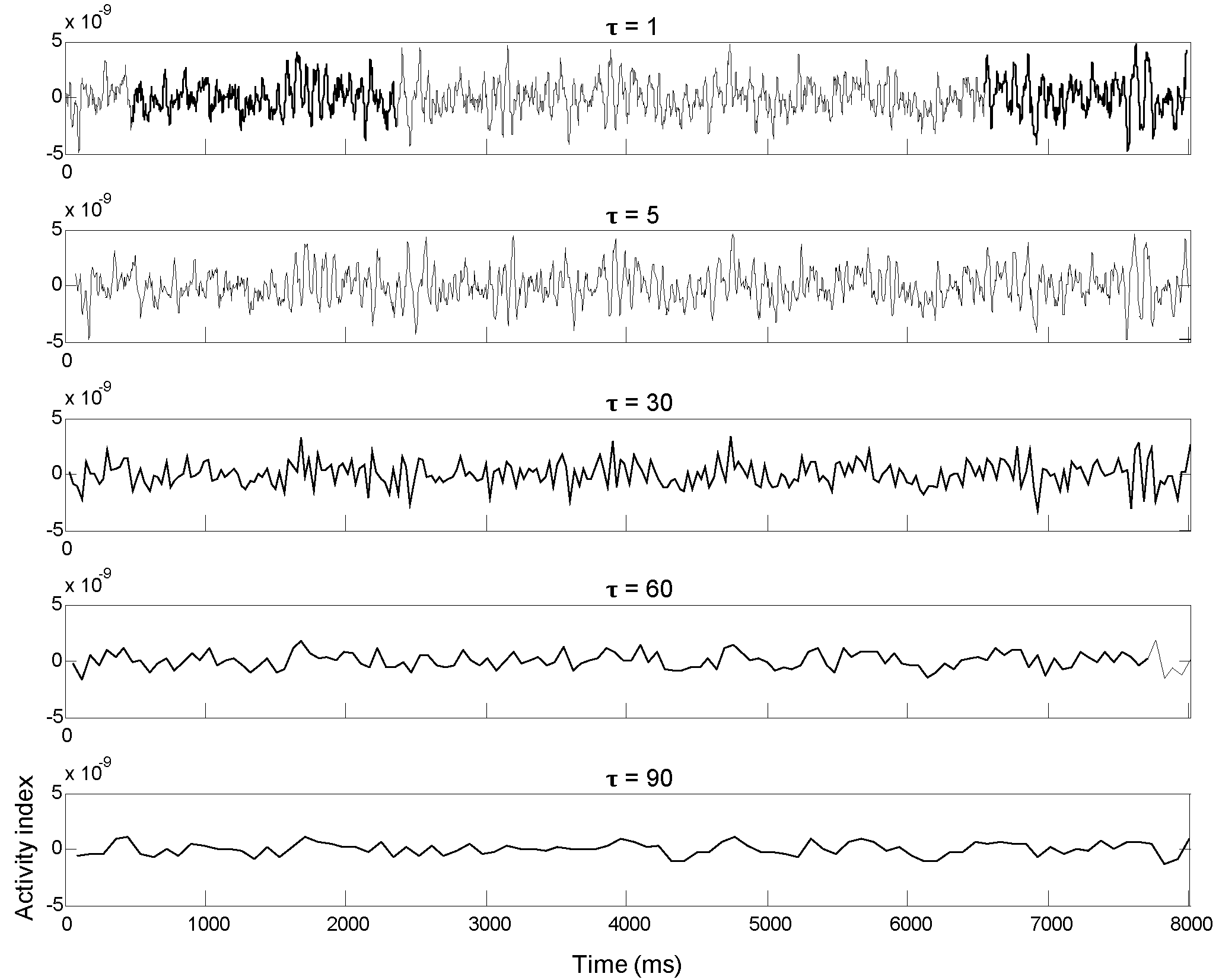
Multiscale Sample Entropy
Shannon Spectral Entropy
Lempel-Ziv Complexity
2.5.2. Feature Extraction of Brain Spectral Features in the Frequency Domain
Relative Band Power
Median Frequency
Spectral Edge Frequency
2.5.3. Regional Features of Resting-State Networks
2.5.4. Asymmetry Features
2.6. Statistical Analyses
2.6.1. Demographic, Menstrual Features, Pain Experiences, and Psychological Characteristics
2.6.2. Brain Features
2.6.3. Correlations between Pain Experiences, Psychological Traits, and Brain Features
3. Results
3.1. Demographic, Menstrual Features, Pain Experiences, and Psychological Characteristics
3.1.1. PDMs and CONs had Similar Demographic Characteristics and Menstrual Features
3.1.2. PDMs Experienced Long-Term Moderate-to-Severe Menstrual Pain
3.1.3. PDMs Displayed Significantly Higher Anxiety, Depression, and Pain Catastrophizing Characteristics than CONs
3.2. Brain Complexity, Spectral, and Hemispheric Asymmetry Features
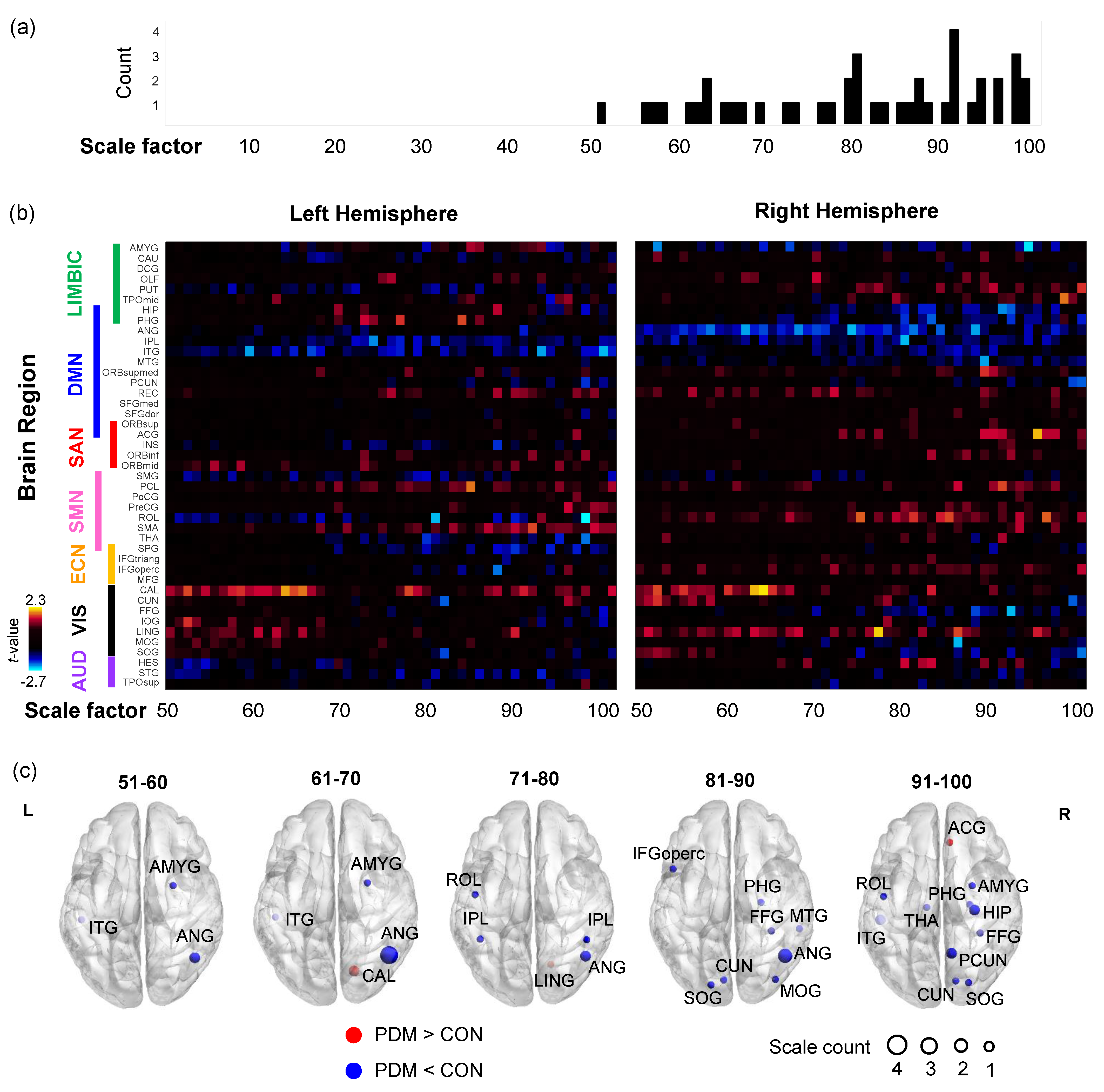
3.2.1. Brain Complexity Feature: Multiscale Sample Entropy
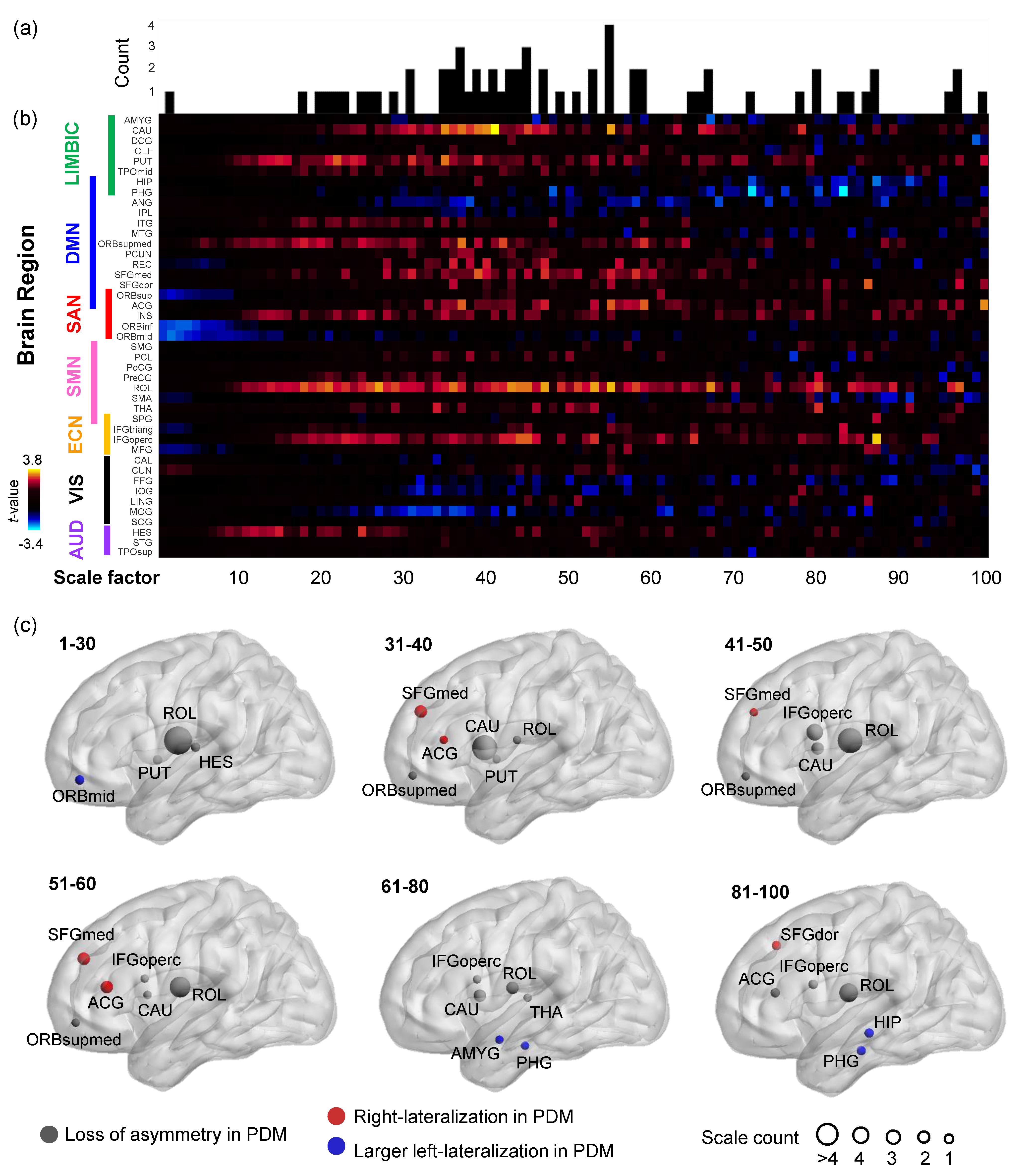
3.2.2. Hemispheric Asymmetry of Multiscale Sample Entropy
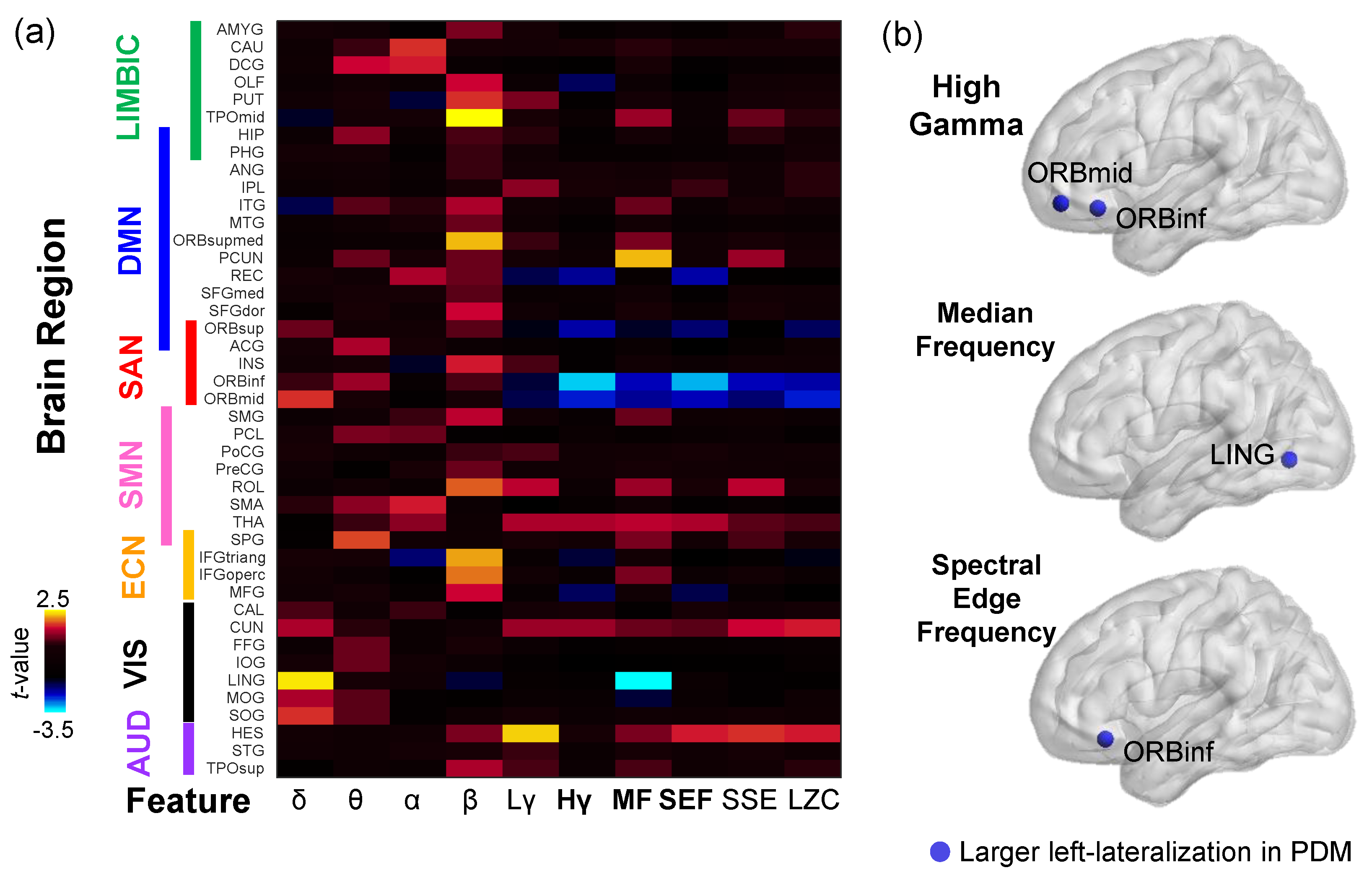
3.2.3. Brain Spectral Features, Other Complexity Features, and Their Hemispheric Asymmetry
3.3. Correlations between Multiscale Sample Entropy and Pain Experiences/Psychological Characteristics
4. Discussion
4.1. Measures of Neural Complexity
4.2. Clinical Implications of Altered Brain Complexity at Rest in Chronic Pain
4.3. Entropy with Multiple Scales Corresponding to Various Ranges of Frequency
4.4. MSE Versus Spectral Analyses
4.5. Limitations
5. Conclusions
Supplementary Materials
Acknowledgments
Author Contributions
Conflicts of Interest
Appendix A
References
- Merskey, H.; Bogduk, N. Classification of Chronic Pain: Descriptions of Chronic Pain Syndromes and Definitions of Pain Terms, 2nd ed.; Harold Merskey, N.B., Ed.; IASP Press: Seattle, WA, USA, 2002. [Google Scholar]
- Treede, R.-D.; Rief, W.; Barke, A.; Aziz, Q.; Bennett, M.I.; Benoliel, R.; Cohen, M.; Evers, S.; Finnerup, N.B.; First, M.B.; et al. A classification of chronic pain for ICD-11. Pain 2015, 156, 1003–1007. [Google Scholar] [CrossRef] [PubMed] [Green Version]
- Baliki, M.N.; Schnitzer, T.J.; Bauer, W.R.; Apkarian, A.V. Brain morphological signatures for chronic pain. PLoS ONE 2011, 6, e26010. [Google Scholar] [CrossRef]
- Smith, D.; Wilkie, R.; Uthman, O.; Jordan, J.L.; McBeth, J. Chronic pain and mortality: A systematic review. PLoS ONE 2014, 9, e99048. [Google Scholar]
- Coco, A.S. Primary dysmenorrhea. Am. Fam. Phys. 1999, 60, 489–496. [Google Scholar]
- Dawood, M.Y. Primary Dysmenorrhea: Advances in Pathogenesis and Management. Obstet. Gynecol. 2006, 108, 428–441. [Google Scholar] [CrossRef] [PubMed]
- Iacovides, S.; Avidon, I.; Baker, F.C. What we know about primary dysmenorrhea today: A critical review. Hum. Reprod. Updat. 2015, 21, 762–778. [Google Scholar] [CrossRef] [PubMed]
- Dawood, M.Y. Dysmenorrhea. J. Reprod. Med. 1985, 30, 154–167. [Google Scholar] [PubMed]
- Proctor, M.L.; Smith, C.A.; Farquhar, C.M.; Stones, R.W. Transcutaneous electrical nerve stimulation and acupuncture for primary dysmenorrhoea. Cochrane Database Syst. Rev. 2002, CD002123. [Google Scholar] [CrossRef]
- IASP Taxonomy Working Group. Visceral and Other Syndromes of the Trunk Apart from Spinal and Radicular Pain. Classification of Chronic Pain, 2nd Edition (Revised). Available online: https://www.iasp-pain.org/files/Content/ContentFolders/Publications2/ClassificationofChronicPain/Part_II-F.pdf (accessed on 12 September 2017).
- Lee, L.-C.; Tu, C.-H.; Chen, L.-F.; Shen, H.-D.; Chao, H.-T.; Lin, M.-W.; Hsieh, J.-C. Association of brain-derived neurotrophic factor gene VAL66MET polymorphism with primary dysmenorrhea. PLoS ONE 2014, 9, e112766. [Google Scholar] [CrossRef]
- Balık, G.; Ustüner, I.; Kağıtcı, M.; Sahin, F.K. Is there a relationship between mood disorders and dysmenorrhea? J. Pediatr. Adolesc. Gynecol. 2014, 27, 371–374. [Google Scholar] [CrossRef] [PubMed]
- Wei, S.-Y.; Chao, H.-T.; Tu, C.-H.; Li, W.-C.; Low, I.; Chuang, C.-Y.; Chen, L.-F.; Hsieh, J.-C. Changes in functional connectivity of pain modulatory systems in women with primary dysmenorrhea. Pain 2015, 157, 1. [Google Scholar] [CrossRef] [PubMed]
- Staud, R. Abnormal endogenous pain modulation is a shared characteristic of many chronic pain conditions. Expert Rev. Neurother. 2012, 12, 577–585. [Google Scholar] [CrossRef] [PubMed]
- Li, W.C.; Tu, C.H.; Chao, H.T.; Yeh, T.C.; Chen, L.F.; Hsieh, J.C. High prevalence of incidental brain findings in primary dysmenorrhoea. Eur. J. Pain 2015, 19, 1071–1074. [Google Scholar] [CrossRef] [PubMed]
- Liu, P.; Yang, J.; Wang, G.; Liu, Y.; Liu, X.; Jin, L.; Liang, F.; Qin, W.; Calhoun, V.D. Altered regional cortical thickness and subcortical volume in women with primary dysmenorrhoea. Eur. J. Pain 2016, 20, 512–520. [Google Scholar] [CrossRef] [PubMed]
- Tu, C.H.; Niddam, D.M.; Chao, H.T.; Chen, L.F.; Chen, Y.S.; Wu, Y.T.; Yeh, T.C.; Lirng, J.F.; Hsieh, J.C. Brain morphological changes associated with cyclic menstrual pain. Pain 2010, 150, 462–468. [Google Scholar] [CrossRef]
- Tu, C.-H.; Niddam, D.M.; Yeh, T.-C.; Lirng, J.-F.; Cheng, C.-M.; Chou, C.-C.; Chao, H.-T.; Hsieh, J.-C. Menstrual pain is associated with rapid structural alterations in the brain. Pain 2013, 154, 1718–1724. [Google Scholar] [CrossRef] [PubMed]
- Liu, P.; Liu, Y.; Wang, G.; Li, R.; Wei, Y.; Fan, Y.; Yu, Y.; Deng, D.; Qin, W. Changes of functional connectivity of the anterior cingulate cortex in women with primary dysmenorrhea. Brain Imaging Behav. 2017, 1–8. [Google Scholar] [CrossRef] [PubMed]
- Tu, C.H.; Niddam, D.M.; Chao, H.T.; Liu, R.S.; Hwang, R.J.; Yeh, T.C.; Hsieh, J.C. Abnormal cerebral metabolism during menstrual pain in primary dysmenorrhea. Neuroimage 2009, 47, 28–35. [Google Scholar] [CrossRef] [PubMed]
- Vincent, K.; Warnaby, C.; Stagg, C.J.; Moore, J.; Kennedy, S.; Tracey, I. Dysmenorrhoea is associated with central changes in otherwise healthy women. Pain 2011, 152, 1966–1975. [Google Scholar] [CrossRef] [PubMed]
- Wei, S.-Y.; Chao, H.-T.; Tu, C.-H.; Lin, M.-W.; Li, W.-C.; Low, I.; Shen, H.-D.; Chen, L.-F.; Hsieh, J.-C. The BDNF Val66Met polymorphism is associated with the functional connectivity dynamics of pain modulatory systems in primary dysmenorrhea. Sci. Rep. 2016, 6, 23639. [Google Scholar] [CrossRef] [PubMed]
- Wei, S.-Y.; Chen, L.-F.; Lin, M.-W.; Li, W.-C.; Low, I.; Yang, C.-J.; Chao, H.-T.; Hsieh, J.-C. The OPRM1 A118G polymorphism modulates the descending pain modulatory system for individual pain experience in young women with primary dysmenorrhea. Sci. Rep. 2017, 7, 39906. [Google Scholar] [CrossRef] [PubMed]
- Wu, T.-H.; Tu, C.-H.; Chao, H.-T.; Li, W.-C.; Low, I.; Chuang, C.-Y.; Yeh, T.-C.; Cheng, C.-M.; Chou, C.-C.; Chen, L.-F.; et al. Dynamic changes of functional pain connectome in women with primary dysmenorrhea. Sci. Rep. 2016, 6, 24543. [Google Scholar] [CrossRef] [PubMed]
- Zhang, B.; Xu, Y.; He, W.; Wang, J.; Chai, H.; Shen, C.; Zhu, Q.; Wang, W.; Beales, D.; Wutzler, A.; et al. Intensity dependence of auditory evoked potentials in primary dysmenorrhea. J. Pain 2017, 151, 27–43. [Google Scholar] [CrossRef] [PubMed]
- Kuo, P.C.; Chen, Y.T.; Chen, Y.S.; Chen, L.F. Decoding the perception of endogenous pain from resting-state MEG. Neuroimage 2017, 144, 1–11. [Google Scholar] [CrossRef] [PubMed]
- Stam, C.J. Nonlinear dynamical analysis of EEG and MEG: Review of an emerging field. Clin. Neurophysiol. 2005, 116, 2266–2301. [Google Scholar] [CrossRef] [PubMed]
- McDonough, I.M.; Nashiro, K.; Nagarajan, S.S.; Chang, C.; Gorgolewski, K. Network complexity as a measure of information processing across resting-state networks: Evidence from the Human Connectome Project. Front. Hum. Neurosci. 2014, 8, 1–15. [Google Scholar] [CrossRef] [PubMed]
- Tononi, G.; Sporns, O.; Edelman, G.M. A measure for brain complexity: Relating functional segregation and integration in the nervous system. Proc. Natl. Acad. Sci. USA 1994, 91, 5033–5037. [Google Scholar] [CrossRef] [PubMed]
- Friston, K.J. The labile brain. I. Neuronal transients and nonlinear coupling. Philos. Trans. R. Soc. Lond. Ser. B Biol. Sci. 2000, 355, 215–236. [Google Scholar] [CrossRef] [PubMed]
- Friston, K.J. The labile brain. II. Transients, complexity and selection. Philos. Trans. R. Soc. Lond. B Biol. Sci. 2000, 355, 237–252. [Google Scholar] [CrossRef] [PubMed]
- Nakagawa, T.T.; Jirsa, V.K.; Spiegler, A.; McIntosh, A.R.; Deco, G. Bottom up modeling of the connectome: Linking structure and function in the resting brain and their changes in aging. Neuroimage 2013, 80, 318–329. [Google Scholar] [CrossRef] [PubMed]
- Garrett, D.D.; Samanez-Larkin, G.R.; MacDonald, S.W.S.; Lindenberger, U.; McIntosh, A.R.; Grady, C.L. Moment-to-moment brain signal variability: A next frontier in human brain mapping? Neurosci. Biobehav. Rev. 2013, 37, 610–624. [Google Scholar] [CrossRef] [PubMed]
- Richman, J.S.; Moorman, J.R. Physiological time-series analysis using approximate entropy and sample entropy. Am. J. Physiol. Heart. Circ. Physiol. 2000, 278, H2039–H2049. [Google Scholar] [CrossRef] [PubMed]
- Yang, A.C.; Tsai, S.-J. Is mental illness complex? From behavior to brain. Prog. Neuro-Psychopharmacol. Biol. Psychiatry 2013, 45, 253–257. [Google Scholar] [CrossRef]
- Costa, M.; Goldberger, A.L.; Peng, C.-K. Multiscale entropy analysis of complex physiologic time series. Phys. Rev. Lett. 2002, 89, 6–9. [Google Scholar] [CrossRef] [PubMed]
- Costa, M.; Goldberger, A.L.; Peng, C.K. Multiscale entropy analysis of biological signals. Phys. Rev. E 2005, 71, 1–18. [Google Scholar] [CrossRef] [PubMed]
- Hu, M.; Liang, H. Multiscale Entropy: Recent Advances. In Complexity and Nonlinearity in Cardiovascular Signals; Springer International Publishing: Cham, Switzerland, 2017; pp. 115–138. [Google Scholar]
- Humeau-Heurtier, A. The multiscale entropy algorithm and its variants: A review. Entropy 2015, 17, 3110–3123. [Google Scholar] [CrossRef] [Green Version]
- Park, J.-H.; Kim, S.; Kim, C.-H.; Cichocki, A.; Kim, K. Multiscale entropy analysis of EEG from patients under different pathological conditions. Fractals 2007, 15, 399. [Google Scholar] [CrossRef]
- Heisz, J.J.; McIntosh, A.R. Applications of EEG neuroimaging data: Event-related potentials, spectral power, and multiscale entropy. J. Vis. Exp. 2013, 1–8. [Google Scholar] [CrossRef] [PubMed]
- Yang, A.C.; Wang, S.J.; Lai, K.L.; Tsai, C.F.; Yang, C.H.; Hwang, J.P.; Lo, M.T.; Huang, N.E.; Peng, C.K.; Fuh, J.L. Cognitive and neuropsychiatric correlates of EEG dynamic complexity in patients with Alzheimer’s disease. Prog. Neuro-Psychopharmacol. Biol. Psychiatry 2013, 47, 52–61. [Google Scholar] [CrossRef] [PubMed]
- Takahashi, T.; Cho, R.Y.; Mizuno, T.; Kikuchi, M.; Murata, T.; Takahashi, K.; Wada, Y. Antipsychotics reverse abnormal EEG complexity in drug-naive schizophrenia: A multiscale entropy analysis. Neuroimage 2010, 51, 173–182. [Google Scholar] [CrossRef] [PubMed]
- Liu, Q.; Chen, Y.F.; Fan, S.Z.; Abbod, M.F.; Shieh, J.S. EEG artifacts reduction by multivariate empirical mode decomposition and multiscale entropy for monitoring depth of anaesthesia during surgery. Med. Biol. Eng. Comput. 2017, 55, 1435–1450. [Google Scholar] [CrossRef] [PubMed]
- Liu, Q.; Wei, Q.; Fan, S.Z.; Lu, C.W.; Lin, T.Y.; Abbod, M.F.; Shieh, J.S. Adaptive computation of multiscale entropy and its application in EEG signals for monitoring depth of anesthesia during surgery. Entropy 2012, 14, 978–992. [Google Scholar] [CrossRef]
- Mizuno, T.; Takahashi, T.; Cho, R.Y.; Kikuchi, M.; Murata, T.; Takahashi, K.; Wada, Y. Assessment of EEG dynamical complexity in Alzheimer’s disease using multiscale entropy. Clin. Neurophysiol. 2010, 121, 1438–1446. [Google Scholar] [CrossRef] [PubMed]
- Hornero, R.; Abasolo, D.; Escudero, J.; Gomez, C. Nonlinear analysis of electroencephalogram and magnetoencephalogram recordings in patients with Alzheimer’s disease. Philos. Trans. R. Soc. A Math. Phys. Eng. Sci. 2009, 367, 317–336. [Google Scholar] [CrossRef] [PubMed] [Green Version]
- Jaworska, N.; Wang, H.; Smith, D.M.; Blier, P.; Knott, V.; Protzner, A.B. Pre-treatment EEG signal variability is associated with treatment success in depression. NeuroImage Clin. 2018, 17, 368–377. [Google Scholar] [CrossRef] [PubMed]
- Grandy, T.H.; Garrett, D.D.; Schmiedek, F.; Werkle-Bergner, M. On the estimation of brain signal entropy from sparse neuroimaging data. Sci. Rep. 2016, 6, 23073. [Google Scholar] [CrossRef] [PubMed]
- Polizzotto, N.R.; Takahashi, T.; Walker, C.P.; Cho, R.Y. Wide range multiscale entropy changes through development. Entropy 2016, 18. [Google Scholar] [CrossRef]
- Catarino, A.; Churches, O.; Baron-Cohen, S.; Andrade, A.; Ring, H. Atypical EEG complexity in autism spectrum conditions: A multiscale entropy analysis. Clin. Neurophysiol. 2011, 122, 2375–2383. [Google Scholar] [CrossRef] [PubMed]
- McIntosh, A.R.; Kovacevic, N.; Itier, R.J. Increased Brain Signal Variability Accompanies Lower Behavioral Variability in Development. PLoS Comput. Biol. 2008, 4, e1000106. [Google Scholar] [CrossRef] [PubMed]
- Sitges, C.; Bornas, X.; Llabrés, J.; Noguera, M.; Montoya, P. Linear and nonlinear analyses of EEG dynamics during non-painful somatosensory processing in chronic pain patients. Int. J. Psychophysiol. 2010, 77, 176–183. [Google Scholar] [CrossRef] [PubMed]
- Takahashi, T.; Cho, R.Y.; Murata, T.; Mizuno, T.; Kikuchi, M.; Mizukami, K.; Kosaka, H.; Takahashi, K.; Wada, Y. Age-related variation in EEG complexity to photic stimulation: A multiscale entropy analysis. Clin. Neurophysiol. 2009, 120, 476–483. [Google Scholar] [CrossRef] [PubMed]
- Wang, C.H.; Tsai, C.L.; Tseng, P.; Yang, A.C.; Lo, M.T.; Peng, C.K.; Wang, H.Y.; Muggleton, N.G.; Juan, C.H.; Liang, W.K. The association of physical activity to neural adaptability during visuo-spatial processing in healthy elderly adults: A multiscale entropy analysis. Brain Cogn. 2014, 92, 73–83. [Google Scholar] [CrossRef] [PubMed]
- Hornero, R.; Escudero, J.; Fernández, A.; Poza, J.; Gómez, C. Spectral and nonlinear analyses of MEG background activity in patients with Alzheimer’s disease. IEEE Trans. Biomed. Eng. 2008, 55, 1658–1665. [Google Scholar] [CrossRef] [PubMed]
- Hu, P.-C.; Kuo, P.-C.; Chen, L.-F.; Chen, Y.-S. Objective assessment of menstrual pain scale from resting brain signals. In Digest of Technical Papers—IEEE International Conference on Consumer Electronics; IEEE: New York, NY, USA, 2014; pp. 167–168. [Google Scholar]
- Brookes, M.J.; Hall, E.L.; Robson, S.E.; Price, D.; Palaniyappan, L.; Liddle, E.B.; Liddle, P.F.; Robinson, S.E.; Morris, P.G. Complexity measures in magnetoencephalography: Measuring “disorder” in schizophrenia. PLoS ONE 2015, 10, e0120991. [Google Scholar] [CrossRef] [PubMed]
- Misic, B.; Mills, T.; Taylor, M.J.; McIntosh, A.R. Brain noise is task dependent and region specific. J. Neurophysiol. 2010, 104, 2667–2676. [Google Scholar] [CrossRef] [PubMed]
- Yang, A.C.; Huang, C.C.; Yeh, H.L.; Liu, M.E.; Hong, C.J.; Tu, P.C.; Chen, J.F.; Huang, N.E.; Peng, C.K.; Lin, C.P.; et al. Complexity of spontaneous BOLD activity in default mode network is correlated with cognitive function in normal male elderly: A multiscale entropy analysis. Neurobiol. Aging 2013, 34, 428–438. [Google Scholar] [CrossRef] [PubMed]
- Yang, A.C.; Hong, C.J.; Liou, Y.J.; Huang, K.L.; Huang, C.C.; Liu, M.E.; Lo, M.T.; Huang, N.E.; Peng, C.K.; Lin, C.P.; et al. Decreased resting-state brain activity complexity in schizophrenia characterized by both increased regularity and randomness. Hum. Brain Mapp. 2015, 36, 2174–2186. [Google Scholar] [CrossRef] [PubMed]
- Goldberger, A.L.; Peng, C.K.; Lipsitz, L.A. What is physiologic complexity and how does it change with aging and disease? Neurobiol. Aging 2002, 23, 23–26. [Google Scholar] [CrossRef]
- Hager, B.; Yang, A.C.; Brady, R.; Meda, S.; Clementz, B.; Pearlson, G.D.; Sweeney, J.A.; Tamminga, C.; Keshavan, M. Neural complexity as a potential translational biomarker for psychosis. J. Affect. Disord. 2017, 216, 89–99. [Google Scholar] [CrossRef] [PubMed]
- Poza, J.; Hornero, R.; Abásolo, D.; Fernández, A.; García, M. Extraction of spectral based measures from MEG background oscillations in Alzheimer’s disease. Med. Eng. Phys. 2007, 29, 1073–1083. [Google Scholar] [CrossRef] [PubMed] [Green Version]
- Escudero, J.; Abásolo, D.; Hornero, R.; Espino, P.; López, M. Analysis of electroencephalograms in Alzheimer’s disease patients with multiscale entropy. Physiol. Meas. 2006, 27, 1091–1106. [Google Scholar] [CrossRef] [PubMed]
- Melzack, R. The McGill Pain Questionnaire: Major properties and scoring methods. Pain 1975, 1, 277–299. [Google Scholar] [CrossRef]
- Melzack, R. The McGill Pain Questionnaire. Pain Measurement and Assessment; Raven Press: New York, NY, USA, 1983; ISBN 978-0-89-004893-1. [Google Scholar]
- Tseng, H.M.; Lu, J.F.; Gandek, B. Cultural issues in using the SF-36 Health Survey in Asia: Results from Taiwan. Heal. Qual. Life Outcomes 2003, 1, 72. [Google Scholar] [CrossRef] [PubMed] [Green Version]
- Ware, J.E.; Snow, K.K.; Kosinski, M.; Gandek, B. SF-36 Health Survey: Manual and Interpretation Guide; Health Institute, Tufts Medical Center: Boston, MA, USA, 1993. [Google Scholar]
- Holden, R.R.; Fekken, G.C.; Reddon, J.R.; Helmes, E.; Jackson, D.N. Clinical reliabilities and validities of the Basic Personality Inventory. J. Consult. Clin. Psychol. 1988, 56, 766–768. [Google Scholar] [CrossRef] [PubMed]
- Spielberger, C.D.; Gorsuch, R.L.; Lushene, R.; Vagg, P.R.; Jacobs, G.A. Manual for the State-Trait Anxiety Inventory (form Y); Consulting Psychologists Press: Palo Alto, CA, USA, 1983. [Google Scholar]
- Beck, A.T.; Rush, A.J.; Shaw, B.F.; Emery, G. Cognitive Therapy of Depression; Guilford Press: New York, NY, USA, 1979; ISBN 0-898-62919-5. [Google Scholar]
- Beck, A.T.; Steer, R.A. Manual for the Beck Anxiety Inventory; Psychological Corporation: San Antonio, TX, USA, 1993. [Google Scholar]
- Sullivan, M.J.; Bishop, S.P.J. The pain catastrophizing scale: Development and validation. Psychol. Assess. 1995, 7, 524–532. [Google Scholar] [CrossRef]
- Uusitalo, M.A.; Ilmoniemi, R.J. Signal-space projection method for separating MEG or EEG into components. Med. Biol. Eng. Comput. 1997, 35, 135–140. [Google Scholar] [CrossRef] [PubMed]
- Chen, Y.S.; Cheng, C.Y.; Hsieh, J.C.; Chen, L.F. Maximum contrast beamformer for electromagnetic mapping of brain activity. IEEE Trans. Biomed. Eng. 2006, 53, 1765–1774. [Google Scholar] [CrossRef] [PubMed]
- Tikhonov, A.N.; Arsenin, V.Y. Solution of Ill-Posed Problems; Scripta Series in Mathematics; VH Winston: Washington, DC, USA, 1977; ISBN 0-470-99124-0. [Google Scholar]
- Pincus, S.; Goldberger, A. Physiological time-series analysis: What does regularity quantify? Am. J. Physiol. 1994, 266, H1643–H1656. [Google Scholar] [PubMed]
- McIntosh, A.R.; Vakorin, V.; Kovacevic, N.; Wang, H.; Diaconescu, A.; Protzner, A.B. Spatiotemporal dependency of age-related changes in brain signal variability. Cereb. Cortex 2014, 24, 1806–1817. [Google Scholar] [CrossRef] [PubMed]
- Shannon, C.E. A mathematical theory of communication. Bell Syst. Tech. J. 1948, 27, 379–423. [Google Scholar] [CrossRef]
- Lempel, A.; Ziv, J. On the Complexity of Finite Sequences. IEEE Trans. Inf. Theory 1976, 22, 75–81. [Google Scholar] [CrossRef]
- Schwender, D.; Daunderer, M.; Mulzer, S.; Klasing, S.; Finsterer, U.; Peter, K. Spectral edge frequency of the electroencephalogram to monitor “depth” of anaesthesia with isoflurane or propofol. Br. J. Anaesth. 1996, 77, 179–184. [Google Scholar] [CrossRef] [PubMed]
- Tzourio-Mazoyer, N.; Landeau, B.; Papathanassiou, D.; Crivello, F.; Etard, O.; Delcroix, N.; Mazoyer, B.; Joliot, M. Automated anatomical labeling of activations in SPM using a macroscopic anatomical parcellation of the MNI MRI single-subject brain. Neuroimage 2002, 15, 273–289. [Google Scholar] [CrossRef] [PubMed]
- Rorden, C.; Brett, M. Stereotaxic display of Brain lesions. Behav. Neurol. 2000, 12, 191–200. [Google Scholar] [CrossRef] [PubMed]
- Alemán-Gómez, Y.; Melie-García, L.; Valdés-Hernandez, P. IBASPM: Toolbox for automatic parcellation of brain structures. In Proceedings of the 12th Annual Meeting of the Organization for Human Brain Mapping, Florence, Italy, 11–15 June 2006. [Google Scholar]
- He, Y.; Wang, J.; Wang, L.; Chen, Z.J.; Yan, C.; Yang, H.; Tang, H.; Zhu, C.; Gong, Q.; Zang, Y.; et al. Uncovering intrinsic modular organization of spontaneous brain activity in humans. PLoS ONE 2009, 4, e5226. [Google Scholar] [CrossRef] [PubMed]
- Kim, M.J.; Gee, D.G.; Loucks, R.A.; Davis, F.C.; Whalen, P.J. Anxiety Dissociates dorsal and ventral medial prefrontal cortex functional connectivity with the amygdala at rest. Cereb. Cortex 2011, 21, 1667–1673. [Google Scholar] [CrossRef] [PubMed]
- Cox, C.L.; Uddin, L.Q.; Dimartino, A.; Castellanos, F.X.; Milham, M.P.; Kelly, C. The balance between feeling and knowing: Affective and cognitive empathy are reflected in the brain’s intrinsic functional dynamics. Soc. Cogn. Affect. Neurosci. 2012, 7, 727–737. [Google Scholar] [CrossRef] [PubMed]
- Buckner, R.L.; Andrews-Hanna, J.R.; Schacter, D.L. The brain’s default network: Anatomy, function, and relevance to disease. Ann. N. Y. Acad. Sci. 2008, 1124, 1–38. [Google Scholar] [CrossRef] [PubMed]
- VanDijk, K.R.A.; Hedden, T.; Venkataraman, A.; Evans, K.C.; Lazar, S.W.; Buckner, R.L. Intrinsic functional connectivity as a tool for human connectomics: Theory, properties, and optimization. J. Neurophysiol. 2010, 103, 297–321. [Google Scholar] [CrossRef] [PubMed]
- Uddin, L.Q.; Kelly, A.M.C.; Biswal, B.B.; Castellanos, F.X.; Milham, M.P. Functional connectivity of default mode network components: Correlation, anticorrelation, and causality. Hum. Brain Mapp. 2009, 30, 625–637. [Google Scholar] [CrossRef] [PubMed]
- Raichle, M.E. The restless brain. Brain Connect. 2011, 1. [Google Scholar] [CrossRef] [PubMed]
- Castellanos, F.X.; Proal, E. Large-scale brain systems in ADHD: Beyond the prefrontal-striatal model. Trends Cogn. Sci. 2012, 16, 17–26. [Google Scholar] [CrossRef] [PubMed]
- Fox, M.D.; Snyder, A.Z.; Vincent, J.L.; Corbetta, M.; VanEssen, D.C.; Raichle, M.E. The human brain is intrinsically organized into dynamic, anticorrelated functional networks. Proc. Natl. Acad. Sci. USA 2005, 102, 9673–9678. [Google Scholar] [CrossRef] [PubMed]
- Fransson, P. Spontaneous low-frequency BOLD signal fluctuations: An fMRI investigation of the resting-state default mode of brain function hypothesis. Hum. Brain Mapp. 2005, 26, 15–29. [Google Scholar] [CrossRef] [PubMed]
- Toro, R.; Fox, P.T.; Paus, T. Functional coactivation map of the human brain. Cereb. Cortex 2008, 18, 2553–2559. [Google Scholar] [CrossRef] [PubMed]
- Rosazza, C.; Minati, L. Resting-state brain networks: Literature review and clinical applications. Neurol. Sci. 2011, 32, 773–785. [Google Scholar] [CrossRef] [PubMed]
- Song, X.W.; Dong, Z.Y.; Long, X.Y.; Li, S.F.; Zuo, X.N.; Zhu, C.Z.; He, Y.; Yan, C.G.; Zang, Y.F. REST: A Toolkit for resting-state functional magnetic resonance imaging data processing. PLoS ONE 2011, 6, e25031. [Google Scholar] [CrossRef] [PubMed]
- Tewarie, P.; Schoonheim, M.M.; Stam, C.J.; van derMeer, M.L.; vanDijk, B.W.; Barkhof, F.; Polman, C.H.; Hillebrand, A. Cognitive and clinical dysfunction, altered MEG resting-state networks and thalamic atrophy in multiple sclerosis. PLoS ONE 2013, 8. [Google Scholar] [CrossRef] [PubMed]
- Greicius, M.D.; Krasnow, B.; Reiss, A.L.; Menon, V. Functional connectivity in the resting brain: A network analysis of the default mode hypothesis. Proc. Natl. Acad. Sci. USA 2003, 100, 253–258. [Google Scholar] [CrossRef] [PubMed]
- Christoff, K.; Irving, Z.C.; Fox, K.C.R.; Spreng, R.N.; Andrews-Hanna, J.R. Mind-wandering as spontaneous thought: A dynamic framework. Nat. Rev. Neurosci. 2016, 17, 718–731. [Google Scholar] [CrossRef] [PubMed]
- Seeley, W.W.; Menon, V.; Schatzberg, A.F.; Keller, J.; Glover, G.H.; Kenna, H.; Reiss, A.L.; Greicius, M.D. Dissociable intrinsic connectivity networks for salience processing and executive control. J. Neurosci. 2007, 27, 2349–2356. [Google Scholar] [CrossRef] [PubMed]
- Deco, G.; Corbetta, M. The dynamical balance of the brain at rest. Neuroscientist 2011, 17, 107–123. [Google Scholar] [CrossRef] [PubMed]
- Spreng, R.N.; Stevens, W.D.; Chamberlain, J.P.; Gilmore, A.W.; Schacter, D.L. Default network activity, coupled with the frontoparietal control network, supports goal-directed cognition. Neuroimage 2010, 53, 303–317. [Google Scholar] [CrossRef] [PubMed]
- Niendam, T.A.; Laird, A.R.; Ray, K.L.; Dean, Y.M.; Glahn, D.C.; Carter, C.S. Meta-analytic evidence for a superordinate cognitive control network subserving diverse executive functions. Cogn. Affect. Behav. Neurosci. 2012, 12, 241–268. [Google Scholar] [CrossRef] [PubMed]
- Vossel, S.; Geng, J.J.; Fink, G.R. Dorsal and ventral attention systems. Neuroscience 2014, 20, 150–159. [Google Scholar] [CrossRef] [PubMed]
- Corbetta, M.; Patel, G.; Shulman, G.L. The reorienting system of the human brain: From environment to theory of mind. Neuron 2008, 58, 306–324. [Google Scholar] [CrossRef] [PubMed]
- DeLuca, M.; Beckmann, C.F.; DeStefano, N.; Matthews, P.M.; Smith, S.M. fMRI resting state networks define distinct modes of long-distance interactions in the human brain. Neuroimage 2006, 29, 1359–1367. [Google Scholar] [CrossRef] [PubMed]
- Engel, A.K.; Gerloff, C.; Hilgetag, C.C.; Nolte, G. Intrinsic coupling modes: Multiscale interactions in ongoing brain activity. Neuron 2013, 80, 867–886. [Google Scholar] [CrossRef] [PubMed]
- Ghasemi, A.; Zahediasl, S. Normality tests for statistical analysis: A guide for non-statisticians. Int. J. Endocrinol. Metab. 2012, 10, 486–489. [Google Scholar] [CrossRef] [PubMed]
- Maris, E.; Oostenveld, R. Nonparametric statistical testing of EEG- and MEG-data. J. Neurosci. Methods 2007, 164, 177–190. [Google Scholar] [CrossRef] [PubMed]
- Xia, M.; Wang, J.; He, Y. BrainNet Viewer: A network visualization tool for human brain connectomics. PLoS ONE 2013, 8, e68910. [Google Scholar] [CrossRef] [PubMed]
- Chung, C.C.; Kang, J.H.; Hu, C.J. Measuring entropy in functional neuroscience: Pathophysiological and clinical applications. Neurosci. Neuroecon. 2016, 5, 45–53. [Google Scholar] [CrossRef]
- Labate, D.; Foresta, F.L.; Morabito, G.; Palamara, I.; Morabito, F.C. Entropic measures of EEG complexity in alzheimer’s disease through a multivariate multiscale approach. IEEE Sens. J. 2013, 13, 3284–3292. [Google Scholar] [CrossRef]
- Labate, D.; LaForesta, F.; Palamara, I.; Morabito, G.; Bramanti, A.; Zhang, Z.; Morabito, F.C. EEG complexity modifications and altered compressibility in mild cognitive impairment and Alzheimer’s Disease. In Smart Innovation, Systems and Technologies; Springer: Cham, Switzerland, 2014; Volume 26, pp. 163–173. [Google Scholar]
- Friston, K.J. Brain function, nonlinear coupling, and neuronal transients. Neuroscientist 2001, 7, 406–418. [Google Scholar] [CrossRef] [PubMed]
- Hemington, K.S.; Wu, Q.; Kucyi, A.; Inman, R.D.; Davis, K.D. Abnormal cross-network functional connectivity in chronic pain and its association with clinical symptoms. Brain Struct. Funct. 2016, 221, 4203–4219. [Google Scholar] [CrossRef] [PubMed]
- Baliki, M.N.; Mansour, A.R.; Baria, A.T.; Apkarian, A.V. Functional reorganization of the default mode network across chronic pain conditions. PLoS ONE 2014, 9, e106133. [Google Scholar] [CrossRef] [PubMed]
- Hwang, R.J.; Chen, L.F.; Yeh, T.C.; Tu, P.C.; Tu, C.H.; Hsieh, J.C. The resting frontal alpha asymmetry across the menstrual cycle: A magnetoencephalographic study. Horm. Behav. 2008, 54, 28–33. [Google Scholar] [CrossRef] [PubMed]
- Hwang, R.J.; Wu, C.H.; Chen, L.F.; Yeh, T.C.; Hsieh, J.C. Female menstrual phases modulate human prefrontal asymmetry: A magnetoencephalographic study. Horm. Behav. 2009, 55, 203–209. [Google Scholar] [CrossRef] [PubMed]
- Vachon-Presseau, E.; Roy, M.; Martel, M.O.; Caron, E.; Marin, M.F.; Chen, J.; Albouy, G.; Plante, I.; Sullivan, M.J.; Lupien, S.J.; et al. The stress model of chronic pain: Evidence from basal cortisol and hippocampal structure and function in humans. Brain 2013, 136, 815–827. [Google Scholar] [CrossRef] [PubMed]
- Phillips, K.; Clauw, D. Central pain mechanisms in chronic pain states–maybe it is all in their head. Best Pract. Res. Clin. Rheumatol. 2011, 25, 141–154. [Google Scholar] [CrossRef] [PubMed]
- Kuner, R.; Flor, H. Structural plasticity and reorganisation in chronic pain. Nat. Rev. Neurosci. 2016, 18, 20–30. [Google Scholar] [CrossRef] [PubMed]
- Yen, C.T.; Lu, P.L. Thalamus and pain. Acta Anaesthesiol. Taiwanica 2013, 51, 73–80. [Google Scholar] [CrossRef] [PubMed]
- Rainville, P.; Duncan, G.H.; Price, D.D.; Carrier, B.; Bushnell, M.C. Pain affect encoded in human anterior cingulate but not somatosensory cortex. Science 1997, 277, 968–971. [Google Scholar] [CrossRef] [PubMed]
- Bushnell, M.C.; Čeko, M.; Low, L.A. Cognitive and emotional control of pain and its disruption in chronic pain. Nat. Rev. Neurosci. 2013, 14, 502–511. [Google Scholar] [CrossRef] [PubMed]
- Liu, Q.; Chen, Y.F.; Fan, S.Z.; Abbod, M.F.; Shieh, J.S. EEG signals analysis using multiscale entropy for depth of anesthesia monitoring during surgery through artificial neural networks. Comput. Math. Methods Med. 2015, 2015, 1–16. [Google Scholar] [CrossRef] [PubMed]
- Valencia, J.F.; Melia, U.S.P.; Vallverdú, M.; Borrat, X.; Jospin, M.; Jensen, E.W.; Porta, A.; Gambús, P.L.; Caminal, P. Assessment of nociceptive responsiveness levels during sedation-analgesia by entropy analysis of EEG. Entropy 2016, 18. [Google Scholar] [CrossRef] [Green Version]
- Courtiol, J.; Perdikis, D.; Petkoski, S.; Müller, V.; Huys, R.; Sleimen-Malkoun, R.; Jirsa, V.K. The multiscale entropy: Guidelines for use and interpretation in brain signal analysis. J. Neurosci. Methods 2016, 273, 175–190. [Google Scholar] [CrossRef] [PubMed]
- Ahmed, M.U.; Li, L.; Cao, J.; Mandic, D.P. Multivariate multiscale entropy for brain consciousness analysis. In Proceedings of the Annual International Conference of the IEEE Engineering in Medicine and Biology Society, EMBS, Boston, MA, USA, 6–18 June 2011; Volume 2011, pp. 810–813. [Google Scholar]
- Dos Santos Pinheiro, E.S.; de Queirós, F.C.; Montoya, P.; Santos, C.L.; do Nascimento, M.A.; Ito, C.H.; Silva, M.; Nunes Santos, D.B.; Benevides, S.; Miranda, J.G.V.; et al. Electroencephalographic patterns in chronic pain: A systematic review of the literature. PLoS ONE 2016, 11, e0149085. [Google Scholar] [CrossRef]
- Ploner, M.; Sorg, C.; Gross, J. Brain Rhythms of Pain. Trends Cogn. Sci. 2016, 21, 100–110. [Google Scholar] [CrossRef] [PubMed]
- Nagarajan, R. Quantifying physiological data with Lempel-Ziv complexity—Certain issues. IEEE Trans. Biomed. Eng. 2002, 49, 1371–1373. [Google Scholar] [CrossRef] [PubMed]
- Lapidoth, A.; Ziv, J. On the universality of the LZ-based decoding algorithm. IEEE Trans. Inf. Theory 1998, 44, 1746–1755. [Google Scholar] [CrossRef]
- Zhang, X.S.; Roy, R.J.; Jensen, E.W. EEG complexity as a measure of depth of anesthesia for patients. IEEE Trans. Biomed. Eng. 2001, 48, 1424–1433. [Google Scholar] [CrossRef] [PubMed]


| Demographic and Clinical Manifestations | PDM (n = 80) | CON (n = 76) | Between-Group (p) | Mann-Whitney U |
|---|---|---|---|---|
| Demographic characteristics | ||||
| Age (y/o) | 22.78 (22–25) | 23.87 (22–26) | 0.036 | 2449.5 |
| BMI | 20.11 (19–22) | 20.55 (19–23) | 0.560 | 2142.5 |
| Edinburgh handedness (%) | 86.67 (70–100) | 89.00 (70–100) | 0.723 | 2867.5 |
| Menstrual features | ||||
| Age at menarche (y/o) | 12 (11–13) | 12 (12–13) | 0.131 | 2591.0 |
| Years of menstruating (y) | 10 (9–12) | 11 (10–13) | 0.248 | 2679.5 |
| Menstrual cycle length (d) | 30 (28–30) | 30 (29–30) | 0.627 | 2796.5 |
| Menstrual pain experiences | ||||
| Age of PDM onset (y/o) | 14.5 (13–16) | - | - | - |
| Menstrual pain history (y) | 8 (6–10) | - | - | - |
| Menstrual pain duration (d) | 2 (1–3) | - | - | - |
| Absenteeism (%) | 54.7 | - | - | - |
| Medication (%) | 54.8 | - | - | - |
| Menstrual pain recalled score (0–10) | 7 (6–8) | - | - | - |
| MPQ: Recalled PPI (1–5) | 3 (2–4) | - | - | - |
| MPQ: Recalled PRI—Total (0–78) | 36 (28–45) | - | - | - |
| Sensory (0–42) | 18 (13–24) | - | - | - |
| Affective (0–14) | 4 (2–9) | - | - | - |
| Evaluative (0–5) | 4.5 (1–5) | - | - | - |
| Miscellaneous (0–17) | 9 (6–12) | - | - | - |
| Psychological Assessment | PDM (n = 80) | CON (n = 76) | Between-Group (p) | Mann-Whitney U |
|---|---|---|---|---|
| Quality of life | ||||
| SF-36 total scores (0–200) | 96.25 (83–104) | 111.35 (106–115) | <0.00001 † | 960.00 |
| PCS (Physical; 0–100) | 50.14 (43–54) | 55.00 (52–58) | <0.00001 † | 1387.0 |
| MCS (Mental; 0–100) | 46.88 (41–54) | 56.66 (50–61) | <0.00001 † | 1617.5 |
| Personality traits | ||||
| BPI: Personal emotional adjustment scale cluster | ||||
| Anxiety (0–14) | 5 (3–8) | 4 (2–6) | 0.00068 * | 2055.5 |
| Depression (0–14) | 2.5 (1–6) | 1 (0–2) | 0.00003 * | 1844.5 |
| Hypochondriasis (0–14) | 5 (3–7) | 2 (1–3) | <0.00001 † | 1420.5 |
| Depressive mood | ||||
| BDI (0–63) | 4 (1–11) | 3 (1–6) | 0.01307 | 2344.0 |
| Anxiety | ||||
| BAI (0–63) | 5 (2–10) | 2 (1–5) | 0.00014 * | 1969.5 |
| STAI total scores (40–160) | 83 (71–91) | 70 (62–77) | <0.00001 † | 1471.0 |
| State anxiety (20–80) | 37 (33–42) | 32 (28–36) | 0.00007 * | 1888.0 |
| Trait anxiety (20–80) | 45 (38–50) | 37 (32–41) | <0.00001 † | 1403.5 |
| Pain catastrophizing | ||||
| PCS total score (0–52) | 17 (9–24) | 3 (0–8) | <0.00001 † | 1121.0 |
| Pain helplessness (0–16) | 7 (4–12) | 1 (0–4) | <0.00001 † | 1172.0 |
| Pain magnification (0–24) | 3 (1–4) | 1 (0–2) | <0.00001 † | 1595.0 |
| Pain rumination (0–12) | 7 (3–10) | 1 (0–3) | <0.00001 † | 1116.0 |
| Contrast/RSN | Brain Region | L/R | Abbr. | Count | Scale Factor | t Score (Range) | p Value (Range) |
|---|---|---|---|---|---|---|---|
| PDM < CON | |||||||
| LIMBIC | Amygdala | R | AMYG.R | 3 | 52, 67, 94 | −2.601~−2.058 | 0.0110~0.0400 |
| Hippocampus | R | HIP.R | 2 | 91~92 | −2.238, −2.136 | 0.0244, 0.0364 | |
| Parahippocampal g. | R | PHG.R | 2 | 83, 92 | −2.085, 1.985 | 0.0392, 0.0450 | |
| DMN | Angular g. | R | ANG.R | 11 | 57~58, 62, 64, 68, 70, 74, 78, 81, 84, 88 | −2.418~−1.988 | 0.0146~0.0462 |
| IPL | L | IPL.L | 1 | 73 | −2.283 | 0.0252 | |
| R | IPL.R | 1 | 80 | −2.406 | 0.0196 | ||
| Precuneus | R | PCUN.R | 2 | 99~100 | −2.124~−2.034 | 0.0338, 0.0430 | |
| ITG | L | ITG.L | 4 | 59, 66, 92, 99 | −2.368~−2.031 | 0.0162~0.0424 | |
| MTG | R | MTG.R | 1 | 89 | −2.331 | 0.0220 | |
| SMN | Rolandic operculum | L | ROL.L | 2 | 80, 97 | −2.690~−2.406 | 0.0044~0.0172 |
| Thalamus | L | THA.L | 1 | 97 | −2.222 | 0.0274 | |
| ECN/VAN | IFG, opercular | L | IFGoperc.L | 1 | 87 | −2.175 | 0.0278 |
| VIS | Cuneus | L | CUN.L | 1 | 81 | −2.118 | 0.0322 |
| R | CUN.R | 1 | 99 | −2.087 | 0.0368 | ||
| Fusiform g. | R | FFG.R | 2 | 88, 92 | −2.452, −2.017 | 0.0140, 0.0412 | |
| MOG | R | MOG.R | 1 | 86 | −2.407 | 0.0178 | |
| SOG | L | SOG.L | 1 | 81 | −2.024 | 0.0424 | |
| R | SOG.R | 1 | 100 | −2.186 | 0.0314 | ||
| PDM > CON | |||||||
| SAN | ACC | R | ACG.R | 1 | 95 | 2.061 | 0.0404 |
| VIS | Calcarine | R | CAL.R | 2 | 63~64 | 2.015~2.188 | 0.0256~0.0446 |
| Lingual g. | R | LING.R | 1 | 77 | 2.111 | 0.0350 | |
| Contrast/ RSN | Brain Region | Abbr. | Count | Scale Factor | t Score (Range) | p Value (Range) |
|---|---|---|---|---|---|---|
| LIMBIC | Parahippocampal g. | PHG | 2 | 72, 83 | −3.393, −3.201 | 0.0012, 0.0016 |
| Amygdala | AMYG | 1 | 67 | −2.518 | 0.0080 | |
| Hippocampus | HIP | 1 | 87 | −2.693 | 0.0092 | |
| Caudate | CAU | 11 | 35~41, 45, 55, 66, 78 | 2.613~3.752 | 0.0004~0.0096 | |
| Putamen | PUT | 3 | 22, 31, 35 | 2.663~3.020 | 0.0034~0.0080 | |
| DMN | SFG, medial orbital | ORBsupmed | 3 | 37, 42, 59 | 2.660~3.050 | 0.0016~0.0088 |
| SFG, dorsolateral | SFGdor | 2 | 86, 97 | 2.512, 2.958 | 0.0026, 0.0090 | |
| SFG, medial | SFGmed | 5 | 31, 39, 47, 55, 58 | 2.544~2.918 | 0.0056~0.0098 | |
| SAN | ACC | ACG | 4 | 37, 55, 59, 100 | 2.876~3.139 | 0.0012~0.0050 |
| MFG, orbital | ORBmid | 1 | 2 | −2.671 | 0.0076 | |
| SMN | Rolandic operculum | ROL | 23 | 18, 20~21, 23, 26~27, 29, 36, 41, 43~45, 47, 49, 51, 53, 55, 58, 67, 80, 84, 96~97 | 2.589~3.390 | 0.0006~0.0086 |
| Thalamus | THA | 1 | 80 | 2.722 | 0.0072 | |
| ECN/VAN | IFG, opercular | IFGoperc | 6 | 43~45, 53, 65, 87 | 2.608~3.474 | 0.0010~0.0094 |
| AUD | Heschl g. | HES | 1 | 25 | 2.695 | 0.0068 |
| Feature/Contrast | Brain Region | Abbr. | Count | t Score (Range) | p Value (Range) |
|---|---|---|---|---|---|
| Hemispheric asymmetry | |||||
| High gamma | |||||
| PDM < CON | IFG, orbital | ORBinf | 1 | −3.258 | 0.0014 |
| PDM < CON | MFG, orbital | ORBmid | 1 | −2.600 | 0.0094 |
| Median frequency | |||||
| PDM < CON | Lingual g. | LING | 1 | −3.463 | 0.0004 |
| Spectral edge frequency | |||||
| PDM < CON | IFG, orbital | ORBinf | 1 | −3.130 | 0.0016 |
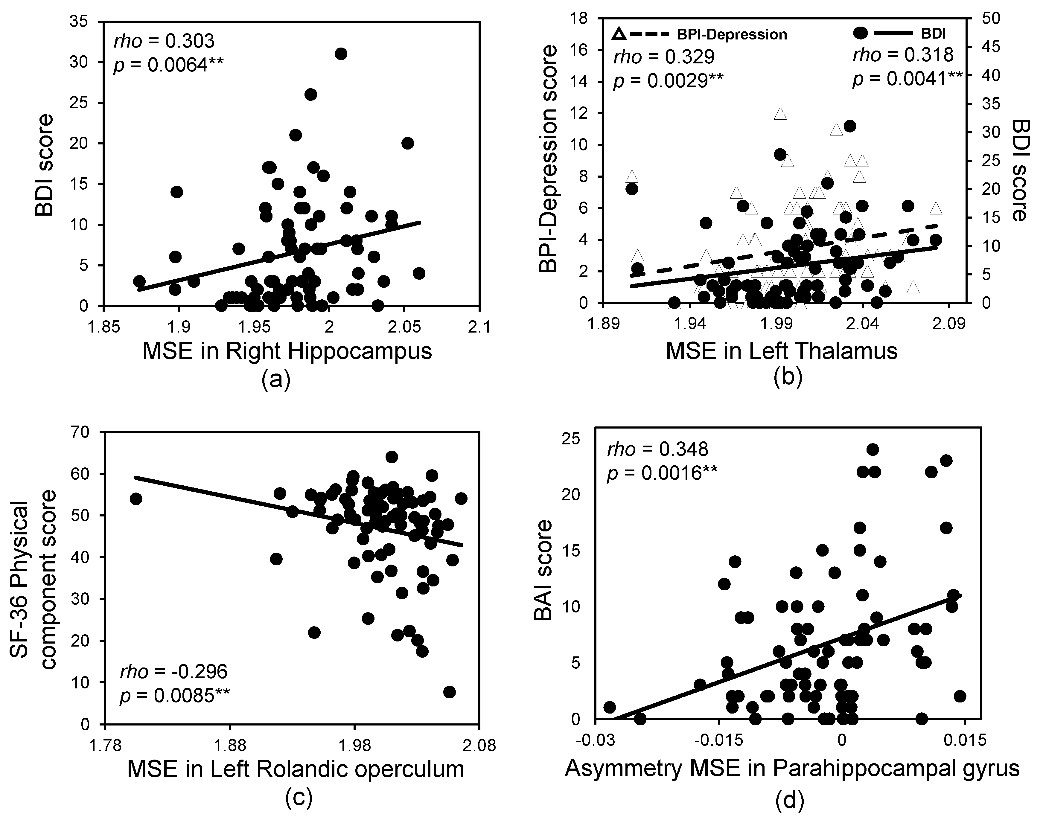
| Feature | Brain Region | RSN | Psychological Score | Scale Factor | Rho | p Value |
|---|---|---|---|---|---|---|
| MSE | Hippocampus (R) | LIMBIC | BDI | 91 | 0.303 | 0.0064 |
| Thalamus (L) | SMN/DMN/LIMBIC | BPI-Depression | 97 | 0.329 | 0.0029 | |
| BDI | 97 | 0.318 | 0.0041 | |||
| ITG (L) | DMN | Pain recalled score | 95 | 0.323 | 0.0045 | |
| Rolandic operculum (L) | SMN | SF-36, Physical component | 97 | −0.296 | 0.0085 | |
| Angular g. (R) | DMN/ECN/AN | BDI | 84 | 0.304 | 0.0060 | |
| BDI | 88 | 0.291 | 0.0087 | |||
| Fusiform g. (R) | VIS | PCS-Magnification | 88 | 0.300 | 0.0072 | |
| asymMSE | Parahippocampal g. | DMN/LIMBIC | BAI | 72 | 0.348 | 0.0016 |
| Rolandic operculum | SMN | BPI-Depression | 84 | −0.304 | 0.0061 | |
| SFG, medial | DMN | BPI-Depression | 55 | −0.321 | 0.0037 |
© 2017 by the authors. Licensee MDPI, Basel, Switzerland. This article is an open access article distributed under the terms and conditions of the Creative Commons Attribution (CC BY) license (http://creativecommons.org/licenses/by/4.0/).
Share and Cite
Low, I.; Kuo, P.-C.; Liu, Y.-H.; Tsai, C.-L.; Chao, H.-T.; Hsieh, J.-C.; Chen, L.-F.; Chen, Y.-S. Altered Brain Complexity in Women with Primary Dysmenorrhea: A Resting-State Magneto-Encephalography Study Using Multiscale Entropy Analysis. Entropy 2017, 19, 680. https://doi.org/10.3390/e19120680
Low I, Kuo P-C, Liu Y-H, Tsai C-L, Chao H-T, Hsieh J-C, Chen L-F, Chen Y-S. Altered Brain Complexity in Women with Primary Dysmenorrhea: A Resting-State Magneto-Encephalography Study Using Multiscale Entropy Analysis. Entropy. 2017; 19(12):680. https://doi.org/10.3390/e19120680
Chicago/Turabian StyleLow, Intan, Po-Chih Kuo, Yu-Hsiang Liu, Cheng-Lin Tsai, Hsiang-Tai Chao, Jen-Chuen Hsieh, Li-Fen Chen, and Yong-Sheng Chen. 2017. "Altered Brain Complexity in Women with Primary Dysmenorrhea: A Resting-State Magneto-Encephalography Study Using Multiscale Entropy Analysis" Entropy 19, no. 12: 680. https://doi.org/10.3390/e19120680
APA StyleLow, I., Kuo, P.-C., Liu, Y.-H., Tsai, C.-L., Chao, H.-T., Hsieh, J.-C., Chen, L.-F., & Chen, Y.-S. (2017). Altered Brain Complexity in Women with Primary Dysmenorrhea: A Resting-State Magneto-Encephalography Study Using Multiscale Entropy Analysis. Entropy, 19(12), 680. https://doi.org/10.3390/e19120680