Article

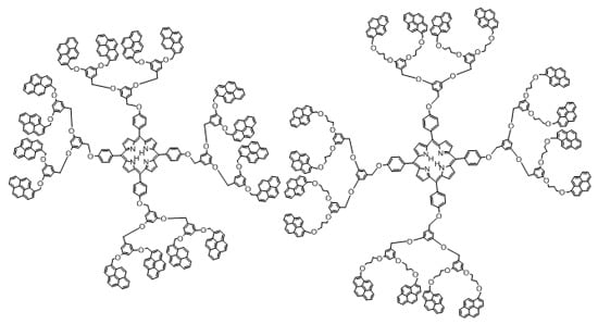
Synthesis of Porphyrin-Dendrimers with a Pyrene in the Periphery and Their Cubic Nonlinear Optical Properties

by
Eric G. Morales-Espinoza
1,
Irina V. Lijanova
2,
Omar G. Morales-Saavedra
3,
Vícente Torres-Zuñiga
3,
Simon Hernandez-Ortega
1 and
Marcos Martínez-García
1,*
1
Instituto de Química, Universidad Nacional Autónoma de México, Cd. Universitaria, Circuito Exterior, Coyoacán, C.P., México D.F. 04510, Mexico
2
Instituto Politécnico Nacional, CIITEC, Cerrada Cecati S/N, Colonia Santa Catarina de Azcapotzalco, C.P., México D.F. 02250, Mexico
3
Centro de Ciencias Aplicadas y Desarrollo Tecnológico, Universidad Nacional Autónoma de México, CCADET-UNAM, Circuito exterior S/N, Ciudad Universitaria C.P., México D.F. 04510, Mexico
*
Author to whom correspondence should be addressed.
Molecules 2011, 16(8), 6950-6968; https://doi.org/10.3390/molecules16086950
Submission received: 9 June 2011 / Revised: 3 August 2011 / Accepted: 5 August 2011 / Published: 15 August 2011
(This article belongs to the Special Issue Tetrapyrroles, Porphyrins and Phthalocyanines)

Abstract

:
Dendrons of pyrene derivatives were attached to a porphyrin core. A marked effect in solution for the dendrimers was observed in the absorption spectra. All the compounds obtained were characterized by 1H-, 13C-NMR, FTIR, UV-vis, MALDI-TOF or FAB+ mass spectrometry and elemental analysis. The cubic nonlinear optical behavior of some the synthesized compounds was tested via Z-Scan measurements in spin-coated film samples.

Graphical Abstract

1. Introduction

Dendrimers are highly branched polymers with a well defined structure, uniform size and molecular weight [1,2,3,4,5,6,7,8]. They are composed by a multifunctional central core to which dendritic branches are connected [9,10,11,12,13]. According to their shape, dendrimers are divided into two types. The first one has a circular or elliptic shape in which repeat units are regularly stretched from a core [14,15], and the second type has a conic shape with repeated units directionally stretched from a core [16,17,18]. Since it is possible to control the dendrimer’s size and structure, and at the same time introduce different kinds of functional groups into their terminal groups or core, numerous applications for these molecules have been found in different fields such as catalysis [19,20,21,22], biomedicine, energy or charge-transfer systems [23,24,25,26,27], sensors [28], charge transfer of light-emitting layer in organic light emitting diodes (OLEDs) [29,30,31], etc. In addition to the above advantages, dendrimers manifest a highly ordered structure and produce thin films with various functional groups on the top surface of the substrate, making possible the creation of unique materials in which surface characteristics are controlled at a molecular level. In the present work we report the synthesis of two generations of dendrimers with molecules of pyrene on the periphery and a tetraphenylporphyrin core.

2. Results and Discussion

The synthesis of the two pyrene-derivative dendrimer series was carried out applying a convergent pathway that consists of two steps, the first one being the synthesis of the two dendrons, and the second one the O-alkylation of the dendrons to the porphyrin. Following this order, dendrons containing pyrenebenzyloxy- and pyrene-1-methyl-3-chloropropoxybenzyloxy groups were prepared according to the convergent Fréchet approach [32] starting from a reduction reaction from the commercially available 1-pyrenecarboxaldehyde 1 via the reduction of the aldehyde 1 to obtain the pyren-1-ylmethanol 2 and its chlorination with SOCl2 and pyridine to obtain compound 3 (Scheme 1) or its O-alkylation with 1-bromo-3-chloropropane in acetone and K2CO3 at reflux to obtain the compound 4 (Scheme 1).
The following signals were observed in the 1H-NMR spectrum of compound 2: one singlet at δH 1.83 for the OH proton, one singlet at δH 5.36 due to the CH2 group and for the pyrene group (multiplets at δH 7.96–8.34). The structure of the pyren-1-ylmethanol compound 2 was confirmed by X-ray diffraction studies (Figure 1). In the crystal structure, a hydrogen bridge was observed between two pyren-1-ylmethanol moieties and the crystal packing of compound 2 showed strong interactions between the four pyrene molecules.
The chemical structure of the 1-(chloromethyl)pyrene 3 was confirmed by 13C-NMR spectroscopy. In the spectrum, the most significant resonance signals were those observed at δc 44.77 of the CH2-Cl group, and the aromatic signals due to the pyrene carbons.
In the 1H-NMR spectrum of compound 4, the following signals were observed: one multiplet at δ 2.03 due to the CH2 group, two multiplets at δ 3.63 and at δ 4.09 assigned to the CH2-Cl and CH2-O groups with a coupling constants J = 6.4 and J = 1.6 Hz, respectively. One singlet at δ 5.12 due to the CH2-O joined to the pyrene, and finally the characteristic signals due to the pyrene moiety. The structure of the compound 4 was further confirmed by mass spectrometry.
Chlorides 3 and 4 were attached to the 3,5-dihydroxybencyl alcohol 5 in DMF, cesium carbonate at 60 °C to give alcohol intermediates 6, 7, which were converted into chlorides 8 and 9 upon treatment with thionyl chloride and pyridine in dichloromethane (Scheme 2). Dendrons 8 and 9 are the first generation dendrons of the pyrene derivative families.
The second generation dendrons 12 and 13 were obtained from the dendrons 8 and 9 following the same methodology. First they were attached to 3,5-dihydroxybenzyl alcohol (5) in DMF using cesium carbonate at 60 °C to give alcohols 10, 11, which were converted into chlorides 12 and 13 upon treatment with thionyl chloride and pyridine in dichloromethane. Dendrons 12 and 13 are the second generation dendrons of the pyrene derivatives families.
The structures of 12 and 13 was confirmed by 1H-NMR spectroscopy. Signals at δH 4.60, 5.76 (dendron 12) and δH 4.35, 5.15 (dendron 13) corresponded to the methylene protons Ar-CH2-Cl and Pyrene-CH2-O. Compound 13 showed three signals at δH 2.03, 3.73 and 4.59 due to the propyloxy groups. The aromatic protons were observed as one singlet at δH 6.84 and 6.80, for compound 12 and 13, respectively. The characteristic signals for the pyrene moiety also were observed.
The synthesis of the dendrimers is depicted in Scheme 3. This synthesis involves only one step, an O-alkylation between dendrons 8, 12 or 9, 13 and porphyrin 14. The reaction was carried out in dimetylformamide and Cs2CO3 at 60 °C for 3 days and the dendrimers were obtained in good yields.
The 1H-NMR spectra of the dendrimers 15 and 16 (Figure 2) showed one broad signal at δH −2.23 due to the N-H protons, three multiplets at δH 2.17, 3.57 and 3.97 assigned to the CH2 protons of the propyl group, two singlets at δH 4.77 and δH 4.98 due to the Ar-CH2-O and Pyrene-CH2-O. The aromatic protons at the dendron were observed as two broad signals at δH 6.25 and δH 6.36. The characteristic signals for the pyrene moiety at δH 7.74 to δH 8.02 were also observed. Two doublets at δH 7.56 and 8.07 were assigned to the aromatic protons with a coupling constant of J = 8.2 Hz joined to the porphyrin. Finally, one broad signal at δH 8.79 was assigned to the pyrrole ring.
The structures of dendrimers 1518 were also confirmed by 13C-NMR, IR and MALDI-TOF mass spectrometry. It was found that all the dendrimers have the expected molecular weight.
In the optical absorption spectra of the dendrimers 1518 (Figure 3) in two different solvents, CH2Cl2 and dimethylformamide (DMF), both Soret and Q-bands do not shift upon increasing the concentration up to 10−6 M, indicating the absence of intermolecular aggregation processes in the ground state. Bands at 329 and 345 nm due to the pyrene molecules and in the case of dendrimers 16 and 18, bands were observed at 243, 277, 328 and 344 nm. The tetraphenylporphyrin clearly shows an intense band at 423 nm and for the dendrimers 16 and 18, new high energy bands were observed at 453 and at 689 nm, along with the Q- bands (see Table 1).

2.1. Linear and Third Order Non-Linear Optical Characterization

Regarding the linear and nonlinear optical properties of the obtained materials, homogeneous film samples were successfully deposited on glass substrates via spin-coating procedures from first and second generation dendrons (compounds 9 and 13, respectively), and from their respective dendrimer systems (compounds 16 and 18, respectively). Selected samples were chosen for cubic NLO-characterization via the Z-Scan technique.
Figure 4a shows the linear absorption coefficients evaluated within the visible range for the deposited samples. Indeed, the Beer-Lambert law applies for such semi-transparent films allowing an adequate data analysis and making these materials potential candidates for optical applications due to their appropriate transparency. It is evident from Figure 4a that the highest absorptive properties occur within the 370–550 nm spectral range, which may indicate additional conjugation of delocalized π-electrons and multi-directional charge transfer provided by the higher content of dendrons within the dendrimer-based film samples. This suggestion will be explored by means of NLO Z-Scan experiments as explained below. Under this framework, the available laser excitation line for Z-Scan experiments (λZ−Scan = 632.8 nm) is also depicted in this figure (vertical dashed line). At this wavelength, low absorption conditions occur, allowing non-resonant NLO-characterizations of the samples which are a critical point when working with photodegradable organic materials. In fact, relatively small linear absorption coefficients (see Table 2) were evaluated for the studied samples at λZ-Scan. These values are very useful for the determination of the nonlinear refraction and absorption coefficients according to the Z-Scan technique. The thicknesses and estimated linear refractive indices of the studied samples are also shown in Table 2.
Z-Scan measurements were performed at room conditions on the developed films. The observed non-local effect of these samples is shown in Figures 4(b-d). A rigorous theoretical fitting was performed in order to evaluate both the nonlinear absorptive and refractive properties of these samples. The NLO-response of the developed dendron- and dendrimer-based films was characterized by varying the polarization input planes of the He-Ne laser system in order to explore microscopic material asymmetries or anisotropies throughout the sample structure. In general, since all NLO-measurements were systematically performed with different laser input polarization states (from 0 to 90°: s- to p-polarization, respectively) and the obtained curves are quite similar in each sample, the film structures do not seem to show any significant anisotropic behaviour, thus confirming their amorphous nature. As shown in Figure 4(b) and Table 2, the Z-Scan curves obtained from the pristine, highly transparent dendron-based films (compounds 9 and 13), exhibit negligible nonlinear refraction and absorption when compared to the curves obtained for the dendrimer films under the same laser power regime. Indeed, compound 9 shows a flat Z-Scan curve and only compound 13 exhibits a typical valley-to-peak Z-Scan transmittance curve. Taking into account the theory developed by Sheik-Bahae et al. and Liu et al. [33,34,35,36,37,38] it is observed from our measurements that the nonlinear refractive response of the studied samples can be unambiguously determined by these typical Z-Scan transmittance curves. Hence, one can immediately observe that the dendron (13) and dendrimer-based films exhibit positive NLO-refraction coefficients (γ or n2 > 0).
The respective theoretical fits (TFs) to the obtained Z-Scan transmission data (solid lines) are also shown in Figures 4(b-d). In order to perform TFs according to previous theoretical models, the normalized Z-Scan transmittance (TN) can be determined as a function of the dimensionless sample position (x = z/z0), where z0 is the Rayleigh range and z is the Z-Scan sample position. Hence, the TFs were obtained according to the following equation, considering both nonlinear refraction and absorption effects [38]:
TN ≈ 1+[4x/(1+x2)(9+x2)]ΔΦ−[2(x2+3)/(1+x2)(9+x2)]ΔΨ
Here, the first term is related to NLO-refractive effects whereas the second one is associated to NLO-absorptive phenomena. Indeed, since some of the obtained Z-Scan curves clearly exhibit a valley-to-peak transmittance asymmetry, NLO absorption effects are also expected [16]. The fitting parameters are in this case the induced phase shifts ΔΦ or ΔΨ, respectively. In the former case, the phase shift is given by ΔΦ=2πγI0Leff/λ, [38] from which the NLO refractive index (γ- coefficient) can be obtained. In the latter case, the phase shift is provoked by the NLO-absorption and is given by ΔΨ=βI0LEff/2, [38] allowing the evaluation of the NLO absorption (β-coefficient), either due to two photon (TPA) or multi-photon absorption (β > 0), and/or saturable absorption (β < 0). In these equations, λ is the laser wavelength, I0 is the input beam intensity (at focal spot: z = 0) and Leff is the effective thickness of the film sample, defined as Leff = ⎾1−(eα0Ls)⏋(α0)−1, where α0 represents the linear absorption coefficient. All these equations are well established and have been proven in early Z-Scan works [33,34,35,36,37,38]. The theoretical restrictions imposed by these formulas in order to apply such expressions at optimal conditions (|ΔΦ0| < π, S ≈ 20%, etc.), are not always fully satisfied in our experimental results due to the large phase shifts and huge nonlinearities. Nevertheless, in most cases (mainly in the case of well defined γ > 0 or γ < 0 curves); our results nearly satisfy these conditions and can be conveniently fitted according to these theoretical formulas. Thus, for comparison purposes and in order to be consistent with the estimation of the γ- and β-values, we assumed their applicability and used them in our experimental results. According to Table 1 the TFs allowed us to evaluate a positive NLO-refractive coefficient in the order of γ = +0.684 × 10⁻⁸ m2W−1 (or n2 = +2.56 × 10⁻² esu) for the dendron-13 based film sample. In contrast, NLO-refractive coefficients as high as n2 = +9.27 × 10⁻² esu and n2 = +104 × 10−2 esu were evaluated for the dendrimer-based films (compounds 16 and 18, respectively). The obtained γ/n2-values are very large, many orders of magnitude larger than those observed for typical glass substrates or for the classical CS2 standard reference material: +1.2 × 10⁻¹¹ esu (Z-Scan at λ = 10.6 μm) or 6.8 × 10⁻¹³ esu (DFWM at λ = 532 nm) [33,35].
On the other hand, only compounds 13 and 18 exhibited measurable NLO absorption at the implemented experimental conditions, where the positive sign obtained for the β-coefficients reveal the nature of the NLO absorptive phenomena in these -second generation- based samples; indicating strong multi-photon absorption effects [33,34,35,36,37,38]. This fact points to the onset of thermal effects during Z-Scan experiments due to long cw-laser irradiation. Indeed, Z-Scan experiments were performed at low laser energy conditions (~6.6 mW) in order to avoid as much as possible photodegradation of the samples.

2.2. Crystal Structure Determination

A suitable crystal of compound 2 (obtained by crystallization from CH2Cl2 at room temperature) was rolled in epoxy resin and mounted on a glass fiber. Bruker Apex AXS CCD area detector X-Ray diffractometer was the instrument used for the determination. The data were first reduced and corrected for absorption using psi-scans, and then solved using the program SHELL-XS. All nonhydrogen atoms were refined with anisotropic thermal parameters and the hydrogen atoms were refined at calculated positions with thermal parameters constrained to the carbon atom on which they were attached. A summary of the key crystallographic information is given in Table 3. CCDC 838806 contains the supplementary crystallographic data for this paper. These data can be obtained free of charge via www.ccdc.cam.ac.uk/conts/retrieving.html (or from the CCDC, 12 Union Road, Cambridge CB2 1EZ, UK; Fax: +44 1223 336033; E-Mail: [email protected])

3. Experimental

3.1. General

Solvents and reagents were purchased as reagent grade and used without further purification. Acetone was distilled over calcium chloride. Tetrahydrofuran was distilled from sodium and benzophenone. Column chromatography was performed on Merck silica gel 60Å (70–230 mesh). 1H- and 13C-NMR were recorded on a Varian-Unity-200 MHz with tetramethylsilane (TMS) as an internal reference. Infrared (IR) spectra were measured on a spectrophotometer Nicolet FT-SSX. Elemental analysis was determined by Galbraith Laboratories, Inc. (Knoxville, TN, USA). FAB+ mass spectra were taken on a JEOL JMS AX505 HA instrument. MALDI-TOF mass spectra were taken on a Bruker Omni FLEX instrument.

3.2. Synthesis of Dendrons and Dendrimers

1-Pyrenemethanol (2): 97% Lithium aluminum hydride (1.7 mmol) was dissolved in dry THF (50 mL). To this solution, compound 1 (6 mmol) dissolved in dry THF (15 mL) was added dropwise using an addition funnel. The reaction was carried at 0 °C for 4 h. After this time, water (10 mL) was added and the reaction mixture was filtered through Celite®. The solvent was evaporated and the residue was dissolved in dichloromethane. The resulting solution was dried with sodium sulfate, filtered and the product was vacuum dried and purified by column chromatography (Al2O3, hexane), as a yellow powder, yield 5.82 mmol (97%). m.p. 123–125 °C. FTIR (KBr pellet, cm−1): 3278, 1594, 1180, 1064, 1008, 842, 705. UV-vis (CHCl3, nm) λmax: 344, 328, 314, 277, 266, 252, 243. 1H-NMR (CDCl3), δ (ppm): 1.83 (s, 1H, OH), 5.36 (s, 2H, Py-CH2-O), 7.96-8.34 (m, 9H, HPy). 13C-NMR (CDCl3), δ (ppm): 63.8 (CH2), 122.9 (CPy), 124.6 (CPy), 124.7 (CPy), 124.9 (CPy), 125.2 (CPy), 125.9 (CPy), 127.3 (CPy), 127.4 (CPy), 127.8 (CPy), 128.8 (CPy), 130.7 (CPy), 131.2 (CPy), 133.7 (Cipso). MS (m/z): 232 [M]+. Calc. for C17H12O: C, 87.90; H, 5.21 (%); Found: C, 87.91; H, 5.20 (%).
1-Chloromethylpyrene (3): A solution of pyridine (6.1 mmol) and SOCl2 (6.1 mmol) in dry CH2Cl2 (50 mL) was added to 2 (6 mmol), and then this mixture was cooled to −10 °C. The reaction was carried out in nitrogen atmosphere for 7 h. After this, the solvent was evaporated and the resulting oil was dried and purified by silica gel chromatography using a mixture of hexane-dichloromethane 2:1 as an eluent to afford 1-chloromethylpyrene (3) as a brownish powder, yield 5.76 mmol (96%). m.p. 148–149 °C. FTIR (KBr pellet, cm−1): 3423, 3042, 1593, 1253, 1182, 841, 757, 689. UV-vis (CHCl3, nm) λmax: 349, 339, 332, 279, 272, 268, 254, 244. 1H-NMR (CDCl3) δ (ppm): 5.25 (s, 2H, Py-CH2-Cl), 7.97–8.19 (m, 9H, Hpyr). 13C-NMR (CDCl3) δ (ppm): 44.7 (CH2), 122.7 (CPy), 124.5 (CPy), 124.7 (CPy), 125.0 (CPy), 125.6 (CPy), 126.1 (CPy), 127.2 (CPy), 127.6 (CPy), 127.9 (CPy), 128.3 (CPy), 129.1 (CPy), 130.2 (CPy), 130.6 (CPy), 131.1 (CPy), 131.9 (Cipso). MS (m/z): 250 [M]+. Calc. for C17H11Cl: C, 81.44; H, 4.42; Cl, 14.14 (%); found: C, 81.44; H, 4.42(%).
1-((3-Chloropropoxy)methyl)pyrene (4): A mixture of 1-pyrenemethanol (2, 6 mmol), potassium carbonate (18 mmol) in dry acetone (100 mL) was heated to reflux and stirred vigorously in nitrogen atmosphere for 30 min. Then, 1-bromo-3-chloro propane (6.1 mmol) dissolved in dry acetone (40 mL) was added dropwise and the reaction was continued for 7 days. The mixture was allowed to cool and the precipitate was filtered. The filtrate was evaporated to dryness under reduced pressure. The residue, dissolved in diethyl ether, was washed with an aqueous solution of 5% Na2CO3 (three times). The organic layer was dried and evaporated to dryness and purified by silica gel chromatography using a mixture of hexane-dichloromethane 2:1 as an eluent to afford 1-[(3-chloropropoxy)methyl]pyrene (4), as a yellow oil, yielding 2.34 mmol (39%). FTIR (KBr pellet, cm−1): 3042, 2859, 1077, 845, 708. UV-vis (CHCl3, nm) λmax: 341, 332, 325, 316, 312, 275, 269, 264, 250, 241, 214, 204. 1H-NMR (CDCl3) δ (ppm): 2.03 (m, 2H, CH2), 3.63 (m, 2H, Ar-CH2-Cl, J = 6.4 Hz), 4.09 (m, 2H, Ar-CH2-O, J = 1.6 Hz), 5.12 (s, 2H, Py-CH2-O), 7.90–8.32 (m, 9H, Ar-H). 13C-NMR (CDCl3) δ (ppm): 32.7 (CH2), 42.0 (CH2-Cl), 66.6 (CH2-O), 71.2 (Py-CH2-O), 117.3 (CPy), 122.3 (CPy), 124.4 (CPy), 124.6 (CPy), 124.8 (CPy), 125.1 (CPy), 125.8 (CPy), 126.8 (CPy), 127.3 (CPy), 127.6 (CPy), 129.2 (CPy), 130.7 (CPy), 131.1 (CPy), 131.2 (CPy), 131.3 (CPy), 134.7 (Cipso). MS (m/z): 308 [M]+. Calc. for C20H17ClO: C, 77.79; H, 5.55 (%); found: C, 77.77; H, 5.57 (%).

3.2.1. General Procedure

A mixture of 3, 4, 8 or 9 (6 mmol) and cesium carbonate (12 mmol) in DMF (20 mL) was stirred vigorously in nitrogen atmosphere for 30 min. 3,5-Dihydroxybenzyl alcohol (5, 2.9 mmol) dissolved in DMF (10 mL) was added dropwise and the reaction was continued for 3 days. The mixture was filtered. The filtrate was evaporated to dryness under reduced pressure. The organic layer was dried and evaporated to dryness and purified by silica gel chromatography using a mixture of hexane-dichloromethane 2:1 as an eluent.
(3,5-bis(Pyren-1-ylmethoxy)phenyl)methanol (6): Yellow powder, yield 1.11 mmol (38 %). FTIR (KBr pellet, cm−1): 3430, 3039, 1593, 1448, 1352, 1287, 1165, 1021, 842, 707. UV-vis (CHCl3, nm) λmax: 344, 336, 328, 319, 314, 294, 277, 271, 267, 253, 244. 1H-NMR (CDCl3), δ (ppm): 1.62 (s, 1H, OH), 4.69 (s, 2H, Ar-CH2-OH), 5.72 (s, 4H, Py-CH2-O), 6.81 (s, 3H, Ar), 7.97–8.29 (m, 18H, HPy). 13C-NMR (CDCl3), δ (ppm): 65.3 (Ar-CH2-O), 68.8 (Py-CH2-O), 101.4 (CAr), 106.0 (CAr), 122.9 (CPy), 124.6 (CPy), 124.9 (CPy), 125.4 (CPy), 126.0 (CPy), 126.8 (CPy), 127.3 (CPy), 127.6 (CPy), 128.0 (CPy), 129.2 (CPy), 129.5 (CPy), 130.7 (CPy), 131.1 (CPy), 131.5 (CipsoPy ), 143.5 (Cipso Ar), 160.3 (Cipso Ar). MS (m/z): 568 [M]+. Calc. for C41H28O3: C, 86.60; H, 4.96 (%); Found: C, 86.60; H, 4.98 (%).
(3,5-bis(3-(pyren-1-ylmethoxy)propoxy)phenyl)methanol (7): Yellow powder, yield 0.72 mmol (25%). FTIR (KBr pellet, cm−1): 3350, 3038, 2946, 2857, 1599, 1497, 1165, 1088, 1054, 844, 704. UV-vis (CH3OH, nm) λmax: 341, 333, 326, 292, 275, 251, 241, 217, 207. 1H-NMR (CDCl3) δ (ppm): 1.68 (br, 1H, OH), 2.03 (m, 4H, CH2), 3.73 (t, 4H, CH2-O, J = 6.0 Hz), 3.90 (t, 4H, CH2-O, J = 6.0 Hz), 4.35 (s, 2H, Ar-CH2-O), 5.15 (s, 4H, Py-CH2-O), 6.11 (t, 1H, Ar, J = 2.2 Hz), 6.21 (d, 2H, Ar, J = 2.2 Hz), 7.88–8.27 (m, 18H, Py). 13C-NMR (CDCl3) δ (ppm): 29.6 (CH2), 64.4 (CH2-O), 65.1 (CH2-O), 66.4 (Ar-CH2-O), 71.6 (Py-CH2-O), 100.0 (CAr), 104.7 (CAr), 123.4–130.7 (Cpy), 131.3 (Cipso Py), 142.8 (Cipso Ar), 160.0 (Cipso Ar). MS (m/z): 684 (M). Calc. for C47H40O5: C, 82.43; H, 5.89 (%); found: C, 82.45; H, 5.85 (%).
(3,5-bis((3,5-bis(Pyren-1-ylmethoxy)benzyl)oxy)phenyl)methanol (10): Yellow powder, yield 1.02 mmol (35%). FTIR (KBr pellet, cm−1): 3438, 2958, 2926, 2856, 1732, 1596, 1462, 1271, 1160, 845, 738, 707. UV-vis (CHCl3, nm) λmax: 346, 330, 315, 278, 269, 244. 1H-NMR (CDCl3), δ (ppm): 2.19 (s, 1H, OH), 4.58 (s, 2H, Ar-CH2-OH), 5.03 (s, 4H, Ar-CH2-O), 5.75 (s, 8H, Py-CH2-O), 6.85 (m, 9H, Ar), 7.97–8.32 (m, 36H, HPy). 13C-NMR (CDCl3), δ (ppm): 64.5 (Ar-CH2-O), 68.1 (CH2-O-Ar), 70.4 (Py-CH2-O), 100.1 (HCAr), 101.8 (HCAr), 105.0 (HCAr), 106.5 (HCAr), 122.9–130.8 (Cpy), 131.1 (Cipso Py), 131.2 (Cipso), 132.2 (Cipso), 138.2 (Cipso), 141.33 (Cipso), 160.3 (Cipso). MALDI-TOF MS (m/z): 1240 (M). Calc. for C89H60O7: C, 86.11; H, 4.87 (%); found: C, 86.12; H, 4.85 (%).
(3,5-bis((3,5-bis(3-(Pyren-1-ylmethoxy)propoxy)benzyl)oxy)phenyl)methanol (11): White powder, yield 1.44 mmol (49%). FTIR (KBr pellet, cm−1): 3421, 3008, 2957, 2926, 2870, 1729, 1597, 1164, 1078, 847, 756, 708. UV-vis (CHCl3, nm) λmax: 344, 328, 316, 277, 266, 245. 1H-NMR (CDCl3) δ (ppm): 2.16 (s, 1H, OH), 2.03 (m, 8H, CH2), 3.78 (m, 16H, CH2-O), 4.56 (s, 2H, CH2-OH), 5.18 (s, 4H, CH2-O), 5.21 (s, 8H, Py-CH2-O), 6.20 (br, 3H, Ar), 6.41 (br, 6H, Ar), 7.96–8.37 (m, 36H, Py). 13C-NMR (CDCl3), δ (ppm): 29.6 (CH2), 61.8 (CH2-O), 64.5 (CH2-O), 66.5 (CH2-O), 69.3 (CH2-O), 71.8 (Py-CH2-O), 100.4 (HCAr), 104.7 (HCAr), 105.3 (HCAr), 105.7 (HCAr), 123.1–129.3 (Cpy), 130.7 (Cipso), 131.2 (Cipso), 131.3 (Cipso), 160.1 (Cipso). MALDI-TOF MS (m/z): 1472 (M). Calc. for C101H84O11: C, 82.31; H, 5.75 (%); found: C, 82.33; H, 5.75 (%).

3.2.2. General Procedure

A solution of pyridine (6.1 mmol) and SOCl2 (6.1 mmol) in dry CH2Cl2 (100 mL) was added to 6, 7, 10 or 11 (6 mmol), and then this mixture was cooled to 0 °C. The reaction was carried out in nitrogen atmosphere for 7 h. After this, the solvent was evaporated and the resulting oil was dried and purified by silica gel chromatography using a mixture of hexane-dichloromethane 2:1 as an eluent.
1,1′-(((5-(Chloromethyl)-1,3-phenylene)bis(oxy))bis(methylene))dipyrene (8): Yellow powder, yield 5.28 mmol (88%). FTIR (KBr pellet, cm−1): 3442, 3043, 1593, 1463, 1381, 1322, 1172, 1043, 844, 712. UV-vis (CHCl3, nm) λmax: 344, 335, 328, 318, 314, 293, 276, 270, 265, 259, 242, 219, 204. 1H-NMR (CDCl3 + DMSO-d6), δ (ppm): 4.60 (s, 2H, CH2-Cl), 5.76 (s, 4H, Py-CH2-O), 6.84 (s, 3H, Ar, J = 2.2 Hz), 8.00-8.33 (m, 18H, HPy). 13C-NMR (CDCl3), δ (ppm): 45.7 (Ar-CH2-Cl), 68.3 (Py-CH2-O), 101.5 (CAr), 107.4 (CAr), 122.4 (Cpy), 124.1 (Cpy), 124.9 (Cpy), 125.6 (Cpy), 126.5 (Cpy), 126.8 (Cpy), 127.1 (Cpy), 127.5 (Cpy), 128.9 (Cpy), 130.0 (Cpy), 130.5 (Cpy), 130.9 (Cipso Py), 139.2 (Cipso Ar), 159.6 (Cipso Ar). MS (m/z): 586 [M]+. Calc. for C41H27ClO2: C, 83.88; H, 4.64 (%); Found: C, 83.86; H, 4.67 (%).
1,1′-(((((5-(Chloromethyl)-1,3-phenylene)bis(oxy))bis(propane-3,1-diyl))bis(oxy))bis(methylene))di-pyrene (9): Yellow powder, yield 4.80 mmol (80%). FTIR (KBr pellet, cm−1): 3350, 3038, 2946, 2857, 1599, 1497, 1165, 1088, 1054, 844, 704. UV-vis (CH3OH, nm) λmax: 341, 333, 326, 292, 275, 251, 241, 217, 207. 1H-NMR (CDCl3) δ (ppm): 2.03 (m, 4H, CH2), 3.73 (t, 4H, CH2-O, J = 6.0 Hz), 3.90 (t, 4H, CH2-O, J = 6.0 Hz), 4.35 (s, 2H, Ar-CH2-OH), 5.15 (s, 4H, Py-CH2-O), 6.11 (t, 1H, Ar, J = 2.2 Hz), 6.21 (d, 2H, Ar, J = 2.2 Hz), 7.88–8.27 (m, 18H, Py). 13C-NMR (CDCl3) δ (ppm): 29.6 (CH2), 46.3 (Ar-CH2-Cl), 64.4 (CH2-O), 65.1 (CH2-O), 71.6 (Py-CH2-O), 100.0 (H-CAr), 104.7 (H-CAr), 123.4–130.7 (Cpireno), 131.31 (Cipso py), 142.86 (Cipso Ar), 160.01 (Cipso Ar). MS (m/z): 702 (M). Calc. for C47H39ClO4: C, 80.27; H, 5.59 (%); found: C, 80.25; H, 5.58 (%).
1,1′,1″,1‴-((((((5-(Chloromethyl)-1,3-phenylene)bis(oxy))bis(methylene))bis(benzene-5,3,1-triyl))tetrakis(oxy))tetrakis(methylene))tetrapyrene (12): Yellow powder, yield 2.52 mmol (42%). FTIR (KBr pellet, cm−1): 3443, 3041, 1596, 1466, 1378, 1321, 1170, 1043, 844, 711. UV-vis (CHCl3, nm) λmax: 344, 335, 328, 318, 314, 293, 276, 270, 265, 259, 242, 219, 204. 1H-NMR (CDCl3 + DMSO-d6), δ (ppm): 4.61 (s, 2H, CH2-Cl), 5.75 (s, 12H, Py-CH2-O), 6.83 (s, 9H, Ar, J = 2.2 Hz), 8.01-8.35 (m, 36H, HPy). 13C-NMR (CDCl3), δ (ppm): 45.7 (Ar-CH2-Cl), 68.3 (Py-CH2-O), 101.5 (CAr), 107.4 (CAr), 122.4 (Cpy), 124.1 (Cpy), 124.9 (Cpy), 125.6 (Cpy), 126.5 (Cpy), 126.8 (Cpy), 127.1 (Cpy), 127.5 (Cpy), 128.9 (Cpy), 130.0 (Cpy), 130.5 (Cpy), 130.9 (Cipso Py), 139.2 (Cipso Ar), 159.6 (Cipso Ar). MS (m/z): 586 [M]+. Calc. for C89H59ClO6: C, 84.85; H, 4.72 (%); Found: C, 84.87; H, 4.69 (%).
1,1′,1″,1‴-((((((((5-(Chloromethyl)-1,3-phenylene)bis(oxy))bis(methylene))bis(benzene-5,3,1-triyl))tetrakis(oxy))tetrakis(propane-3,1-diyl))tetrakis(oxy)) tetrakis(methylene)) tetrapyrene (13): Yellow powder, yield 4.0 mmol (67%): FTIR (KBr pellet, cm−1): 3206, 3129, 3049, 2937, 2863, 2082, 1970, 1732, 1599, 1528, 1479, 1329, 1295, 1162, 1055, 845, 752, 680. UV-vis (CH2Cl2, nm) λmax: 345, 329, 278, 244. 1H-NMR (CDCl3) δ (ppm): 2.01 (m, 8H, CH2), 3.77 (m, 16H, CH2-O), 4.25 (s, 2H, CH2-Cl), 5.16 (s, 4H, CH2-O), 5.20 (s, 8H, Py-CH2-O), 6.21 (br, 3H, Ar), 6.42 (br, 6H, Ar), 7.94-8.33 (m, 36H, Py). 13C-NMR (CDCl3), δ (ppm): 29.4 (CH2), 46.4 (CH2-Cl), 64.6 (CH2-O), 66.7 (CH2-O), 69.4 (CH2-O), 71.7 (Py-CH2-O), 100.3 (HCAr), 104.6 (HCAr), 105.3 (HCAr), 105.7 (HCAr), 123.3-129.5 (Cpy), 130.5 (Cipso), 131.1 (Cipso), 131.4 (Cipso), 160.2 (Cipso). MALDI-TOF MS (m/z): 1490 (M). Calc. for C101H83ClO10: C, 81.30; H, 5.61 (%); found: C, 81.33; H, 5.60 (%).

3.2.3. General Procedure

A mixture of compound 8, 9, 12 or compound 13 (5.4 mmol) and cesium carbonate (10.1 mmol) in DMF (20 mL) was stirred vigorously in nitrogen atmosphere for 30 min. The porphyrin 14 (2.7 mmol) dissolved in DMF (10 mL) was added dropwise and the reaction was continued for 3 days. The mixture was filtered. The filtrate was evaporated to dryness under reduced pressure. The organic layer was dried and evaporated to dryness and purified by silica gel chromatography using a mixture of hexane-ethyl acetate 2:1 as an eluent.
Dendrimer 15: Brown powder, yield 0.26 mmol (19%). FTI. (KBr pellet, cm−1): 3313, 3037, 2953, 2869, 1601, 1504, 1467, 1263, 1172, 1007, 842, 800, 707. UV-vis (DMF, nm) λmax: 329, 345, 423,518, 557, 597, 653. 1H-NMR (CDCl3), δ (ppm): −2.18 (br, 2H, NH), 5.39 (s, 8H, CH2-Ar), 5.92 (s, 16H, CH2-Py), 6.22 (br, 4H, Ar), 6.42 (br, 8H, Ar), 7.79 (d, 8H, Ar, J = 8.4 Hz), 7.97–8.43 (m, 72H, Py), 8.62 (d, 8H, Ar, J = 8.4 Hz), 8.97 (br, 8H, Porph). 13C-NMR (THF-d8/DMSO-d6), δ (ppm): 64.4 (CH2-O-Ar), 68.5 (CH2-O-Ar), 70.6 (Py-CH2-O), 100.3 (HCAr), 101.6 (HCAr), 105.1 (HCAr), 114.6(CAr,porph), 122.8-130.6 (Cpy), 127.2 (Cpyrrol), 128.1 (CAr,porph), 131.3 (Cipso py), 131.1 (Cipso), 132.4 (Cipso), 136.2 (CAr,porph), 138.2 (Cipso), 141.5 (Cpyrrol), 160.6 (Cipso). MALDI-TOF MS (m/z): 2879 (M+). Calc. for C208H134N4O12: C, 86.70; H, 4.69, N 1.94 (%); found: C, 86.73; H, 4.68 (%).
Dendrimer 16: Brown powder, yield 0.23 mmol (17%). FTIR (KBr pellet, cm−1): 3359, 3194, 2921, 2852, 1730, 1658, 1466, 1271, 1167, 1136, 847, 809, 721. UV-vis (DMF, nm) λmax: 243, 277, 328, 344, 422, 453, 518, 551, 595, 651, 689. 1H-NMR (CDCl3), δ (ppm): −2.23 (br, 2H, NH), 2.17 (m, 16H, CH2), 3.57 (m, 16H, CH2-O), 3.79 (m, 16H, CH2-O), 4.77 (s, 8H, CH2-Ar), 4.98 (s, 16H, CH2-Py), 6.25 (br, 4H, Ar), 6.36 (br, 8H, Ar), 7.56 (d, 8H porph, J = 8.2 Hz), 7.74–8.02 (m, 72H, Py), 8.07 (d, 8H, Ar, J = 8.2 Hz), 8.79 (br, 8H, Ar). 13C-NMR (THF-d8/DMSO-d6), δ (ppm): 64.8 (CH2-O-Ar), 68.1 (CH2-O-Ar), 70.9 (Py-CH2-O), 100.4 (HCAr), 101.6 (HCAr), 105.5 (HCAr), 114,3 (CAr,porph), 122.8-130.0 (Cpy), 127.2 (Cpyrrol), 128.5 (CAr,porph), 131.2 (Cipso py), 131.3 (Cipso), 132.6 (Cipso), 136.6 (CAr,porph), 138.3 (Cipso), 141.9 (Cpyrrol), 161.1 (Cipso). MALDI-TOF MS (m/z): 3343 (M+). Calc. for C232H182N4O20: C, 83.28; H, 5.48 (%); found: C, 83.25; H, 5.43, N 1.67 (%).
Dendrimer 17: Black powder, yield 0.11 mmol (8%). FTIR (KBr pellet, cm−1): 3313, 3038, 2923, 1602, 1503, 1470, 1233, 1173, 965, 842, 799, 708. UV-vis (DMF, nm) λmax: 315, 329, 345, 375, 423, 519, 555, 595, 652. 1H-NMR (THF-d8/DMSO-d6) δ (ppm): −2.23 (br, 2H, NH), 4.63 (s, 32H, CH2-Py), 5.66 (br, 16H, CH2-O), 5.77 (br, 8H, CH2-O), 6.18 (br, 36H, Ar), 7.67 (d, 8H, Ar, J = 8.6 Hz), 7.97–8.50 (m, 144H, Py), 8.69 (d, 8H, Ar, J = 8.6 Hz), 8.96 (s, 8Hpyrrol). 13C-NMR (THF-d8/DMSO-d6), δ (ppm): 64.6 (CH2-O-Ar), 68.3 (CH2-O-Ar), 70.6 (Py-CH2-O), 100.1 (HCAr), 101.8 (HCAr), 105.0 (HCAr), 114,6 (CAr,porph), 122.8-130.9 (Cpy), 127.4 (Cpyrrol), 128.3 (CAr,porf), 131.1 (Cipso py), 131.3 (Cipso), 132.3 (Cipso), 136.8 (CAr,porph), 138.3 (Cipso), 141.4 (Cpyrrol), 160.1 (Cipso). MALDI-TOF MS (m/z): 5567 (M+). Calc. for C400H262N4O28: C, 86.22; H, 4.64, N 1.01 (%); found: C, 86.23; H, 4.62 (%).
Dendrimer 18: Black powder, yield 0.16 mmol (12%). FTIR (KBr pellet, cm−1): 3312, 3039, 2925, 2867, 1901, 1716, 1595, 1460, 1291, 1235, 1164, 1063, 966, 844, 802, 709. UV-vis (DMF, nm) λmax: 244, 278, 329, 345, 423, 455, 519, 557, 594, 652, 693. 1H-NMR (CDCl3/CD3OD) δ (ppm): −2.23 (br, 2H, NH), 1.98 (br, 32H, CH2), 3.63 (br, 32H, CH2-O), 3.97 (br, 32H, CH2-O), 4.61 (br, 16H, CH2-O), 4.82 (br, 8 H, CH2-O), 5.17 (br, 32H, CH2-Py), 6.27 (br, 36H, Ar), 7.22 (br, 16H, Ar,), 7.61–8.34 (m, 144H, Py), 8.84 (br, 8Hpyrrol). 13C-NMR (CDCl3/CD3OD), δ (ppm): 29.5 (CH2), 60.1 (CH2-O), 64.5 (CH2-O), 66.5 (CH2-O), 68.7 (CH2-O), 71.5 (Py-CH2-O), 98.6 (HCAr), 105.8 (HCAr), 107.6 (HCAr), 113.1 (HCAr,porph), 119.6 (Cpyrrol), 123.2-131.1 (Cpy), 135.6 (Cipso), 143.4 (Cpyrrol), 158.6 (Cipso), 159.9 (Cipso), 160.13 (Cipso). MALDI-TOF MS (m/z): 6496 (M+). Calc. for C448H358N4O44: C, 86.76; H, 5.55, N 0.86 (%); found: C, 86.73; H, 5.57 (%).

3.3. Cubic NLO-Characterization

Finally, some of the synthesized dendrons and dendrimers were also studied as active media for cubic χ(3)-nonlinear optical effects such as nonlinear refraction and absorption via Z-Scan measurements (in spin-coated film samples obtained from THF-based dissolutions). The experimental Z-Scan set-up was implemented using an unpolarized laser beam from a 35 mW He-Ne laser system working at 632.8 nm (THORLABS, HRR170-1) [39]. Its energy was carefully monitored and kept constant during long Z-Scan measurements. The spatial mode of the laser beam was close to Gaussian TEM00. The polarization plane of the He-Ne laser beam was adjusted and controlled by means of a linear polarizer mounted on a rotation stage. The polarized laser beam was focused on the sample by means of a positive lens (f = 5 cm), so that a light power density of ~8.53 × 10⁶ W m−2 reached the studied sample at the focal spot. At last, the samples were mounted on a motorized translation stage (25 mm length travel in steps of 2 µm) in order to perform Z-Scan experiments within the optical focal range. A large area Si-photodetector (EOT ET-2040) was located at ~0.96 m from the focusing lens, after a 2.5 mm diameter (~20% transmittance) diaphragm-aperture. All NLO-signals captured from photo-detectors were measured with a digital oscilloscope (Tektronix TDS, 744A), and all motion systems and set-up management were automated via a LabView control program.

4. Conclusions

Outstanding cubic NLO-effects were particularly measured for the dendrimer-18 based film sample via the Z-Scan technique, where a high NLO-refractive coefficient in the order of 10−2 esu was found. This remarkably NLO-activity is mainly due to the high content of pyrene derivatives available in its respective second generation (dendron 13) building units. In contrast, the first generation dendron 9 and the respective dendrimer-16 compound exhibit lower NLO-activity. Accordingly, film samples obtained from compounds 13 and 18 consistently exhibited TPA-NLO absorption effects at same experimental conditions. However, more NLO-investigations should be performed in these materials in order to further understand the electronic and thermal contributions to the cubic nonlinearities.

Acknowledgments

This work was supported by the DGAPA (IN202010). We would also like to thank Rios O.H., Velasco L., Huerta S.E., Patiño M.M.R., Rodriguez-Rosales A.A. and Peña Gonzalez M.A., for technical assistance.

Conflict of Interest

The authors declare no conflict of interest.

References

  1. Zeng, F.; Zimmerman, S.C. Dendrimers in supramolecular chemistry: From molecular recognition to self-assembly. Chem. Rev. 1997, 97, 1681–1712. [Google Scholar] [CrossRef] [PubMed]
  2. Newkome, G.R.; He, E.; Moorefield, C.N. Suprasupermolecules with novel properties: Metallodendrimers. Chem. Rev. 1999, 99, 1689–1746. [Google Scholar] [CrossRef] [PubMed]
  3. Smith, D.K.; Diederich, F. Supramolecular dendrimer chemistry: A journey through the branched architecture. Top. Curr. Chem. 2000, 210, 183–227. [Google Scholar]
  4. Emrick, T.; Fréchet, J.M.J. Self-assembly of dendritic structures. Curr. Opin. Colloid Interf. Sci. 1999, 4, 15–23. [Google Scholar] [CrossRef]
  5. Bosman, A.W.; Janssen, H.M.; Meijer, E.W. About dendrimers: Structure, physical properties, and applications. Chem. Rev. 1999, 99, 1665–1688. [Google Scholar] [CrossRef] [PubMed]
  6. Dendrimers and other Dendritic Polymers; Fréchet, J.M.J.; Tomalia, D.A. (Eds.) Wiley: Chichester, UK, 2001. [Google Scholar]
  7. Newkome, G.R.; Moorefield, C.N.; Vögtle, F. Dendrimers and Dendrons: Concepts, Syntheses, Applications; Wiley-VCH: Weinheim, Germany, 2001. [Google Scholar]
  8. Vögtle, F.; Richardt, G.; Werner, N. Dendrimer Chemistry: Concepts, Syntheses, Properties, Applications; Wiley-VCH: Weinheim, Germany, 2009. [Google Scholar]
  9. Valerio, C.; Fillaut, J.L.; Ruiz, J.; Guittard, J.; Blais, J.C.; Astruc, D. The Dendritic effect in molecular recognition: Ferrocene dendrimers and their use as supramolecular redox sensors for the recognition of small inorganic anions. J. Am. Chem. Soc. 1997, 119, 2588–2589. [Google Scholar] [CrossRef]
  10. Valerio, C.; Alonso, E.; Ruiz, J.; Blais, J.C.; Astruc, D. A polycationic metallodendrimer with 24 [Fe(η5-C5Me5)(η6-N-Alkylaniline)]+ termini that recognizes chloride and bromide anions. Angew. Chem. Int. Ed. 1999, 38, 1747–1751. [Google Scholar] [CrossRef]
  11. Labande, A.; Astruc, D. Colloids as redox sensors: Recognition of H2PO4 and HSO4 by amidoferrocenylalkylthiol–gold nanoparticles. Chem. Commun. 2000, 1007–1008. [Google Scholar] [CrossRef]
  12. Nlate, S.; Ruiz, J.; Sartor, V.; Navarro, R.; Blais, J.C.; Astruc, D. Molecular batteries: Ferrocenylsilylation of dendrons, dendritic cores, and dendrimers: New convergent and divergent routes to ferrocenyl dendrimers with stable redox activity. Chem. Eur. J. 2000, 6, 2544–2553. [Google Scholar] [CrossRef]
  13. Labande, A.; Ruiz, J.; Astruc, D. Supramolecular gold nanoparticles for the redox recognition of oxoanions: Syntheses, titrations, stereoelectronic effects, and selectivity. J. Am. Chem. Soc. 2002, 124, 1782–1789. [Google Scholar] [CrossRef] [PubMed]
  14. Tomalia, D.A.; Naylor, A.M.; Goddard, W.A., III. Starburst dendrimers: Molecular-level control of size, shape, surface chemistry, topology, and flexibility from atoms to macroscopic matter. Angew. Chem. Int. Ed. 1990, 29, 138–175. [Google Scholar] [CrossRef]
  15. Ornelas, C.; Ruiz, A.J.; Cloutet, E.; Alves, S.; Astruc, D. Click assembly of 1,2,3-triazole-linked dendrimers, including ferrocenyl dendrimers, which sense both oxo anions and metal cations. Angwe. Chem. Int. Ed. 2007, 46, 872–877. [Google Scholar] [CrossRef] [PubMed]
  16. Goodson, T., III; Varnavski, O.; Wang, Y. Optical properties and applications of dendrimer-metal nanocomposites. Int. Rev. Phys. Chem. 2004, 23, 109–150. [Google Scholar] [CrossRef]
  17. Wang, W.; Sun, H.; Kaifer, E.A. Redox active, hybrid dendrimers containing Fréchet- and Newkome-type blocks. Org. Lett. 2007, 9, 2657–2660. [Google Scholar] [CrossRef] [PubMed]
  18. Ispasoiu, R.G.; Balogh, L.; Varnavski, O.P.; Tomalia, D.A.; Goodson, T., III. Large optical limiting from novel metal-dendrimer nanocomposite materials. J. Am. Chem. Soc. 2000, 122, 11005–11006. [Google Scholar] [CrossRef]
  19. Samoc, M.; Samoc, A.; Luther-Davies, B.; Humphrey, M.G.; Wong, M.S. Third-order optical nonlinearities of oligomers, dendrimers and polymers derived from solution Z-scan studies. Opt. Mater. 2003, 21, 485–488. [Google Scholar] [CrossRef]
  20. Astruc, D.; Boisselier, E.; Ornelas, C. Dendrimers designed for functions: From physical, photophysical, and supramolecular properties to applications in sensing, catalysis, molecular electronics, photonics, and nanomedicine. Chem. Rev. 2010, 110, 1857–1959. [Google Scholar] [CrossRef] [PubMed]
  21. Boas, U.; Christensen, J.B.; Heegaard, P.M.H. Dendrimers in Medicine and Biotechnology: New Molecular Tools; RSC Publishing: Cambridge, UK, 2006. [Google Scholar]
  22. Mintzer, M.A.; Grinstaff, M.W. Biomedical applications of dendrimers: A tutorial. Chem. Soc. Rev. 2011, 40, 173–190. [Google Scholar] [CrossRef] [PubMed]
  23. Cameron, D.J.A.; Shaver, M.P. Aliphatic polyester polymer stars: Synthesis, properties and applications in biomedicine and nanotechnology. Chem. Soc. Rev. 2011, 40, 1761–1776. [Google Scholar] [CrossRef] [PubMed]
  24. Powell, C.E.; Hurst, S.K.; Morrall, J.P.; Cifuentes, M.P.; Roberts, R.L.; Samoc, M.; Humphrey, M.G. Organometallic complexes for nonlinear optics. 39. Syntheses and third-order nonlinear optical properties of first-generation peripherally metalated arylalkynyl dendrimers. Organometallics 2007, 26, 4456–5576. [Google Scholar] [CrossRef]
  25. Majoral, J.P. State of the art developments in the chemistry and properties of dendrimers and hyperbranched polymers. New J. Chem. 2007, 31, 1039–1040. [Google Scholar]
  26. Al-Jamal, K.T.; Ramaswamy, C.; Florence, A.T. Supramolecular structures from dendrons and dendrimers. Adv. Drug Deliv. Rev. 2005, 57, 2238–2270. [Google Scholar] [CrossRef] [PubMed]
  27. Röglin, L.; Lempens, E.H.M.; Meijer, E.W. A synthetic “tour de force”: Well-defined multivalent and multimodal dendritic structures for biomedical applications. Angew. Chem. Int. Ed. 2011, 50, 102–112. [Google Scholar] [CrossRef] [PubMed]
  28. Cifuentes, M.P.; Powell, C.E.; Morrall, J.P. Electrochemical, spectroelectrochemical, and molecular quadratic and cubic nonlinear optical properties of alkynylruthenium dendrimers. J. Am. Chem. Soc. 2006, 128, 10819–10832. [Google Scholar] [CrossRef] [PubMed]
  29. Kraft, A.; Grimsdale, A.C.; Holmes, A.B. Electroluminescent conjugated polymers-seeing polymers in a new light. Angew. Chem. Int. Ed. 1998, 37, 402–428. [Google Scholar] [CrossRef]
  30. Lo, S.-C.; Burn, P.L. Development of dendrimers: Macromolecules for use in organic light-emitting diodes and solar cells. Chem. Rev. 2007, 107, 1097–1116. [Google Scholar] [CrossRef] [PubMed]
  31. Burn, P.L.; Lo, S.-C.; Samuel, I.D.W. Metallo-supramolecular block copolymers. Adv. Mater. 2007, 19, 1675–1688. [Google Scholar] [CrossRef]
  32. Hawker, C.J.; Fréchet, J.M.J. Preparation of polymers with controlled molecular architecture. A new convergent approach to dendritic macromolecules. J. Am. Chem. Soc. 1990, 112, 7638–7648. [Google Scholar] [CrossRef]
  33. Sheik-Bahae, M.; Said, A.A.; van Stryland, E.W. High-sensitivity, single-beam n2 measurements. Opt. Lett. 1989, 14, 955–957. [Google Scholar] [CrossRef] [PubMed]
  34. Sheik-Bahae, M.; Said, A.A.; Hagan, D.J.; Soileau, M.J.; van Stryland, E.W. Nonlinear refraction and optical limiting in “thick” media. Opt. Eng. 1991, 30, 1228–1235. [Google Scholar] [CrossRef]
  35. Sheik-Bahae, M.; Said, A.A.; Wei, T.-H.; Hagan, D.J.; van Stryland, E.W. Sensitive measurement of optical nonlinearities using a single beam. IEEE J. Quantum Electron. 1990, 26, 760–769. [Google Scholar] [CrossRef]
  36. Xia, T.; Hagan, D.J.; Sheik-Bahae, M.; van Stryland, E.W. Eclipsing Z-scan measurement of λ/104 wave-front distortion. Opt. Lett. 1994, 19, 317–319. [Google Scholar] [CrossRef] [PubMed]
  37. Nonlinear Optics of Organic Molecules and Polymers; Nalwa, H.S.; Miyata, S. (Eds.) CRS Press Inc.: Boca Raton, FL, USA, 1997. [Google Scholar]
  38. Liu, X.; Guo, S.; Wang, H.; Hou, L. Theoretical study on the closed-aperture Z-scan curves in the materials with nonlinear refraction and strong nonlinear absorption. Opt. Commun. 2001, 197, 431–437. [Google Scholar] [CrossRef]
  39. Rodríguez-Rosales, A.A.; Morales-Saavedra, O.G.; Román, C.J.; Ortega-Martínez, R. Variation of nonlinear refractive index in dye-doped liquid crystals by local and nonlocal mechanisms. Opt. Mater. 2008, 31, 350–360. [Google Scholar] [CrossRef]
Sample Availability: Not available
Scheme 1. Synthesis of the pyrene derivatives.
Molecules 16 06950 sch001
Reagents and Conditions: (a) LiAlH4, THF, 0 °C, N2; (b) SOCl2, pyridine, CH2Cl2; and (c) Br(CH2)3Cl, K2CO3, acetone, reflux.
Figure 1. Crystal structure and crystal packing of the 1-(chloromethyl)pyrene 2. Selected bond lengths [Å]: O(1)-C(21) = 1.421, C(21)-C(1) = 1.508, C(1)-C(2) = 1.378. Selected angles (°): O(1)-C(21)-C(1) = 114.2, C(21)-C(1)-C(2) = 120.2, C(1)-C(2)-C(3) = 121.9.
Molecules 16 06950 g001
Scheme 2. Synthesis of dendrons 8, 9 and 12, 13.
Molecules 16 06950 sch002aMolecules 16 06950 sch002b
Scheme 3. Synthesis of dendrimers 1518.
Molecules 16 06950 sch003
Reagents and Conditions: Cs2CO3/DMF, 60 °C.
Figure 2. 1H-NMR spectra of dendrimers 15 (a) and 16 (b) in CDCl3 at room temperature.
Molecules 16 06950 g002aMolecules 16 06950 g002b
Figure 3. UV-vis spectra of dendrimers 1518 in DMF at 1 × 10⁻⁶ M.
Molecules 16 06950 g003
Figure 4. Linear and nonlinear optical measurements obtained for the pristine reference dendrons (9 and 13) and the dendrimer-based film samples (16 and 18): (a) Linear absorption coefficients of the film samples evaluated within the visible range; (b) Closed aperture Z-Scan data obtained at λZ-Scan = 632.8 nm for the first and second generation dendron-based films; and (c-d) closed aperture Z-Scan data obtained for the dendrimer-based films under similar experimental conditions. An estimated experimental error below 5% is also considered for the Z-Scan data (error bars). Theoretical fitting: continuous lines.
Molecules 16 06950 g004
Table 1. UV-vis absorption maxima (λmax) of dendrimers 1518.
Compoundλmax, (nm)
15329, 345, 423,518, 557, 597, 653.
17315, 329, 345, 375, 423, 519, 555, 595, 652
16243, 277, 328, 344, 422, 453, 518, 551, 595, 651, 689
18244, 278, 329, 345, 423, 455, 519, 557, 594, 652, 693
Table 2. Linear and cubic nonlinear optical parameters of the dendron/dendrimer-based films measured according to the Z-Scan technique (Closed aperture Z-Scan measurements, @ λZ-Scan = 633 nm, S ≈ ~18%, Rayleigh range: z0 = 3.1 mm).
Dendron/Dendrimer Film SampleLinear Refractive Index: n0 @ λZ-Scan = 633 nmLinear Absorption Coefficient: α0 (@ 633 nm) [m−1]Sample Thickness [nm]Δφ0ψ0NLO-Refractive Index: γ/n2 Z-Scan @ λ = 632nm × 10−8 [m2 W−1]/ × 102 [esu]NLO-Absorption: β (TPA or SA) [×10−2 m W−1]
Dendron 91.56 ± 0.05254,185.520800.0/0.00.0/0.00.0
Dendron 131.59 ± 0.055117,828.26590+4.2/+0.05+0.684/+2.56+0.162 (TPA)
Dendrimer 161.68 ± 0.047524,837.9124+0.4/0.0+2.49/+9.270.0
Dendrimer 181.74 ± 0.0451,284,314116+3.0/+0.05+27.7/+104+9.15 (TPA)
Table 3. Crystal data and structure refinement.
Empirical formulaC17H12O
Formula weight232.27
Temperature298(2) K
Wavelength0.71073 Å
Crystal systemMonoclinic
Space groupP 21/c
Unit cell dimensionsa = 19.995(3) Å
b = 8.9672(14) Å
c = 13.232(2) Å
Volume2370.8(7) Å3
Z8
Density (calculated)1.301 Mg/m3
Absorption coefficient0.079 mm−1
F(000)976
Crystal size/shape/color0.32 × 0.23 × 0.10 mm/Prism/Colorless
Theta range for data collection2.04 to 25.38°
Index ranges−24 ≤ h ≤24, −10 ≤ k ≤10, −15 ≤ l ≤15
Reflections collected19076
Independent reflections4355 [R(int) = 0.0904]
Completeness to theta = 25.36°99.9%
Absorption correctionNone
Refinement methodFull-matrix least-squares on F2
Data/restraints/parameters4355/2/331
Goodness-of-fit on F20.804
Final R indices [I > 2sigma(I)]R1 = 0.0473, wR2 = 0.0740
R indices (all data)R1 = 0.1546, wR2 = 0.0954
Largest diff. peak and hole0.124 and −0.092 e.Å−3