Article

Accumulation of Unusual Gangliosides GQ3 and GP3 in Breast Cancer Cells Expressing the GD3 Synthase

1
Structural and Functional Glycobiology Unit, UMR CNRS 8576, University of Sciences and Technologies of Lille, 59655 Villeneuve d’Ascq, France
2
INSERM U908, University of Sciences and Technologies of Lille, 59655 Villeneuve d’Ascq, France
*
Author to whom correspondence should be addressed.
These authors contributed equally to this work.
Current address: Cancer Research Program, Garvan Institute of Medical Research, Darlinghurst, NSW 2010, Australia
#
Current address: Department of Rheumatology, Leiden University Medical Center, 2300 RC Leiden, The Netherlands
Molecules 2012, 17(8), 9559-9572; https://doi.org/10.3390/molecules17089559
Submission received: 18 June 2012 / Revised: 20 July 2012 / Accepted: 6 August 2012 / Published: 10 August 2012
(This article belongs to the Special Issue Advances in Carbohydrate Chemistry 2012)

Abstract

:
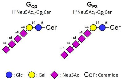
Glycosphingolipids from the ganglio-series are usually classified in four series according to the presence of 0 to 3 sialic acid residues linked to lactosylceramide. The transfer of sialic acid is catalyzed in the Golgi apparatus by specific sialyltransferases that show high specificity toward glycolipid substrates. ST8Sia I (EC 2.4.99.8, SAT-II, SIAT 8a) is the key enzyme controlling the biosynthesis of b- and c-series gangliosides. ST8Sia I is expressed at early developmental stages whereas in adult human tissues, ST8Sia I transcripts are essentially detected in brain. ST8Sia I together with b- and c-series gangliosides are also over-expressed in neuroectoderm-derived malignant tumors such as melanoma, glioblastoma, neuroblastoma and in estrogen receptor (ER) negative breast cancer, where they play a role in cell proliferation, migration, adhesion and angiogenesis. We have stably expressed ST8Sia I in MCF-7 breast cancer cells and analyzed the glycosphingolipid composition of wild type (WT) and GD3S+ clones. As shown by mass spectrometry, MCF-7 expressed a complex pattern of neutral and sialylated glycosphingolipids from globo- and ganglio-series. WT MCF-7 cells exhibited classical monosialylated gangliosides including GM3, GM2, and GM1a. In parallel, the expression of ST8Sia I in MCF-7 GD3S+ clones resulted in a dramatic change in ganglioside composition, with the expression of b- and c-series gangliosides as well as unusual tetra- and pentasialylated lactosylceramide derivatives GQ3 (II3Neu5Ac4-Gg2Cer) and GP3 (II3Neu5Ac5-Gg2Cer). This indicates that ST8Sia I is able to act as an oligosialyltransferase in a cellular context.

Graphical Abstract

Abbreviations:

BSA:
Bovine Serum Albumin
Cer:
ceramide
DMB:
1,2-diamino-4,5-methylenedioxybenzene
DMEM:
Dulbecco’s Modified Eagle’s Medium
EDTA:
Ethylenediaminetetraacetic Acid
DMSO:
Dimethyl Sulfoxide
FBS:
Fetal Bovine Serum
FITC:
Fluorescein Isothiocyanate
FL-HPLC:
Fluorescence Detection High Performance Liquid Chromatography
GD3S:
GD3 synthase
GSL:
glycosphingolipid
HPRT:
Hypoxanthine PhosphoRibosylTransferase
HRP:
horseradish peroxidase
LacCer:
Lactosylceramide
mAb:
monoclonal Antibody
MALDI-TOF:
matrix assisted laser desorption-ionization time-of-flight
MS:
Mass Spectrometry
PBS:
Phosphate Buffered Saline
QPCR:
Quantitative real-time Polymerase Chain Reaction
SDS-PAGE:
Sodium Dodecyl Sulfate Polyacrylamide Gel Electrophoresis
ST3Gal V:
GM3 synthase
ST8Sia I:
GD3 synthase
ST8Sia V:
GT3 synthase; WT: Wild Type

1. Introduction

Glycosphingolipids (GSL) from the ganglio-series are classified in four series according to the presence of 0 to 3 sialic acid residues linked to lactosylceramide (Galβ1-4Glc-Cer, LacCer) [1]. The transfer of sialic acid to LacCer is catalyzed in the Golgi apparatus by specific sialyltransferases (namely ST3Gal V, ST8Sia I and ST8Sia V) that show high specificity toward glycolipid substrates [2]. LacCer, GM3 (Neu5Acα2-3Galβ1-4Glc-Cer, II3Neu5Ac1-Gg2Cer), GD3 (Neu5Acα2-8Neu5Acα2-3Galβ1-4Glc-Cer, II3Neu5Ac2-Gg2Cer) and GT3 (Neu5Acα2-8Neu5Acα2-8Neu5Acα2-3Galβ1-4Glc-Cer, II3Neu5Ac3-Gg2Cer) are therefore the precursors for 0-, a-, b- and c-series gangliosides and the biosynthesis of these compounds determine the relative proportion of gangliosides in each series (Figure 1). Elongation of the precursors can then occur by the sequential action of N-acetyl-galactosaminyltransferase (β4GalNAc T1), galactosyltransferase (β3Gal T4) and sialyltransferases (ST3Gal I, ST3Gal II and ST8Sia V), α-gangliosides deriving from the action of ST6GalNAc III, V or VI on GM1b, GD1a or GT1b (Table 1).
Figure 1. Biosynthesis pathway for gangliosides. The code names of gangliosides are according to Svennerholm [1]. Cer: ceramide.
Molecules 17 09559 g001
The sialyltransferase ST8Sia I (EC 2.4.99.8, SAT-II, SIAT 8a) is the only enzyme known to catalyze the transfer of a sialic acid residue onto GM3 through an α2,8-linkage to synthesize GD3. ST8Sia I and GD3 are expressed in fetal tissues at an early developmental stage [3,4] where they play a key role in cell-cell interaction, cell differentiation and proliferation [5], whereas in adult human tissues, ST8Sia I is essentially detected in the brain [6]. ST8Sia I and GD3 have been also shown to be over-expressed in neuroectoderm-derived malignant tumors such as melanoma, glioblastoma and neuroblastoma, and in estrogen receptor negative breast cancer [7,8,9,10].
Table 1. Glycosyltransferases involved in gangliosides biosynthesis. R = GA3, GM3, GD3 or GT3.
GeneCommon nameMain acceptor(s)Accession #Reference
UGCGGlcCer synthaseCeramideNM_003358[18]
B4GALT6LacCer synthase GlucosylceramideNM_004775[19,20]
ST3GAL5GM3 synthaseLactosylceramideNM_003896[21]
ST8SIA1GD3 synthaseGM3, GD3NM_003034.2[11,12,13]
ST8SIA5GT3 synthaseGD3, GM1b, GD1a, GT1bNM_013305[17]
B4GALNACT1GM2/GD2 synthaseGA3, GM3, GD3, GT3NM_001478.2[22,23,24]
B3GALT4GM1a/GD1b synthaseGA2, GM2, GD2, GT2NM_003782.3[23,25]
ST3GAL1ST3Gal IGalβ1-3GalNAcβ1-4-RNM_003033[26]
ST3GAL2ST3Gal IIGalβ1-3GalNAcβ1-4-RNM_006927[27]
ST6GALNAC3ST6GalNAc IIINeu5Acα2-3Galβ1-3GalNAcβ1-4-RNM_152996[28]
ST6GALNAC5ST6GalNAc VNeu5Acα2-3Galβ1-3GalNAcβ1-4-RNM_030965.1[29]
ST6GALNAC6ST6GalNAc VINeu5Acα2-3Galβ1-3GalNAcβ1-4-RNM_013443.3[29]
The human ST8Sia I cDNA was simultaneously isolated by expression cloning by three research groups [11,12,13]. The ST8SIA1 gene is located on chromosome 12, in p12.1-p11.2 and consists of five coding exons spanning over 135 kbp of genomic DNA [14]. ST8Sia I cDNA encodes a 341 amino acid membrane-bound Golgi enzyme with a 12 amino-acid cytoplasmic tail, a transmembrane domain of about 20 residues and a catalytic domain containing the conserved Sialyl motifs involved in substrate binding and transfer [15].
Whereas ST8Sia I mainly sialylates GM3, Nakayama and co-workers have underlined its ability to synthesize GT3 from GD3 [6]. ST8Sia I was also shown to use GM1b, GD1a or GT1b as acceptor substrates to synthesize GD1c, GT1a or GQ1b, respectively, both in vitro and in vivo [16]. However, the α2,8-sialyltransferase ST8Sia V is a much better candidate for GT1a/GQ1b synthase activity [17] and no ST8Sia V activity was detected toward GM3. Consequently, ST8Sia I is considered as the only GD3 synthase (GD3S) that controls the biosynthesis of gangliosides from the b- and c-series.
By stable transfection of the full-length cDNA of human GD3 synthase, we have isolated cellular clones deriving from MCF-7 breast cancer cells that constitutively express GD3S together with b- and c-series gangliosides. Here, we show by mass spectrometry and HPLC analysis that clones that express a high level of GD3S also accumulate unusual tetra- and pentasialylated derivatives of LacCer, GQ3 (II3Neu5Ac4-Gg2Cer) and GP3 (II3Neu5Ac5-Gg2Cer).

2. Results and Discussion

2.1. Analysis of ST8Sia I Expression by QPCR in Control and GD3S+ MCF-7 Clones

MCF-7 cells were transfected with the pcDNA3-GD3S expression vector containing the full-length cDNA of human GD3S or the empty pcDNA3 vector as control. Transfected cells were cultured 21 days in the presence of 1 mg/mL G418. Individual G418-resistant colonies were isolated by limiting dilution cloning. Forty-four clones were obtained and analyzed for the expression of GD3S. As previously shown [30], QPCR analysis of GD3S expression (Figure 2) indicates that GD3S mRNA is express at a very low level in wild-type and control (empty vector transfected) MCF-7 cells compared to SK-Mel 28 melanoma cells used as positive control [31]. Within the forty-four analyzed clones, three GD3S+ clones (clone #31, #41 and #44) were selected according to the high expression of GD3S compared to SK-Mel 28 (1.3-fold, 2.3-fold and 6.3-fold, respectively) (Figure 2).
Figure 2. QPCR analysis of GD3S expression in control and GD3S+ MCF-7 clones. Quantification of GD3S expression was performed by the method described by Livak and Schmittgen [32] and normalized to HPRT. The expression of GD3S in MCF-7 cells was relative to SK-Mel 28, which was regarded as 100%.
Molecules 17 09559 g002

2.2. Flow Cytometry Analysis of Gangliosides Expression in MCF-7 GD3S+ Clones

The pattern of gangliosides was monitored in the three selected MCF-7 GD3S+ clones (clone #31, #41, #44) by flow cytometry using anti-GD3 R24 and anti-GT3 A2B5 mAbs. As shown in Figure 3, the three GD3S+ clones expressed GD3 and GT3 whereas wild-type and control (empty vector transfected) MCF-7 cells did not expressed complex gangliosides. GT3 is expressed at a similar level in the three GD3S+ clones but a decrease of GD3 is observed in clone #44 compared to clone #31 and #41 whereas the expression level of GD3S was 4.8-fold or 2.7-fold higher in clone #44 compared to clone #31 and #41, respectively (Figure 2). Control cells showed no change in the ganglioside profile compared with wild-type MCF-7 (data not shown).

2.3. MS Analysis of Gangliosides in MCF-7 and GD3S+ Clones

Glycolipids were extracted from cells, purified by reverse phase chromatography and permethylated prior to MS analysis. Mass spectrometry analysis established that glycolipid profiles of MCF-7 WT and GD3S+ clones were characterized by complex patterns of neutral and sialylated glycosphingolipids from globo- and ganglio-series. Profiles of all cell lines were dominated by two signals at m/z 1460 and 1572 both identify based on their MALDI-TOF/TOF fragmentation patterns (data not shown) and in agreement with previously published analyses [33] as mixtures of Gb4 and GA1 differing by the nature of their lipid moieties (d18:1-16:0 or d18:1-24:0). Along these two major components, MS and MS/MS analyses permitted us to identified other minor neutral GSLs including LacCer and Gb3 (Figure 4). The comparison of MS profiles did not show any significant difference in neutral GSLs content between MCF-7 WT and GD3+ clones. On the contrary, the content in sialylated glycolipids varied among the different cell lines. MCF-7 WT cells exhibited monosialylated gangliosides including GM3, GM2, and GM1. The only disialylated GSL observed in MCF-7 WT cells was GD1 at m/z 2182 and 2194. Its sequence analysis by MALDI-TOF/TOF typified it as GD1a, thus lacking disialylated motif (data not shown). GD3+ clones did not show GSLs from the G2 and G1 families but synthesized instead a family of unusual highly sialylated lactosylceramide derivatives substituted by up to 5 Neu5Ac residues tentatively identified as GD3, GT3, GQ3 and GP3. The structure of these four compounds was confirmed by MALDI-TOF/TOF sequencing.
Figure 3. Flow cytometry analysis of gangliosides in pcDNA3-GD3S transfected MCF-7 cells. Immunodetection of GD3 and GT3 was performed using anti-GD3 R24 (dilution 1:100) and anti-GT3 A2B5 (dilution 1:10) mAbs. The gray peaks correspond to the negative controls (incubation with secondary antibody alone, anti-mouse IgM or anti-mouse IgG labeled with Alexa 488).
Molecules 17 09559 g003
The Glycolipid profile of SK-Mel 28 cells was also analyzed in respect to the presence of polysialylated lactosylceramide derivatives. Contrarily to MCF-7 WT cells, GSLs extracted from SK-Mel 28 cells did not contain globo-series but were dominated by ganglio-series, which induces much higher overall sialic acid content (data not shown). In particular, disialylated GD3 appears to be the major component along with monosialylated GM3 and trisialylated GT3. However, the tetra- and pentasialylated gangliosides synthesized by MCF-7 GD3+ clones were not detected in SK-Mel 28.
Figure 4. Comparison of MS profiles of permethylated glycosphingolipids purified from MCF-7 WT and GD3S+ clone #44. All GSLs are present as d18:1/C16:0 (Cer*) and d18:1/C24:0 (Cer**) isomers. Molecules 17 09559 i001, Gal; Molecules 17 09559 i002, Glc; Molecules 17 09559 i003, GalNAc; Molecules 17 09559 i004, Neu5Ac.
Molecules 17 09559 g004
We illustrate the sequence analyses of unusual GQ3 and GP3 in Figure 5. These two molecules differing in the presence of a single Neu5Ac residue were structurally related. They shared the fragmentation pattern of a linear stretch of four sialic acid residues in terminal non-reducing position as [M+Na]+ B-ions at m/z 398, 759, 1120 and 1481 and a linear sequence of Sia4Hex2Cer at reducing end as [M+Na]+ Y-ions at m/z 1106, 1466, 1827 and 2188. GP3 showed additional B and Y ions at m/z 1842 and 2551 typifying a linear Sia5 sequence. Altogether, these data established that MCF-7 GD3+ clone #44 synthesizes an unusual family of oligosialylated lactosylceramide derivatives presented from 2 to 5 Neu5Ac residues. The disappearance of GM2, GM1 and GD1a in MCF-7 GD3+ clone #44 could be explained by the depletion of GM3 substrate caused by the over-expression of the GD3S+ that competes with β-4GalNAcT1 for the use of GM3 substrate.
Figure 5. MS/MS sequencing of permethylated glycosphingolipids. Analysis of GQ3 at m/z 2568 (A) and GP3 at m/z 2929 (B) with ceramide moieties d18:1/C24:0 (Cer**). All fragments are observed as [M+Na]+ adducts except those at m/z at 376 and 660 observed as [M+H]+ adducts. Fragment ions were annotated according to nomenclature of Domon and Costello [34]. Molecules 17 09559 i005, Gal; Molecules 17 09559 i006, Glc; Molecules 17 09559 i007, GalNAc; Molecules 17 09559 i008, Neu5Ac.
Molecules 17 09559 g005

2.4. Quantification of Polysialylation Associated with Gangliosides by HPLC

Because mass spectrometry does not provide reliable quantitative data, we quantified the extent of oligosialylation modifications in GSLs induced by the overexpression of GD3 in MCF-7 by screening all three GD3S+ clones (#31, #41 and #44) (Figure 6). To do that, oligosialylated motifs were released from purified GSLs by mild hydrolysis according to optimized procedures [35] and labeled by DMB before separation and quantification by FL-HPLC [36]. The analysis of relative quantifications of sialic acid chains in GSLs of all MCF-7 GD3S+ clones compared to MCF-7 WT demonstrated a sharp increase of sialylation oligomerization degree, up to five residues, in accordance with mass spectrometry analysis. In MCF-7 WT, no oligosialylation could be observed, in accordance with the sole identification of GD1a by MS. In contrast, all GD3S+ clones contained about 30% of Sia2 and Sia3 motifs, up to 10% of Sia4 and up to 5% of Sia5. Although clones presented similar GSL oligosialylation profiles, small scale quantitative differences could be observed between the three clones, with a prevalence of higher DP values for clone #44 compared to clones #31 and #41.
Figure 6. Relative quantification of oligosialylation sequence on GSL of MCF-7 WT GD3S+ clones #31, clone #41 and clone #44 by FL-HPLC after DMB derivation.
Molecules 17 09559 g006

3. Experimental Section

3.1. Antibodies and Reagents

Anti-GD3 R24 mAb was purchased from Abcam (Cambridge, UK) and anti-GT3 A2B5 mAb was kindly provided by Pr. Jacques Portoukalian (Depart. of Transplantation and Clinical Immunology, Claude Bernard University and Edouard Herriot Hospital, Lyon, France). FITC-conjugated sheep anti-mouse IgG was from GE Healthcare (Templemars, France). FITC-conjugated anti-mouse IgM was purchased from Molecular Probes (Invitrogen, Carlsbad, CA, USA).

3.2. Cell Culture

The breast cancer cell line MCF-7 and the melanoma cell line SK-Mel 28 were obtained from the American Type Cell Culture Collection. Cell culture reagents were purchased from Lonza (Levallois-Perret, France). Cells were routinely grown in monolayer and maintained at 37 °C in an atmosphere of 5% CO2, in Dulbecco’s modified Eagle’s medium (DMEM) supplemented with 10% fetal bovine serum (FBS), 2 mM L-glutamine, and 100 units/mL penicillin-streptomycin. GD3S positive (GD3S+) MCF-7 clones were obtained by stable transfection of the pcDNA3-GD3S expression vector encoding the full-length human GD3 synthase [37] as previously described [33]. Individual resistant colonies were isolated by limit dilution. Three positive clones (#31, #41 and #44), expressing different levels of GD3S were used for further study. Control cells (empty vector transfected) and GD3S+ clones were cultured in the presence of 1 mg/mL G418 (Invitrogen, Cergy-Pontoise, France).

3.3. Quantitative Real-Time-PCR (QPCR) Analysis of GD3 Synthase

Total RNA was extracted using the Nucleospin RNA II kit (Macherey Nagel, Hoerdt, France), quantified using a NanoDrop spectrophotometer (Thermo Scientifics, Wilmington, USA) and the purity of the preparation was checked by ratio of the absorbance at 260 and 280 nm. The cDNA was synthesized using 2 µg of RNA (GE Healthcare). PCR primers for GD3S and Hypoxanthine PhosphoRibosylTransferase (HPRT) were previously described [30,38] and synthesized by Eurogentec (Seraing, Belgium). PCR reactions (25 µL) were performed using 2X SYBR® Green Universal QPCR Master Mix (Stratagene, Amsterdam, The Netherlands), with 2 µL of cDNA solution and 300 nM final concentration of each primer. PCR conditions were as follows: 95 °C for 30 s, 51 °C for 45 s, 72 °C for 30 s (40 cycles). Assays were performed in triplicate and GD3S transcript expression level was normalized to HPRT using the 2ΔΔCt method described by Livak and Schmittgen [32]. Serial dilutions of the appropriate positive control cDNA sample were used to create standard curves for relative quantification and negative control reactions were performed by replacing cDNA templates by sterile water.

3.4. Analysis of Cell Surface Ganglioside by Flow Cytometry

Cells were washed in cold PBS and detached by EDTA 2 mM. Cells were incubated at 4 °C during 1 h with anti-GD3 R24 (1:100) and anti-GT3 A2B5 (1:10), diluted in phosphate buffered saline (PBS) containing 0.5% bovine serum albumin (PBS-BSA) (Sigma-Aldrich). After washing with PBS-BSA, cells were incubated on ice during 1 h with Alexa Fluor 488 anti-IgG or anti-IgM (1:500). After two washes in PBS-BSA, cells were analyzed by flow cytometry (FACScalibur, Becton Dickinson). Control experiments were performed using secondary antibody alone.

3.5. Extraction and Preparation of Glycolipids

Twenty dishes (10 cm diameter) of cultured cells were washed twice with ice-cold PBS and cells were scraped and sonicated on ice in 200 µL of water. The resulting material was dried under vacuum and sequentially extracted by CHCl3/CH3OH (2:1, v/v), CHCl3/CH3OH (1:1, v/v) and CHCl3/CH3OH/H2O (1:2:0.8, v/v/v). Supernatants were pooled, dried and subjected to a mild saponification in 0.1 M NaOH in CHCl3/CH3OH (1:1) at 37 °C for 2 h and then evaporated to dryness [39]. Samples were reconstituted in CH3OH/H2O (1:1, v/v) and applied to a reverse phase C18cartridge (Waters, Milford, MA, USA) equilibrated in the same solvent. After washing with CH3OH/H2O (1:1, v/v), GSLs were eluted by CH3OH, CHCl3/CH3OH (1:1, v/v) and CHCl3/CH3OH (2:1, v/v).

3.6. Mass Spectrometry Analysis of GSL

Prior to mass spectrometry analysis, GSL were permethylated according to Ciucanu and Kerek [40]. Briefly, compounds were incubated 2 h in a suspension of 200 mg/mL NaOH in dry DMSO (300 µL) and CH3I (200 µL). The methylated derivatives were extracted in CHCl3 and washed several times with water. The reagents were evaporated and the sample was dissolved in CHCl3 in the appropriate dilution. MALDI-MS and MS/MS analyses of permethylated GSL were performed on 4800 Proteomics Analyzer (Applied Biosystems, Framingham, MA, USA) mass spectrometer, operated in the reflectron mode. For MS acquisition, 5 µL of diluted permethylated samples in CHCl3 were mixed with 5 µL of 2,5-dihydroxybenzoic acid matrix solution (10 mg/mL dissolved in CHCl3/CH3OH (1:1, v/v)). The mixtures (2 µL) were then spotted on the target plate and air dried. MS survey data comprises a total of 50 sub-spectra of 1500 laser shots. Peaks observed in the MS spectra were selected for further MS/MS. CID MS/MS data comprises a total of 100 sub-spectra of 3000 laser shots. Two or more spectra can be combined post-acquisition with mass tolerance set at 0.1 Da to improve S/N ratio. The potential difference between the source acceleration voltage and the collision cell was set to 1 kV and argon was used as collision gas.

3.7. Analysis of Oligo-Sialylated Sequences by HPLC

In order to minimize internal fragmentation of polysialylated sequences, sialylated glycan samples were directly coupled to 1,2-diamino-4,5-methylenedioxybenzene (DMB) without prior mild hydrolysis [35]. Samples were incubated for 2.5 h at 50 °C in 50 μL of a DMB reagent solution (2.7 mM DMB, 9 mM sodium hydrosulfite, and 0.5 mM β-mercaptoethanol in 20 mM TFA). 10 μL of 1 M NaOH was then added and the reaction mixtures further incubated in the dark at room temperature for 1 h. Samples were stored at 4 °C before analysis. DMB-derivatized sialic acid oligomers were separated on a HPLC apparatus fitted with a CarboPac PA-100 column (Dionex). CarboPac column was eluted at 1 mL/min with a concentration gradient of 2 to 32% of 1 M NaNO3 in water. Elution was monitored by an on line fluorescence detector set at wavelengths of 373 nm for excitation and 448 nm for emission.

4. Conclusions

ST8Sia I is the only sialyltransferase able to transfer a sialic acid residue onto GM3 to synthesize GD3 and is therefore considered as the GD3 synthase. The animal ST8Sia family can be divided in three groups according to the capacity to carry out poly-, oligo- and mono-α2,8-sialylation and phylogenic analyses have clearly associated ST8Sia I to the group of mono-α2,8-sialyltransferase [41]. However, by expression cloning of the human GT3 synthase, Nakayama and co-workers have underlined the ability of ST8Sia I to synthesize GT3 from GD3 [6]. Here, we show, for the first time, that ST8Sia I is able to synthesize unusual highly sialylated lactosylceramide derivatives substituted by up to 5 Neu5Ac residues and identified as GQ3 (II3Neu5Ac4-Gg2Cer) and GP3 (II3Neu5Ac5-Gg2Cer), showing that this enzyme can act as an oligosialyltransferase.
In humans, the ST8SIA1 gene is located on chromosome 12, in p12.1-p11.2 and consists of five coding exons spanning over 135 kbp of genomic DNA [14]. Two initiation codons on the first exon lead to two protein isoforms of 356 or 341 amino acids that differ in their N-terminal part. However, the relative capacity of each isoform to transfer more than two sialic acid residues has not been evaluated. By in vitro sialyltransferase assay, a recombinant soluble form of the human GD3 synthase was shown to synthesize, after a long period of incubation, higher polysialogangliosides, which presumably have more than three sialic acid residues but these compounds were not characterized [6].
From a general point of view, the occurrence of oligosialylation associated with glycolipids has been rarely reported so far. One exception is the recent identification of polysialogangliosides containing α2,8-linked polyNeu5Ac with DPs ranging from 2 to at least 16 in sea urchin sperm head [42]. However, to our knowledge, tetra- and pentasialylated lactosylceramide derivatives have never been described in human tissues and cells. Although SK-Mel 28 cells that constitutively express high levels of GD3S, synthesize high quantities of GD3 and GT3, we were not able to detect these unusual tetra- and pentasialylated gangliosides in this cell line. Indeed, in our cellular model, these unusual structures were obtained after transfection of GD3S cDNA and may not exist in natural conditions, and the depletion of GM3 in GD3S+ MCF-7 clones could explain that GD3S used other gangliosides, such as GT3 or GQ3, as acceptor substrates. Nevertheless, one may thus expect that revisiting the structure of human gangliosides with recent high sensitivity mass spectrometry techniques will uncover oligosialylated glycolipids in cancerous cell lines and in normal or pathological tissues.

Acknowledgments

This work was supported by the University of Sciences and Technologies of Lille, the Association pour la Recherche sur le Cancer (Grant n° 7936 and 5023) et le comité de l’Aisne de La Ligue contre le Cancer. Florent Colomb is supported by a PhD fellowship from “Vaincre la mucoviscidose”.

References

  1. Svennerholm, L. Ganglioside designation. Adv. Exp. Med. Biol. 1980, 125, 11–19. [Google Scholar]
  2. Zeng, G.; Yu, R.K. Cloning and transcriptional regulation of genes responsible for synthesis of gangliosides. Curr. Drug Targets 2008, 9, 317–324. [Google Scholar] [CrossRef]
  3. Yamamoto, A.; Haraguchi, M.; Yamashiro, S.; Fukumoto, S.; Furukawa, K.; Takamiya, K.; Atsuta, M.; Shiku, H.; Furukawa, K. Heterogeneity in the expression pattern of two ganglioside synthase genes during mouse brain development. J. Neurochem. 1996, 66, 26–34. [Google Scholar]
  4. Yu, R.K.; Macala, L.J.; Taki, T.; Weinfield, H.M.; Yu, F.S. Developmental changes in ganglioside composition and synthesis in embryonic rat brain. J. Neurochem. 1988, 50, 1825–1829. [Google Scholar]
  5. Yamashita, T.; Wada, R.; Sasaki, T.; Deng, C.; Bierfreund, U.; Sandhoff, K.; Proia, R.L. A vital role for glycosphingolipid synthesis during development and differentiation. Proc. Natl. Acad. Sci. USA 1999, 96, 9142–9147. [Google Scholar]
  6. Nakayama, J.; Fukuda, M.N.; Hirabayashi, Y.; Kanamori, A.; Sasaki, K.; Nishi, T.; Fukuda, M. Expression cloning of a human GT3 synthase. GD3 and GT3 are synthesized by a single enzyme. J. Biol. Chem. 1996, 271, 3684–3691. [Google Scholar]
  7. Furukawa, K.; Hamamura, K.; Aixinjueluo, W.; Furukawa, K. Biosignals modulated by tumor-associated carbohydrate antigens novel targets for cancer therapy. Ann. NY Acad. Sci. 2006, 1086, 185–198. [Google Scholar] [CrossRef]
  8. Oblinger, J.L.; Pearl, D.K.; Boardman, C.L.; Saqr, H.; Prior, T.W.; Scheithauer, B.W.; Jenkins, R.B.; Burger, P.C.; Yates, A.J. Diagnostic and prognostic value of glycosyltransferase mRNA in glioblastoma multiforme patients. Neuropathol. Appl. Neurobiol. 2006, 32, 410–418. [Google Scholar] [CrossRef]
  9. Ruan, S.; Lloyd, K.O. Glycosylation pathways in the biosynthesis of gangliosides in melanoma and neuroblastoma cells relative glycosyltransferase levels determine ganglioside patterns. Cancer Res. 1992, 52, 5725–5731. [Google Scholar]
  10. Ruckhäberle, E.; Rody, A.; Engels, K.; Gaetje, R.; von Minckwitz, G.; Schiffmann, S.; Grösch, S.; Geisslinger, G.; Holtrich, U.; Karn, T.; Kaufmann, M. Microarray analysis of altered sphingolipid metabolism reveals prognostic significance of sphingosine kinase 1 in breast cancer. Breast Cancer Res. Treat. 2008, 112, 41–52. [Google Scholar] [CrossRef]
  11. Nara, K.; Watanabe, Y.; Maruyama, K.; Kasahara, K.; Nagai, Y.; Sanai, Y. Expression cloning of a CMP-NeuAcNeuAc alpha 2-3Gal beta 1-4Glc beta 1-1'Cer alpha 2,8-sialyltransferase (GD3 synthase) from human melanoma cells. Proc. Natl. Acad. Sci. USA 1994, 91, 7952–7956. [Google Scholar] [CrossRef]
  12. Sasaki, K.; Kurata, K.; Kojima, N.; Kurosawa, N.; Ohta, S.; Hanai, N.; Tsuji, S.; Nishi, T. Expression cloning of a GM3-specific alpha-2,8-sialyltransferase (GD3 synthase). J. Biol. Chem. 1994, 269, 15950–15956. [Google Scholar]
  13. Haraguchi, M.; Yamashiro, S.; Yamamoto, A.; Furukawa, K.; Takamiya, K.; Lloyd, K.O.; Shiku, H.; Furukawa, K. Isolation of GD3 synthase gene by expression cloning of GM3 alpha-2,8-sialyltransferase cDNA using anti-GD2 monoclonal antibody. Proc. Natl. Acad. Sci. USA 1994, 91, 10455–10459. [Google Scholar]
  14. Furukawa, K.; Horie, M.; Okutomi, K.; Sugano, S.; Furukawa, K. Isolation and functional analysis of the melanoma specific promoter region of human GD3 synthase gene. Biochim. Biophys. Acta 2003, 1627, 71–78. [Google Scholar] [CrossRef]
  15. Harduin-Lepers, A.; Vallejo-Ruiz, V.; Krzewinski-Recchi, M.A.; Samyn-Petit, B.; Julien, S.; Delannoy, P. The human sialyltransferase family. Biochimie 2001, 83, 727–737. [Google Scholar]
  16. Nara, K.; Watanabe, Y.; Kawashima, I.; Tai, T.; Nagai, Y.; Sanai, Y. Acceptor substrate specificity of a cloned GD3 synthase that catalyzes the biosynthesis of both GD3 and GD1c/GT1a/GQ1b. Eur. J. Biochem. 1996, 238, 647–652. [Google Scholar]
  17. Kim, Y.J.; Kim, K.S.; Do, S.; Kim, C.H.; Kim, S.K.; Lee, Y.C. Molecular cloning and expression of human alpha2,8-sialyltransferase (hST8Sia V). Biochem. Biophys. Res. Commun. 1997, 235, 327–330. [Google Scholar]
  18. Ichikawa, S.; Sakiyama, H.; Suzuki, G.; Hidari, K.I.; Hirabayashi, Y. Expression cloning of a cDNA for human ceramide glucosyltransferase that catalyzes the first glycosylation step of glycosphingolipid synthesis. Proc. Natl. Acad. Sci. USA 1996, 93, 4638–4643. [Google Scholar]
  19. Nomura, T.; Takizawa, M.; Aoki, J.; Arai, H.; Inoue, K.; Wakisaka, E.; Yoshizuka, N.; Imokawa, G.; Dohmae, N.; Takio, K.; et al. Purification, cDNA cloning, and expression of UDP-Gal: Glucosylceramide beta-1,4-galactosyltransferase from rat brain. J. Biol. Chem. 1998, 273, 13570–13577. [Google Scholar]
  20. Takizawa, M.; Nomura, T.; Wakisaka, E.; Yoshizuka, N.; Aoki, J.; Arai, H.; Inoue, K.; Hattori, M.; Matsuo, N. cDNA cloning and expression of human lactosylceramide synthase. Biochim. Biophys. Acta 1999, 1438, 301–304. [Google Scholar] [CrossRef]
  21. Ishii, A.; Ohta, M.; Watanabe, Y.; Matsuda, K.; Ishiyama, K.; Sakoe, K.; Nakamura, M.; Inokuchi, J.; Sanai, Y.; Saito, M. Expression cloning and functional characterization of human cDNA for ganglioside GM3 synthase. J. Biol. Chem. 1998, 273, 31652–31655. [Google Scholar]
  22. Nagata, Y.; Yamashiro, S.; Yodoi, J.; Lloyd, K.O.; Shiku, H.; Furukawa, K. Expression cloning of beta 1,4 N-acetylgalactosaminyltransferase cDNAs that determine the expression of GM2 and GD2 gangliosides. J. Biol. Chem. 1992, 267, 12082–12089. [Google Scholar]
  23. Iber, H.; Zacharias, C.; Sandhoff, K. The c-series gangliosides GT3, GT2 and GP1c are formed in rat liver Golgi by the same set of glycosyltransferases that catalyse the biosynthesis of asialo-, a- and b-series gangliosides. Glycobiology 1992, 2, 137–142. [Google Scholar] [CrossRef]
  24. Yamashiro, S.; Haraguchi, M.; Furukawa, K.; Takamiya, K.; Yamamoto, A.; Nagata, Y.; Lloyd, K.O.; Shiku, H.; Furukawa, K. Substrate specificity of beta 1,4-N-acetylgalactosaminyltransferase in vitro and in cDNA-transfected cells. GM2/GD2 synthase efficiently generates asialo-GM2 in certain cells. J. Biol. Chem 1995, 270, 6149–6155. [Google Scholar]
  25. Amado, M.; Almeida, R.; Carneiro, F.; Levery, S.B.; Holmes, E.H.; Nomoto, M.; Hollingsworth, M.A.; Hassan, H.; Schwientek, T.; Nielsen, P.A.; et al. A family of human beta3-galactosyltransferases. Characterization of four members of a UDP-galactose: Beta-N-acetyl-glucosamine/beta-N-acetyl-galactosamine beta-1,3-galactosyltransferase family. J. Biol. Chem 1998, 273, 12770–12778. [Google Scholar]
  26. Kitagawa, H.; Paulson, J.C. Differential expression of five sialyltransferase genes in human tissues. J. Biol. Chem. 1994, 269, 17872–17878. [Google Scholar]
  27. Giordanengo, V.; Bannwarth, S.; Laffont, C.; van Miegem, V.; Harduin-Lepers, A.; Delannoy, P.; Lefebvre, J.C. Cloning and expression of cDNA for a human Gal(beta1-3)GalNAc alpha2,3-sialyltransferase from the CEM T-cell line. Eur. J. Biochem. 1997, 247, 558–566. [Google Scholar]
  28. Tsuchida, A.; Ogiso, M.; Nakamura, Y.; Kiso, M.; Furukawa, K.; Furukawa, K. Molecular cloning and expression of human ST6GalNAc III: Restricted tissue distribution and substrate specificity. J. Biochem. 2005, 138, 237–243. [Google Scholar]
  29. Harduin-Lepers, A.; Mollicone, R.; Delannoy, P.; Oriol, R. The animal sialyltransferases and sialyltransferase-related genes: A phylogenetic approach. Glycobiology 2005, 15, 805–817. [Google Scholar] [CrossRef]
  30. Cazet, A.; Groux-Degroote, S.; Teylaert, B.; Kwon, K.M.; Lehoux, S.; Slomianny, C.; Kim, C.H.; Le Bourhis, X.; Delannoy, P. GD3 synthase overexpression enhances proliferation and migration of MDA-MB-231 breast cancer cells. Biol. Chem. 2009, 390, 601–609. [Google Scholar]
  31. Ruan, S.; Raj, B.K.; Lloyd, K.O. Relationship of glycosyltransferases and mRNA levels to ganglioside expression in neuroblastoma and melanoma cells. J. Neurochem. 1999, 72, 514–521. [Google Scholar]
  32. Livak, K.J.; Schmittgen, T.D. Analysis of relative gene expression data using real-time quantitative PCR and the 2(-Delta Delta C(T)) Method. Methods 2001, 25, 402–408. [Google Scholar] [CrossRef]
  33. Steenackers, A.; Cazet, A.; Bobowski, M.; Rombouts, Y.; Lefebvre, J.; Guérardel, Y.; Tulasne, D.; Le Bourhis, X.; Delannoy, P. Expression of GD3 synthase modifies ganglioside profile and increases migration of MCF-7 breast cancer cells. C.R. Chim. 2012, 15, 3–14. [Google Scholar]
  34. Domon, B.; Costello, C.E. A systematic nomenclature for carbohydrate fragmentations in FAB-MS/MS spectra of glycoconjugates. Glycoconjug. J. 1988, 5, 397–409. [Google Scholar] [CrossRef]
  35. Sato, C.; Inoue, S.; Matsuda, T.; Kitajima, K. Fluorescent-assisted detection of oligosialyl units in glycoconjugates. Anal. Biochem. 1999, 266, 102–109. [Google Scholar]
  36. Chang, L.-Y.; Harduin-Lepers, A.; Kitajima, K.; Sato, C.; Huang, C.-J.; Khoo, K.-H.; Guérardel, Y. Developmental regulation of oligosialylation in zebrafish. Glycoconjug. J. 2009, 26, 247–261. [Google Scholar]
  37. Moon, S.K.; Kim, H.M.; Lee, Y.C.; Kim, C.H. Disialoganglioside (GD3) synthase gene expression suppresses vascular smooth muscle cell responses via the inhibition of ERK1/2 phosphorylation, cell cycle progression, and matrix metalloproteinase-9 expression. J. Biol. Chem. 2004, 279, 33063–33070. [Google Scholar]
  38. Zhang, X.; Ding, L.; Sandford, A.J. Selection of reference genes for gene expression studies in human neutrophils by real-time PCR. BMC Mol. Biol. 2005, 6, 4. [Google Scholar]
  39. Schnaar, R.L. Isolation of glycosphingolipids. Methods Enzymol. 1994, 230, 348–370. [Google Scholar]
  40. Ciucanu, I.; Kerek, F. Rapid and simultaneous methylation of fatty and hydroxy fatty acids for gas-liquid chromatographic analysis. J. Chromatogr. 1984, 284, 179–185. [Google Scholar]
  41. Harduin-Lepers, A.; Petit, D.; Mollicone, R.; Delannoy, P.; Petit, J.-M.; Oriol, R. Evolutionary history of the alpha2,8-sialyltransferase (ST8Sia) gene family: Tandem duplications in early deuterostomes explain most of the diversity found in the vertebrate ST8Sia genes. BMC Evol. Biol. 2008, 8, 258. [Google Scholar]
  42. Miyata, S.; Yamakawa, N.; Toriyama, M.; Sato, C.; Kitajima, K. Co-expression of two distinct polysialic acids, α2,8- and α2,9-linked polymers of N-acetylneuraminic acid, in distinct glycoproteins and glycolipids in sea urchin sperm. Glycobiology 2011, 21, 1596–1605. [Google Scholar] [CrossRef]
  • Sample Availability: Not available.