Tumour Cell Labelling by Magnetic Nanoparticles with Determination of Intracellular Iron Content and Spatial Distribution of the Intracellular Iron

Abstract
:1. Introduction
2. Results and Discussion
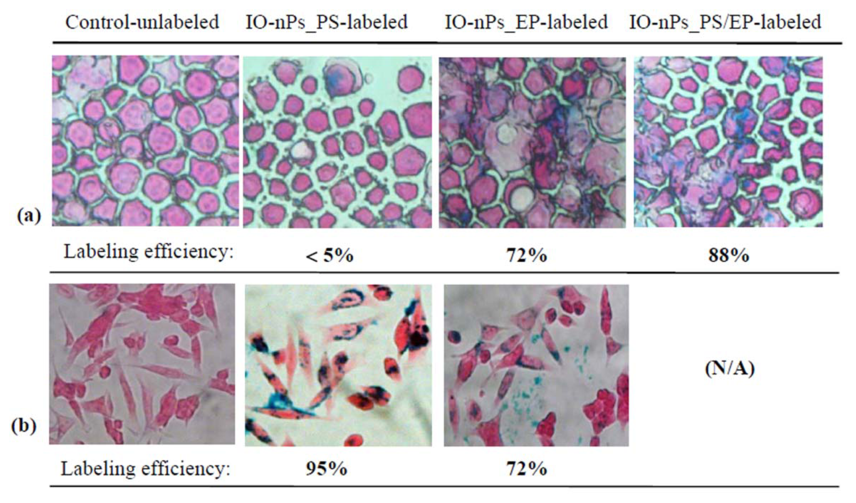
2.1. Magnetic Labelled Cells—Efficiency and Viability
2.2. Assessment Cellular Iron Uptake and Spatial Distribution
2.3. Discussion
3. Experimental Section
3.1. Cell Labelling by Magnetic Nanoparticles
3.1.1. Labelling with Transfection Agent (PS)
3.1.2. Labelling with Electroporation
3.1.3. Labelling with Electroporation in the Presence of PS
3.2. Assessment of Magnetically Labelled Cells
3.2.1. Cell Viability by MTS Assay
3.2.2. Prussian Blue Staining for Assessing Labelling Efficiency
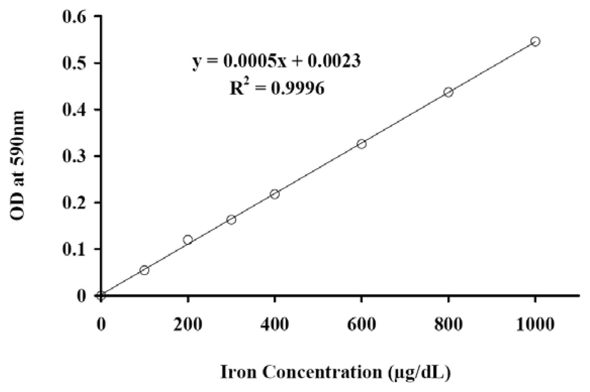
3.2.3. Quantitative Colorimetric Iron Assay
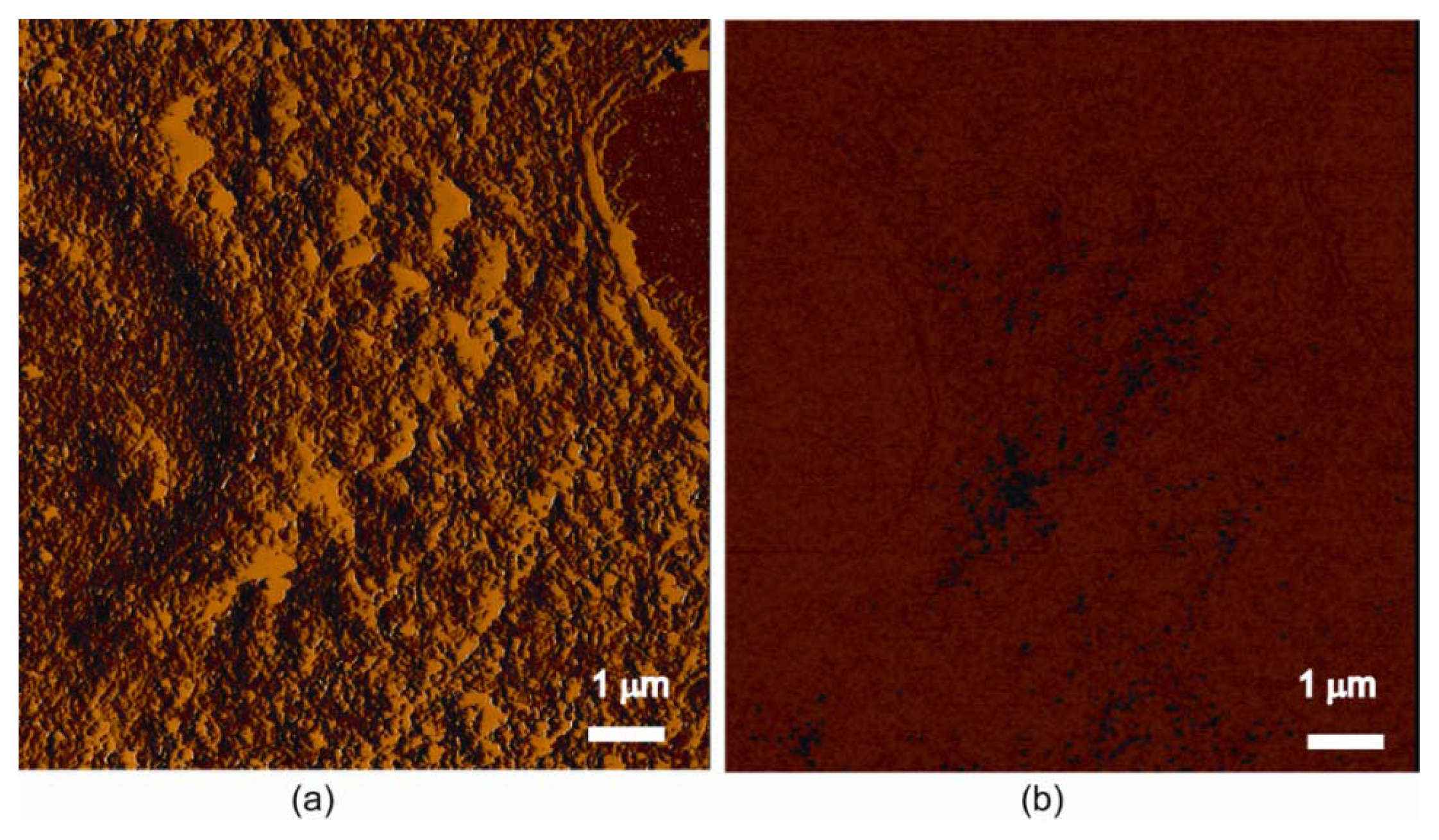
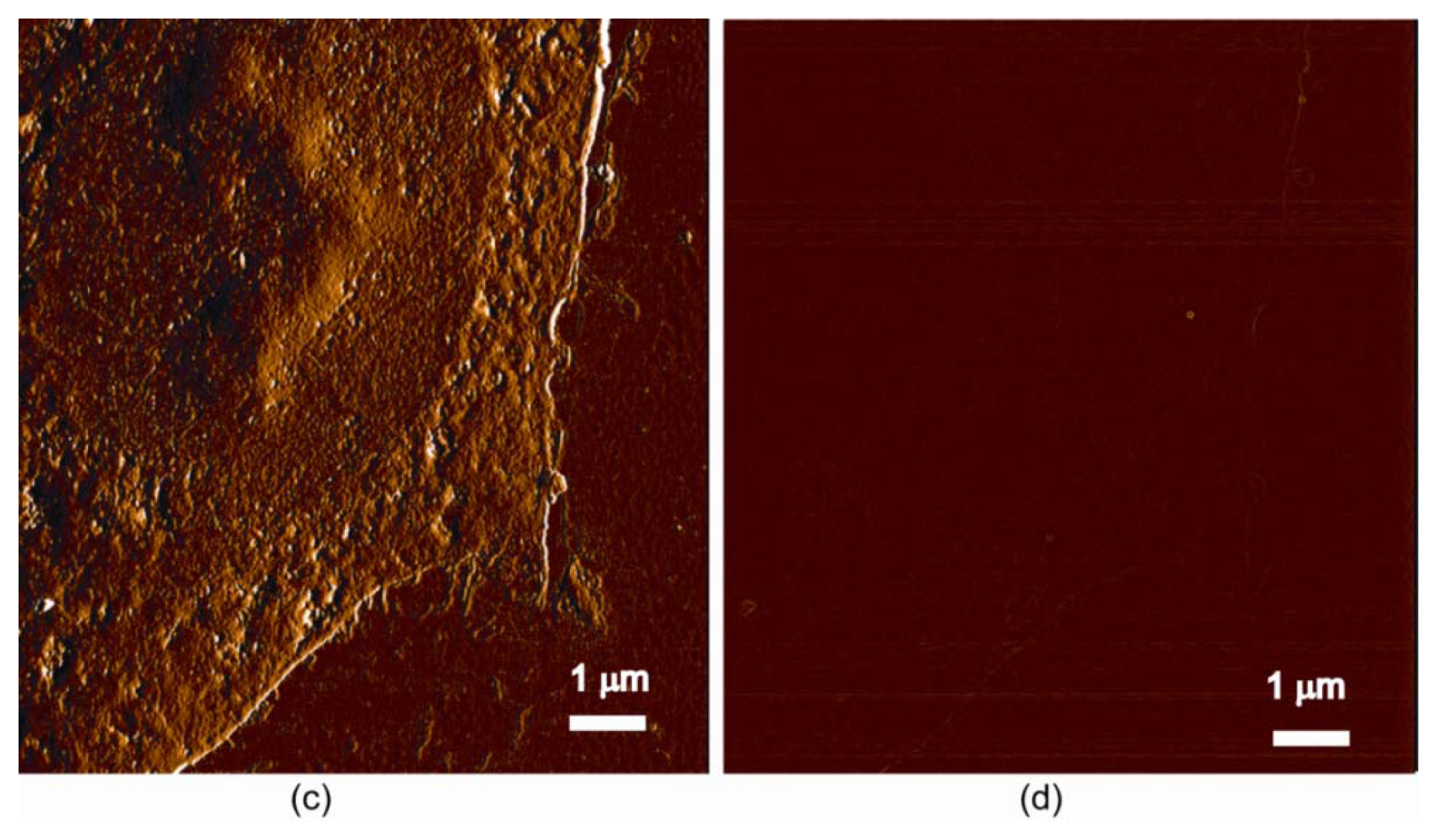
3.3. Magnetic Force Microscopy (MFM) Observation and Assessment
3.3.1. Magnetic Force Microscopy (MFM) Principle
3.3.2. MFM Scans with Image Processing for Single Cell Assessment
4. Conclusions
Acknowledgments
Conflict of Interest
References
- Thorek, D.L.; Chen, A.K.; Czupryna, J.; Tsourkas, A. Superparamagnetic iron oxide nanoparticle probes for molecular imaging. Ann. Biomed. Eng 2006, 34, 23–38. [Google Scholar]
- Pankhurst, Q.A.; Thanh, N.T.K.; Jones, S.K.; Dobson, J. Progress in applications of magnetic nanoparticles in biomedicine. In J. Phys. D Appl. Phys.; 2009; Volume 42. [Google Scholar] [CrossRef]
- Frimpong, R.A.; Hilt, J.Z. Magnetic nanoparticles in biomedicine: synthesis, functionalization and applications. Nanomedicine (Lond) 2010, 5, 1401–1414. [Google Scholar]
- Pinkse, G.G.; Steenvoorde, E.; Hogendoorn, S.; Noteborn, M.; Terpstra, O.T.; Bruijn, J.A.; de Heer, E. Stable transplantation results of magnetically retracted islets: A novel method. Diabetologia 2004, 47, 55–61. [Google Scholar]
- Alexiou, C.; Jurgons, R.; Schmid, R.J.; Bergemann, C.; Henke, J.; Erhardt, W.; Huenges, E.; Parak, F. Magnetic drug targeting—biodistribution of the magnetic carrier and the chemotherapeutic agent mitoxantrone after locoregional cancer treatment. J. Drug Target 2003, 11, 139–149. [Google Scholar]
- Alexiou, C.; Arnold, W.; Klein, R.J.; Parak, F.G.; Hulin, P.; Bergemann, C.; Erhardt, W.; Wagenpfeil, S.; Lubbe, A.S. Locoregional cancer treatment with magnetic drug targeting. Cancer Res 2000, 60, 6641–6648. [Google Scholar]
- Ivkov, R.; DeNardo, S.J.; Daum, W.; Foreman, A.R.; Goldstein, R.C.; Nemkov, V.S.; DeNardo, G.L. Application of high amplitude alternating magnetic fields for heat induction of nanoparticles localized in cancer. Clin. Cancer Res 2005, 11, 7093s–7103s. [Google Scholar]
- Matuszewski, L.; Persigehl, T.; Wall, A.; Schwindt, W.; Tombach, B.; Fobker, M.; Poremba, C.; Ebert, W.; Heindel, W.; Bremer, C. Cell tagging with clinically approved iron oxides: Feasibility and effect of lipofection, particle size, and surface coating on labeling efficiency. Radiology 2005, 235, 155–161. [Google Scholar]
- Oudkerk, M.; van den Heuvel, A.G.; Wielopolski, P.A.; Schmitz, P.I.; Borel Rinkes, I.H.; Wiggers, T. Hepatic lesions: detection with ferumoxide-enhanced T1-weighted MR imaging. Radiology 1997, 203, 449–456. [Google Scholar]
- Zhang, B.; Jiang, B.; Chen, Y.; Huang, H.; Xie, Q.; Kang, M.; Zhang, H.; Zhai, C.; Wu, Y. Detection of viability of transplanted beta cells labeled with a novel contrast agent—polyvinylpyrrolidone-coated superparamagnetic iron oxide nanoparticles by magnetic resonance imaging. Contrast Media Mol. Imaging 2012, 7, 35–44. [Google Scholar]
- Bulte, J.W.; Kraitchman, D.L. Iron oxide MR contrast agents for molecular and cellular imaging. NMR Biomed 2004, 17, 484–499. [Google Scholar]
- Sykova, E.; Jendelova, P. In vivo tracking of stem cells in brain and spinal cord injury. Prog. Brain Res 2007, 161, 367–383. [Google Scholar]
- Jiang, J.; Chen, Y.; Zhu, Y.; Yao, X.; Qi, J. Efficient in vitro labeling of human prostate cancer cells with superparamagnetic iron oxide nanoparticles. Cancer Biother. Radiopharm 2011, 26, 461–467. [Google Scholar]
- Sun, J.H.; Zhang, Y.L.; Nie, C.H.; Qian, S.P.; Yu, X.B.; Xie, H.Y.; Zhou, L.; Zheng, S.S. In vitro labeling of endothelial progenitor cells isolated from peripheral blood with superparamagnetic iron oxide nanoparticles. Mol. Med. Rep 2012, 6, 282–286. [Google Scholar]
- Wang, Y.X. Superparamagnetic iron oxide based MRI contrast agents: Current status of clinical application. Quant. Imaging Med. Surg 2012, 1, 35–40. [Google Scholar]
- Corot, C.; Robert, P.; Idee, J.-M.; Port, M. Recent advances in iron oxide nanocrystal technology for medical imaging. Adv. Drug Deliv. Rev 2006, 58, 1471–1504. [Google Scholar]
- Zhu, X.M.; Wang, Y.X.; Leung, K.C.; Lee, S.F.; Zhao, F.; Wang, D.W.; Lai, J.M.; Wan, C.; Cheng, C.H.; Ahuja, A.T. Enhanced cellular uptake of aminosilane-coated superparamagnetic iron oxide nanoparticles in mammalian cell lines. Int. J. Nanomed 2012, 7, 953–964. [Google Scholar]
- Zhang, Y.; Kohler, N.; Zhang, M. Surface modification of superparamagnetic magnetite nanoparticles and their intracellular uptake. Biomaterials 2002, 23, 1553–1561. [Google Scholar]
- Foster, P.J.; Dunn, E.A.; Karl, K.E.; Snir, J.A.; Nycz, C.M.; Harvey, A.J.; Pettis, R.J. Cellular magnetic resonance imaging: In vivo imaging of melanoma cells in lymph nodes of mice. Neoplasia 2008, 10, 207–216. [Google Scholar]
- Lewin, M.; Carlesso, N.; Tung, C.H.; Tang, X.W.; Cory, D.; Scadden, D.T.; Weissleder, R. Tat peptide-derivatized magnetic nanoparticles allow in vivo tracking and recovery of progenitor cells. Nat. Biotechnol 2000, 18, 410–414. [Google Scholar]
- Buerli, T.; Pellegrino, C.; Baer, K.; Lardi-Studler, B.; Chudotvorova, I.; Fritschy, J.M.; Medina, I.; Fuhrer, C. Efficient transfection of DNA or shRNA vectors into neurons using magnetofection. Nat. Protoc 2007, 2, 3090–3101. [Google Scholar]
- Walczak, P.; Ruiz-Cabello, J.; Kedziorek, D.A.; Gilad, A.A.; Lin, S.; Barnett, B.; Qin, L.; Levitsky, H.; Bulte, J.W.M. Magnetoelectroporation: improved labeling of neural stem cells and leukocytes for cellular magnetic resonance imaging using a single FDA-approved agent. Nanomed. Nanotechnol. Biol. Med 2006, 2, 89–94. [Google Scholar]
- Prentice, P.; Cuschierp, A.; Dholakia, K.; Prausnitz, M.; Campbell, P. Membrane disruption by optically controlled microbubble cavitation. Nat. Phys 2005, 1, 107–110. [Google Scholar]
- Bulte, J.W.M.; Douglas, T.; Witwer, B.; Zhang, S.C.; Strable, E.; Lewis, B.K.; Zywicke, H.; Miller, B.; van Gelderen, P.; Moskowitz, B.M.; et al. Magnetodendrimers allow endosomal magnetic labeling and in vivo tracking of stem cells. Nat. Biotechnol 2001, 19, 1141–1147. [Google Scholar]
- Leung, K. Ferumoxides. Molecular Imaging and Contrast Agent Database (MICAD). Available online: http://www.ncbi.nlm.nih.gov/books/NBK23037/ (accessed on 26 January 2013).
- Frank, J.A.; Miller, B.R.; Arbab, A.S.; Zywicke, H.A.; Jordan, E.K.; Lewis, B.K.; Bryant, L.H., Jr; Bulte, J.W. Clinically applicable labeling of mammalian and stem cells by combining superparamagnetic iron oxides and transfection agents. Radiology 2003, 228, 480–487. [Google Scholar]
- Wang, L.; Wang, Z.; Frank, T.G.; Brown, S.I.; Chudek, S.A.; Cuschieri, A. Rapid and efficient cell labeling with a MRI contrast agent by electroporation in the presence of protamine sulfate. Nanomedicine (Lond) 2009, 4, 305–315. [Google Scholar]
- Amemiya, Y.; Tanaka, T.; Yoza, B.; Matsunaga, T. Novel detection system for biomolecules using nano-sized bacterial magnetic particles and magnetic force microscopy. J. Biotechnol 2005, 120, 308–314. [Google Scholar]
- Shen, H.B.; Long, D.H.; Zhu, L.Z.; Li, X.Y.; Dong, Y.M.; Jia, N.Q.; Zhou, H.Q.; Xin, X.; Sun, Y. Magnetic force microscopy analysis of apoptosis of HL-60 cells induced by complex of antisense oligonucleotides and magnetic nanoparticles. Biophys. Chem 2006, 122, 1–4. [Google Scholar]
- Zhang, Y.; Yang, M.; Ozkan, M.; Ozkan, C.S. Magnetic force microscopy of iron oxide nanoparticles and their cellular uptake. Biotechnol. Prog 2009, 25, 923–928. [Google Scholar]
- Huberman, A.; Perez, C. Nonheme iron determination. Anal. Biochem 2002, 307, 375–378. [Google Scholar]
- Wu, Y.J.; Muldoon, L.L.; Varallyay, C.; Markwardt, S.; Jones, R.E.; Neuwelt, E.A. In vivo leukocyte labeling with intravenous ferumoxides/protamine sulfate complex and in vitro characterization for cellular magnetic resonance imaging. Am. J. Physiol. Cell Physiol 2007, 293, C1698–C1708. [Google Scholar]
- Riemer, J.; Hoepken, H.H.; Czerwinska, H.; Robinson, S.R.; Dringen, R. Colorimetric ferrozine-based assay for the quantitation of iron in cultured cells. Anal. Biochem 2004, 331, 370–375. [Google Scholar]
- Brekke, C.; Morgan, S.C.; Lowe, A.S.; Meade, T.J.; Price, J.; Williams, S.C.; Modo, M. The in vitro effects of a bimodal contrast agent on cellular functions and relaxometry. NMR Biomed 2007, 20, 77–89. [Google Scholar]
- Kamau, S.W.; Hassa, P.O.; Steitz, B.; Petri-Fink, A.; Hofmann, H.; Hofmann-Amtenbrink, M.; von Rechenberg, B.; Hottiger, M.O. Enhancement of the efficiency of non-viral gene delivery by application of pulsed magnetic field. Nucleic Acids Res 2006, 34, e40. [Google Scholar]
- Mykhaylyk, O.; Antequera, Y.S.; Vlaskou, D.; Plank, C. Generation of magnetic nonviral gene transfer agents and magnetofection in vitro. Nat. Protoc 2007, 2, 2391–2411. [Google Scholar]
- Lee, C.H.; Kim, E.Y.; Jeon, K.; Tae, J.C.; Lee, K.S.; Kim, Y.O.; Jeong, M.Y.; Yun, C.W.; Jeong, D.K.; Cho, S.K.; et al. Simple, efficient, and reproducible gene transfection of mouse embryonic stem cells by magnetofection. Stem Cells Dev 2008, 17, 133–141. [Google Scholar]
- Park, H.Y.; Noh, E.H.; Chung, H.M.; Kang, M.J.; Kim, E.Y.; Park, S.P. Efficient generation of virus-free iPS cells using liposomal magnetofection. PLoS One 2012, 7, e45812. [Google Scholar]
- Furlani, E.P.; Xue, X. A model for predicting field-directed particle transport in the magnetofection process. Pharm. Res 2012, 29, 1366–1379. [Google Scholar]
- Hoskins, C.; Wang, L.; Cheng, W.P.; Cuschieri, A. Dilemmas in the reliable estimation of the in vitro cell viability in magnetic nanoparticle engineering: which tests and what protocols? Nanoscale Res. Lett 2012, 7, 77. [Google Scholar]
- Liu, D.; Wang, L.; Wang, Z.; Cuschieri, A. Magnetoporation and magnetolysis of cancer cells via carbon nanotubes induced by rotating magnetic fields. Nano Lett 2012, 12, 5117–5121. [Google Scholar]
- Hong, R.; Cima, M.J.; Weissleder, R.; Josephson, L. Magnetic microparticle aggregation for viscosity determination by MR. Magn. Reson. Med 2008, 59, 515–520. [Google Scholar]


| Labelling method | PS | EP | PS/EP |
|---|---|---|---|
| Final concentration of IO-nPs (μg/mL) | 30 | 100 | 100 |
| Final concentration of PS (μg/mL) | 3.0 | – | 3.0 |
| Duration of procedure | 14–16 h | 30 min | 30 min |
| Labelling efficiency (%) | 95 | 72 | 88 |
| Cell viability (A 375M) | 98.73 ± 5.56 | 73.21 ± 7.21 | 89.34 ± 3.56 |
| Iron uptake | Labelled (pg/cell) | Control (pg/cell) |
|---|---|---|
| A375M (melanoma) | 3.773 ± 0.348 (n = 4) | 0.075 ± 0.130 (n = 4) |
| MCF7 (breast) | 4.115 ± 0.564 (n = 4) | 0.179 ± 0.229 (n = 4) |
| Measurement | Uptake (pg Fe/cell) | Cells # | Magnetic particles & labelling * | References |
|---|---|---|---|---|
| ICP-AES | 16.9 ± 1.1 | CLL-185 | SPIOs (1 mg/mL) and lipofection | [8] |
| ICP-AES | 0.8 ± 0.1 | CLL-185 | SPIOs (10 μg/mL) and lipofection | [8] |
| ICP-MS | 35 | B16F10 | MPIO beads and macrophages | [19] |
| MR relaxometry | 9.3 ± 4.3 | CG-4 | SPIOs (1–25 μg/mL) and dendrimers | [24] |
| Ferrozine assay | 8.5 ± 2.0 | CG-4 | SPIOs (1–25 μg/mL) and dendrimers | [24] |
| MR relaxometry | 13.6 ± 5.5 | HeLa | SPIOs (1–25 μg/mL) and dendrimers | [24] |
| Ferrozine assay | 13.6 ± 2.9 | HeLa | SPIOs (1–25 μg/mL) and dendrimers | [24] |
| Relaxometry/Ferrozine | 3.8 ± 1.2 | CG-4 | Ferumoxides and PLL (25 μg/mL) | [26] |
| Gamma counter and 111In | 10 to 30 | CD34+ | CLIO-Tat peptides (100 μg/mL) | [20] |
| Ferrozine assay | 1 to 5 | NSC (C17.2) | Ferumoxides (2 mg/mL) and EP | [22] |
| Quantichrom assay | 26.0 | Leukocytes | Ferumoxides (50 μg/mL) and PS | [32] |
© 2013 by the authors; licensee MDPI, Basel, Switzerland This article is an open access article distributed under the terms and conditions of the Creative Commons Attribution license (http://creativecommons.org/licenses/by/3.0/).
Share and Cite
Wang, Z.; Cuschieri, A. Tumour Cell Labelling by Magnetic Nanoparticles with Determination of Intracellular Iron Content and Spatial Distribution of the Intracellular Iron. Int. J. Mol. Sci. 2013, 14, 9111-9125. https://doi.org/10.3390/ijms14059111
Wang Z, Cuschieri A. Tumour Cell Labelling by Magnetic Nanoparticles with Determination of Intracellular Iron Content and Spatial Distribution of the Intracellular Iron. International Journal of Molecular Sciences. 2013; 14(5):9111-9125. https://doi.org/10.3390/ijms14059111
Chicago/Turabian StyleWang, Zhigang, and Alfred Cuschieri. 2013. "Tumour Cell Labelling by Magnetic Nanoparticles with Determination of Intracellular Iron Content and Spatial Distribution of the Intracellular Iron" International Journal of Molecular Sciences 14, no. 5: 9111-9125. https://doi.org/10.3390/ijms14059111
APA StyleWang, Z., & Cuschieri, A. (2013). Tumour Cell Labelling by Magnetic Nanoparticles with Determination of Intracellular Iron Content and Spatial Distribution of the Intracellular Iron. International Journal of Molecular Sciences, 14(5), 9111-9125. https://doi.org/10.3390/ijms14059111