Article

Biological Evaluation of Double Point Modified Analogues of 1,25-Dihydroxyvitamin D2 as Potential Anti-Leukemic Agents

1
Faculty of Biotechnology, University of Wroclaw, 14a Joliot-Curie, Wroclaw 50-383, Poland
2
Pharmaceutical Research Institute, 8 Rydygiera, Warsaw 01-793, Poland
3
Department of Biotechnology, Faculty of Engineering, Toyama Prefectural University, 5180 Kurokawa, Imizu, Toyama 939-0398, Japan
4
School of Immunity and Infection, University of Birmingham, Vincent Drive, Edgbaston, Birmingham, West Midlands B15 2TT, UK
*
Author to whom correspondence should be addressed.
Int. J. Mol. Sci. 2016, 17(2), 91; https://doi.org/10.3390/ijms17020091
Submission received: 27 November 2015 / Revised: 29 December 2015 / Accepted: 4 January 2016 / Published: 1 February 2016

Abstract

:
Structurally similar double-point modified analogues of 1,25-dihydroxyvitamin D2 (1,25D2) were screened in vitro for their pro-differentiating activity against the promyeloid cell line HL60. Their affinities towards human full length vitamin D receptor (VDR) and metabolic stability against human vitamin D 24-hydroxylase (CYP24A1) were also tested. The analogues (PRI-1730, PRI-1731, PRI-1732, PRI-1733 and PRI-1734) contained 5,6-trans modification of the A-ring and of the triene system, additional hydroxyl or unsaturation at C-22 in the side chain and reversed absolute configuration (24-epi) at C-24 of 1,25D2. As presented in this paper, introduction of selected structural modifications simultaneously in two distinct parts of the vitamin D molecule resulted in a divergent group of analogues. Analogues showed lower VDR affinity in comparison to that of the parent hormones, 1,25D2 and 1,25D3, and they caused effective HL60 cell differentiation only at high concentrations of 100 nM and above. Unexpectedly, introducing of a 5,6-trans modification combined with C-22 hydroxyl and 24-epi configuration switched off entirely the cell differentiation activity of the analogue (PRI-1734). However, this analogue remained a moderate substrate for CYP24A1, as it was metabolized at 22%, compared to 35% for 1,25D2. Other analogues from this series were either less (12% for PRI-1731 and PRI-1733) or more (52% for PRI-1732) resistant to the enzymatic deactivation. Although the inactive analogue PRI-1734 failed to show VDR antagonism, when tested in HL60 cells, its structure might be a good starting point for our design of a vitamin D antagonist.

Graphical Abstract

1. Introduction

1,25-Dihydroxyvitamin D3 (1,25D3) belongs to the family of steroid/thyroid hormones. Initially 1,25D3 was discovered as a potent anti-rachitic agent [1], but its roles in immunomodulation, cell proliferation and differentiation, as well as cancer prevention were later discovered [2,3]. Vitamin D3, a precursor of 1,25D3, is produced from 7-dehydrocholesterol in human skin, when exposed to UV-light. In many regions, there is a lack of sunlight in the wintertime, which leads to vitamin D3 deficiency. In order to counteract this deficiency, it is necessary to incorporate foods with supplementary vitamin D into the diet. Food from animal sources contains vitamin D3, while from plants, vitamin D2.
1,25D2 differs from 1,25D3 by the additional unsaturation at C-22 and a methyl at C-24 (presented in Figure 1); whether these two forms are equivalent when used to prevent rachitis and osteoporosis has been highly debated [4]. Because of their multiple functions, analogues of 1,25D3 are currently used to treat psoriasis and secondary hyperparathyroidism associated with a chronic renal disease, and are also in clinical trials against prostate cancer and acute myeloid leukemia [5,6,7]. 1,25D3 itself is not suitable for most therapeutic purposes, because of its high potential to mobilise calcium from intestines, kidneys, and bones to the blood serum, which may cause calcification of soft tissues. Therapeutic analogues must therefore be selective in their activities, with reduced calcemic activity and enhanced differentiation-inducing potential. In our laboratories, many new analogues were tested in order to select analogues with the desired properties.
Figure 1. Structures of 1,25D2 and 1,25D3.
Ijms 17 00091 g001
Our previous experiments documented that analogues of 1,25D2 with one-carbon unit extended and rigidified (PRI-1906), and additionally homologated at both terminal carbons (PRI-1907) aliphatic side-chain revealed particularly strong differentiation-inducing potential [8,9]. In a later attempt to lower the toxicity of PRI-1907, new 1,25D2 analogues were designed, which were modified in two distinct parts of the molecule. These structures combined the optimized side-chains of PRI-1906 and PRI-1907 with the known 19-nor modification and, as a result, new analogues PRI-5201 and PRI-5202 were obtained. These analogues had reduced calcaemic activities and general toxicity for mice, and strongly enhanced differentiation-inducing potential, when compared to 1,25D3 [10]. 1,25D3, 1,25D2 and analogues in the target cells bind to the nuclear vitamin D receptor (VDR) [11]. VDR, once bound by the ligand in the cytosol, is transported to the cell nucleus, where it acts as a ligand-activated transcription factor in complex with other regulators of transcription [12]. The biological activities of analogues are regulated at multiple levels and depend on the availability of the given analogue in blood serum, its effective transport to the cells, efficient binding to VDR and rate of degradation to inactive metabolites. CYP24A1, an enzyme located in the inner membrane of mitochondria, is responsible for degradation of the compound to its inactive metabolite, calcitroic acid [13]. Therefore, in our current paper, we describe the biological evaluation that has allowed us to describe the structure-function relationships of our new double point modified analogues of 1,25D2.

2. Results

2.1. Differentiation of HL60 Cells

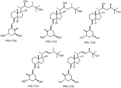
We have previously reported the pro-differentiating activities of double-point modified analogues of 1,25D2 [10]. Here, we use the same acute myeloid leukemia (AML) cell line HL60 to examine the pro-differentiating activities of the new analogues: PRI-1730, PRI-1731, PRI-1732, PRI-1733, PRI-1734 [14]. The structures of these analogues are presented in Figure 2.
Figure 2. Structures of double point modified analogues of 1,25D2 including 22-hydroxy analogues PRI-1730, PRI-1732 and PRI-1734, as well as analogue PRI-1731 with D2-like side-chain and its 24-epi analogue PRI-1733.
Ijms 17 00091 g002
Cells were treated with analogues at varying concentrations (0.1, 1, 10, 100 nM and 1 μM) for 96 h after which the expression of the monocyte/macrophage markers CD11b and CD14 were studied using flow cytometry. The data obtained (Figure 3) show that the new analogues have much lower pro-differentiating activities in comparison to that of 1,25D2 and 1,25D3. In particular, analogue PRI-1734 appears to be completely inactive as there was no upregulation of either CD11b or CD14 at any of the concentrations tested.

2.2. Nuclear Translocation of VDR

We analyzed VDR levels in the nuclear fractions of HL60 cells exposed to analogues at either 10 or 100 nM and at various time points. For these experiments, actin was used as a control. 1,25D2 and 1,25D3 induced a significant increase in VDR levels at both 10 and 100 nM following 24 h treatment. PRI-1731 and PRI-1733 elevated VDR at similar levels at both 10 nM and 100 nM, but to much lower levels than provoked by 1,25D2 or 1,25D3 by 24 h. PRI-1733 and PRI-1734 did not appear to elevate VDR at either concentration or at either of the 24 and 72 h time points. The amount of VDR appears to correlate with the overall differentiation effect (Figure 4).
Figure 3. Preliminary screening of pro-differentiating activities of analogues studied. HL60 cells were exposed to 1,25D2, 1,25D3 and analogues at concentrations of: 0.1, 1, 10, 100 nM, and 1 μM. The expression levels of both (A) CD11b and (B) CD14 were studied. Percentages of positive cells are presented in the y-axis.
Ijms 17 00091 g003aIjms 17 00091 g003b
Figure 4. Nuclear localization of VDR protein in HL60 exposed to 1,25D2, 1,25D3 or analogues. HL60 cells were exposed to compounds at 10 nM and 100 nM concentration for 24 h (A) and 72 h (B) and then expression of VDR was determined in the nuclear fractions.
Ijms 17 00091 g004

2.3. Activation of C/EBPβ by 1,25D2, 1,25D3 and Analogues

We also determined the ability of each analogue to upregulate expression of C/EBPβ protein. Here, we determined C/EBPβ levels in the nucleus of HL60 cells following 72 h treatment with 10 or 100 nM 1,25D2, 1,25D3 and analogues. Both 1,25D2 and 1,25D3 strongly upregulated C/EBPβ2 and C/EBPβ3 at both 10 and 100 nM (Figure 5).
Figure 5. C/EBPβ isoforms in HL60 cells treated with 1,25D2, 1,25D3 and analogues at 10 nM (A) and 100 nM (B) concentrations. HL60 cells were treated for 72 h with 1,25D2, 1,25D3 and analogues. The nuclear fraction was separated by electrophoresis and transferred onto PVDF membrane and probed with antibodies against C/EBPβ, and β-actin as fractionation/loading controls. In addition to the three C/EBPβ isoforms, unidentified bands, possibly cleavage products of C/EBPβ, are present.
Ijms 17 00091 g005

2.4. Binding Affinity of Analogues to VDR

Next, the affinities of the analogues to VDR were assessed using a fluorescence polarization (FP)-based competition assay. The affinity of analogues for VDR was checked using a wide range of concentrations and compared to that of 1,25D2 and 1,25D3. Dose-response curves were plotted, and IC50 values were calculated from these dose-response curves (Table 1).
Table 1. The vitamin D receptor (VDR) binding affinity expressed as IC50 and percentage activity.
1,25D31,25D2PRI-1730PRI-1731PRI-1732PRI-1733PRI-1734
IC504.494 × 10−91.466 × 10−82.006 × 10−72.218 × 10−82.138 × 10−74.386 × 10−62.641 × 10−6
RBA a10030.662.17520.262.030.1020.170
a The potency of 1,25D3 is normalised to 100; RBA: relative binding affinity.
Binding affinities for the analogues were shown to be lower than that of 1,25D3 and 1,25D2, with analogue PRI-1731, the most biologically active of these analogues, displaying the highest affinity to VDR. We have previously noted that there appears to be no correlation with differentiation abilities and affinity to the VDR [10].

2.5. Metabolic Resistance of Analogs to CYP24A1

The catabolism of each analogue by human CYP24A1 was analyzed using the membrane fraction prepared from the recombinant Escherichia coli cells expressing hCYP24A1 [15,16]. Figure 6 shows the HPLC profile of each analogue and the metabolites from catabolism by hCYP24A1. Table 2 shows the conversion ratio of each substrate into the metabolites. The metabolic conversion of analogue PRI-1731 was only 12%, which was much lower than that of native 1,25D2, 35%. The only structural difference between PRI-1731 and 1,25D2 is the altered geometry of the triene system i.e., a 5,6-trans instead of 5,6-cis. This finding suggests that 5,6-trans modification is contributing substantially to the increased stability of the analogue in a series of analogues with the side-chain of the regular length. Unexpectedly, reversing the chirality at C-24 in the 5,6-trans analogue PRI-1731 from the natural (24S) into the (24R) configuration (PRI-1733) did not have any effect on metabolic conversion, which remained equal to 12%, while it did for functional activity of other 1,25D2 analogues (PRI-1732 and PRI-1734). In a series of 5,6-trans analogues of 1,25D2 with the natural configuration at C-24, the addition of C-22 hydroxyl with saturated C22-C23 bond resulted in a dramatic loss of metabolic resistance from 12% for PRI-1731 to 52% for its 22-hydroxy derivative PRI-1732. We suspect that high loss of metabolic resistance of PRI-1732 may be due to the electronic and steric effects of interaction of this analogue with the hydroxylating enzyme. Preparation, isolation and structure identification of the major metabolite of PRI-1732 by HPLC/MS/MS is currently underway in our laboratories. The decrease of metabolic resistance was also observed from 12% for PRI-1733 to 22% for PRI-1734. Addition of C-22 hydroxyl is not that influential for natural 5,6-cis vitamins, as the conversion of 22-hydroxy analogue PRI-1730 is 31% as compared to 35% for parent 1,25D2. For C-22 hydroxy analogues PRI-1730 and PRI-1732, 5,6-trans modification decreased metabolic resistance. Interestingly, consistently in our studies [17], 1,25D2 is more stable than 1,25D3 and, therefore, 1,25D2 analogues should be preferred in this respect (Table 2).
Table 2. Metabolic conversion of active forms of vitamin D and its analogues by human CYP24A1 (%).
Compound1,25D31,25D2PRI-1730PRI-1731PRI-1732PRI-1733PRI-1734
Metabolic conversion (%)44353112521222
Data represent means of at least 3 independent experiments.
Figure 6. HPLC profiles of 1,25D2 and of its analogues and their metabolites by human CYP24A1. The peaks with arrows indicate putative metabolites. The HPLC profile of 1,25D3 is shown in Figure S1 in supplementary information of this paper. The metabolic profile of native 1,25D2 by human CYP24A1 was nearly the same as in our previous reports [15,16]. ∆A indicates the absorbance difference at 265 nm.
Ijms 17 00091 g006

2.6. Identifying If PRI-1734 Has Antagonistic Activity

Given that PRI-1734 appeared to be inactive in promoting differentiation in HL60 cells, we hypothesized that PRI-1734 may have properties antagonistic to 1,25D2 and 1,25D3. Thus, HL60 cells were exposed to PRI-1734 at increasing concentrations in combination with either 10 nM 1,25D2 or 10 nM 1,25D3. Results presented in Figure 7 show that PRI-1734 does not appear to have antagonistic properties, as the differentiation induced by both 1,25D2 and 1,25D3 remained unaffected.
Figure 7. Effect of PRI-1734 in combination with 1,25D3 or 1,25D2 on differentiation of HL60 cells. HL60 cells were exposed to 10 nM of either 1,25D2 or 1,25D3 in combination with PRI-1734 at different concentrations of: 1 nM, 10 nM, 100 nM, and 1 μM. The expression levels of both CD11b and CD14 were studied. Percentages of positive cells are presented in the y-axis. Ctrl indicates untreated control cells, Eth indicates ethanol (vehicle) treated control cells.
Ijms 17 00091 g007

3. Discussion

To enhance the therapeutic properties of 1,25D3, it is necessary to obtain analogues that have increased benefits to risk ratios, as compared to the parent compound. Thousands of different analogues have been synthesized in the past, but the optimal structure-function relationship has yet to be defined [18]. In previous research, we documented that analogues in which the side-chain has unsaturation at C-22 and methyl at C-24, and which is extended and branched with methyl or ethyl groups have increased differentiation-inducing properties [8,9]. Addition of 19-nor modification has led to reduced calcaemic activities and general toxicity for mice, and an even stronger differentiation-inducing potential [10].
Here we have demonstrated for the first time that introducing the 24-epi modification into the structure of 22-hydroxy-5,6-trans analogue (PRI-1732) resulted in a complete lack of differentiation-inducing activity of the compound. The inactive analogue PRI-1734 was a good substrate for CYP24A1 and was oxidized by this enzyme into the three more polar metabolites in a total of 22% comparing to 35% for 1,25D2 and 52% for the parent analogue PRI-1732.
Previous research has shown that the overall biological activity of a given analogue depends on interplay between various properties. For example, affinity of the analogue to VDR rarely correlates with differentiation-inducing potential [18]. The speed of catabolism also cannot explain all the differences in the activities of analogues [17]. Our studies have shown that the ability of a given analogue to induce nuclear accumulation of VDR and C/EBPβ transcription factors in AML cells is the best predictor of differentiation-inducing properties [10,19,20].

4. Materials and Methods

4.1. Chemicals and Antibodies

1,25D3, 1,25D2 and analogues were manufactured at the Pharmaceutical Research Institute (Warsaw, Poland). The compounds were placed in glass ampoules at and kept −20 °C. Analogues were dissolved in absolute ethanol at 100 μM and further diluted in culture medium for each required experimental concentration. Antibodies for flow cytometry CD14-PE and CD11b-FITC were from ImmunoTools (Friesoythe, Germany). Antibodies for Western blotting and chemiluminescence blotting substrate were from Santa Cruz Biotechnology Inc. (Santa Cruz, CA, USA).

4.2. Cell Lines

HL60 cells were from the Institute of Immunology and Experimental Therapy in Wroclaw, Poland. The cells were cultured in RPMI 1640 medium containing 10% fetal calf serum (FCS, Sigma, St. Louis, MO, USA), 100 units/mL penicillin and 100 μg/mL streptomycin (Sigma). The cells were cultured at standard cell culture conditions, i.e., humidified atmosphere of 95% air and 5% CO2 at 37 °C. The cell number and viability were determined by haemocytometer counts and trypan blue (0.4%) exclusion. For all experiments, the cells were suspended in fresh medium containing 1,25D2, 1,25D3, analogue or the equivalent volume of ethanol as a vehicle control.

4.3. Determination of Cell Differentiation by Flow Cytometry

The expression of cell surface markers of monocytic differentiation was determined by flow cytometry. The cells were treated with compounds at various concentrations for 96 h and then stained with CD11b and CD14 antibodies. Cells were washed twice in 500 μL PBS and incubated for 1 h on ice with 1 μL CD14-PE and 1 μL CDllb-FITC. Cells were washed three times with PBS containing 0.1% BSA and suspended in 400 μL PBS prior to analysis on the Becton Dickinson Accuri C6 (San Jose, CA, USA). Data analysis was performed using Becton Dickinson Accuri C6 software.

4.4. Preparation of Cell Lysates

Cells (5 × 10⁶) were washed three times with PBS and lysed for 20 min on ice in 80 μL of lysis buffer pH 7.5 (20 mM Tris, 1% Triton X-100,150 mM NaCl, 1 mM EDTA, 1 mM EGTA) with added protease inhibitor cocktail (Roche Diagnostics, Mannheim, Germany). The lysates were centrifuged for 5 min, at 18,000× g, at 4 °C in order to separate them. Supernatants were designated cytoplasmic (C) fractions and the nuclei remaining in pellets were washed and sonicated for 5 s in the same lysis buffer as before. In order to obtain the nucler (N) fraction, samples were centrifuged for 5 min at 18,000× g, at 4 °C. Twenty microliters of 5× SDS sample buffer were added to each fraction and boiled for 10 min in order to denature the proteins.

4.5. Western Blotting

Twelve-percent SDS-PAGE gels were used to separate 30 μL of cell lysates (derived from 5 × 10⁶ cells) and transferred to the PVDF membranes. The membranes were incubated with primary antibody (2 h), followed by horseradish peroxidise-conjugates secondary antibody (1 h). Chemiluminescence substrate was used to identify protein bands. Membranes were stripped, and reprobed with additional antibodies. The loading control for all experiments was actin. Membranes were scanned and bands quantified using Image J 1.34s software (freeware by Wayne Rasband, NIH).

4.6. Human VDR Binding Assay

Binding affinity to VDR was evaluated using a Polarscreen Vitamin D receptor competitor assay, under manufacturer conditions (Life Technologies, Carlsbad, CA, USA). The polarised fluorescence was measured using Envision (Perkin-Elmer, Waltham, MA, USA). All compounds were evaluated within the range 10–11 to 10–5 M, half maximal inhibitory concentration (IC50) values were calculated using the average of measured values.

4.7. Metabolic Resistance of Analogues to CYP24A1

Metabolism of each analogue by recombinant human CYP24A1 was examined using the reconstituted system containing the membrane fraction prepared from the recombinant , Escherichia coli cells expressing human CYP24A1 [15,16]. The reaction mixture containing 2.0 μM bovine adrenodoxin (ADX), 0.2 μM bovine adrenodoxin reductase (ADR), 20 nM CYP24A1, 5 µM each analogue, 1 mM NADPH, 100 mM Tris-HCl (pH 7.4), and 1 mM EDTA was incubated for 15 min at 37 °C. Four-volumes of chloroform/methanol (3:1) were used to terminate each reaction in combination with vigorous shaking. Following this, the organic phase was retrieved and dried. The resultant residue was dissolved in acetonitrile, and was centrifuged at 20,000× g for 15 min. The resultant supernatant was submitted to High Performance Liquid Chromatography (HPLC) according to the conditions described here: column, YMC-pack ODS-AM (4.6 mm × 300 mm) (YMC Co., Tokyo, Japan); UV detection, 265 nm; flow late, 1.0 mL/min; column temperature, 40 °C; linear gradients of 20%–100% acetonitrile aqueous solution per 25 min followed by 100% acetonitrile for 5 min.

5. Conclusions

Data presented here document that the modifications introduced to our new double point modified 1,25D2 analogues have not increased their differentiation-inducing properties. Nevertheless, the modifications introduced resulted in a very divergent group of analogs having moderate to completely abolished cell differentiation activity. These analogues had affinities to VDR similar or lower than 1,25D2, and were less efficient in inducing differentiation of AML cells. Based on flow cytometry assays, we can order their differentiation-inducing activities as follows: 1,25D3 > 1,25D2 > PRI-1731 > PRI-7130 ≈ PRI-1733 > PRI-1732 > PRI-1734. They induced nuclear accumulation of VDR and C/EBPβ less efficiently than 1,25D3 and 1,25D2. Since the analogues PRI-1732 and PRI-1734 were the least active of all tested compounds, we hypothesize that the combination of a 22-hydroxyl with 5,6-trans modification has led either to decreased transport of the analogues into the cells or to a diminished interaction with VDR inside the cells.

Supplementary Materials

Supplementary materials can be found at https://www.mdpi.com/1422-0067/17/2/91/s1.

Acknowledgments

This project has received funding from the European Union’s Seventh Framework Programme for Research, Technological Development and Demonstration under grant agreement no. 315902. Aoife Corcoran and Sharmin Nadkarni gratefully acknowledge receipt of a Marie Curie Research Fellowships. Ewa Marcinkowska, Geoffrey Brown and Andrzej Kutner are partners within the Marie Curie Initial Training Network DECIDE (Decision-making within cells and differentiation entities therapies).

Author Contributions

Aoife Corcoran performed the experiments using cell lines, analyzed and interpreted their results, and wrote the article; Sharmin Nadkarni and Andrzej Kutner designed, synthesized and described the analogues, Kaori Yasuda and Toshiyuki Sakaki designed, performed and described metabolic studies of the analogues, Geoffrey Brown contributed to analysis of the data, took part in writing the paper and acquired grant, Ewa Marcinkowska conceptualized the hypothesis, interpreted the data and wrote the article.

Conflicts of Interest

The authors declare no conflict of interest.

References

  1. Holick, M.F. Vitamin D and bone health. J. Nutr. 1996, 126, 1159–1164. [Google Scholar]
  2. Trump, D.L.; Deeb, K.; Johnson, C.S. Vitamin D: Considerations in the continued development as an agent for cancer prevention and therapy. Cancer J. 2010, 16, 1–9. [Google Scholar] [CrossRef] [PubMed]
  3. Van Etten, E.; Mathieu, C. Immunoregulation by 1,25-dihydroxyvitamin D3: Basic concepts. J. Steroid Biochem. Mol. Biol. 2005, 97, 93–101. [Google Scholar] [CrossRef] [PubMed]
  4. Houghton, L.A.; Vieth, R. The case against ergocalciferol (vitamin D2) as a vitamin supplement. Am. J. Clin. Nutr. 2006, 84, 694–697. [Google Scholar] [PubMed]
  5. Nuijten, M.; Andress, D.L.; Marx, S.E.; Sterz, R. Chronic kidney disease Markov model comparing paricalcitol to calcitriol for secondary hyperparathyroidism: A US perspective. Curr. Med. Res. Opin. 2009, 25, 1221–1234. [Google Scholar] [CrossRef] [PubMed]
  6. Koeffler, H.P.; Aslanian, N.; O'Kelly, J. Vitamin D2 analog (Paricalcitol; Zemplar) for treatment of myelodysplastic syndrome. Leuk. Res. 2005, 29, 1259–1262. [Google Scholar] [CrossRef] [PubMed]
  7. Harrison, J.S.; Bershadskiy, A. Clinical experience using vitamin D and analogs in the treatment of myelodysplasia and acute myeloid leukemia: A review of the literature. Leuk. Res. Treat. 2012. [CrossRef] [PubMed]
  8. Baurska, H.; Kiełbiński, M.; Biecek, P.; Haus, O.; Jaźwiec, B.; Kutner, A.; Marcinkowska, E. Monocytic differentiation induced by side-chain modified analogs of vitamin D in ex vivo ex vivo cells from patients with acute myeloid leukemia. Leuk. Res. 2014, 38, 638–647. [Google Scholar] [CrossRef] [PubMed]
  9. Baurska, H.; Klopot, A.; Kielbinski, M.; Chrobak, A.; Wijas, E.; Kutner, A.; Marcinkowska, E. Structure-function analysis of vitamin D2 analogs as potential inducers of leukemia differentiation and inhibitors of prostate cancer proliferation. J. Steroid Biochem. Mol. Biol. 2011, 126, 46–54. [Google Scholar] [CrossRef] [PubMed]
  10. Corcoran, A.; Bermudez, M.A.; Seoane, S.; Perez-Fernandez, R.; Krupa, M.; Pietraszek, A.; Chodyński, M.; Kutner, A.; Brown, G.; Marcinkowska, E. Biological evaluation of new vitamin D2 analogues. J. Steroid Biochem. Mol. Biol. 2015. [Google Scholar] [CrossRef] [PubMed]
  11. Pike, J.W.; Meyer, M.B.; Lee, S.M. The vitamin D receptor: Biochemical, molecular, biological, and genomic era investigations. Vitam. D 2011, 1, 97–135. [Google Scholar]
  12. Pike, J.W.; Meyer, M.B. Fundamentals of vitamin D hormone-regulated gene expression. J. Steroid Biochem. Mol. Biol. 2014, 114, 5–11. [Google Scholar] [CrossRef] [PubMed]
  13. Horváth, H.C.; Khabir, Z.; Nittke, T.; Gruber, S.; Speer, G.; Manhardt, T.; Bonner, E.; Kallay, E. CYP24A1 splice variants--implications for the antitumorigenic actions of 1,25-(OH)2D3 in colorectal cancer. J. Steroid Biochem Mol. Biol. 2010, 121, 76–79. [Google Scholar] [CrossRef] [PubMed]
  14. Nadkarni, S.; Chodyński, M.; Krajewski, K.; Cmoch, P.; Marcinkowska, E.; Brown, G.; Kutner, A. Convergent synthesis of double point modified analogs of 1α,25-dihydroxyvitamin D2 for biological evaluation. J. Steroid Biochem. Mol. Biol. 2015. [Google Scholar] [CrossRef] [PubMed]
  15. Kusudo, T.; Sakaki, T.; Abe, D.; Fujishima, T.; Kittaka, A.; Takayama, H.; Hatakeyama, S.; Ohta, M.; Inouye, K. Metabolism of A-ring diastereomers of 1α,25-dihydroxyvitamin D3 by CYP24A1. Biochem. Biophys. Res. Commun. 2014, 321, 774–782. [Google Scholar] [CrossRef] [PubMed]
  16. Urushino, N.; Yasuda, K.; Ikushiro, S.; Kamakura, M.; Ohta, M.; Sakaki, T. Metabolism of 1α,25-dihydroxyvitamin D2 by human CYP24A1. Biochem. Biophys. Res. Commun. 2009, 384, 144–148. [Google Scholar] [CrossRef] [PubMed]
  17. Bolla, N.R.; Corcoran, A.; Yasuda, K.; Chodyński, M.; Krajewski, K.; Cmoch, P.; Marcinkowska, E.; Brown, G.; Sakaki, T.; Kutner, A. Synthesis and evaluation of geometric analogs of 1α,25-dihydroxyvitamin D2 as potential therapeutics. J. Steroid Biochem. Mol. Biol. 2015. [Google Scholar] [CrossRef] [PubMed]
  18. Nadkarni, S.; Chodynski, M.; Corcoran, A.; Marcinkowska, E.; Brown, G.; Kutner, A. Double point modified analogs of vitamin d as potent activators of vitamin D receptor. Curr. Pharm. Des. 2015, 21, 1741–1763. [Google Scholar] [CrossRef] [PubMed]
  19. Gocek, E.; Kielbinski, M.; Wylob, P.; Kutner, A.; Marcinkowska, E. Side-chain modified vitamin D analogs induce rapid accumulation of VDR in the cell nuclei proportionately to their differentiation-inducing potential. Steroids 2008, 73, 1359–1366. [Google Scholar] [CrossRef] [PubMed]
  20. Baurska, H.; Marchwicka, A.; Klopot, A.; Kutner, A.; Marcinkowska, E. Studies on the mechanisms of superagonistic pro-differentiating activities of side-chain modified analogs of vitamin D2. Oncol. Rep. 2012, 28, 1110–1116. [Google Scholar] [PubMed]