Article

Whole-Genome Re-Alignment Facilitates Development of Specific Molecular Markers for Races 1 and 4 of Xanthomonas campestris pv. campestris, the Cause of Black Rot Disease in Brassica oleracea

1
Department of Horticulture, Sunchon National University, 255, Jungang-ro, Suncheon 57922, Korea
2
School of Life Sciences, University of Warwick, Wellesbourne Campus, Warwick CV35 9EF, UK
*
Authors to whom correspondence should be addressed.
Int. J. Mol. Sci. 2017, 18(12), 2523; https://doi.org/10.3390/ijms18122523
Submission received: 4 November 2017 / Revised: 20 November 2017 / Accepted: 21 November 2017 / Published: 24 November 2017
(This article belongs to the Section Molecular Plant Sciences)

Abstract

:
Black rot, caused by Xanthomonas campestris pv. campestris (Xcc), is a seed borne disease of Brassicaceae. Eleven pathogenic races have been identified based on the phenotype interaction pattern of differential brassica cultivars inoculated with different strains. Race 1 and 4 are the two most frequent races found in Brassica oleracea crops. In this study, a PCR molecular diagnostic tool was developed for the identification of Xcc races 1 and 4 of this pathogen. Whole genomic sequences of races 1, 3, 4 and 9 and sequences of three other Xanthomonas pathovars/species (X. campestris pv. incanae (Xci), X. campestris pv. raphani (Xcr) and X. euvesicatoria (Xev) were aligned to identify variable regions among races. To develop specific markers for races 1 and 4, primers were developed from a region where sequences were dissimilar in other races. Sequence-characterized amplified regions (SCAR) and insertion or deletion of bases (InDel) were used to develop each specific set of primers. The specificity of the selected primers was confirmed by PCR tests using genomic DNA of seven different Xcc races, two strains of X. campestris pathovars and other species of bacteria. Bacterial samples of the races 1 and 4 isolates were collected from artificially inoculated cabbage leaves to conduct bio-PCR. Bio-PCR successfully detected the two Xcc isolates. By using our race-specific markers, a potential race 1 strain from the existing Korean Xcc collection was identified. The Xcc race 1 and 4-specific markers developed in this study are novel and can potentially be used for rapid detection of Xcc races through PCR.

Graphical Abstract

1. Introduction

Black rot, a seed-borne disease caused by Xanthomonas campestris pv. campestris (Xcc), is one of the most important diseases of plants of the Brassicaceae family [1]. Black rot is a global problem which can reduce more than fifty percent yield under severe attacks in favorable conditions [2]. This pathogen is found all over the world, showing diversity in different countries and provinces of the same country [3,4,5,6].
Xcc is a small, rod-shaped, mobile, aerobic, gram-negative, non-spore forming obligate bacterium [7]. The pathogen enters the vascular tissues mainly through the hydathodes, but also through wounded tissues and stomata and is able to develop disease symptoms systemically. This disease is favored by warm, humid conditions and can spread rapidly by rain dispersal and irrigation water [8]. Infected seeds and plants, crop debris and cruciferous weeds are potential inoculum sources of this disease.
Xcc was grouped into six races based on a postulated gene-for-gene model [3,9]. Later, this model was expanded to include nine races [1,10]. Seven pathogenic races were identified on the basis of interaction between differential cultivars of Brassica species and different strains of Xcc [5]. Two novel races of this organism, race 10 and race 11, have been reported in Portugal this year [11]. Among these races, races 1 and 4 are the two most frequently found races all over the world largely because of their severe infections in Brassica oleracea crops [12].
Management of black rot is very difficult. The disease can be controlled by using healthy seeds, crop rotation and resistant cultivars [2,13], but in practice these methods have frequently not been efficiently used. In order to design effective control strategies, including seed health tests and development of resistant varieties against black rot disease, it is important to know the race of the pathogens present in different crops and areas. To date, a total of twenty-seven race-specific resistant genotypes have been developed in USA, Russia, UK, and Portugal (Table S1 [3,10,11,14,15]). No race-specific resistant cultivars of Brassica oleracea species have been developed for Korea as the disease has not been studied in this country yet. The identification of Xcc races and thereafter development of resistant cultivars of Brassica oleracea to black rot have become a priority for the vegetable breeders in Korea.
Developing race-specific markers is not only required to identify the races in a particular location, but also important for breeding programmes that aim to develop race-specific B. oleracea resistant genotypes for each area. Race-specific markers might also enable researchers to identify the presence of Xcc at an early stage. There are several methods used for the detection of Xcc such as selective and semi-selective media, plant bioassays [16,17] and serological techniques [18,19]. These techniques, however, were developed for Xcc detection and identification of Xcc at the pathovar and species level only. In addition, these methods are time consuming and labour intensive [20]. Recently, DNA-based techniques have been developed for Xcc detection. PCR is a very powerful, fast, reliable, and comparatively low cost method for the specific identification of plant pathogenic bacteria, yeast and also other organisms [21,22,23,24,25]. Sequence-characterized amplified region (SCAR) analysis can be a source of specific sequences for developing diagnostic molecular markers for detecting Xcc races 1 and 4 [26,27]. Other reliable molecular markers can be based on InDels, either insertions or deletions, that can be useful especially in phylogenetic studies in natural populations and also for species-identification procedures [28,29,30,31].
To date, a single method has yet to be developed for race-specific detection of Xcc. Therefore, it is important to develop race-specific markers that can rapid and efficiently identify Xcc races 1 and 4 from plant samples.
Until now, only pathogenicity tests on differential cultivars are being used for Xcc race-specific detection across the world (Table S2 [3,5,9,10,11,32]). PCR with race-specific markers could be used for faster determination of races. The objective of this work was to develop race 1 and 4 specific markers for identifying these races of Xcc. To achieve this goal, available Xcc and Xanthomonas species sequences were retrieved and aligned and race 1 and 4 specific primers were developed. The developed markers were validated for specificity to race 1 and 4 including bacterial DNA pools of Xcc races, X. campestris pathovars, other bacterial species and a protist.

2. Results

2.1. Specificity of Primers

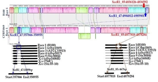
Each of the similar blocks representing sequence homology were shown by a single colour (Figure 1 and Figure 2). Any dissimilarity between sequences was targeted to develop markers (Figure 1 and Figure 2). The primer pair Xcc_47R1 (forward and reverse), designed from Xcc race 1 sequences, amplified a fragment of 1089 bp from only one sample tested (Xcc race 1 strain, Lane 1, Figure 3a). In contrast, another primer combination, Xcc_85R1, amplified all DNA samples, from all races of Xcc (Figure 3b). However, the DNA sample from race 1 produced an expected amplicon size of 467 bp, whereas those of other six races was close to 900 bp (expected product size is 872 bp as per calculations) and were identical to each other (Figure 3b). In both cases, none of the other tested DNA samples of X. campestris pathovars, other bacteria species, fungal species and protist produced any visible amplicons (Figure 3a,b).
Two primer pairs designed from an Xcc race 4 sequence, Xcc1_46R4 and Xcc2_46R4, also amplified only from the DNA sample of race 4 (Xcc race 4 strain, Lane 4, Figure 4a,b). None of the other samples were amplified by these two sets of primers.

2.2. Validation of Markers through Bio-PCR in Inoculated Cabbage Leaves

The primer pair Xcc_47R1 designed for race 1 showed positive bio-PCR reaction only for the three samples that were infected with race 1 out of 21 inoculated leaf samples (3 samples from plants inoculated with each race) (Figure 5a). The expected amplicons of 1089 bp specific for the Xcc race 1 were observed (Figure 5a) in three race 1 inoculated cabbage leaf samples. Other 18 samples that were inoculated with six other Xcc races did not produce any observable amplicons. The positive control DNA sample (in Lane 22) derived from Xcc race 1 produced identical amplicons as the amplicons obtained from Xcc race 1 inoculated cabbage leaf samples (Figure 5a,b). Another primer combination, Xcc_85R1, produced the expected 467 bp amplicons for three bacterial samples from plants inoculated with race 1; bacterial samples from the plants inoculated with the other six races resulted in identical amplicons of expected 872 bp in size (Figure 5b).
Similarly, in a separate Bio-PCR with primers designed for race 4, only three samples of plants inoculated with race 4 were amplified and none of the other 18 samples that were infected with six other different races yielded any bands (Figure 6a,b). As expected, the control DNA sample was amplified that confirmed the presence of a race 4 sample.

2.3. Race Determination

Two race 1 specific markers (Xcc_47R1 and Xcc_85R1) amplified Korean KACC10377 strain with specific bands similar to the control DNA samples of HRIW-3811 strain (Figure 7a,b). Two primer pairs designed for race 4 (Xcc_47R1 and Xcc_85R1) did not amplify from any of the tested unknown strains (Figure 7c,d).

3. Discussion

3.1. PCR-Based Markers Specifically Detected Xcc Races 1 and 4

The purpose of this study was to develop PCR-based SCAR markers for the identification of Xcc races 1 and 4 strains. Molecular methods of race identification have been used for other pathogens. For example, K3a race was identified for Xanthomonas oryzae pv. oryzae (Xoo) bacterial blight pathogen of rice by using an AFLP-derived marker [33]. SCAR markers were found highly reliable to detect CYR32 and CYR33 races of wheat stripe rust, caused by Puccinia striiformis f. sp. tritici, in China [34]. A modified SCAR marker, inter-retrotransposon Sequence-Characterized Amplified Regions (IR-SCAR), was used to detect race 1 of Fusarium oxysporum f. sp. lactucae in Lettuce [35]. To date, no race specific markers have been reported for detecting any of the Xcc races. Therefore, the development of PCR markers which can be comparatively cheap, reliable and effective for the rapid detection of bacterial pathogens, was the main aim of this study.

3.2. Whole Genome Sequences of Xcc Enabled Developing Race-Specific Novel Markers

Whole genome sequences of four races of Xcc (1, 3, 4, race 9), two other pathovars of X. campestris (Xci, Xcr) and another species of Xanthomonas (Xev) were used for in silico analysis (www.ncbi.nlm.nih.gov) and enabled us to realign, compare and find variable regions present among them. These genome sequences are a valuable resource for comparative analysis among the different races of Xcc (Table 1 [36,37,38,39,40,41,42], Figure 1 and Figure 2). Sequences of Xcc races were generally highly conserved. When the homology of different races and pathovars was compared by using a comprehensive suite of molecular biology and NGS analysis tools, our target races, (Xcc races 1 and 4) showed some unique sequences at different locations compared to all other races and pathovars. The variable sequences facilitated developing Xcc races 1 and 4 markers.
One set of SCAR markers (Xcc_47R1) and one set of InDel marker (Xcc_85R1) have been developed for specific detection of race 1. Similarly, two other SCAR markers Xcc1_46R4 and Xcc2_46R4 were able to distinguish Xcc race 4 from all other races of Xcc and X. campestris pathovars. Thus, three SCAR markers and an InDel marker appeared to be unique for the detection of the Xcc race 1 and race 4 isolates. PCR amplification exhibited positive amplicons only when DNA of the targeted Xcc race 1 and 4 were used and was negative for other Xcc races, X. campestris pathovars and other species. The sequences used for primer development were not from the strains used in the tests presented here. Thus, the developed SCAR and InDel markers allowed the identification of a strain of race 1 and a strain of race 4 and they may generally act as a diagnostic marker for Xcc races 1 and 4. These markers will have to be extensively tested with a larger number of strains of each Xcc race, to assess specificity and determine if they detect all Xcc race 1 and Xcc race 4 strains or just a subset of strains of these races.
There is no information on Xcc races in Korea, but Xcc is prevalent in this country. We believe that these PCR-based assays with race-specific SCAR and InDel markers have potential for identification of Xcc races 1 and 4 of this pathogen from black rot infected plant samples that might also be contaminated with several other bacteria, fungi and microbes.

3.3. Direct and Rapid Detection Tool for Xcc Race 4 Developed

A Bio-PCR method was previously exploited for the amplification of a member of the rhs family gene for detecting X. oryzae pv. oryzae (Xoo) at the pathovar and species levels [43]. In our study, we tested this method in cabbage leaves inoculated with seven different races. This approach was found very effective and accurate. These results suggested that the PCR-based technique can be used directly to detect and identify Xcc races 1 and 4 pathogens in infected cabbage leaf samples without isolating the bacteria from the infected leaves. Thus, this method could potentially be used in disease forecasting to assist cabbage growers.

3.4. Potential Applications of Race 4 Specific Markers

The race-specific markers can identify races through Bio-PCR within a few hours whereas use of differential cultivars for race determination requires several months [20]. Thus, in a given crop season, a whole crop might fail even before the causal Xcc race is identified by using a conventional race-determination method that uses differential cultivars. The bio-PCR assay based on SCAR and InDel markers could be useful and invaluable for the identification of race 1 and 4 strains and can assist in programmes that aim at developing resistant cultivars for the effective control of black rot disease.
Two race 1-specific markers amplified from the Korean strain KACC10377 indicating that this strain might belong to Xcc race 1. This is the first identified race in Korea. In Asia, race 1, race 4 and race 6 are dominant in Nepal [5] and India [32]. Thus, the Xcc strain identified in this study could be part of an important Xcc race in Korea.
The markers developed will need to be tested with a large collection of Xcc isolates to assess their specificity and the method should also be tested with plants at different stages of growth and infection. It will be particularly important to determine if the strains can be detected early after infection and before symptom development.

4. Materials and Methods

4.1. Bacterial Strains and Culture Conditions

Representative strains of Xcc races (races 1 to 7) and X. campestris pathovars HRIW-6377 (Xci), HRIW-8305 (Xcr) were obtained from the culture collection of the School of Life Sciences, Wellesbourne Campus, University of Warwick, UK (HRIW). Three Xcc isolates ICMP8, ICMP13051 and ICMP12464 were obtained from the Landcare Research, Auckland, New Zealand (Table 2 [3,44]). Other eight Xcc (KACC19132, KACC19133, KACC19134, KACC19135, KACC19136, KACC10377 and KACC17966) and X. campestris isolates (KACC11153, KACC11154, KACC17821, KACC17126, KACC10491, KACC10490) were obtained from the Korean Agriculture Culture Collection (KACC) (Table 2 [3,44]). All bacterial isolates were cultured on King’s medium B [45] and incubated at 30 °C for 48 h.

4.2. Isolation of Total DNA

Genomic DNA of all bacteria isolates was extracted using a DNeasy Plant Mini Kit (QIAGEN, Hilden, Germany) following the Manufacturer’s instructions. The concentration and purity of the extracted DNA was then measured using a Nanodrop ND-1000 spectrophotometer (NanoDrop, Wilmington, DE, USA).

4.3. Sequence Retrieval and Alignment

In silico analysis was conducted for designing primers for specific detection of strains of Xcc races 1 and 4. For comparative genome analyses, whole genome sequences of Xcc strains of B100 (race 1), CFBP1869 (race 1), ATCC33913 (race 3), CFBP5817 (race 4) and 8004 (race 9) and other three Xanthomonas pathovars/species (Xanthomonas campestris pv. incanaeXci (CFBP1606R), X. campestris pv. raphani‒Xcr (756C) and X. euvesicatoria‒Xev (strain 85-10) were downloaded from NCBI [46]. The genome size of Xcc races and X. campestris pathovars ranged between 4.91 Mbp and 5.17 Mbp (Table 2) and their GC content ranged between 64.2 and 65.3 indicating that there might be large nucleotide sequence variation among them. For comparative purpose, the strain B100 (race 1) was considered as a reference genome for designing primers. Mauve (version 2.4.0) and Geneious (Free trial version) were used to align the genomes and to identify the homology between sequence blocks among the races of Xcc (shown by differential colors); this facilitated the identification of differential regions among these isolates that were useful to develop primers for specific races.

4.4. Primer Design and PCR Conditions

The primers were designed from whole genome sequences of bacteria using Primer3 software [47] and checked for specificity in silico using ‘In Silico simulation of molecular biology experiments software’ (www.insilico.ehu.es). Highly conserved regions were excluded when primers were designed. Only variable regions were selected as they may provide race-specific signature sequences required for the identifying Xcc races 1 and 4. Four sets of potential race-specific primers were designed, two sets for race 1 and two sets for race 4. Primers Xcc_47R1 and Xcc_85R1 were designed to detect race 1 and primers Xcc1_46R4 and Xcc2_46R4 were designed to detect race 4 (Table 3). The race 1 primers were: Xcc_47R1 between 498412-4985901 and Xcc_85R1 between 4836126–4836592; these two primers were expected to amplify 1089 and 467 base pairs sequences, respectively. The race 4 specific primers were: Xcc1_46R4 designed between 1843518 and 1843057 and Xcc1_46R4 primer designed between 1842956 to 1842379; these two primers were expected to amplify 462 and 578 base pairs, respectively (Table 3).
Emerald PCR master mix (Takara, Shiga, Japan) was used in Polymerase Chain Reaction (PCR) for amplification of the target regions with respective markers. A 20.0 μL of PCR reaction mixture containing forward and reverse primers (1.0 μL each), Emerald PCR master mix (9.0 μL), ultra-pure water (8 μL) and 1.0 μL DNA was used for PCR amplification. PCR was performed using the following conditions in a thermo cycler (Takara, Shiga, Japan): denaturation at 95 °C for 5 min followed by 20 cycles of amplification at 95 °C for 30 s, annealing at 65 °C for 40 s and 72 °C for 45 s, and terminated by a final elongation at 72 °C for 5 min, but in case of race 4-specific amplification annealing was carried out at 66 °C for 40 s. Electrophoresis was done using 1.5% agarose gel at 100 V for 30 min, visualized on ENDUROTM GDS Gel Documentation system under UV light (302 nm).

4.5. Testing the Specificity of Primers

At first, the specificity of the designed primers was checked by using BLAST tool [48]. Wet lab validation was carried out by performing PCR with genomic DNA (1 μL of a suspension approximately at 60 ng μL−1) of Xcc races (races 1 to 7) and eight Xcc strains (of unknown race), two other pathovars of X. campestris (Xci and Xcr), five strains of other Xanthomonas species (two strains of X. euvesicatoria, X. campestris pv. zinniae, X. axonopodis pv. dieffenbachiae and X. axonopodis pv. glycines) and strains of other test-bacteria species e.g., Pseudomonas syringae pv. maculicola, Erwinia carotovora subsp. carotovora, one fungal species, Didymella bryoniae, and a protist Plasmodiophora brassicae. The precision and specificity of the developed primers Xcc_47R1 and Xcc_85R1 for race 1, and Xcc1_46R4 and Xcc2_46R4 for race 4 detection (Table 2 [3,44]) were assessed. Negative controls were performed with the DNA template replaced with a 1.0 μL DDW.

4.6. Detection of the Race 1 and 4-Specific Pathogen by PCR in Artificially Infected Cabbage Leaves

The sensitivity of the four specific markers (i.e., their efficiency) in direct PCR based assay was evaluated using artificially infected leaves. For evaluation of these primers, 35-days-old cabbage inbred line (BN3122, Asia Seed Co. Ltd., Seoul, Korea) plants, susceptible against black rot disease, were inoculated with seven races of Xcc (Table 1) in a glasshouse with artificially controlled temperature (26 ± 2 °C) and relative humidity (70–80%). Three different individual plants were inoculated with each race. The inoculated plants that had visible and conspicuous black rot symptoms at 14 days after inoculation (DAI) were sampled. V-shaped lesions with blackened veins of about 2 cm were cut into small pieces and each cut piece of sample was soaked in 200 μL ultra-pure water. After 40 min, 10 μL water containing bacterial cells was taken for PCR reactions. The bio-PCR was performed following the method as described in ‘PCR amplification’ with an exception in number of amplification cycles (30 cycles).

4.7. Race Identification

Genomic DNA samples of eight Xcc strains of unknown race (ICMP8, KACC19132, KACC19133, KACC19134, KACC19135, KACC19136, KACC17966 and KACC10377) were used for race identification. PCR amplification was done using developed molecular markers for both race 1 and 4 for specific detection of races of Xcc. Two strains of two known races, strain HRIW-3811 of race 1 and strain HRIW-1279A of race 4 were used as controls.

5. Conclusions

This study exploited the variation within the whole genome sequences of Xcc races and pathovars for developing Xcc races 1 and 4 specific markers. Markers developed based on in silico analysis were further tested for their specificity through BLAST search. Four markers were able to distinguish Xcc races 1 and 4 from all other Xcc races and pathovars. The developed markers have potential to identify Xcc races 1 and 4 from any infected cabbage leaf samples rapidly and effectively without deploying a DNA isolation procedure. This is the first report that develops Xcc race 1 and 4-specific markers for the direct and rapid detection of black rot disease. These markers could be utilized worldwide for early detection of black rot disease in infected fields and for quick identification of Xcc races.

Supplementary Materials

The following are available online at www.mdpi.com/1422-0067/18/12/2523/s1.

Acknowledgments

We thank the School of Life Sciences, Wellesbourne Campus, University of Warwick, UK for providing control races of Xcc and Xci and Xcr isolates. We thank the Korean Agriculture Culture Collection (KACC), Korea and the ICMP collection from New Zealand for providing isolates. This study was supported by the Center for Horticultural Seed Development (Golden Seed Project No. 213007-05-2-SB510) of the Ministry of Agriculture, Food and Rural Affairs in the Republic of Korea (MAFRA).

Author Contributions

Ill-Sup Nou, Jong-In Park and Hoy-Taek Kim conceived and designed the study. Sathishkumar Natarajan conducted the in silico analysis. Mehede Hassan Rubel prepared and cultured the samples, isolated DNA and performed wet lab validation. Joana G. Vicente selected control isolates for this study. Mehede Hassan Rubel, Arif Hasan Khan Robin and Joana G. Vicente wrote the manuscript and created the tables and figures. All authors read and approved the final draft of the manuscript.

Conflicts of Interest

The authors declare no conflict of interest.

Abbreviations

XccXanthomonas campestris pv. campestris
XciXanthomonas campestris pv. incane
XcrXanthomonas campestris pv. raphani
PCRPolymerase chain reaction
SCARSequence-characterized amplified region
InDelInsertions or deletions

References

  1. Vicente, J.G.; Holub, E.B. Xanthomonas campestris pv. campestris (cause of black rot of crucifers) in the genomic era is still a worldwide threat to brassica crops. Mol. Plant Pathol. 2013, 14, 2–18. [Google Scholar]
  2. Williams, P.H. Black rot: A continuing threat to world crucifers. Plant Dis. 1980, 64, 736–742. [Google Scholar] [CrossRef]
  3. Vicente, J.G.; Conway, J.; Roberts, S.J.; Taylor, J.D. Identification and origin of Xanthomonas campestris pv. campestris races and related pathovars. Phytopathology 2001, 91, 492–499. [Google Scholar] [PubMed]
  4. Silva, M.R. Genetic Diversity of Xanthomonas campestris pv. campestris in Brazil. Master’s Thesis, Universidade Federal de Vicosa, Vicosa, Brazil, 2006. [Google Scholar]
  5. Jensen, B.D.; Vicente, J.G.; Manandhar, H.K.; Roberts, S.J. Occurrence and diversity of Xanthomonas campestris pv. campestris in vegetable Brassica fields in Nepal. Plant Dis. 2010, 94, 298–305. [Google Scholar]
  6. Popović, T.; Jošić, D.; Starović, M.; Milovanović, P.; Dolovac, N.; Poštić, D.; Stanković, S. Phenotypic and Genotypic Characterization of Xanthomonas campestris pv. campestris Isolated from Cabbage, Kale and Broccoli. Arch. Biol. Sci. 2013, 65, 585–593. [Google Scholar]
  7. Sewariya, V.K.; Shrivastava, R.; Prasad, G.B.K.S.; Arora, K. In-Vitro Evaluation of Novel Synthetic Compounds against Xanthomonas campestris pv. campestris. Int. J. Pharma Biol. Sci. 2012, 3, 441–453. [Google Scholar]
  8. Kocks, C.G.; Zadoks, J.C.; Ruissen, M.A. Spatio-temporal development of black rot (X. campestris pv. campestris) in cabbage in relation to initial inoculums levels in field plots in The Netherlands. Plant Pathol. 1999, 48, 176–188. [Google Scholar]
  9. Kamoun, S.; Kamdar, H.V.; Tola, E.; Kado, C.I. Incompatible interactions between crucifers and Xanthomonas campestris involve a vascular hypersensitive response: Role of the hrpX locus. Mol. Plant-Microbe Interact. 1992, 5, 22–33. [Google Scholar] [CrossRef]
  10. Fargier, E.; Manceau, C. Pathogenicity assays restrict the species Xanthomonas campestris into three pathovars and reveal nine races within X. campestris pv. campestris. Plant Pathol. 2007, 56, 805–818. [Google Scholar] [CrossRef]
  11. Cruz, J.; Tenreiro, R.; Cruz, L. Assessment of Diversity Xanthomonas campestris Pathovars Affecting Cruciferous Plants in Portugal and Disclosure of two novel X. campestris pv. campestris races. J. Plant Pathol. 2017, 99. [Google Scholar] [CrossRef]
  12. Taylor, J.D.; Conway, J.; Roberts, S.J.; Astley, D.; Vicente, J.G. Sources and origin of resistance to Xanthomonas campestris pv. campestris in Brassica genomes. Phytopathology 2002, 92, 105–111. [Google Scholar] [CrossRef] [PubMed]
  13. Griesbach, E.; Loptien, H.; Miersch, U. Resistance to Xanthomonas campestris pv. campestris (Pammel) Dowson in cabbage Brassica oleracea L. Resistance genes Xanthomonas campestris pv. campestris (Pammel) Dowson im Kohl Brassica oleracea L. J. Plant Dis. Protect. 2003, 5, 461–475. [Google Scholar]
  14. Dickson, M.D.; Hunter, J.E. Inheritance of resistance in cabbage seedlings to black rot. Hort. Sci. 1987, 22, 108–109. [Google Scholar]
  15. Ignatov, A.; Hida, K.; Kuginuki, Y. Black rot of crucifers and sources of resistance in Brassica crops. Jpn. Agric. Res. Quart. 1998, 32, 167–172. [Google Scholar]
  16. Roberts, S.J.; Brough, J.; Everett, B.; Redstone, S. Extraction methods for Xanthomonas campestris pv. campestris from brassica seed. Seed Sci. Technol. 2004, 32, 439–453. [Google Scholar] [CrossRef]
  17. Koenraadt, H.; van Bilsen, J.G.P.M.; Roberts, S.J. Comparative test of four semi-selective agar media for the detection of Xanthomonas campestris pv. campestris in brassica seeds. Seed Sci. Technol. 2005, 33, 115–125. [Google Scholar] [CrossRef]
  18. Alvarez, A.M.; Lou, K. Rapid identification of Xanthomonas campestris pv. campestris by ELISA. Plant Dis. 1985, 69, 1082–1086. [Google Scholar] [CrossRef]
  19. Chitarra, L.G.; Langerak, C.J.; Bergervoet, J.H.W.; Van den Bulk, R.W. Detection of the plant pathogenic bacterium Xanthomonas campestris pv. campestris in seed extracts of Brassica sp. Applying fluorescent antibodies and flow Cytometry. Cytometry 2002, 47, 118–126. [Google Scholar]
  20. Singh, D.; Raghavendra, B.T.; Singh, R.P.; Singh, H.; Raghuwanshi, R.; Singh, R.P. Detection of black rot disease causing pathogen Xanthomonas campestris pv. campestris by bio-PCR from seeds and plant parts of cole crop. Seed Sci. Technol. 2014, 42, 36–46. [Google Scholar]
  21. Alvarez, A.M.; Benedict, A.A.; Mizumoto, C.Y.; Hunter, J.E.; Gabriel, D.W. Serological, pathological and genetic diversity among strains of Xanthomonas campestris infecting crucifers. Phytopathology 1994, 84, 1449–1457. [Google Scholar] [CrossRef]
  22. Berg, T.; Tesoriero, L.A.; Hailstones, D.L. PCR-based detection of Xanthomonas campestris pathovars in Brassica seed. Plant Pathol. 2005, 54, 416–427. [Google Scholar] [CrossRef]
  23. Leibner-Ciszak, J.; Dobrowolska, A.; Krawczyk, B.; Kaszuba, A.; Staczek, P. Evaluation of a PCR melting profile method for intraspecies differentiation of Trichophyton rubrum and Trichophyton interdigitale. J. Med. Microbiol. 2010, 59, 185–192. [Google Scholar] [CrossRef] [PubMed]
  24. Kałużna, M.; Puławska, J.; Sobiczewski, P. The use of PCR melting profile for typing of Pseudomonas syringae isolates from stone fruit trees. Eur. J. Plant Pathol. 2010, 126, 437–443. [Google Scholar] [CrossRef]
  25. Kałużna, M.; Puławska, J.; Waleron, M.; Sobiczewski, P. The genetic characterization of Xanthomonas arboricola pv. juglandis, the causal agent of walnut blight in Poland. Plant Pathol. 2014, 63, 1404–1416. [Google Scholar]
  26. Tsygankova, S.V.; Ignatov, A.N.; Boulygina, E.S.; Kuznetsov, B.B. Genetic relationships among strains of Xanthomonas campestris pv. campestris revealed by novel rep-PCR primers. Eur. J. Plant Pathol. 2004, 110, 845–853. [Google Scholar]
  27. Causin, R.; Scopel, C.; Grendene, A.; Montecchio, L. An Improved Method for the Detection of Phytophthora cactorum (L.C.) Schröeter in Infected Plant Tissues Using Scar Markers. J. Plant Pathol. 2005, 87, 25–35. [Google Scholar]
  28. Väli, U.; Brandström, M.; Johansson, M.; Ellegren, H. Insertion-deletion polymorphisms (indels) as genetic markers in natural populations. BMC Genet. 2008, 9, 8. [Google Scholar] [CrossRef] [PubMed]
  29. Erixon, P.; Oxelman, B. Whole-gene positive selection, elevated synonymous substitution rates, duplication, and indel evolution of the chloroplast clpP1 gene. PLoS ONE 2008, 3, e1386. [Google Scholar] [CrossRef] [PubMed]
  30. Nakamura, H.; Muro, T.; Imamura, S.; Yuasa, I. Forensic species identification based on size variation of mitochondrial DNA hypervariable regions. Int. J. Legal Med. 2009, 123, 177–184. [Google Scholar] [CrossRef] [PubMed]
  31. Pereira, F.; Carneiro, J.; Matthiesen, R.; van Asch, B.; Pinto, N.; Gusmao, L.; Amorim, A. Identification of species by multiplex analysis of variable-length sequences. Nucleic Acids Res. 2010, 38, e203. [Google Scholar] [CrossRef] [PubMed]
  32. Singh, D.; Rathaur, P.S.; Vicente, J.G. Characterization, genetic diversity and distribution of Xanthomonas campestris pv. campestris races causing black rot disease in cruciferous crops of India. Plant Pathol. 2016, 65, 1411–1418. [Google Scholar]
  33. Song, E.S.; Kim, S.Y.; Noh, T.H.; Cho, H.; Chae, S.C.; Lee, B.M. PCR-Based Assay for Rapid and Specific Detection of the New Xanthomonas oryzae pv. oryzae K3a Race Using an AFLP-Derived Marker. Microbiol. Biotechnol. 2014, 24, 732–739. [Google Scholar]
  34. Wang, B.; Hu, X.; Li, Q.; Hao, B.; Zhang, B.; Li, G.; Kang, Z. Development of race-specific SCAR markers for detection of Chinese races CYR32 and CYR33 of Puccinia striiformis f. sp. tritici. Plant Dis. 2010, 94, 221–228. [Google Scholar] [CrossRef]
  35. Pasquali, M.; Dematheis, F.; Gullino, M.L.; Garibaldi, A. Identification of race 1 of Fusarium oxysporum f. sp. lactucae on lettuce by inter-retrotransposon sequence-characterized amplified region technique. Phytopathology 2007, 97, 987–996. [Google Scholar] [CrossRef] [PubMed]
  36. Da Silva, A.C.; Ferro, J.A.; Reinach, F.C.; Farah, C.S.; Furlan, L.R.; Quaggio, R.B.; Monteiro-Vitorello, C.B.; Van Sluys, M.A.; Almeida, N.F.; Alves, L.M.; et al. Comparison of the genomes of two Xanthomonas pathogens with differing host specificities. Nature 2002, 417, 459–463. [Google Scholar] [CrossRef] [PubMed]
  37. Qian, W.; Jia, Y.; Ren, S.X.; He, Y.Q.; Feng, J.X.; Lu, L.F.; Sun, Q.; Ying, G.; Tang, D.J.; Tang, H.; et al. Comparative and functional genomic analyses of the pathogenicity of Phytopathogen Xanthomonas campestris pv. campestris. Genome Res. 2005, 15, 757–767. [Google Scholar] [CrossRef] [PubMed]
  38. Vorholter, F.J.; Schneiker, S.; Goesmann, A.; Krause, L.; Bekel, T.; Kaiser, O.; Linke, B.; Patschkowski, T.; Ruckert, C.; Schmid, J.; et al. The genome of Xanthomonas campestris pv. campestris B100 and its use for the reconstruction of metabolic pathways involved in xanthan biosynthesis. J. Biotechnol. 2008, 134, 33–45. [Google Scholar] [PubMed]
  39. Bolot, S.; Cerutti, A.; Carrère, S.; Arlat, M.; Saux, M.F.; Portier, P.; Poussier, S.; Jacques, M.; Noëla, L.D. Genome Sequences of the Race 1 and Race 4 Xanthomonas campestris pv. campestris Strains CFBP 1869 and CFBP 5817. Genome Announc. 2015, 3. [Google Scholar] [CrossRef]
  40. Roux, B.; Bolot, S.; Guy, E.; Denance, N.; Lautier, M.; Jardinaud, M.F.; Fischer-Le Saux, M.; Portier, P.; Jacques, M.A.; Gagnevin, L.; et al. Genomics and transcriptomics of Xanthomonas campestris species challenge the concept of core type III effectome. BMC Genom. 2015, 16, 975. [Google Scholar] [CrossRef] [PubMed]
  41. Bogdanove, A.J.; Koebnik, R.; Lu, H.; Furutani, A.; Angiuoli, S.V.; Patil, P.B.; Van Sluys, M.A.; Ryan, R.P.; Meyer, D.F.; Han, S.W.; et al. Two new complete genome sequences offer insight into host and tissue specificity of plant pathogenic Xanthomonas spp. J. Bacteriol. 2011, 193, 5450–5464. [Google Scholar] [CrossRef] [PubMed]
  42. Thieme, F.; Koebnik, R.; Bekel, T.; Berger, C.; Boch, J.; Büttner, D.; Caldana, C.; Gaigalat, L.; Goesmann, A.; Kay, S.; et al. Insights into genome plasticity and pathogenicity of the plant pathogenic bacterium Xanthomonas campestris pv. vesicatoria revealed by the complete genome sequence. J. Bacteriol. 2005, 187, 54–66. [Google Scholar]
  43. Cho, M.S.; Kang, M.J.; Kim, C.K.; Seol, Y.J.; Hhan, J.H.; Park, S.C. Sensitive and specific detection of Xanthomonas oryzae pv. oryzae by real-time bio-PCR using pathovars specific primers based on an rhs family gene. Plant Dis. 2011, 95, 589–594. [Google Scholar]
  44. Laila, R.; Robin, A.H.K.; Yang, K.; Choi, G.J.; Park, J.I.; Nou, I.S. Detection of Ribosomal DNA Sequence Polymorphisms in the Protist Plasmodiophora brassicae for the Identification of Geographical Isolates. Int. J. Mol. Sci. 2017, 18, 84. [Google Scholar] [CrossRef] [PubMed]
  45. King, E.O.; Ward, M.K.; Raney, D.R. Two simple media for the demonstration of pyrocanin and fluorescin. J. Lab. Clin. Med. 1954, 44, 301–307. [Google Scholar] [PubMed]
  46. NCBI. Available online: https://www.ncbi.nlm.nih.gov (accessed on 22 November 2017).
  47. Primer3Plus. Available online: http://www.bioinformatics.nl/cgi-bin/primer3plus/primer3plus.cgi (accessed on 12 October 2017).
  48. Xanthomonas campestris pv. campestris (Xcc)—BLAST Tool. Available online: http://210.110.86.160/lab/home.html (accessed on 12 October 2017).
Figure 1. Alignment of whole genome sequences of four Xanthomonas campestris pv. campestris races, two other X. campestris pathovars (incanae and raphani) and another Xanthomonas species (X. euvesicatoria). (a) A multiple alignment of whole-genome of four races (races 1, 3, 4 and 9) of Xcc and three X. campestris pathovars/species (Xci, Xcr and Xev) consists of several rearranged pieces larger than 1 Kb. Each genome is laid out horizontally with homologous segments (LCBs) outlined as colored rectangles. Regions inverted relative to race 1 (B100) are set below those that match in the forward orientation. Lines collate aligned segments between genomes. Average sequence similarities within an LCB, measured in sliding windows, are proportional to the heights of interior colored bars. Large sections of white within blocks and gaps between blocks indicate lineage specific sequence; (b) Comparative homology among four races and three X. campestris pathovars/species using Mauve tool, version 2.4.0 and Geneious software (Free trial version).
Ijms 18 02523 g001
Figure 2. (a) A Mauve tool (version 2.4.0)-based visualization of two strains of Xanthomonas campestris pv. campestris race 1 (B100 and CFBP1869). Comparative homology of published genome sequences of Xcc and other Xanthomonas campestris pathovars developed using Geneious 8.0 (free trial) software for two race 1 specific designed primers (b) Xcc_47R1 and (c) Xcc_85R1.
Ijms 18 02523 g002
Figure 3. Agarose gel electrophoresis of PCR products from genomic DNA of X. campestris pv. campestris (Xcc) races, X. campestris pathovars (incanae and raphani) and other bacteria, protist and fungus. Reactions were performed using the primer pairs (a) Xcc_47R1_F, Xcc_47R1_R and (b) Xcc_85R1_F, Xcc_85R1_R. DNA ladder-100 bp; Lanes 1–7: Xcc race 1 to race 7; Lane 8: Xci (WHRI-6377); Lane 9: Xcr (WHRI-8305); Lane 10: Xcz (KACC17126); Lane 11: Xc (KACC10490); Lane 12: Xev (KACC11153); Lane 13: Xad (KACC17821); Lane 14: Xag (KACC10491); Lane 15: Pseudomonas syringae pv. maculicola (ICMP13051); Lane 16: Erwinia carotovora subsp. carotovora (ICMP12464); Lane 17: Plasmodiophora brassicae, Lane 18: Didymella bryoniae and Lane 19: Negative control (DDW); gDNA concentration of all samples was 60 ng µL−1.
Ijms 18 02523 g003
Figure 4. Agarose gel electrophoresis of Sequence Characterized Amplified Regions (SCAR) PCR products from genomic DNA of X. campestris pv. campestris races and X. campestris pathovars and other bacteria and protist. Reactions were performed using the primer pairs (a) Xcc1_46R4_F, Xcc1_46R4_R and (b) Xcc2_46R4_F, Xcc2_46R4_R. DNA ladder-100 bp; Lanes 1–7: Xcc race 1 to race 7; Lane 8: WHRI-6377 (Xci); Lane 9: WHRI-8305 (Xcr); Lane 10: Xcz (KACC17126); Lane 11: Xc (KACC10490); Lane 12: Xev (KACC11153); Lane 13: Xad (KACC17821); Lane 14: Xag (KACC10491); Lane 15: Pseudomonas syringae pv. maculicola (ICMP13051); Lane 16: Erwinia carotovora subsp. carotovora (ICMP12464); Lane 17: Plasmodiophora brassicae, Lane 18: Didymella bryoniae and Lane 19: Negative control (DDW). g DNA concentration of all samples was 60 ng µL−1.
Ijms 18 02523 g004
Figure 5. Bio-PCR detection of Xcc race 1 in cabbage leaves using two race 1 specific markers (a) Xcc_47R1 and (b) Xcc_85R1; Lanes 1–21: Three inoculated leaf samples against each of seven Xcc races (races 1–7); Lane 22: Control HRIW-3811-genomic DNA (race 1) and Lane 23: Negative control (DDW).
Ijms 18 02523 g005
Figure 6. Bio-PCR detection of Xcc race 4 in cabbage leaves shown using two race 4 specific markers (a) Xcc1_46R4 and (b) Xcc2_46R4; bp; Lanes 1–21: Three inoculated leaf samples against each of seven Xcc races (race 1–7); Lane 22: genomic DNA (race 4, HRIW-1279A) and Lane 23: Negative control (DDW).
Ijms 18 02523 g006
Figure 7. PCR amplification of race-unknown strains of X. campestris pv. campestris for race determination with two race 1-specifc markers: (a) Xcc_47R1 and (b) Xcc_85R1; Lane1: HRIW-3811 (Xcc race 1 control); and with two race 4-specific markers (c) Xcc1_46R4 and (d) Xcc2_46R4 Lane1: HRIW-1279A (Xcc race 4 control).
Ijms 18 02523 g007
Table 1. Published whole genome sequences of Xcc races and Xc pathovars with accession number, genome size and GC content.
StrainsAccessionRacesGenome Size (bp)G + C Content (%)Reference
ATCC 33913 (UK) Xanthomonas campestris pv. campestrisAE00892235,076,18865.1[36]
Strain 8004 Xanthomonas campestris pv. campestrisNC_00708695,148,70865.0[37]
B100 (UK) Xanthomonas campestris pv. campestrisAM92068915,079,00265.0[38]
CFBP1869 (France) Xanthomonas campestris pv. campestrisNZ_CM00254515,008,83265.0[39]
CFBP 5817 (France) Xanthomonas campestris pv. campestrisNZ_CM00267344,918,95565.2[39]
CFBP1606R Xanthomonas campestris pv. incanaeNZ_CM002635-4,967,28865.0[40]
756C Xanthomonas campestris pv. raphaniNC_017271-4,941,21465.3[41]
Strain 85–10 Xanthomonas euvesicatoriaNC_007508.1-5,178,46664.7[42]
Table 2. Plant pathogenic bacterial races of Xanthomonas campestris pv. campestris, X. campestris pathovars/species and other strains used in this study with origin, host and year of collection.
SL.Bacterial Strains *RacesHostCountryCollection YearReference
1X. campestris pv. campestris (HRIW-3811)1B. oleraceaUS2017[3]
2X. campestris pv. campestris (HRIW-3849A)2B. oleracea var. botrytisUS2017[3]
3X. campestris pv. campestris (HRIW-5212)3B. oleracea var. gemmiferaUK2017[3]
4X. campestris pv. campestris (HRIW-1279A)4B. oleracea var. capitataUK2017[3]
5X. campestris pv. campestris (HRIW-3880)5B. oleracea var. capitataAustralia2017[3]
6X. campestris pv. campestris (HRIW-6181)6B. rapaPortugal2017[3]
7X. campestris pv. campestris (HRIW-8450A)7B. oleracea var. capitataUK2017[3]
8X. campestris pv. campestris (ICMP8)-Brassica oleracea var. capitataNew Zealand2016This work
9X. campestris pv. campestris (KACC19132)-B. rapa (Pyeongchang)South Korea2017This work
10X. campestris pv. campestris (KACC19133)-B. rapa (Gangneung)South Korea2017This work
11X. campestris pv. campestris (KACC19134)--South Korea2017This work
12X. campestris pv. campestris (KACC19135)--South Korea2017This work
13X. campestris pv. campestris (KACC19136)--South Korea2017This work
14X. campestris pv. campestris (KACC10377)-Brassica oleracea var. capitataSouth Korea2017This work
15X. campestris pv. campestris (KACC17966)--South Korea2017This work
16X. campestris pv. incane (WHRI-6377)-Matthiola incanaUK2017[3]
17X. campestris pv. raphanin (WHRI-8305)2B. rapa var. perviridisUK2017[3]
18X. campestris (KACC10490)--South Korea2017This work
19Pseudomonas syringae pv. maculicola (ICMP13051)-Brassica oleracea var. capitataNew Zealand2016This work
20Erwinia carotovora subsp. carotovora (ICMP12464)-Brassica oleracea var. capitataNew Zealand2016This work
21Plasmodiophora brassicae (Pathotype1)-Gangneung-1-B. rapaSouth Korea2016[44]
22X. euvesicatoria (KACC11153)--South Korea2017This work
23X. axonopodis pv. dieffenbachiae (KACC17821)-Anthurium andraeanum (Yongin)South Korea2017This work
24X. campestris pv. zinniae (KACC17126)-Zinnia elegans (Suwon)South Korea2017This work
25X. axonopodis pv. glycines (KACC10491)-Glycine maxSouth Korea2017This work
26Didymella bryoniae (NIHHS1326)-Cucumis melonSouth Korea2016This work
* ICMP: International Collection of Microorganisms from Plants (ICMP), Landcare Center, Auckland, New Zealand; KACC: Korean Agricultural Culture Collection, Korea; NIHHS: National Institute of Horticultural and Herbal Science, Korea; HRIW: collection from the School of Life Sciences, Wellesbourne Campus, The University of Warwick, UK.
Table 3. Primer sets used for Xcc race 1 and race 4-specific PCR amplification.
Primer NameSequences (5’…3’)Genomic PositionGene NameDescriptionBase Pair (bp)Annealing Temperature
Xcc_47R1_FCCTCCTGAGTCATGGCAATGGC498412-4985901xcc-b100_4389Peptidoglycan binding Protein108965 °C for 40 s
Xcc_47R1_RTAGCAGGGGAGTGCTGCTTGC
Xcc_85R1_FGCGGCTCGGCTTCACGGTCAGC4836126-4836592xcc-b100_4275Membrane protein with arac family transcriptional regulator and peptidase domain467
Xcc_85R1_RGCCCAGGATGCAGCGCAGCGT
Xcc1_46R4_FGGCATGGGGAATGATCGTTGAC1843518-1843057Intergenic-46266 °C for 40 s
Xcc1_46R4_RATGCGGGCGATGGGATGGCCA
Xcc2_46R4_FGCGTAGCGAAAACTGGTAGTTC1842956-1842379Intergenic-578
Xcc2_46R4_RGCACAGGCGCACCAGCATATGGC