Article

Oncotoxic Properties of Serotonin Transporter Inhibitors and 5-HT1A Receptor Ligands

by
Jarosław Walory
1,*,
Lidia Mielczarek
1,2,
Małgorzata Jarończyk
1,
Mirosława Koronkiewicz
1,
Jerzy Kossakowski
2,
Ryszard Bugno
3,
Andrzej J. Bojarski
3 and
Zdzisław Chilmonczyk
1
1
Department of Drug Biotechnology and Bioinformatics, National Medicines Institute, Chełmska 30/34, 00-725 Warsaw, Poland
2
Warsaw University of Medicine, Banacha 1, 02-097 Warsaw, Poland
3
Department of Medicinal Chemistry, Institute of Pharmacology, Polish Academy of Sciences, Smętna 12, 31-343 Cracow, Poland
*
Author to whom correspondence should be addressed.
Int. J. Mol. Sci. 2018, 19(10), 3260; https://doi.org/10.3390/ijms19103260
Submission received: 20 September 2018 / Revised: 15 October 2018 / Accepted: 18 October 2018 / Published: 20 October 2018
(This article belongs to the Section Molecular Pharmacology)

Abstract

:
The cytotoxic activity of several serotonin transporter (SERT) inhibitors and subtype of serotonin receptor 1A (5-HT1A receptor) ligands have been examined in androgen-insensitive human PC-3 prostate and neuroblastoma SH-SY5Y cancer cells. Almost all of the studied compounds (except 5-HT1A receptor agonist (2R)-(+)-8-Hydroxy-2-(di-n-propylamino)tetralin hydrobromide (8-OH-DPAT)) exhibited absolute cytotoxic activity against the examined cancer cells. The compound 4-Fluoro-N-[2-[4-(7-methoxy-1-naphthalenyl)-1-piperazinyl]ethyl]benzamide hydrochloride (S14506) that showed highest activity against neuroblastoma tumors was the 5-HT1A receptor agonist (although not alike other 5-HT1A receptor agonists). On the other hand, the compound 6-nitro-2-(4-undecylpiperazin-1-yl)quinoline hydrochloride (AZ07) that had the highest activity against PC-3 prostate cancer cells was a compound exhibiting antagonistic activity against the 5-HT1A receptor. Thus, compounds of oncotoxic properties S14506 and AZ07 should be evaluated further for their potential use in the prevention and treatment of cancer. Most of the 15 compounds tested exhibited either agonistic or antagonistic activity for both the cyclic adenosine monophosphate (cAMP) and extracellular signal-regulated kinase 1 and 2 (ERK1/2) pathways in human embryonic kidney 293 (HEK293) cells that overexpress the 5HT1AR gene. However, compounds paroxetine, N-Ac-paroxetine and 2-[4-(cyclobutylmethyl)piperazin-1-yl]-6-nitroquinoline hydrochloride (AB22) simultaneously exhibited antagonistic activity on the cAMP pathway and agonistic activity on the ERK1/2 pathway. Fluoxetine relative to compound AZ07 had almost three times lower cytotoxic activity against PC-3 prostate cancer cells. However, the proapoptotic activity of fluoxetine compared to compound AZ07 is almost two times higher which would suggest that the cytotoxic activity of both compounds may be dependent on different cell death mechanisms. Compound S14506 was found to be an antagonist of the serine-threonine protein kinase B (Akt) pathway. Prosurvival Akt activity may be reversed by Akt antagonists. Therefore, the antagonistic activity of S14506 on the Akt pathway may evoke caspase-3 expression and cytotoxicity. It appears that one should not expect a straightforward relationship between the activation of particular serotonergic pathways by selective serotonin reuptake inhibitors (SSRIs) and 5-HT1A receptor ligands and their cytotoxic or cytoprotective activity. Additionally, nuclear transcription factor κB (NF-κB), which may be involved in 5-HT-dependent biochemical pathways by coordinating different subunits in the formation of a dimer, may regulate the transcription of different transduction pathways. Therefore, it can be suggested that the mechanism of the cytotoxic activity of certain compounds (serotonergic against nonserotonergic) may depend on the compound and cancer type being examined. Docking studies showed that S14506, buspirone and spiperone bind in similar ways in the 5-HT1A receptor model and interacted with similar 5-HT1A receptor residues. S14506 and spiperone were found to be located closer to both phenylalanines in TM6 than buspirone, thus exhibiting more antagonist binding modes.

Graphical Abstract

1. Introduction

Selective serotonin reuptake inhibitors (SSRIs) are among the most commonly used antidepressant drugs [1]. It has been shown that some antidepressant drugs and some subtype of serotonin 1A (5-HT1A) receptor ligands may exhibit neuroprotective activity, which could be connected to their antidepressant activity. On the other hand, it was reported that the very same group of compounds may induce apoptosis in some cancer cell lines (for a review see [2]). This activity could be connected to the compounds’ serotonergic activity since it was found that 5-hydroxytryptamine (5-HT) induced proliferation and migration of PC-3 and DU-145 prostate cells (but not androgen-dependent LNCaP prostate cells). The action of 5-HT was inhibited to varying degrees by the inhibition of mitogen activated protein kinase (MAPK) and phosphatidylinositol-3-kinase (PI3K) as well as by a 5-HT1A receptor antagonist [3].
The clinically used SSRIs paroxetine (2) and fluvoxamine (4) (Figure 1) were found to induce apoptosis in some cancer cell lines. It was argued that their antiproliferative activity could not be connected to serotonergic pathway since their acetylation abrogated the ability of these compounds to inhibit 5-HT uptake, but did not impair their cytotoxic activity (it was also suggested that the compounds are unlikely to represent useful lead compounds for inducing apoptosis in B-cell-derived tumors) [4,5].
In this paper, we examined the cytotoxic activity of several compounds endowed with serotonergic activity including known compounds—fluoxetine (1), paroxetine (2), fluvoxamine (4) (and acetylated derivatives of paroxetine (3) and fluvoxamine (5)) (Figure 1), compounds (68) from our previous studies [6] and new synthesized compounds (912) as serotonin transporter (SERT) inhibitors (Table 1) as well as 5-HT1A receptor agonists (2R)-(+)-8-Hydroxy-2-(di-n-propylamino)tetralin hydrobromide (8-OH-DPAT) (13), 4-Fluoro-N-[2-[4-(7-methoxy-1-naphthalenyl)-1-piperazinyl]ethyl]benzamide hydrochloride (S 14506) (14) and antagonist N-[2-[4-(2-Methoxyphenyl)-1-piperazinyl]ethyl]-N-2-pyridinylcyclohexanecarboxamide maleate (WAY100635) (15) (Figure 2). The antiproliferative activity of the compounds was examined in SH-SY5Y and PC-3 cancer cell lines (with Balb/c 3T3 cells as a reference).

2. Results

2.1. Experimental Studies

New SERT inhibitors, compounds KW117 (6), AZ05 (7), AZ07 (8), AB5A (9), AB9 (10), AB21 (11) and AB22 (12), possessed a nanomolar affinity for SERT (Table 1). Compounds KW117 (6), AZ05 (7) and AZ07 (8) exhibited moderate antidepressant activity in the in vivo Porsolt forced swim test [6]. It appeared that almost all of the examined compounds (besides 5-HT1A receptor agonist 8-OH-DPAT, SSRI fluvoxamine against SH-SY5Y and AB9 against both tumor lines) exhibited moderate to high cytotoxic activity at the micromolar level against neuroblastoma and prostate cancer cells (Table 2).
Compound AZ07 (IC50 = 5.74 ± 1.02 μM) showed the highest absolute cytotoxicity to PC-3 prostate cancer cells, and the 5-HT1A receptor agonist S14506 (IC50 = 4.27 ± 0.41 μM) had the highest cytotoxicity to neuroblastoma SH-SY5Y cells. Of the known SERT inhibitors, paroxetine had the highest cytotoxicity, and fluvoxamine had the lowest. Acetylation of fluvoxamine and paroxetine diminished the cytotoxicity of these derivatives (not alike in B- and T-cell derived tumors [4]) both to PC-3 (IC50 ratio fluvoxamine/Ac-fluvoxamine—0.52; paroxetine/Ac-paroxetine—0.27) and SH-SY5Y (IC50 ratio fluvoxamine/Ac-fluvoxamine—0.80; paroxetine/Ac-paroxetine—0.28) tumors.
The influence of the examined compounds on the cyclic adenosine monophosphate (cAMP) and extracellular signal-regulated kinase 1 and 2 (ERK1/2) pathways in human embryonic kidney 293 (HEK293) cells that overexpress the 5-HT1AR gene has also been determined. Most of the compounds exhibited either agonistic or antagonistic activity on both the cAMP and MAPK/ERK pathways (Table 3). However, some of them—AB22 (8), paroxetine (2) and Ac-paroxetine (3)—behaved similar to mixed agonists–antagonists, exhibiting antagonistic activity on the cAMP pathway and agonistic activity on the MAPK/ERK pathway.
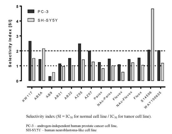
It is reasonable to express the cytotoxic activity of a compound against cancer cells as a selectivity index (SI) calculated as (IC50 control cells/IC50 cancer cells) (normal murine Balb/c 3T3 fibroblast cells were used as a control, Table 2, Figure 3). It is assumed that a compound with SI ≥ 2 may be considered a potential anticancer drug, since a high SI value reflects selectivity towards cancer cells while a SI value < 2 reflects general toxicity [7]. The compounds that had the desired SI values against SH-SY5Y cells were S14506 (SI 4.82) and AB5A (SI 2.15); those with desired SI values against PC-3 cells were S14506 (SI 2.06), WAY100635 (SI 2.03), KW117 (SI 2.64), AZ05 (SI 2.48), and AZ07 (SI 2.00). Only compound S14506 had a SI higher than 2 in both cell lines. For both tumor lines, lack of selective cytotoxicity showed AB9. Compound S14506 had the highest absolute activity against neuroblastoma tumor cells, and compound AZ07 had the highest absolute activity against PC-3 prostate cancer cells.
The ability of on fluoxetine (1, commonly used antidepressant drug) and compound AZ07 (8) to induce apoptosis as a cytotoxic mechanism was evaluated. Fluoxetine dose-dependently induced substantial apoptosis in PC-3 prostate cancer cells (Figure 4, 21, 60, 80% apoptosis at 25, 50 and 75 μM respectively, cytotoxic activity against PC-3 cells IC50 = 17.10 µM). The proapoptotic activity of compound AZ07 (exhibiting the highest absolute cytotoxicity against prostate cancer cells, IC50 = 5.74 µM) was almost twice smaller even if it was examined at 75 μM (20, 35, 44% apoptosis at 25, 50 and 75 μM respectively). This result could suggest that, in the case of compound AZ07 (maybe in other cases as well), the cell death may be induced through pathways other than apoptosis [8].
S14506, an atypical selective agonist of the 5-HT1A receptor, demonstrated significant inhibitory effects on PC-3 cells (IC50 = 9.98 ± 1.26 μM), and had the highest activity on SH-SY5Y cells (4.27 ± 0.47 μM). In HEK293 cells that overexpress the 5-HT1AR gene, it was found that S14506 acted as a cAMP pathway agonist (dose-dependently diminishing cAMP levels, EC50 = 25.4 pM) and a MAPK/ERK1/2 pathway agonist (elevating phosphorylated ERK1/2 levels, EC50 = 93.0 pM) (Figure 5 and Figure 6). It should be noted, however, that S14506 was found to be an Akt pathway antagonist in HEK293 cells that overexpress the 5-HT1AR gene (Figure 7). Akt may activate nuclear translocation of NF-κB, leading to caspase-3 inhibition and cell survival. The prosurvival activity of Akt may be reversed by Akt antagonists [9,10]. Therefore, the antagonistic activity of S14506 on Akt may induce caspase-3 activity and cytotoxicity.
The cytotoxic activity of S14506 against prostate cancer PC-3 cells (but not against neuroblastoma SH-SY5Y cells, Figure 8 and Figure 9) was reversed by treatment with the 5-HT1A receptor antagonist WAY100635 and inverse agonist spiperone.
It was also found that S14506 activated the cAMP biochemical pathway in PC-3 cells (IC50 = 0.32 µM, Figure 10) but not in SH-SY5Y cells.
Compound S14506, although structurally related to the 5-HT1A receptor inverse agonist spiperone, has been found to be one of the most potent agonists of the receptor, with very high affinity (Kd = 0.79 ± 0.2 nM, compared to 8-OH-DPAT Kd = 1.5 ± 0.5 nM). Additionally, the affinity of S14506 (but not of 8-OH-DPAT) was reduced by divalent manganese, magnesium and calcium ions. The presence of sodium ions markedly reduced the binding of 8-OH-DPAT but not the binding of S14506 [11]. S14506 potently reduced the duration of immobility in the forced swim test in rats at the minimal effective dose (MED) 0.01 mg/kg, s.c. (MED for 8-OH-DPAT was 0.63 mg/kg). The action of S14506 was blocked by the 5-HT1A receptor antagonist WAY100135. It was proposed that the antidepressant action of the compound is conveyed by postsynaptic 5-HT1A receptors [12]. It was also found that compound S14506 exhibited the properties of a dopamine D2 receptor antagonist [13].

2.2. Molecular Modelling

After docking, the ligands (buspirone, S14506, and spiperone) bound in a similar mode to the pocket formed by transmembrane helices (TM): TM3, TM5, TM6 and TM7 (Figure 11). The binding energies for buspirone, S14506 and spiperone were similar: −19.46, −22.46 and −21.21 kcal/mol, respectively. The charged piperazine nitrogen atom of the compounds interacted with residue Asp116 in TM3, which is the key recognition site for monoamine G-protein coupled receptor (GPCR) ligands [14]. The docking studies indicated that buspirone binds to the 5-HT1A receptor model in a similar manner as described earlier [15]. Interactions between the pyrimidine moiety of buspirone and TM3, TM5, TM6 were observed. The azaspirone portion of buspirone was close to TM2 and TM7, forming a hydrogen bond with Asn386 in TM7. Compound S14506, similar to buspirone, interacted with the 5HT1A receptor model at transmembrane helices TM3, TM5, TM6, and TM7 as well as with the extracellular loop 2 (ECL2) (Figure 11). S14506 is in a position with the naphthyl moiety located inside the binding pocket of the 5HT1A receptor and the fluoro group atom near the extracellular part of the receptor. The S14506 flouro group interacts with amino acids from TM2 only, and the amido moiety can form a hydrogen bond with Asn386 in TM7 in the extracellular part of the receptor. The S14506 methoxy group interacts with Ser199 and Thr200 in TM5. The aromatic interactions of the naphthyl part of S14506 with residues Phe361 and Phe362 in TM6 were also observed. The close interactions of spiperone with Asn386 in TM7 and Thr121 in TM5 were observed. In addition, spiperone and S14506 are located closer to both phenylalanines in TM6 than buspirone. Site-directed mutagenesis indicated that the affinity of aryloxypropanolamines was strongly affected by the Asn386Val mutation and that the oxygen atoms of the oxypropanol moiety are essential for the interaction with the 5-HT1A receptor [16]. The hydroxyl group present in the chemical structure of many neurotransmitters e.g., serotonin, appears to form hydrogen bonds with a series of Ser/Thr residues in TMH5 [17]. A serine (Ser199) and threonine (Thr200) residue in TMH5 of the 5-HT1A receptor were both capable of forming hydrogen bonds with the 5-hydroxy group of 5-HT. The importance of Thr200 for 5-HT binding was supported by site-directed mutagenesis studies [18]. Aromatic residues such as Phe361 and Phe362 in TM6 are part of an aromatic cluster that most likely regulates receptor activation [19].
It should be noted that somewhat different results were previously obtained by Milligan et al. [11], which is not surprising since the homology model of the 5-HT1A receptor based on the electron-crystallographic structure of bacteriorhodopsin was used [20]. The only interactions observed in both docking studies were between the S14506 methoxy group and Ser199 and Thr200 of the 5HT1A receptor models. The fluoro group was directed to the intracellular part of the receptor binding pocket, and the naphthyl moiety was directed to the opposite (extracellular) part. The fluoro group interacted with the Asn396 side chain in TM7 that, together with other residues, forms a part of the Asp-Arg-Tyr (DRY) signaling microdomain located in the intracellular part of the receptor (Asp1333.49, Arg1343.50, Asn3967.49). It was suggested that S14506 spans between the 5-HT recognition site and the DRY domain and may activate the interaction of the receptor with G proteins. On the basis of their results, Milligan et al. [11] suggested that the binding of [3H]-S14506 to the 5-HT1A receptor appears to be more similar to that of an antagonist, or an inverse agonist, than that of an agonist and that the efficacy of the drug at the receptor may be a consequence of a direct interaction with the signaling residues.

3. Discussion

It should be noted that several SSRIs (as well as some 5-HT1A receptor antagonists) consistently exhibit cytotoxic and proapoptotic activity against different cancer cell lines. The blockade of SERT in general results in 5-HT transport inhibition into the presynaptic neuron, thus elevating serotonergic function. The growth stimulatory effect of 5-HT has been described in different cell types and carcinomas, and 5-HT is thought to participate in the proliferation of different cancer types; moreover, known 5-HT1A receptor agonists were not found to exhibit cytotoxic activity in cancer cells (for a review, see [2]). The activity of 5-HT was shown to be connected to biochemical pathways such as those of MAPK(MEK)/ERK, Akt and NF-κB. It should be noted that NF-κB (formed as a dimer of specific subunits), by coordinating different subunits in the formation of a dimer, may regulate the transcription of different genes including those relating to apoptotic, antiapoptotic and inflammatory processes; the effects of NF-κB on cell survival or death will depend on the activation of distinct NF-κB factors [21,22]. It was suggested that the activation of different serotonergic pathways and the activation or blockade of 5-HT1A receptors may result in either prosurvival or proapoptotic activity [15,23,24]. Additionally, considering that several biochemical pathways may be directed by the 5-HT1A receptor [25], one should not expect a straightforward relationship between the serotonergic activity of SSRIs and 5-HT1A receptor ligands and their cytotoxic or cytoprotective activity.
Our results appear to support such an assumption. Most of the compounds (115) exhibited either agonistic or antagonistic activity on both the cAMP and pERK1/2 pathways in HEK293 cells that overexpress the 5-HT1AR gene. However, some of the compounds (2, 3 and 12) behaved similarly to mixed agonist–antagonists, exhibiting antagonistic activity on the cAMP pathway and agonistic activity on the ERK1/2 pathway. This result suggests that the cytotoxic activity of the compounds examined may be dependent on the activation of different biochemical pathways.
It was found that compound AZ07 had higher cytotoxic activity than that of fluoxetine in prostate PC-3 cancer cells. It should be noted, however, that fluoxetine had high proapototic activity and that compound AZ07 had low proapototic activity in PC-3 cancer cells. This observation suggests that the cytotoxic activity of compound AZ07 may be dependent on mechanisms other than apoptosis [26].
The acetylation of fluvoxamine and paroxetine diminished their cytotoxic activity (unlike in B- and T-cell-derived tumors [4]) both on PC-3 (IC50 ratio fluvoxamine/Ac-fluvoxamine—0.52; paroxetine/Ac-paroxetine—0.27) and SH-SY5Y (IC50 ratio fluvoxamine/Ac-fluvoxamine—0.80; paroxetine/Ac-paroxetine—0.28) cells.
The cytotoxic activity of compound S14506, an 5-HT1A receptor agonist, was consistently reversed by 5-HT1A receptor antagonists (WAY100635 and spiperone) in prostate but not in neuroblastoma cancer cells. Additionally, it was found that compound S14506 is an agonists of the cAMP and ERK1/2 pathways exhibited but is an antagonist of the prosurvival Akt pathway, which would explain its cytotoxic activity. This result suggests that either the cytotoxic activity of S14506 may be connected to the activation of the 5-HT1A receptor biochemical transduction pathway in the prostate, but not in neuroblastoma cancer cells, or that a receptor antagonist may be transduction pathway-specific.
Combining our results (particularly those concerning the cytotoxicity of and the biochemical pathways activated by compounds 8, 14 and the acetylated paroxetine and fluvoxamine derivatives) with the results of Schuster et al. [4] suggests that the mechanism of a compound’s cytotoxic activity (serotonergic against nonserotonergic) may depend on the compound and cancer type being examined.
On the basis of the S14506 previous docking studies to the bacteriorhodopsin homology-based 5-HT1A receptor, model microenvironments of the receptor activation made by the hydrophilic-conserved residues of the adjacent Arg3.50, Asp/Glu3.49, Asn1.50, Asp2.50, Asn7.49 and Tyr7.53 were identified [11]. It was also suggested that the compound may bind preferentially to an uncomplexed receptor (whereas 8-OH-DPAT binds preferentially to a receptor with prebound trimeric G protein), thus following the classic scheme for the binding of inverse agonists (spiperone, a 5-HT1A receptor inverse agonist, binds preferentially to the free receptor rather than to the coupled receptor). The experimental data indicate that S14506 has preferential affinity for the non-functional antagonist state of the receptor, but after binding, the S14506-receptor complex signals the G protein more efficiently than do other ligand-receptor complexes [11]. Our results suggest that S14506, buspirone and spiperone occupy a similar binding pocket and interact with similar 5-HT1A receptor residues. We did not observe the interaction of either compound with the conserved DRY microdomain. It is therefore difficult to determine if the S14506 binding mode is agonist- or antagonist-like. Although our arrangement of S14506 in the 5-HT1A receptor model is different to that of Milligan et al. [11] (different templates were used for homology model construction), the ligand occupies more or less the same binding pocket. Yin et al. [27] proposed that ring-like groups of inverse agonist/antagonists shift towards TM6 relative to agonists, leading to the outward movement of TM6, coupled with an inward shift of the intracellular end of this helix. According to our simulations, spiperone and S14506 are located closer to both phenylalanines in TM6 than buspirone, exhibiting a more antagonist-like binding mode. By interacting with the aromatic residues in TM6, the compounds could form an inactivating complex, but both S14506 and buspirone are cAMP pathway agonists (S14506 was also found to be an agonist of the ERK1/2 pathway). It should be noted that S14506 was found to have antagonistic activity on the Akt pathway.

4. Materials and Methods

4.1. Compounds

Fluvoxamine (4) and paroxetine (2) were isolated from their dosage forms Fevarin (Abbott Healthcare Products B.V., Weesp, The Netherlands) and Rexetin (Gedeon Richter Plc., Budapest, Hungary), respectively. Tablets were crushed into powder, and the salts contained in the tablet mass (fluvoxamine maleate and paroxetine hydrochloride) were converted into their free base form (15% NaOH aq); then, the aqueous mixtures were extracted with chloroform, and extracts were purified by silica gel flash chromatography (9:1, chloroform/methanol). Acetylated derivatives of fluvoxamine (5) and paroxetine (3) were obtained by acetylation with acetic anhydride according to a previously published procedure [4]. Fluoxetine (1) was purchased from Sigma-Aldrich, St. Louis, MO, USA. 8-OH-DPAT (13), S14506 (14), WAY100635 (15) and spiperone (16) were purchased from Tocris Bioscience, Bristol, UK.
KW117 (6-nitro-2-(4-octylpiperazin-1-yl)quinoline hydrochloride) (6), AZ05 (6-nitro-2-(4-nonylpiperazin-1-yl)quinolone hydrochloride) (7), and AZ07 (6-nitro-2-(4-undecylpiperazin-1-yl)quinoline hydrochloride) (8) were obtained as described in Gabrielsen et al. [6]. New compounds: AB5A (2-[4-(4-fluorobenzyl)piperazin-1-yl]-6-nitroquinoline hydrochloride) (9), AB9 (6-nitro-2-[4-(2-phenylethyl)-piperazin-1-yl]-6-nitroquinoline hydrochloride) (10), AB21 (2-[4-(cyclohexylmethyl)piperazin-1-yl]-6-nitroquinoline hydrochloride) (11), and AB22 (2-[4-(cyclobutylmethyl)piperazin-1-yl]-6-nitroquinoline hydrochloride) (12) were synthesized as described below in Section 6—Experimental procedures.

4.2. Cell Lines

4.2.1. PC-3 and SH-SY5Y, as well as HEK293 and Balb/c 3T3 Cells as a Reference

SH-SY5Y is a human neuroblastoma-like cell line often used as an in vitro model to study the function and differentiation of neurons. These cells produce serotonin and express major serotonergic proteins and mRNA including receptor 5-HT1A [28]. PC-3 is an androgen-independent neuroendocrine tumor line, does not form glands, is negative for androgen receptor (AR), prostate-specific antigen (PSA) and expresses serotonergic receptor subtype 1 (5-HTR1A) [29]. PC-3 is more diverse and aggressive than the DU-145 prostate cancer cell line.
Transformed human embryonic kidney cells (HEK293) were used to determine the functional activity of 5-HT1A receptor ligands. The Balb/c 3T3 cell line is a standard cell line often used as a model in studies of normal fibroblast cells. Lines were obtained from American Type Culture Collection (ATCC) Manassas, VA, USA.

4.2.2. Preparation of a Stable Transfectant 5-HT1A—HEK293 Cell Line

The transfectant’s parent line, Flp-In T-Rex-293, is a HEK293 (CRL-1573) cell line that was obtained from ATCC. Flp-In T-Rex-293 cells (Cat. No. R780-07), which stably express the lacZ-Zeocin gene fusion and Tet repressor with regulatory pcDNA6/TR plasmid, are part of the System (Cat. No. K6500-01) Life Technology (Carlsbad, CA, USA).
Lines were obtained by cotransfection with a pcDNA5/FRT/TO plasmid containing the gene encoding the serotonin receptor 5-HT1A and a pOG44 vector containing Flp recombinase. The Flp recombinase plasmid inserts the 5-HT1A gene into the genome at Flp Recombination Target (FRT) sites. After the cultivation of stable lines, gene expression can be induced by the addition of tetracycline. The enhanced green fluorescent protein (EGFP) sequence was also introduced into the system next to the appropriate receptor protein. Ligations were performed using a ligase from New England Biolabs (T4 DNA Ligase M0202S, Ipswich, MA, USA).
Transformations were performed with One Shot® TOP10 chemically competent E. coli C4040-10, Life Technologies. Selection of clones possessing the plasmid was carried out using their antibiotic (ampicillin) resistance. The resulting clones were checked for the presence of a recombinant gene by sequencing. Transfections were performed using Lipofectamine® 3000 transfection reagent (L3000-008, Life Technologies, Carlsbad, CA, USA). Antibiotic selection was performed using hygromycin and blasticidin.
5-HT1A receptor expression in the selected 10 clones of cells transfected with different plasmids was confirmed by Western blot. The level of expression and cellular localization of receptor proteins was monitored by immunofluorescence or detection by sodium dodecyl sulfate polyacrylamide gel electrophoresis (SDS-PAGE) protein electrophoresis. Enhanced Green Fluorescent Protein (EGFP) fluorescence allowed measurement and visualization.

4.3. Cell Cultures

Cultures of PC-3 cells were grown in RPMI 1640 medium (Sigma-Aldrich, St. Louis, MO, USA), Balb/c 3T3 in Dulbecco’s modified Eagle’s medium (DMEM), SH-SY5Y in DMEM: F12 (Sigma-Aldrich, St. Louis, MO, USA), and HEK293 cells in DMEM (Gibco, Grand Island, NY, USA). All cell lines were grown at 37 °C in a humidified atmosphere with 5% CO2. All media contained 1% GlutaMax (Gibco, Grand Island, NY, USA), 1% Antibiotic Antimycotic Solution (10,000 units/mL penicillin G, 10 mg/mL streptomycin sulfate, 25 mg/mL amphotericin B, Sigma-Aldrich, St. Louis, MO, USA) and 10% heat-inactivated and sterile-filtered fetal bovine serum (FBS) (Gibco, Grand Island, NY, USA), except Balb/c 3T3 cells, whose media was supplemented with 10% calf serum (Biowest, Courtaboeuf, France).
Cell lines were passaged every 3–4 days by trypsinization with 0.25% trypsin solution, with or without EDTA (2–5 min) (Thermo Fisher Scientific, Waltham, MA, USA). Cells used for experiments were derived from a continuous culture and were in the exponential growth phase. The cells were counted using a Handheld Automated Cell Counter – Scepter (Merck Millipore, Billerica, MA, USA).

4.4. Study of In Vitro Binding Affinity of Compounds for Serotonin Transporter (SERT)

The experiments were performed according to the method described by Owens et al. [30] Rat cerebral cortex was homogenized in 30 volumes of ice-cold 50 mM Tris-HCl containing 150 mM NaCl and 5 mM KCl, pH 7.7 at 25 °C and centrifuged at 20,000 × g for 20 min. The supernatant was decanted, and the pellet was resuspended in 30 volumes of buffer and centrifuged three times. The final pellet was suspended in Tris-HCl buffer at a proportion of approximately 5 mg wet weight/mL. An increasing concentration of the analyzed compounds (30 μL) was added to the incubation mixture containing 240 μL of tissue suspension and 30 μL of 1 nM [3H]-citalopram (used for labeling 5-HT-transporter). Non-specific binding was determined in the presence 30 μL of 1 μM imipramine (displacer). Duplicate samples were incubated at room temperature for 1 h. The incubation was terminated by rapid filtration over fiberglass filters FilterMate B (Perkin Elmer, Waltham, MA, USA) using a 96-well FilterMate harvester (PerkinElmer, Waltham, MA, USA). The radioactivity of the filters was measured with a MicroBeta TriLux 1450 scintillation counter. Ki values were calculated from the Cheng–Prusoff equation [31]:
K i = IC 50 1 + L o K D
where Lo is the labeled ligand concentration, and KD is the dissociation constant of the labeled ligand.

4.5. Examination of the Functional Activity of 5-HT1A Receptor Ligands in Transformed HEK293

The examination of the functional activity read out as cAMP level inhibition and ERK1/2 and Akt1 phosphorylation was performed according to the instructions of the One-plate Cisbio Bioassays cAMP dynamic 2, phospho-ERK (Thr202/Tyr204) and Akt1 phospho- S473 Cellular Assay Kits respectively, using a HEK293 human fibroblast cell line that overexpresses 5-HT1A receptors. The measurements were performed in 384-well microtiter plates using 200,000, 1,875,000 or 2,000,000 cells/mL. The cAMP levels in PC-3 cells were determined using 600,000 cells/mL. Receptor activation with the appropriate ligands was carried out for 30 min, and then the cells were lysed. The results were analyzed with GraphPad Prism v 6.0 (GraphPad Software, San Diego, CA, USA) using its in-built functions. Ligand activity was determined using a 4-parameter equation of the dose-response function. The response curves were obtained by plotting the HTRF® ratio (signal ratio 665/620 nm × 10,000) relative to the log concentration of the [compound] M.
All compounds were tested with both agonists and antagonists. To test the response of cAMP to an agonist the cells were activated with forskolin (1 µM) followed by agonist addition. Antagonist activity (both for cAMP and pERK1/2 determination) was measured against the reference agonist (5-carboxamidotryptamine)-pretreated cells (0.1 μM of 5-CT = EC80, for cAMP measurements, forskolin-stimulated cells were used) and expressed as IC50 (antagonist concentration showing 50% inhibition of maximal response). Data are an average of minimum three independent experiments in duplicate. The control of specific activity was measured under the same conditions on cells lacking the 5-HT1A receptor. The results were expressed as % of the reference agonist activity. In the antagonist mode 100% of the reference agonist activity (EC80) corresponded to 0% antagonist activity.

4.6. Determination of the Effect of Test Compounds on Cell Survival

The effects of the test compounds on the survival of PC-3, SH-SY5Y, Balb/c 3T3 and HEK293 cells was measured with the aid of the 3-(4,5-dimethylthiazol-2-yl)-2,5-diphenyltetrazolium bromide (MTT) assay. The cells were cultured in 96-well plates (100 μL/well) for 72 h. The optimized initial densities were the following: for PC-3—60,000 cells/mL, SH-SY5Y and Balb/c 3T3—50,000 cells/mL. The concentration of the starter culture was determined in a preliminary experiment to provide optimum cell growth during the experiment, and to ensure continuous exponential growth of cells and prevent overcrowding throughout the experimental incubation, 70% confluence was determined to be optimal for a 72 h test.
The activity of the compounds was determined at the following concentrations: 0.01; 0.1; 1.0; 2.5; 5.0; 10; 15; 25; 50; 75 and 100 μM (additionally 7.5 μM for compound AB9). After 20 h preincubation and evaluation of the uniformity of cell growth, the medium was aspirated, and the respective solutions of the substances were added to the culture medium. Control cells were only treated the solvent used for the compounds and medium alone. The positive control of cytotoxicity for the control of the system was fluoxetine.
The MTT assay is a colorimetric method that measures cell viability by revealing the metabolic activity of cells in culture. MTT (3-(4,5-dimethylthiazol-2-yl)-2,5-diphenyltetrazolium bromide) is reduced by intracellular dehydrogenase, which is present only in live cells (mitochondria), to purple, water-insoluble formazan. After it is dissolved, formazan absorbance of the resulting solution is directly proportional to the number of living cells in culture.
After appropriate incubation, the culture medium was removed. Next, 50 μL of MTT solution (0.5 mg/mL) diluted in PBS to 0.5 mg/mL was added to each well. The plates were incubated for 3 h at 37 °C. Then, the precipitated formazan was dissolved by the addition of 200 μL isopropanol. After a 24-hour incubation at 4 °C in the dark, the absorbance of the dissolved formazan was measured at 570 nm (the maximum absorption of formazan) and at 690 nm (reference wavelength) using a Power Wave XS microplate spectrophotometer (Bio-Tek, Winooski, VT, USA). The plates were sealed to reduce evaporation before incubation. The test was performed in three to five independent experiments with four times the measured concentration tested. The cell survival percentage was determined as % of controls, which were cells treated with the test compound solvent at the highest concentration. The MTT data were analyzed to determinate the IC50 values (concentration required to reduce the viability of cells by 50% compared with the control cells) of each compound.

4.7. Apoptosis Determination

Apoptosis was measured using the FITC Annexin V Apoptosis Detection Kit I (BD Biosciences, San Jose, CA, USA). Cells, after incubations 72 h with tested agents, were centrifuged, rinsed twice with cold phosphate-buffered saline (PBS) and suspended in binding buffer at 1 × 10⁶ cells/mL. Then, 100-μL volumes of the cell suspensions were labeled according to the manufacturer’s instructions. Flow cytometry analysis was performed within 1 h after labeling.

4.8. Flow Cytometry

Flow cytometry analyses was run on a FACSCanto II flow cytometer (BD Biosciences, San Jose CA, USA) and analyzed using BD FACSDiva software.

4.9. Molecular Modeling

The homology model of the 5-HT1A serotonin receptor is based on the X-ray structure of the serotonin 1B receptor, (PDB ID 4IAR), and the crystal structure was employed [15,26,32]. The start and end positions of the transmembrane helices (TM) in the models were found to be the following: Val31-Val51 (TM1), Leu78-Leu99 (TM2), Gly105-Cys128 (TM3), Ser158-Ser168 (TM4), Asp192-Val213 (TM5), Met351-Val367 (TM6), and Leu380-Leu395 (TM7).
The structures of the compounds (buspirone, S 14509 and spiperone) were taken from the PubChem database. The ligands were protonated at the nitrogen atom in the piperazine ring. ICM/Molsoft software was used for docking studies [23]. Ligands were docked to 5HT1A-serotonin receptor models using the usual rigid protein-flexible ligand approach [23]. The binding poses were evaluated with an all-atom ICM ligand binding score calculated using the VLS (virtual ligand screening) scoring function [33]. The VLS scoring function is the sum of the internal force field energy of the ligand and the ligand-receptor interaction energy that includes several weighted terms: van der Waals term, a hydrophobicity term, an electrostatic solvation term, hydrogen bonding interactions, and an entropic term proportional to the number of flexible torsions in the ligand.

4.10. Data Analysis

All experiments were performed in triplicate, and the results were obtained from at least three independent experiments. The data are expressed as the mean ± SEM (standard error of the mean). The statistical significance was calculated using an unpaired t-test. Differences were considered statistically significant at p ≤ 0.05. The experimental data were analyzed using GraphPad Prism 6.01 for Windows (GraphPad Software, San Diego, CA, USA, www.graphpad.com).
Curve fitting and calculation of IC50 values was performed by the GraphPad Prism program from the best-fit (R2 > 0.95) of the Hill slope curve to experimental data using nonlinear regression analysis according to the following formula:
Y = 100 1 + 10 ( ( LogIC 50 X ) *   Hill   slope )
where X is the log of dose, Y is the growth inhibition value normalized to the control, and Hill slope is the unitless slope factor or Hill slope.

5. Conclusions

The cytotoxic activity of several SERT inhibitors and 5-HT1A receptor ligands was examined. Almost all of the examined compounds (except for 5-HT1A receptor agonists 8-OH-DPAT and 5-CT, but including agonist S14506) exhibited cytotoxic activity against neuroblastoma and prostate cancer cells. Compound AZ07 had the highest absolute cytotoxic activity against PC-3 prostate cancer cell lines (IC50 = 5.74 μM) and the 5-HT1A receptor agonist S14506 had the highest cytotoxicity against neuroblastoma SH-SY5Y cells (IC50 = 4.27 μM). Compound AZ07 in amounts up to 75 µM did not exhibit substantial proapoptotic activity, which could suggest that its cytotoxic activity may be connected to a different pathway. Only compound S14506 (14) had a SI higher than 2 in both cell lines. The highest activity against neuroblastoma tumor and androgen-insensitive human prostate cancer was found to be with compound S14506 and compound AZ07 (8), respectively. Therefore, further studies for compounds S14506 and AZ07 for their potential use in the prevention and treatment of cancer are needed. Most of the compounds (115) exhibited either agonistic or antagonistic activity on both the cAMP and pERK1/2 pathways in HEK293 cells that overexpress the 5-HT1AR gene. However compounds 2, 3 and 12 exhibited antagonistic activity on the cAMP pathway and agonistic activity on the ERK1/2 pathway. Thus it maybe suggested that the cytotoxic activity of the examined compounds may be connected to the activation of a particular biochemical pathway. In spite of the higher cytotoxicity of compound AZ07 compared to that of fluoxetine, the proapoptotic activity of fluoxetine was high, while the activity of compound AZ07 was low, in prostate PC-3 cancer cells. This finding suggests that the cytotoxic activity of compound AZ07 may be dependent on a mechanism different than apoptosis. Compound S14506 was found to be an antagonist of the Akt pathway. Akt may activate the nuclear translocation of NF-κB, leading to caspase-3 inhibition and cell survival. The prosurvival activity of Akt may be reversed by Akt antagonists. Therefore, the antagonistic activity of S14506 on the Akt may induce caspase-3 activity and cytotoxicity.
Combining our above results (particularly those concerning the cytotoxicity of and the biochemical pathways activated by compounds 8, 14 and the acetylated paroxetine and fluvoxamine derivatives) with the results of Schuster et al. [4], one should not expect a straightforward relationship between the activation of particular serotonergic pathways by SSRIs and 5-HT1A receptor ligands and their cytotoxic or cytoprotective activity. It was also found that NF-κB, which may be involved in 5-HT-dependent biochemical pathways by coordinating different subunits to form a dimer, may regulate the transcription of different transduction pathways, and the effects of NF-κB on cell survival or death depend on the activation of distinct NF-κB factors. Therefore, it can be suggested that the mechanism of a compound’s cytotoxic activity (serotonergic against nonserotonergic) may depend on the compound and cancer type being examined.
The results of molecular modeling suggest that S14506, buspirone and spiperone occupy similar binding pockets and interact with similar 5-HT1A receptor residues. We did not observe the interaction of either compound with the conserved DRY microdomain. S14506 and spiperone were found to be located closer to both phenylalanines in TM6 than buspirone was, exhibiting a more antagonist-like binding mode. By interacting with aromatic residues in TM6, the compounds could form inactivated complex, but both S14506 and buspirone are agonists for the cAMP pathway (and S14596 was also found to be an agonist of the ERK1/2 pathway). It should be noted that S14506 was found to be an AKT pathway antagonist.

6. Experimental Procedures

6.1. Preparation of N-Alkylaryl-6-nitroquipazines Derivatives 9 (AB5A) and 10 (AB9)

The synthesis was done according to the following scheme:
Ijms 19 03260 i008
2-[4-(4-Fluorobenzyl)piperazin-1-yl]-6-nitroquinoline (9) (AB5A). A slurry of sodium hydride in mineral oil (100 mg, 2.47 mmol) was added to a solution of 6-nitroquipazine (0.2 g, 0.77 mmol) in acetonitrile cooled to 0 °C. After 30 min, 1-(bromomethyl)-4-fluorobenzene (0.146 g, 0.77 mmol) and 3 drops of 1,8-diazabicyclo(5.4.0)undec-7-ene (DBU) were added followed by an incubation at room temperature for 3 h. After this, the solution was filtered and evaporated under reduced pressure. The reaction mixture was purified by column chromatography (2% MeOH/CHCl3), yielding yellow-orange crystals.
C20H19FN4O2. M = 366.39 g/mol. Yield 60%. m.p.: 96.5–98 °C. 1H nuclear magnetic resonance (NMR) (300 MHz, CDCl3): δ 8.52 (d, 1H, J = 2.7 Hz, Harom), 8.29 (dd, 1H, J1 = 2.4 Hz, J2 = 9.3 Hz, Harom), 7.94 (d, 1H, J = 9.3 Hz, Harom), 7.88 (d, 1H, J = 9.0 Hz, Harom), 7.57 (m, 4H, Hbenzyl), 7.32 (dd, 1H, J1 = 5.4 Hz, J2 = 8.4 Hz, Harom), 3.85 (t, 2H, J = 4.9 Hz, CH2-Ph), 3.72 (t, 2H, J = 5.1 Hz, N(CH2)piper-Ph), 3.02 (t, 2H, J = 5.1 Hz, N(CH2)piper-Ph), 2.56 (t, 2H, J = 4.9 Hz, N(CH2)piper-CH2), 1.73 (s, 2H, N(CH2)piper-CH2). Electrospray ionisation mass spectrometry (ESI-MS) m/z: found 367.1 [M + H]+ (100%), calculated for [C20H20FN4O2]+ 367.2.
2-[4-(2-Phenylethyl)piperazin-1-yl]-6-nitroquinoline (10) (AB9). A slurry of sodium hydride in mineral oil (100 mg, 2.47 mmol) was added to a solution of 6-nitroquipazine (0.16 g, 0.62 mmol) in acetonitrile cooled to 0 °C. After 30 min, 1-(bromoethyl)- benzene (0.115 g, 0.62 mmol) and 5 drops of DBU were added, and then, the mixture was heated in a water bath under reflux for 20 h. After this step, the solution was filtered and evaporated under reduced pressure. The reaction mixture was purified by column chromatography (2% MeOH/CHCl3), yielding yellow-orange crystals.
C21H22N4O2. M = 362.43 g/mol. Yield 60%. m.p.: 143–145 °C. 1H NMR (300 MHz, CDCl3): δ 8.60 (d, 1H, J = 2.7 Hz, Harom), 8.25 (dd, 1H, J1 = 2.7 Hz, J2 = 9.3 Hz, Harom), 8.10 (d, 1H, J = 9.3 Hz, Harom), 7.61 (d, 1H, J = 9.3 Hz, Harom), 7.24 (m, 6H, Harom, Hphenyl), 5.24 (d, 2H, J = 1.8 Hz, N(CH2)piper-Ph), 5.19 (d, 2H, J = 1.8 Hz, N(CH2)piper-Ph), 5.07 (d, 2H, J = 1.8 Hz, N(CH2)piper-CH2), 5.03 (d, 2H, J = 1.8 Hz, N(CH2)piper-CH2), 2.84 (t, 2H, J = 7.6 Hz, CH2Ph), 2.64 (m, 2H, NCH2). ESI-MS m/z: found 363.1 [M + H]+ (100%), calculated for [C21H23N4O2]+ 363.2.

6.2. Preparation of 6-Nitroquipazine N-Cycloalkyl Derivatives 11 (AB21) and 12 (AB22)

The synthesis was done according to the following scheme:
Ijms 19 03260 i009
A solution of lithium hydride (100 mg, 12.5 mmol) was added to a solution of 6-nitroquipazine (100 mg, 2.47 mmol) in acetone cooled to 0 °C. After 30 min, cycloalkyl halide (1.88 mmol) and 10 drops of DBU (1,8-diazabicyclo[5.4.0]undec-7-ene) were added, and then, the mixture was heated on a water bath under reflux for 72 h. After this step, the solution was filtered and evaporated under reduced pressure. The reaction mixture was purified by column chromatography (1–4% MeOH/CHCl3), yielding yellow-orange crystals. The resulting N-cyclic derivative of quipazine was directly subjected to a nitration reaction. The derivative (0.71 mmol) was cooled to 0 °C and dissolved in concentrated H2SO4 (1.83 cm3), after which concentrated HNO3 (0.35 cm3) was added dropwise. After approximately 1 h, the mixture was poured onto crushed ice and basified with 12 M NaOH to pH = 11. The mixture was then extracted several times with methylene chloride, and the extract was dried over anhydrous MgSO4 for 24 h. Next, the solvent was filtered and evaporated. A pure brownish-orange-colored product was obtained by column chromatography (1–4% MeOH/CHCl3).
2-[4-(Cyclohexylmethyl)piperazin-1-yl]-6-nitroquinoline (11) (AB21)
C20H26N4O2. M = 354.45 g/mol. Yield 60%. m.p. 107–108 °C. 1H NMR (300 MHz, CDCl3): δ 8.53 (d, 1H, J = 2.7 Hz, Harom), 8.29 (dd, 1H, J1 = 2.7 Hz, J2 = 9.3 Hz, Harom), 7.94 (d, 1H, J = 9.3 Hz, Harom), 7.65 (d, 1H, J = 9.3 Hz, Harom), 7.05 (d, 1H, J = 9.3 Hz, Harom), 3.85 (t, 4H, J = 5.0 Hz, N(CH2)2piper-Ph), 2.53 (t, 4H, J = 4.4 Hz, N(CH2)2piper-CH2), 2.19 (d, 2H, J = 6.9 Hz, NCH2), 1.78 (m, 4H, 2 × (CH2)cyclohexyl), 1.53 (m, 4H, 2 × (CH2)cyclohexyl), 1.22 (m, 1H, CHcyclohexyl), 0.90 (m, 2H, (CH2)cyclohexyl). ESI-MS m/z: found 355.1 [M + H]+ (100%), calculated for [C20H27N4O2]+ 355.2.
2-[4-(Cyclobutylmethyl)piperazin-1-yl]-6-nitroquinoline (12) (AB22)
C18H22N4O2. M = 326.39 g/mol. Yield 50%. M. m.p. 99–101 °C. 1H NMR (300 MHz, CDCl3): δ 8.53 (d, 1H, J = 2.7 Hz, Harom), 8.29 (dd, 1H, J1 = 2.7 Hz, J2 = 9.0 Hz, Harom), 7.95 (d, 1H, J = 9.3 Hz, Harom), 7.65 (d, 1H, J = 9.3 Hz, Harom), 7.05 (d, 1H, J = 9.3 Hz, Harom), 3.89 (t, 4H, J = 4.8 Hz, N(CH2)2piper-Ph), 2.64 (m, 5H, N(CH2)2piper-CH2, CHcyclobutyl), 2.52 (m, 2H, NCH2), 2.11 (m, 2H, CH2cyclobutyl), 1.82 (m, 4H, 2 x CH2cyclobutyl). ESI-MS m/z: found 327.1 [M + H]+ (100%), calculated for [C18H23N4O2]+ 327.2.

Author Contributions

J.W. and Z.C. designed the experiments, analyzed the data and prepared the manuscript; M.J. performed molecular modelling studies; J.W., L.M. and M.K. performed the experiments; R.B., J.K. and A.J.B. performed the synthesis and purification experiments; A.J.B and Z.C. supervised the project.

Funding

This research was partially funded by Narodowe Centrum Badań i Rozwoju (Pol-Nor/198887/73/2013).

Conflicts of Interest

The authors declare no conflict of interest.

Abbreviations

Aktserine-threonine protein kinase B
pAkt1phospho-Akt isoform 1
Balb/c 3T3murine fibroblast cell line
cAMPcyclic adenosine monophosphate
5-CT5-carboxamidotryptamine
8-OH-DPAT(R)-(+)-8- hydroxy-(2-di-n-propylamino)tetralin
DBU1,8-diazabicyclo(5.4.0)undec-7-ene
DU-145androgen-independent human prostate cancer cell line
EC50concentration inducing 50% of maximal response
ECLextracellular loop
EDTAdisodium ethylenediaminetetraacetic acid
ERK1/2extracellular signal-regulated kinase 1 and 2
pERK1/2phospho-ERK1/2
EGFPenhanced green fluorescent protein
HEKhuman embryonic kidney cell line
5-HT5-hydroxytryptamine (serotonin)
5-HT1A receptorsubtype of serotonin receptor 1A
IC50concentration showing 50% inhibition of maximal response
LNCaPandrogen-sensitive human prostate adenocarcinoma
MAPKmitogen activated protein kinase
MEKacronym from MAPK/ERK kinase
MTT(3-(4,5-dimethylthiazol-2-yl)-2,5-diphenyltetrazolium bromide)
NF-κBnuclear transcription factor κB
PC-3androgen-independent human prostate cancer cell line
PI3Kphosphatidylinositol-3-kinase
SEMstandard error of the mean
SERTserotonin transporter
SH-SY5Yhuman neuroblastoma-like cell line
SIselectivity index
SSRIselective serotonin reuptake inhibitor
TMHtransmembrane helices
VLSvirtual ligand screening

References

  1. Frick, L.R.; Rapanelli, M.; Arcos, M.L.; Cremaschi, G.A.; Genaro, A.M. Oral administration of fluoxetine alters the proliferation/apoptosis balance of lymphoma cells and upregulates T cell immunity in tumor-bearing mice. Eur. J. Pharmacol. 2011, 659, 265–272. [Google Scholar] [CrossRef] [PubMed]
  2. Chilmonczyk, Z.; Bojarski, A.J.; Pilc, A.; Sylte, I. Serotonin tranporter and receptor ligands with antidepressant activity as neuroprotective and proapoptotic agents. Pharm. Rep. 2017, 69, 469–478. [Google Scholar] [CrossRef] [PubMed]
  3. Dizeyi, N.; Hedlund, P.; Bjartell, A.; Tinzl, M.; Ustild-Tasken, K.; Abrahamsson, P.A. Serotonin activates MAP kinase and PI3K/Akt signaling pathways in prostate cancer cell lines. Urol. Oncol. 2011, 29, 436–445. [Google Scholar] [CrossRef] [PubMed]
  4. Schuster, C.; Fernbach, N.; Rix, U.; Superti-Furga, G.; Holy, M.; Freissmuth, M.; Sitte, H.H.; Sexl, V. Selective serotonin reuptake inhibitors—A new modality for the treatment of lymphoma/leukaemia? Biochem. Pharmacol. 2007, 74, 1424–1435. [Google Scholar] [CrossRef] [PubMed]
  5. Rosetti, M.; Frasnelli, M.; Tesei, A.; Zoli, W.; Conti, M. Cytotoxicity of different selective serotonin reuptake inhibitors (SSRIs) against cancer cells. J. Exp. Ther. Oncol. 2006, 6, 23–29. [Google Scholar] [PubMed]
  6. Gabrielsen, M.; Wołosewicz, K.; Zawadzka, A.; Kossakowski, J.; Nowak, G.; Wolak, M.; Stachowicz, K.; Siwek, A.; Ravna, A.W.; Kufareva, I.; et al. Synthesis, antidepressant evaluation and docking studies of long-chain alkylnitroquipazines as serotonin transporter inhibitors. Chem. Biol. Drug Des. 2013, 81, 695–706. [Google Scholar] [CrossRef] [PubMed]
  7. Badisa, R.B.; Darling-Reed, S.F.; Joseph, P.; Cooperwood, J.S.; Latinwo, L.M.; Goodman, C.B. Selective cytotoxic activities of two novel synthetic drugs on human breast carcinoma MCF-7 Cells. Anticancer Res. 2009, 29, 2993–2996. [Google Scholar] [PubMed]
  8. Milczarek, M.; Wiktorska, K.A.; Mielczarek, L.; Koronkiewicz, M.; Dąbrowska, A.; Lubelska, K.; Matosiuk, D.; Chilmonczyk, Z. Autophagic cell death and premature senescence: New mechanism of 5-fluorouracil and sulforaphane synergic anticancer effect in MDA-MB-231 triple negative breast cancer cell line. Food Chem. Toxicol. 2018, 111, 1–8. [Google Scholar] [CrossRef] [PubMed]
  9. Hsiung, S.C.; Tamir, H.; Franke, T.F.; Liu, K.P. Roles of extracellular signal-regulated kinase and Akt signaling in coordinating nuclear transcription factor- kappaB-dependent cell survival after serotonin 1A receptor activation. J. Neurochem. 2005, 95, 1653–1666. [Google Scholar] [CrossRef] [PubMed]
  10. Adayev, T.; Ray, I.; Sondhi, R.; Sobocki, T.; Banerjee, P. The G protein-coupled 5- HT1A receptor causes suppression of caspase-3 through MAPK and protein kinase Calpha. Biochim. Biophys. Acta 2003, 1640, 85–96. [Google Scholar] [CrossRef]
  11. Milligan, G.; Kellett, E.; Dacquet, C.; Dubreuil, V.; Jacoby, E.; Millan, M.J.; Lavielle, G.; Spedding, M. S14506: Novel receptor coupling at 5-HT1A receptors. Neuropharmacology 2001, 40, 334–344. [Google Scholar] [CrossRef]
  12. Schreiber, R.; Brocco, M.; Gobert, A.; Veiga, S.; Millan, M.J. The potent activity of the 5-HT1A receptor agonists, S 14506 and S 14671, in the rat forced swim test is blocked by 5-HT1A receptor antagonists. Eur. J. Pharmacol. 1994, 271, 537–541. [Google Scholar] [CrossRef]
  13. Protais, P.; Chagraoui, A.; Arbaoui, J.; Mocaër, E. Dopamine receptor antagonist properties of S 14506, 8-OH-DPAT, raclopride and clozapine in rodents. Eur. J. Pharmacol. 1994, 271, 167–177. [Google Scholar] [CrossRef]
  14. Sylte, I.; Edvardsen, O.; Dahl, S.G. Molecular modeling of UH-301 and 5-HT(1a) receptor interactions. Protein Eng. 1996, 9, 149–160. [Google Scholar] [CrossRef] [PubMed]
  15. Warszycki, D.; Rueda, M.; Mordalski, S.; Kristiansen, K.; Satała, G.; Rataj, K.; Chilmonczyk, Z.; Sylte, I.; Abagyan, R.; Bojarski, A.J. From Homology Models to a Set of Predictive Binding Pockets-a 5-HT1A Receptor Case Study. J. Chem. Inf. Model. 2017, 57, 311–321. [Google Scholar] [CrossRef] [PubMed]
  16. Kuipers, W.; Link, R.; Standaar, P.J.; Stoit, A.R.; Van Wijngaarden ILeurs, R.; Ijzerman, A.P. Study of the Interaction Between Aryloxypropanolamines and Asn386 in Helix VII of the Human 5-Hydroxytryptamine1A Receptor. Mol. Pharmacol. 1997, 51, 889–896. [Google Scholar] [CrossRef] [PubMed]
  17. Strader, C.D.; Candelore, M.R.; Hill, W.S.; Sigal, I.S.; Dixon, R.A. Identification of two serine residues involved in agonist activation of the beta-adrenergic receptor. J. Biol. Chem. 1989, 264, 13572–13578. [Google Scholar] [PubMed]
  18. Ho, B.Y.; Karschin, A.; Branchek, T.; Davidson, N.; Lester, H.A. The role of conserved aspartate and serine residues in ligand binding and in function of the 5-HT1A receptor: A site-directed mutation study. FEBS Lett. 1992, 31, 259–262. [Google Scholar] [CrossRef]
  19. Strader, C.D.; Sigal, I.S.; Dixon, R.A. Structural basis of beta-adrenergic receptor function. FASEB J. 1989, 3, 1825–1832. [Google Scholar] [CrossRef] [PubMed]
  20. Grigorieff, N.; Ceska, T.; Downing, K.; Baldwin, J.; Henderson, R. Electron crystallographic structure of bacteriorhodopsin. J. Mol. Biol. 1996, 259, 393–421. [Google Scholar] [CrossRef] [PubMed]
  21. Yang, L.; Tao, L.Y.; Chen, X.P. Roles of NF-κB in central nervous system damage and repair. Neurosci. Bull. 2007, 23, 307–313. [Google Scholar] [CrossRef] [PubMed] [Green Version]
  22. Shih, R.H.; Wang, C.Y.; Yang, C.M. NF-kappaB signaling pathways in neurological inflammation: A mini review. Front. Mol. Neurosci. 2015, 8, 77. [Google Scholar] [CrossRef] [PubMed]
  23. Abagyan, R.; Totrov, M.; Kuznetsow, D.N. ICM: A new method for protein modeling and design: Applications to docking and structure prediction from the distorted native conformation. J. Comput. Chem. 1994, 15, 488–506. [Google Scholar] [CrossRef]
  24. Daniele, S.; Da Pozzo, E.; Zappelli, E.; Martini, C. Trazodone treatment protects neuronal-like cells from inflammatory insult by inhibiting NF-kappaB, p38 and JNK. Cell Signal. 2015, 27, 1609–1629. [Google Scholar] [CrossRef] [PubMed]
  25. Chilmonczyk, Z.; Bojarski, A.J.; Pilc, A.; Sylte, I. Functional selectivity and antidepressant activity of serotonin 1A receptor ligands. Int. J. Mol. Sci. 2015, 16, 18474–18506. [Google Scholar] [CrossRef] [PubMed] [Green Version]
  26. Wang, C.; Jiang, Y.; Ma, J.; Wu, H.; Wacker, D.; Katritch, V.; Han, G.W.; Liu, W.; Huang, X.-P.; Vardy, E.; et al. Structural Basis for Molecular Recognition at Serotonin Receptors. Science 2013, 340, 610–614. [Google Scholar] [CrossRef] [PubMed] [Green Version]
  27. Yin, W.; Zhou, X.E.; Yang, D.; de Waal, P.W.; Wang, M.; Dai, A.; Cai, X.; Huang, C.H.-Y.; Liu, P.; Wang, X.; et al. Crystal structure of the human 5-HT1B serotonin receptor bound to an inverse agonist. Cell Discov. 2018, 4, 12. [Google Scholar] [CrossRef] [PubMed]
  28. Adeosun, S.O.; Albert, P.R.; Austin, M.C.; Iyo, A.H. 17β-estradiol-induced regulation of the novel 5-HT1A-related transcription factors NUDR and Freud-1 in SH SY5Y Cells. Cell. Mol. Neurobiol. 2012, 32, 517–521. [Google Scholar] [CrossRef] [PubMed]
  29. Dizeyi, N.; Bjartell, A.; Nilsson, E.; Hansson, J.; Gadaleanu, V.; Cross, N.; Abrahamsson, P.A. Expression of serotonin receptors and role of serotonin in human prostate cancer tissue and cell lines. Prostate 2004, 59, 328–336. [Google Scholar] [CrossRef] [PubMed]
  30. Owens, M.J.; Morgan, W.N.; Plott, S.J.; Nemeroff, C.B. Neurotransmitter receptor and transporter binding profile of antidepressants and their metabolites. J. Pharmacol. Exp. Ther. 1997, 283, 1305–1322. [Google Scholar] [PubMed]
  31. Cheng, Y.; Prusoff, W.H. Relationship between the inhibition constant (K1) and the concentration of inhibitor which causes 50 percent inhibition (I50) of an enzymatic reaction. Biochem. Pharmacol. 1973, 22, 3099–3108. [Google Scholar] [PubMed]
  32. Cherezov, V.; Rosenbaum, D.M.; Hanson, M.A.; Rasmussen, S.G.F.; Thian, F.S.; Kobilka, T.S.; Choi, H.-J.; Kuhn, P.; Weis, W.I.; Kobilka, B.K.; et al. High-Resolution Crystal Structure of an Engineered Human beta2-Adrenergic G Protein-Coupled Receptor. Science 2007, 318, 1258–1265. [Google Scholar] [CrossRef] [PubMed]
  33. Schapira, M.; Totrov, M.; Abagyan, R. Prediction of the binding energy for small molecules, peptides and proteins. J. Mol. Recognit. 1999, 12, 177–190. [Google Scholar] [CrossRef]
Figure 1. Structures of fluoxetine (1), paroxetine (2), fluvoxamine (4) and acetylated derivatives of paroxetine (3) and fluvoxamine (5).
Ijms 19 03260 g001
Figure 2. Structures of 5-HT1A receptor agonists (13, 14, and 17), antagonist (15) and inverse agonist (16).
Ijms 19 03260 g002
Figure 3. Selectivity index (SI = IC50 for normal cell line/IC50 for tumor cell line).
Ijms 19 03260 g003
Figure 4. Induction of apoptosis (sum of early and late apoptosis) and necrosis in PC-3 prostate cancer cells after 72 h of incubation with fluoxetine (A) and compound AZ07 (B).
Ijms 19 03260 g004
Figure 5. Influence of S14506 on the (1 µM) forskolin-stimulated cAMP level in HEK293 cells that overexpress the 5-HT1AR gene.
Ijms 19 03260 g005
Figure 6. Influence of S14506 on pERK1/2 level in HEK293 cells that overexpress the 5-HT1AR gene.
Ijms 19 03260 g006
Figure 7. Influence of S14506 on pAkt level in HEK293 cells that overexpress the 5-HT1AR gene.
Ijms 19 03260 g007
Figure 8. Cytotoxicity of S14506 on PC-3 cells in the presence of WAY100635 (5 µM) and spiperone (5 µM). * p < 0.05 vs. control.
Ijms 19 03260 g008
Figure 9. Cytotoxicity of S14506 on NH-SY5Y cells in the presence of WAY100635 (5 µM) and spiperone (5 µM).
Ijms 19 03260 g009
Figure 10. Influence of S14506 on the cAMP level in PC-3 prostate cancer cells (1 µM forskolin).
Ijms 19 03260 g010
Figure 11. (A) The binding pose of S14506 (yellow), buspirone (cyan) and spiperone (red) in the 5HT1A receptor model (colored from N-terminal (violet) to C-terminal (red)) and main interactions in the binding pocket of the 5-HT1A receptor; (B) interaction of S14506, buspirone and spiperone with the residues in the 5-HT1A binding pocket.
Ijms 19 03260 g011aIjms 19 03260 g011b
Table 1. Structures and binding affinities of new serotonin transporter (SERT) inhibitors.
CompoundStructureSymbolSERT Ki ± SEM, nM
6 Ijms 19 03260 i001KW11720.8 ± 3.8 [6]
7 Ijms 19 03260 i002AZ051.8 ± 0.2 [6]
8 Ijms 19 03260 i003AZ0712.6 ± 1.3 [6]
9 Ijms 19 03260 i004AB5A7.4 ± 2.4
10 Ijms 19 03260 i005AB91.3 ± 0.6
11 Ijms 19 03260 i006AB2114.9 ± 1.6
12 Ijms 19 03260 i007AB227.6 ± 0.8
SEM—standard error of the mean.
Table 2. Effect of new and classic selective serotonin reuptake inhibitors (SSRIs) and ligands of 5-HT1A receptor on PC-3, SH-SY5Y and Balb/c 3T3 cells.
No.LigandCellsIC50 ± SEM (µM)Selectivity Index
1.Fluoxetine (1)PC-317.10 ± 1.38 *1.53
SH-SY5Y25.30 ± 4.411.04
Balb/c 3T326.20 ± 2.71
2.Paroxetine (2)PC-312.10 ± 0.37 *1.23
SH-SY5Y18.10 ± 0.48 *0.82
Balb/c 3T314.90 ± 0.24
3.N-Ac-Paroxetine (3)PC-345.30 ± 1.44 *1.46
SH-SY5Y65.60 ± 1.011.01
Balb/c 3T366.17 ± 2.35
4.Fluvoxamine (4)PC-347.80 ± 2.851.09
SH-SY5Y89.40 ± 3.64 *0.58
Balb/c 3T352.00 ± 5.40
5.N-Ac-Fluvoxamine (5)PC-392.23 ± 9.651.43
SH-SY5Y112.00 ± 3.851.21
Balb/c 3T3135.95 ± 3.95
6.KW117 (6)PC-314.40 ± 1.70 *2.65
SH-SY5Y25.40 ± 2.661.50
Balb/c 3T338.10 ± 6.54
7.AZ05 (7)PC-39.85 ± 0.79 *2.48
SH-SY5Y17.30 ± 1.85 *1.41
Balb/c 3T324.40 ± 1.49
8.AZ07 (8)PC-35.74 ± 1.02 *2.00
SH-SY5Y9.15 ± 0.741.26
Balb/c 3T311.50 ± 0.62
9.AB5A (9)PC-327.20 ± 3.20 *1.43
SH-SY5Y18.07 ± 1.68 *2.15
Balb/c 3T338.90 ± 4.08
10.AB9 (10)PC-375.80 ± 6.46 *0.29
SH-SY5Y39.33 ± 1.360.56
Balb/c 3T322.20 ± 6.21
11.AB21 (11)PC-318.00 ± 1.881.14
SH-SY5Y21.60 ± 1.070.95
Balb/c 3T320.50 ± 3.50
12.AB22 (12)PC-323.50 ± 2.211.51
SH-SY5Y35.00 ± 8.961.01
Balb/c 3T335.50 ± 8.71
13.8-OH-DPAT (13)PC-3n.c.n.c.
SH-SY5Yn.c.n.c.
Balb/c 3T3n.c.
14.S14506 HCl (14)PC-39.98 ± 1.26 *2.06
SH-SY5Y4.27 ± 0.414.82
Balb/c 3T320.60 ± 2.58
15.WAY100635 (15)PC-343.30 ± 4.05 *2.03
SH-SY5Y74.30 ± 11.801.18
Balb/c 3T388.00 ± 13.53
n.c.—IC50 not calculable; * p < 0.05 vs. Balb/c 3T3.
Table 3. Functional activity of new and classic ligands of 5-HT1A receptor in HEK293 cells that overexpress the 5-HT1AR gene.
EntryAgonistscAMP EC50 (M)ERK1/2 EC50 (M)
1.5-CT8.20 × 10−911.50 × 10⁻⁹
2.8-OH-DPAT29.30 × 10−991.00 × 10⁻⁹
3.S14506 HCl25.40 × 10−1262.02 × 10−12
AB22A13.80 × 10−9
ParoxetineA0.15 × 10−6
N-Ac-ParoxetineA4.23 × 10−6
AntagonistscAMP IC50 (M)ERK1/2 IC50 (M)
4.WAY1006351.40 × 10−98.23 × 10⁻⁹
5.Spiperone6.80 × 10−62.77 × 10⁻⁶
6.Fluoxetine0.54 × 10−60.83 × 10⁻⁶
7.KW117n.c.7.18 × 10−6
8.AB5A96.90 × 10−617.90 × 10⁻⁶
9.AB911.08 × 10−65.00 × 10⁻⁶
10.AB212.00 × 10−90.78 × 10⁻⁶
11.AB2276.08 × 10−6b
12.AZ059.70 × 10−61.28 × 10⁻⁶
14.AZ0773.00 × 10−80.90 × 10⁻⁶
15.Paroxetine51.10 × 10−9b
16.N-Ac-Paroxetine1.00 × 10−6b
17.Fluvoxamine8.24 × 10−610.60 × 10⁻⁶
18.N-Ac-Fluvoxamine20.10 × 10−611.20 × 10⁻⁶
a—compound exhibits antagonistic activity on the cAMP pathway; b—compound exhibits; agonistic activity on the ERK1/2 pathway; n.c. —not calculable.